Definition of terms: Joint, Articulation
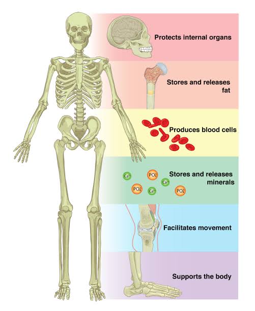
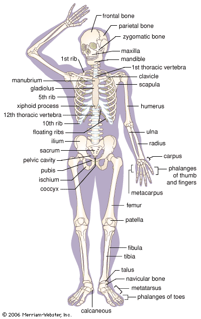
Definitions of Terms: Joint & Articulation A Comprehensive Clinical Reference Guide covering joint anatomy, structural and functional classifications, detailed synovial structures, and pathological correlations . 1. Definition of a Joint A joint (Latin: articulus) is the precise site where two or more bones meet or articulate. Joints are the fundamental units that provide mobility and stability to the human skeleton, making all body movements possible. The human skeleton contains 206 bones that serve as structural scaffolding; their interaction at joints facilitates locomotion while ensuring the protection of internal organs through the immobility of specific adjacent bones (e.g., cranial sutures protecting the brain). Clinical Note Joint dysfunction is among the most common reasons for patient visits in primary care. An accurate clinical assessment requires a deep understanding of both the structural components (what the joint is made of) and the functional capabilities (how the joint moves). 2. Definition of Articulation Articulation refers specifically to the coming together of two bone surfaces at a joint. While often used interchangeably with “joint,” the term articulation emphasizes the functional relationship and the point of contact between bone surfaces rather than just the structural assembly. Quality of Articulation: Determines the range and type of movement possible. Fit vs. Mobility: A “poor” articulation fit, such as the glenohumeral (shoulder) joint, allows for maximum mobility but sacrifices stability. Conversely, an “excellent” fit, such as the hip joint, provides high stability at the cost of reduced mobility. 3. Key Terminology for Clinical Officers The following terms are essential for diagnostic and therapeutic practice regarding musculoskeletal health: Term Definition Clinical Relevance Articular cartilage Hyaline cartilage covering bone ends at synovial joints; provides a smooth, slippery surface. Prevents friction and absorbs shock; damaged in osteoarthritis; avascular. Joint capsule Fibrous sleeve enclosing the joint; consists of outer fibrous and inner synovial layers. Inflamed in capsulitis; provides structural integrity; continuous with periosteum. Synovial membrane Inner layer of capsule secreting synovial fluid; highly vascularized serous tissue. Site of inflammation in Rheumatoid Arthritis; produces hyaluronan and lubricin. Synovial fluid Viscous, non-Newtonian fluid filling the joint cavity; yolk-like consistency. Provides lubrication and nutrients; altered in septic arthritis. Ligament Dense regular connective tissue connecting bone to bone. Frequently torn in sports injuries (sprains); can be extrinsic or intrinsic. Articular disc / Meniscus Fibrocartilage pad between articulating surfaces (C-shaped or oval). Torn meniscus is a common knee injury; smooths bone movement. Bursa Fluid-filled sac lined by synovial membrane. Reduces friction at pressure points; inflamed in bursitis. Tendon Dense connective tissue attaching muscle to bone. Acts as a “dynamic ligament”; prone to tendonitis and rupture. Articular cavity Fluid-filled space within synovial joints. Site of joint effusion and hematoma; target for arthrocentesis. Clinical Pearl Articular cartilage is avascular (lacks blood vessels) and aneural. It receives nutrients primarily through passive diffusion from synovial fluid. This extremely limited blood supply means that cartilage injuries heal very poorly, and degenerative changes like osteoarthritis are largely irreversible. 4. Joint Classification Joints are classified via two complementary systems: Structural (the binding tissue) and Functional (the range of motion). Rule of Classification Always distinguish between the Structural classification (what it is made of) and the Functional classification (how much it moves). For example, the knee is structurally a synovial joint but functionally a diarthrosis. 4.1 Structural Classification Type Binding Tissue Examples Mobility Fibrous Dense fibrous connective tissue; no joint cavity. Cranial sutures, syndesmosis, teeth (gomphosis). Immovable (Synarthrosis) Cartilaginous Hyaline cartilage (Primary) or Fibrocartilage (Secondary). Epiphyseal plates, pubic symphysis, IV discs. Slightly movable (Amphiarthrosis) Synovial Not directly joined; enclosed by an articular capsule. Shoulder, hip, knee, elbow, wrist, ankle. Freely movable (Diarthrosis) 4.2 Functional Classification Synarthrosis: No movement permitted. Mostly corresponds to fibrous joints (e.g., skull sutures). Amphiarthrosis: Slight or partial movement. Mostly corresponds to cartilaginous joints (e.g., intervertebral discs). Diarthrosis: Free movement in one or more planes. All synovial joints are diarthroses. Functional Classification by Axes of Movement (Diarthroses): Classification Axes Movement Types Examples Uniaxial One axis Flexion/Extension only Hinge (knee); Pivot (atlantoaxial) Biaxial Two axes Flexion/Extension + Abduction/Adduction Condyloid (wrist); Saddle (thumb) Polyaxial Three axes All movements including rotation Ball-and-socket (hip, shoulder) 4.3 Subtypes of Synovial Joints Synovial joints are the most movable and are divided into six distinct structural subtypes: Type Description Movement Example Plane (Gliding) Flat or slightly curved articulating surfaces. Sliding/gliding Intercarpal joints, vertebral facet joints. Hinge Convex surface fits into a concave surface. Uniaxial: flexion/extension. Elbow, knee, ankle. Pivot Rounded bone rotates within a ring of ligament/bone. Uniaxial: rotation only. Atlantoaxial joint (C1-C2). Condyloid Oval-shaped condyle fits into an elliptical cavity. Biaxial: flexion/extension, abd/add. Radiocarpal (wrist) joint. Saddle Both surfaces are concave and convex (saddle-shaped). Biaxial: all movements except rotation. 1st carpometacarpal (thumb) joint. Ball-and-Socket Spherical head fits into a cup-like socket. Polyaxial: all movements in all planes. Shoulder, Hip. 5. SYNOVIAL JOINT STRUCTURE IN DETAIL 5.1 Articular Capsule The capsule is continuous with the periosteum and consists of two layers: Fibrous Layer (Outer): Dense white fibrous tissue (capsular ligament). It holds the joint together and is highly innervated but lacks blood vessels. Synovial Layer (Inner): Also known as the synovium. A highly vascularized layer that secretes and absorbs synovial fluid. — Type A synoviocytes: Macrophagic cells that remove debris. — Type B synoviocytes: Fibroblastic cells that manufacture hyaluronan and lubricin. 5.2 Articular Cartilage Hyaline cartilage serves to minimize friction and absorb shock. It is organized into four zones: Superficial zone: Collagen fibers are parallel to the surface. Transitional zone: Randomly oriented fibers. Deep zone: Fibers are perpendicular to the surface. Tidemark: The calcified interface separating cartilage from subchondral bone. 5.3 Synovial Fluid A non-Newtonian ultrafiltrate of plasma. Viscosity is maintained by hyaluronan molecules. Its three primary functions are: Lubrication: Reduces friction during movement. Nutrient Distribution: Vital for avascular articular cartilage. Shock Absorption: Cushions impact during loading. 5.5 Innervation and Vasculature Hilton’s Law: States that the nerves supplying a joint also supply the muscles moving that joint and the skin covering their distal attachments. Articular nerves transmit proprioceptive (position) and nociceptive (pain) data. Vasculature: Arterial supply comes from articular