Accessory Organs of the Gastrointestinal Tract
A comprehensive anatomical study covering the liver, gallbladder, biliary tree, pancreas, and spleen, including morphology, segmentation, neurovascular supply, and clinical application.
CHAPTER 1: Introduction and General Organization
The gastrointestinal tract, while primarily concerned with the mechanical and chemical processing of ingested food, depends critically upon a group of extrinsic organs that secrete essential digestive fluids into the lumen of the alimentary canal. These accessory organs — the liver, gallbladder, pancreas, and spleen — are developmentally, anatomically, and functionally distinct from the tubular digestive tract itself, yet their integrated activity is indispensable for normal digestion, metabolism, and homeostasis.
- The Liver: The largest gland in the human body, performs over 500 distinct biochemical functions, including bile synthesis, carbohydrate metabolism, protein synthesis, and detoxification.
- The Gallbladder: Serves as a reservoir and concentrator of hepatic bile, releasing it in a regulated manner in response to dietary stimuli.
- The Pancreas: Exhibits a unique dual endocrine-exocrine nature, with its exocrine acinar cells producing the most potent digestive enzyme cocktail in the body, while its endocrine islets regulate systemic glucose homeostasis.
- The Spleen: Though not a secretory organ of digestion, it is anatomically and vascularly integrated with the portal system and performs vital hematological and immunological functions.
CHAPTER 2: The Liver — Gross Anatomy, Lobes, and Segmentation
2.1 Surface Projections and General Morphology
The liver is the largest solid organ in the body, weighing approximately 1.2–1.5 kg in the adult, and occupying the right hypochondrium and epigastrium. It is wedge-shaped, with a broad right surface adapted to the contour of the right dome of the diaphragm, and a tapering left extremity extending toward the left hypochondrium.
- Superior (Diaphragmatic) Surface: Convex and related to the diaphragm and anterior abdominal wall.
- Inferior (Visceral) Surface: Concave and related to the stomach, duodenum, hepatic flexure of the colon, right kidney, and right suprarenal gland.
- Glisson's Capsule: A thin fibrous capsule covering the entire organ. Except at the bare area and porta hepatis, the liver is invested by visceral peritoneum.
The surface projection of the liver can be mapped by drawing a line from the right fifth intercostal space at the midclavicular line to the left fifth intercostal space at the midclavicular line, with the inferior border descending to the right costal margin and crossing the midline at the level of the xiphoid process.
2.2 Anatomical Lobes
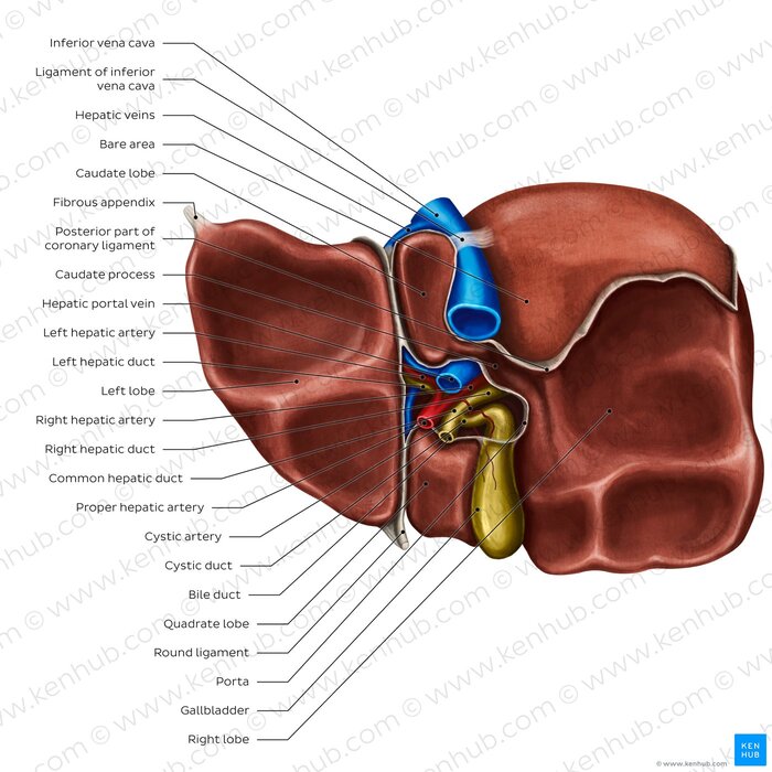
Traditionally, the liver is divided into four anatomical lobes based on external landmarks visible on the visceral surface:
- Right Lobe and Left Lobe: Separated anteriorly by the falciform ligament and posteriorly by the fissure for the ligamentum venosum.
- Quadrate Lobe: Located between the gallbladder fossa and the fissure for the ligamentum teres.
- Caudate Lobe: Situated on the posterior surface between the inferior vena cava groove and the fissure for the ligamentum venosum.
Note: These anatomical lobes do not correspond to functional divisions. The quadrate and caudate lobes receive blood from both the right and left branches of the portal vein and hepatic artery.
2.3 Functional Lobation and the Couinaud Classification
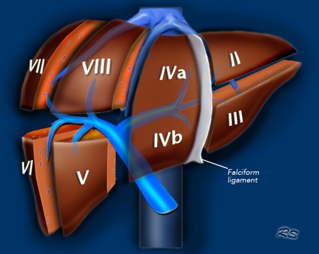
Modern functional understanding ( revolutionized by Claude Couinaud in 1957) divides the liver into eight functionally independent segments. Each possesses its own portal triad (portal vein branch, hepatic artery branch, and bile duct) and is drained by a specific tributary of the hepatic veins.
The eight segments are numbered clockwise in the transverse plane, beginning with segment I (the caudate lobe) in the posterior-superior position. Segments II and III constitute the left lateral sector; segment IV (subdivided into IVa and IVb) constitutes the left medial sector. Segments V and VI form the right anterior sector, while segments VII and VIII form the right posterior sector.
Segment I (Caudate Lobe) is unique in receiving portal blood supply from both the right and left portal branches and draining directly into the inferior vena cava via emissary veins.
| Feature | Anatomical Lobation | Functional Lobation (Couinaud) |
|---|---|---|
| Basis of division | External landmarks (fissures, ligaments) | Vascular and biliary branching patterns |
| Number of lobes | 4 (right, left, quadrate, caudate) | 8 segments (I–VIII) |
| Blood supply | Not segment-specific | Each segment has independent portal triad |
| Venous drainage | Via hepatic veins (not segment-specific) | Each segment drains to specific hepatic vein tributary |
| Clinical relevance | Limited surgical utility | Basis for segmental hepatic resection |
| Key landmark | Falciform ligament | Cantlie's line (middle hepatic vein plane) |
CHAPTER 3: The Liver — Peritoneal Relations, Ligaments, and Neurovascular Architecture
3.1 Peritoneal Relations and Ligaments
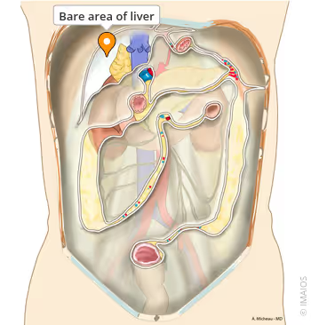
The liver is almost entirely covered by visceral peritoneum, with the exception of the bare area, a large triangular region on the posterior-superior surface bounded by the coronary ligament. The bare area is in direct contact with the diaphragm and is devoid of peritoneal covering, allowing the hepatic veins to enter the IVC without traversing the peritoneal cavity.
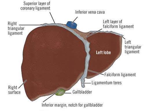
- Coronary Ligament: Consists of anterior and posterior layers that meet laterally to form the right and left triangular ligaments.
- Falciform Ligament: Extends from the umbilicus to the liver, attaching it to the anterior abdominal wall and diaphragm. Its free inferior margin contains the ligamentum teres (obliterated left umbilical vein), dividing the liver into anatomical right and left lobes.
- Ligamentum Venosum: Remnant of the fetal ductus venosus; lies in the fissure on the posterior surface and separates the caudate lobe from the left lobe.
3.2 The Porta Hepatis
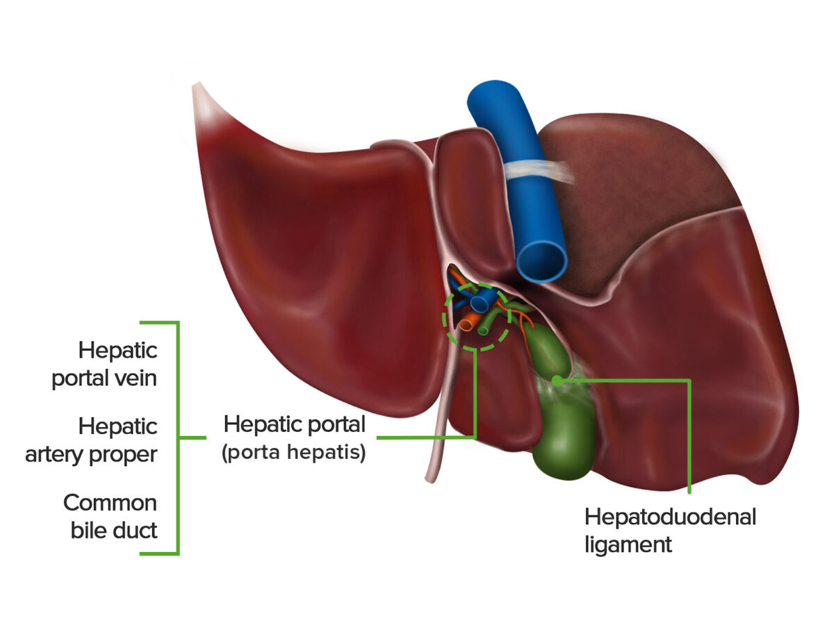
The porta hepatis is the hilum of the liver, located on the inferior surface in the fissure between the quadrate lobe anteriorly and the caudate lobe posteriorly. It transmits the portal triad:
- Hepatic Portal Vein (posteriorly).
- Proper Hepatic Artery (medially).
- Common Hepatic Duct (anteriorly and to the right).
Additionally, the porta hepatis transmits lymphatic vessels, autonomic nerve fibers, and the ligamentum teres.
3.3 Vascular Supply and Drainage
The liver enjoys a unique dual blood supply:
- Hepatic Portal Vein (~75%): Conveys nutrient-rich but deoxygenated blood from the gastrointestinal tract, spleen, pancreas, and gallbladder.
- Hepatic Artery Proper (~25%): Oxygenated blood from the celiac trunk.
- Hepatic Sinusoids: Where blood is processed by hepatocytes before draining into central veins and eventually the hepatic veins.
- Venous Exit: The hepatic veins (right, middle, and left) drain directly into the inferior vena cava at the superior posterior aspect of the liver, within the bare area.
— Right hepatic vein: drains segments V, VI, VII, and VIII.
— Middle hepatic vein: drains segments IV, V, and VIII.
— Left hepatic vein: drains segments II and III.
— Caudate lobe (segment I): drains directly into the IVC via short emissary veins.
3.4 Innervation and Lymphatic Drainage
Innervation: Receives sympathetic and parasympathetic fibers via the hepatic plexus within the lesser omentum. Sympathetic fibers originate from the celiac plexus (T7–T9), while parasympathetic fibers are derived from the anterior vagal trunk (C3–C5 via the vagus nerve). Afferent (pain) fibers from the capsule travel with sympathetics to enter the spinal cord at T7–T9, explaining referred pain to the RUQ and epigastrium.
Lymphatic Pathways:
1. Superficial lymphatics: in Glisson's capsule; drain to hepatic nodes at porta hepatis, then celiac lymph nodes.
2. Deep lymphatics: accompany hepatic veins to posterior mediastinal nodes and thoracic duct.
The liver produces approximately one-third of the body's lymph.
CHAPTER 4: The Gallbladder and Extrahepatic Biliary Tree
4.1 Gross Anatomy of the Gallbladder
A pear-shaped, distensible organ measuring 7–10 cm in length, capable of storing 30–50 mL of concentrated bile. It is situated in a shallow fossa on the inferior surface of the liver, between the right and quadrate lobes. It is divided into four regions:
- Fundus: Blind, rounded extremity projecting beyond the inferior margin of the liver (contact with anterior wall at right 9th costal cartilage).
- Body: The main portion.
- Infundibulum (Neck): Tapers to join the cystic duct.
- Hartmann's Pouch: Small outpouching at the neck/cystic duct junction; a common site for gallstone impaction.
The mucosal surface features deep folds known as Rokitansky-Aschoff sinuses.
4.2 The Extrahepatic Biliary Tree
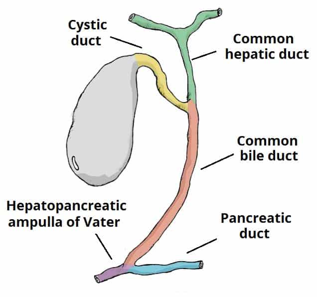
Bile is collected by canaliculi -> interlobular ducts -> segmental ducts -> Right and Left Hepatic Ducts. These merge at the porta hepatis to form the Common Hepatic Duct (~3 cm long). The CHD is joined by the Cystic Duct to form the Common Bile Duct (CBD) (~8 cm long, 6-8 mm diameter).
The CBD has four parts:
- Supraduodenal part: descends in the free edge of the lesser omentum.
- Retroduodenal part: passes posterior to the 1st part of the duodenum.
- Pancreatic part: grooves or is embedded in the posterior pancreatic head.
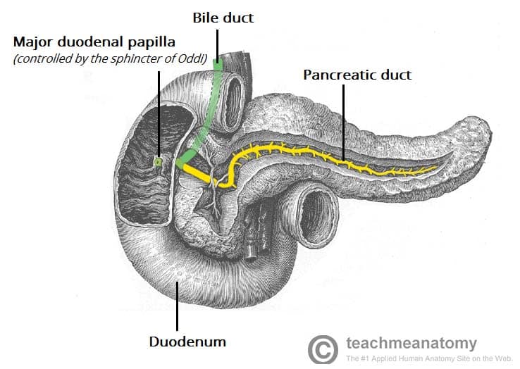
- Intramural part: traverses the duodenal wall to open at the major duodenal papilla.
The CBD joins the main pancreatic duct (Wirsung) at the hepatopancreatic ampulla (ampulla of Vater), approximately 8–10 cm distal to the pylorus. The flow is regulated by the sphincter of Oddi.
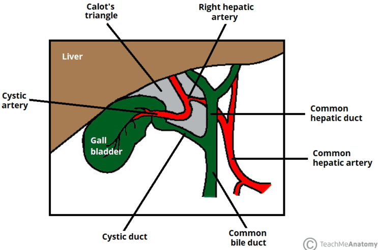
4.3 Calot's Triangle (Cystohepatic Triangle)
A surgically critical space bounded by:
- Common hepatic duct (medially).
- Cystic duct (laterally).
- Inferior surface of the liver (superiorly).
The triangle contains the cystic artery and the cystic lymph node (of Lund).
CHAPTER 5: The Pancreas — Topography, Ductal System, and Vascular Relations
5.1 Topography and Subdivisions
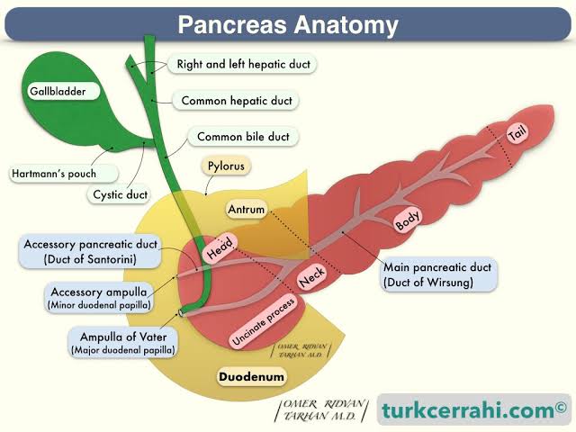
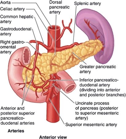
The pancreas is a retroperitoneal, elongated gland (12–15 cm) extending transversely across the posterior wall from the C-loop of the duodenum to the splenic hilum. It has five parts:
- Head: Nestled within the C-loop of the duodenum.
- Uncinate Process: Hook-like extension passing posterior to the superior mesenteric vessels.
- Neck: Constricted portion anterior to the portal vein confluence.
- Body: Extends across the aorta and left renal vessels.
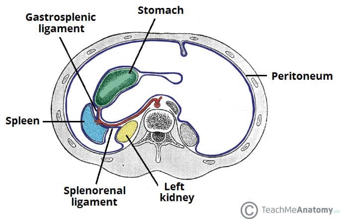
- Tail: Narrow left extremity approaching the splenic hilum within the splenorenal ligament.
5.2 Ductal System
- Main Pancreatic Duct (Wirsung): Begins in the tail; courses to the head to join the CBD at the ampulla of Vater.
- Accessory Pancreatic Duct (Santorini): Drains the superior head; usually opens at the minor duodenal papilla (2 cm proximal to the major papilla).
- Pancreas Divisum: Occurs in ~10% of individuals when the two ducts fail to fuse.
5.3 Vascular Supply
Arterial: Derived from celiac trunk and SMA. The head/uncinate receive supply from pancreaticoduodenal arcades. The body/tail are supplied by branches of the splenic artery (dorsal, greater, and caudal pancreatic arteries).
Venous Drainage: Corresponds to arteries. The portal vein forms posterior to the neck of the pancreas by the union of the superior mesenteric vein and the splenic vein.
CHAPTER 6: The Spleen — Physical Projections and Peritoneal Attachments
6.1 Surface Projections and Morphology
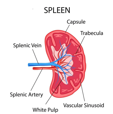
The largest lymphoid organ (12 cm length, 7 cm width, 3–4 cm thickness, 150–200 g). Located in the left hypochondrium, deep to the 9th, 10th, and 11th ribs. Morphology:
- Superior Border: Characteristically notched (distinguishes it from the kidney on palpation).
- Visceral Surface: Concave with impressions for the stomach, kidney, colon, and pancreas.
- Peritoneum: Completely invested except at the hilum.
6.2 Peritoneal Attachments and 6.3 Vascular Supply
- Gastrosplenic Ligament: Greater curvature to hilum; transmits short gastric and left gastroepiploic vessels.
- Splenorenal (Lienorenal) Ligament: Left kidney to hilum; transmits splenic artery/vein and tail of the pancreas.
- Splenic Artery: Largest branch of the celiac trunk; tortuous course along superior pancreatic border.
CHAPTER 7: Portal-Systemic (Portacaval) Anastomoses
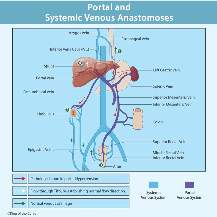
Sites where the portal venous system communicates with systemic circulation. In portal hypertension (cirrhosis), blood shunts from high-pressure portal to low-pressure systemic veins.
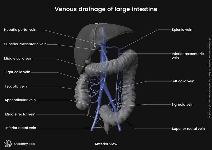
| Site | Portal Tributary | Systemic Vein | Clinical Manifestation |
|---|---|---|---|
| Lower Esophagus | Left gastric (coronary) vein | Azygos / Hemiazygos veins | Esophageal varices; hematemesis |
| Anal Canal | Superior rectal vein | Middle & Inferior rectal veins | Internal / External hemorrhoids |
| Paraumbilical | Paraumbilical veins | Superficial epigastric veins | Caput medusae |
| Retroperitoneal | Colic & Duodenal veins | Lumbar, renal, phrenic veins | Retroperitoneal varices; Ascites |
CHAPTER 8: Clinical and Applied Anatomy
Portal Hypertension
Defined as portal pressure > 5 mmHg (Normal 5–10 mmHg). Consequences include collateral circulation, splenomegaly, ascites, and hepatic encephalopathy. Management: beta-blockers, band ligation, or TIPS (Transjugular Intrahepatic Portosystemic Shunt).
Cholelithiasis
Gallstones classified as cholesterol (radiolucent), pigment (opaque), or mixed. "5 Fs": female, fertile, forty, fat, family history. Complications include cholecystitis, choledocholithiasis, and pancreatitis.
Pancreatic Carcinoma: Head vs. Tail
- Head (60%): Presents early with painless obstructive jaundice and palpable gallbladder (Courvoisier's sign). Treatment: Whipple procedure.
- Body/Tail (40%): Presents late with vague pain and weight loss; often unresectable.
Splenic Rupture
Most common solid organ injury in blunt trauma (ribs 9-11). Features include LUQ pain, Kehr's sign (referred pain to L shoulder), and shock. Vaccination against encapsulated organisms (Strep. pneumoniae, H. influenzae, N. meningitidis) is mandatory post-splenectomy to prevent OPSI (Overwhelming Post-Splenectomy Infection).
Referred Pain Pathways Summary
| Condition | Anatomical Basis | Key Clinical Feature |
|---|---|---|
| Referred hepatic pain | T7–T9 Sympathetics; Phrenic (C3–C5) | Pain to R upper quadrant and R shoulder |
| Referred pancreatic pain | T5–T9 Sympathetics | Epigastric pain radiating to the back |
| Kehr's sign | T9–T10; Phrenic irritation | LUQ pain referred to L shoulder |
