Classifications of Bones
A comprehensive guide detailing the six categories of bones, their morphological characteristics, anatomical examples, and high-yield clinical correlations.
Overview of Bone Types
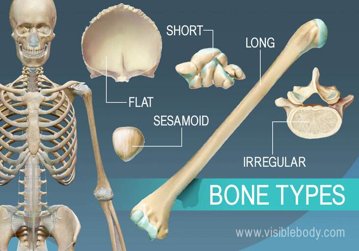
The human skeleton is composed of bones that are classified based on their shape and function. These classifications provide insight into the mechanical demands placed on the bone and the specific pathologies that affect them.
| Type | Examples | Key Characteristics |
|---|---|---|
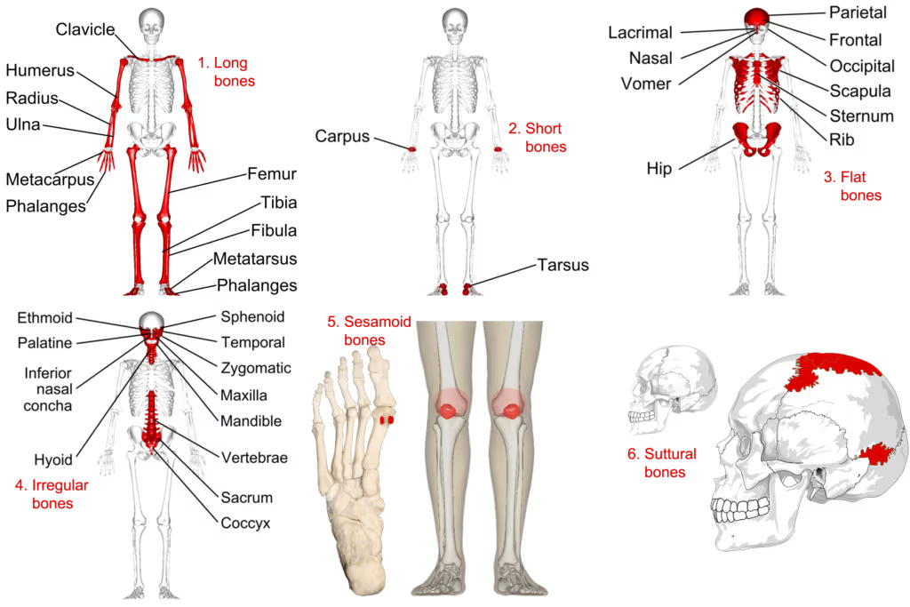
| Long Bones | Femur, Tibia, Humerus, Radius, Ulna, Phalanges, Metacarpals, Metatarsals | Longer than wide; consist of a shaft plus two ends; contain a medullary cavity; formed via endochondral ossification. |
| Short Bones | Carpals (8), Tarsals (7) | Cube-shaped; compact shell over spongy bone; no medullary cavity; serve as shock absorbers. |
| Flat Bones | Skull bones, Sternum, Ribs, Scapulae | Thin and flattened; "sandwich" structure (tables + diploe); formed via intramembranous ossification. |
| Irregular Bones | Vertebrae, Sacrum, Coccyx, Mandible, Maxilla, Ethmoid, Sphenoid, Hyoid | Complex shapes; spongy bone covered by thin compact bone; often pneumatized (contain air sinuses). |
| Sesamoid Bones | Patella, Pisiform, Fabella, 1st MTP sesamoids | Embedded within tendons; increase mechanical advantage and reduce friction. |
| Accessory Bones | Os trigonum, Os naviculare, Os peroneum, Os vesalianum | Extra bones resulting from failed fusion; usually asymptomatic but can mimic fractures. |
1. LONG BONES
Definition: Bones that are longer than they are wide, consisting of a shaft (diaphysis) and two ends (epiphyses). They contain a medullary cavity with yellow marrow in the diaphysis and red marrow in the epiphyses during development.
Key Anatomical Examples
| Bone | Location | Approx. Length | Key Features | Common Fractures |
|---|---|---|---|---|
| Radius | Lateral forearm | ~26 cm | Crosses over ulna during pronation; distal end articulates with scaphoid and lunate. | Colles', Smith's, radial head. |
| Ulna | Medial forearm | ~28 cm | Olecranon process (triceps insertion); trochlear notch articulates with humerus. | Olecranon, coronoid process, Monteggia. |
| Metacarpals | Palm | 5–9 cm | Numbered I–V; base articulates with carpals; head with phalanges. | Boxer's (5th neck), Bennett's (1st base). |
| Phalanges | Fingers/Toes | 2–5 cm | 14 per hand/foot; proximal, middle, distal (thumb/hallux lack middle). | Tuft fracture, Mallet finger. |
- Salter-Harris Injuries: The epiphyseal (growth) plate is the weakest point in the pediatric skeleton. Type II is the most common (~75%). Type V (crush) has the highest risk of growth arrest.
- Osteosarcoma: Most common primary bone malignancy in children. Typically arises in the metaphysis (distal femur > proximal tibia). X-ray shows Codman's triangle and "sunburst" pattern.
- Ewing Sarcoma: Second most common malignancy; involves the diaphysis. X-ray shows "onion-skin" periosteal reaction. Associated with t(11;22) translocation.
- Pathological Fractures: Occur through pre-existing lesions. In adults: metastatic disease (breast, prostate, lung) or multiple myeloma. In children: unicameral bone cysts.
2. SHORT BONES
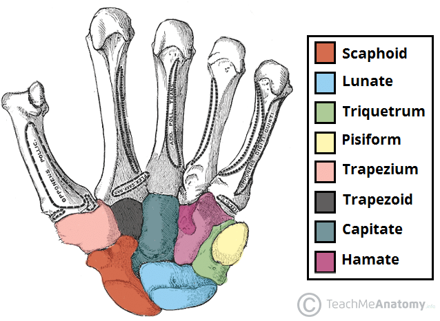
Definition: Approximately cube-shaped bones where length, width, and thickness are roughly equal. They lack a medullary cavity and consist of a thin compact shell surrounding a cancellous core.
Carpal Bones (8 per hand)
| Row | Bone | Description | Clinical Notes |
|---|---|---|---|
| Proximal | Scaphoid | Boat-shaped | 70% of carpal fractures. Blood supply is distal-to-proximal (retrograde); high risk of Avascular Necrosis (AVN). |
| Proximal | Lunate | Moon-shaped | Dislocates anteriorly. AVN of lunate is called Kienböck's Disease. |
| Proximal | Pisiform | Sesamoid | Inside flexor carpi ulnaris tendon; protects ulnar nerve in Guyon's canal. |
| Distal | Trapezium | Saddle joint | Articulates with 1st metacarpal; critical for thumb mobility. |
| Distal | Capitate | Largest | The central keystone of the carpus. |
| Distal | Hamate | Hook process | Hook of hamate fractures are common in golfers and racket sports. |
Tarsal Bones (7 per foot)
| Bone | Key Features | Clinical Relevance |
|---|---|---|
| Talus | Entirely covered by cartilage; no muscle attachments. | Hawkins classification for neck fractures; high AVN risk. |
| Calcaneus | Largest tarsal bone; forms the heel. | Lover's fracture (high-energy fall); check for associated lumbar spine fractures. Bohler's angle < 20° is diagnostic. |
| Navicular | Articulates with talus and 3 cuneiforms. | Stress fractures in athletes; tenderness at the 'N spot'. |
| Cuboid | Lateral foot bone. | Cuboid syndrome: subluxation causing lateral foot pain. |
- Carpal Tunnel Syndrome: Median nerve compression under the flexor retinaculum. Causes numbness in the radial 3.5 fingers.
- Chopart Joint: Dislocation of the talonavicular and calcaneocuboid joints (midtarsal).
- Lisfranc Injury: Disruption of the tarsometatarsal joints.
3. FLAT BONES
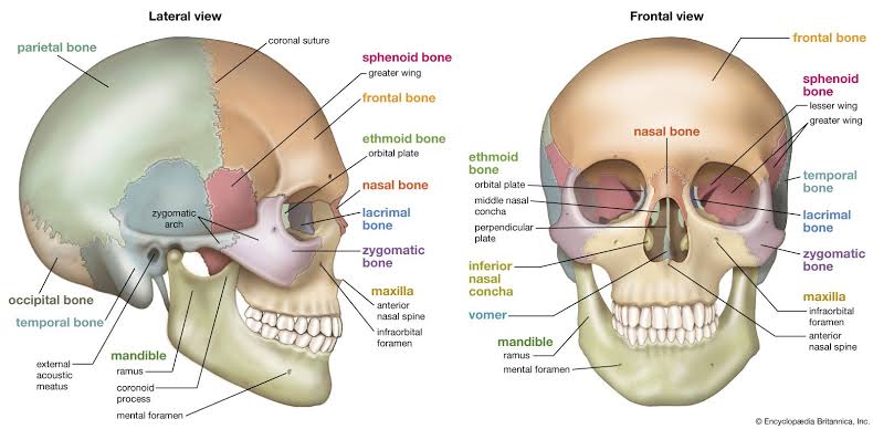
Definition: Thin, flattened, and usually curved bones. They feature a sandwich-like structure: two layers of compact bone (outer and inner tables) with a layer of spongy bone (diploe) between them. They protect internal organs and provide broad areas for muscle attachment.
Skull and Thoracic Flat Bones
| Bone | Distinguishing Features | Clinical Notes |
|---|---|---|
| Frontal | Forehead and superior orbits; contains frontal sinuses. | Fractures involve anterior cranial fossa; risk of CSF rhinorrhea. |
| Occipital | Posterior base; contains foramen magnum. | Basilar fractures can cause lower cranial nerve palsies (CN IX–XII). |
| Sternum | Manubrium, body, and xiphoid process. | The Angle of Louis (manubriosternal joint) marks the 2nd rib and T4/T5 level. |
| Ribs | 12 pairs; flat and curved. | 1st/2nd rib fractures indicate high-energy trauma (brachial plexus risk). Flail chest is ≥3 adjacent ribs fractured in ≥2 places. |
- Craniosynostosis: Premature fusion of sutures. Sagittal fusion → Scaphocephaly (long/narrow head). Coronal fusion → Brachycephaly.
- Le Fort Fractures: Complex facial fractures. Type I: Horizontal (above teeth). Type II: Pyramidal (through maxilla/nose). Type III: Craniofacial dysjunction.
- Sternal Foramen: A congenital defect in the sternal body that can be mistaken for a lytic lesion on X-ray.
4. IRREGULAR BONES
Definition: Bones with complex shapes that do not fit other categories. They consist of cancellous bone enclosed by a thin layer of compact bone.
Vertebrae (26 in adults)
| Region | Count | Distinguishing Features |
|---|---|---|
| Cervical (C1–C7) | 7 | Transverse foramina (vertebral artery); bifid spines. C1 (Atlas) lacks a body; C2 (Axis) has the Dens. |
| Thoracic (T1–T12) | 12 | Costal facets for ribs; heart-shaped bodies; long downward-pointing spinous processes. |
| Lumbar (L1–L5) | 5 | Large kidney-shaped bodies; triangular vertebral foramina; bear the most weight. |
| Sacrum | 1 (5 fused) | Triangular; articulates with ilia. Contains sacral hiatus for caudal anesthesia. |
Other Irregular Bones
- Ethmoid: Between the orbits; contains the cribriform plate. Fractures cause anosmia (loss of smell).
- Sphenoid: The "keystone" of the cranium. Contains the sella turcica for the pituitary gland.
- Mandible: The only movable skull bone. Condylar fractures can cause malocclusion.
- Hyoid: U-shaped bone in the neck; does not articulate with any other bone. Fractures suggest strangulation.
- Spinal Stenosis: Narrowing of the canal. Causes neurogenic claudication (leg pain relieved by forward flexion).
- Cauda Equina Syndrome: Compression of nerve roots below L1–L2. Surgical Emergency: Symptoms include saddle anesthesia and bowel/bladder dysfunction.
5. SESAMOID BONES
Definition: Small, round bones developed within tendons (intramembranous ossification) in response to strain. They act as pulleys to increase mechanical leverage.
| Sesamoid | Location | Function | Clinical Significance |
|---|---|---|---|
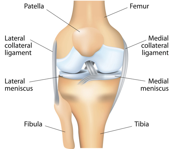
| Patella | Anterior knee | Increases quadriceps moment arm. | Dislocation is usually lateral (valgus stress). |
| Pisiform | Palmar wrist | Leverage for Flexor Carpi Ulnaris. | Protects ulnar nerve in Guyon's canal. |
| Fabella | Posterior knee | In lateral gastrocnemius head. | Present in ~25%; can cause peroneal nerve compression. |
| 1st MTP | Plantar big toe | Bears weight during push-off. | Sesamoiditis in dancers and runners. |
6. ACCESSORY (SUPERNUMERARY) BONES
Definition: Extra bones resulting from the failure of ossification centers to fuse. Usually discovered incidentally on X-ray.
| Bone | Location | Prevalence | Clinical Relevance |
|---|---|---|---|
| Os Trigonum | Posterior talus | 7–14% | Causes Posterior Ankle Impingement in dancers. |
| Os Naviculare | Medial navicular | 4–14% | Mistaken for navicular fracture; causes medial foot pain. |
| Os Peroneum | Lateral cuboid | ~20% | Found in peroneus longus tendon; can fracture during inversion. |
| Os Vesalianum | 5th metatarsal base | Rare | Must differentiate from a Jones fracture. |
- Accessory Bones: Feature smooth, corticated margins; often bilateral (~50%).
- Fractures: Feature irregular, non-corticated margins and may show displacement.
- Rule: Always compare with the contralateral side on X-ray.
