The Peritoneum
A Comprehensive Anatomical Notes Covering Structural Organization, Peritoneal Reflections and Folds, Subdivisions of the Peritoneal Cavity, Spaces and Gutters, Neurovascular Supply, and Clinical Applied Anatomy.
CHAPTER 1: INTRODUCTION AND FUNCTIONAL OVERVIEW
1.1 Functional Significance of the Peritoneum
The peritoneum is a continuous, glistening serous membrane that lines the abdominal cavity and invests the viscera contained within it. It represents the largest and most complexly arranged serous membrane in the human body. The peritoneum acts to minimize friction between moving organs, transmit neurovascular structures, and serve as a dynamic defensive immunological barrier within the abdominopelvic cavity.
The peritoneum is not merely a passive lining; it is an active, dynamic organ with significant physiological and immunological functions. It secretes serous fluid that lubricates the surfaces of the abdominal organs, allowing them to move freely against one another during peristalsis, respiration, and changes in body position. The peritoneum also plays a critical role in immune defense, with mesothelial cells capable of phagocytosis and the production of inflammatory mediators. The peritoneal cavity serves as a potential space that can accommodate pathological processes such as infection, hemorrhage, and malignancy, making understanding of its anatomy essential for surgical and diagnostic procedures.
The peritoneum is ultimately one continuous sheet, but for descriptive purposes, it is divided into two layers: the parietal peritoneum that lines the abdominal wall and the visceral peritoneum that covers the abdominal organs. Between these two layers lies the peritoneal cavity, a potential space that normally contains only a thin film of serous fluid (approximately 50-100 mL) that lubricates the surfaces and reduces friction.
The Peritoneum as a Functional Unit
The peritoneum operates not as a passive membrane but as an active biological interface. The mesothelial cells that line the peritoneum secrete lubricating fluid, participate in immune surveillance, and can undergo metaplasia in response to chronic irritation. The peritoneal cavity, though normally a potential space, can expand significantly to accommodate pathological fluid collections (ascites), air (pneumoperitoneum), or blood (hemoperitoneum). Understanding the three-dimensional anatomy of the peritoneum and its reflections is therefore fundamental to abdominal surgery, interventional radiology, and emergency medicine.
CHAPTER 2: STRUCTURAL ORGANIZATION AND LAYERS
2.1 Parietal Peritoneum
The parietal peritoneum is the portion of the peritoneum that lines the internal surface of the abdominopelvic wall. It is derived embryologically from the somatic mesoderm, which also gives rise to the body wall muscles, bones, and connective tissue. This embryological origin determines its innervation by somatic nerves and its sensitivity to the same stimuli as the skin: pain, pressure, temperature, and laceration.
The parietal peritoneum is firmly attached to the transversalis fascia of the abdominal wall by loose areolar tissue. It lines the anterior and lateral abdominal walls, the pelvic walls, the inferior surface of the diaphragm, and the anterior surface of the retroperitoneal organs. The parietal peritoneum reflects onto the viscera at various points, becoming continuous with the visceral peritoneum.
2.2 Visceral Peritoneum
The visceral peritoneum is the portion of the peritoneum that directly invests the abdominal organs. It is derived embryologically from the splanchnic mesoderm, which also gives rise to the smooth muscle and connective tissue of the gastrointestinal tract. This embryological origin determines its innervation by autonomic (visceral) nerves and its relative insensitivity to most stimuli.
The visceral peritoneum covers the organs almost completely (in the case of intraperitoneal organs) or only on their anterior surface (in the case of retroperitoneal organs). At the points where organs are attached to the body wall or to other organs by mesenteries, omenta, or ligaments, the visceral peritoneum is continuous with the parietal peritoneum or with the visceral peritoneum of adjacent organs.
2.3 The Peritoneal Cavity
The peritoneal cavity is the potential space between the parietal and visceral layers of the peritoneum. It is not a true "cavity" in the sense of being empty; rather, it is a potential space that normally contains only a thin film of serous fluid (approximately 50-100 mL) that lubricates the peritoneal surfaces and allows the organs to move freely against one another.
The peritoneal cavity is completely closed in males, forming a sealed potential space. In females, however, the peritoneal cavity communicates with the external environment via the uterine tubes (fallopian tubes), uterus, and vagina. This communication pathway is clinically significant because it provides a potential route for the spread of infection from the exterior to the peritoneal cavity (e.g., ascending pelvic inflammatory disease) and explains why pneumoperitoneum can occur in females after sexual intercourse or vaginal procedures without necessarily having a perforated viscus.
Sexual Dimorphism of the Peritoneal Cavity
The open communication between the peritoneal cavity and the exterior in females (via the uterine tubes, uterus, and vagina) has several clinical implications:
- It provides a route for ascending infection, which is why pelvic inflammatory disease (PID) can lead to peritonitis.
- It explains why females can develop pneumoperitoneum after sexual intercourse or vaginal procedures without necessarily having a perforated viscus.
- It provides a potential route for the spread of malignancy from the genital tract to the peritoneal cavity.
In contrast, the sealed peritoneal cavity in males means that any pneumoperitoneum is highly suggestive of a perforated viscus or recent surgery.
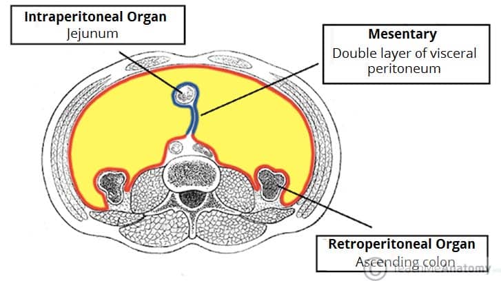
2.4 Organ Relationship Classifications
Abdominal organs are classified based on their relationship to the peritoneum into three categories: intraperitoneal, primarily retroperitoneal, and secondarily retroperitoneal. This classification has profound implications for surgical approach, organ mobility, and the patterns of disease spread.
2.4.1 Intraperitoneal Organs
Intraperitoneal organs are almost entirely covered by visceral peritoneum and are suspended into the peritoneal cavity by mesenteries. They are mobile and have a greater range of movement than retroperitoneal organs. Intraperitoneal organs include:
| Organ | Mesentery | Notes |
|---|---|---|
| Stomach | Lesser and greater omenta | Highly mobile; attached at lesser and greater curvatures |
| Spleen | Gastrosplenic and splenorenal ligaments | Intraperitoneal but relatively fixed by ligaments |
| Liver | Falciform, coronary, triangular ligaments | Mostly intraperitoneal except bare area |
| Jejunum and Ileum | Mesentery of small intestine | Highly mobile; long mesentery allows wide movement |
| Transverse colon | Transverse mesocolon | Mobile; suspended from posterior abdominal wall |
| Sigmoid colon | Sigmoid mesocolon | Mobile; variable length of mesentery |
| First part of duodenum | None (short segment) | Only the first 2 cm (duodenal cap) is intraperitoneal |
| Fourth part of duodenum | None (short segment) | Short intraperitoneal segment before duodenojejunal flexure |
| Appendix | Mesoappendix | Intraperitoneal; mobile within right iliac fossa |
| Cecum (variable) | None or short mesocecum | May be retrocecal or intraperitoneal |
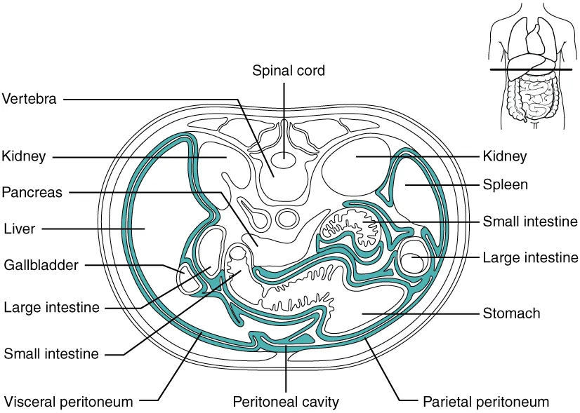
2.4.2 Primarily Retroperitoneal Organs
Organs that developed and remain outside the peritoneum throughout embryological development. They are located in the retroperitoneal space and are covered by parietal peritoneum only on their anterior surface. These organs include:
- Kidneys and ureters
- Suprarenal (adrenal) glands
- Abdominal aorta and inferior vena cava
- Esophagus (abdominal portion)
- Rectum (lower two-thirds)
2.4.3 Secondarily Retroperitoneal Organs
Organs that were initially intraperitoneal during embryological development but lost their mesenteries and became retroperitoneal as their mesenteries fused with the posterior abdominal wall. These organs are covered by parietal peritoneum only on their anterior surface. They include:
- Pancreas (except the tail, which remains intraperitoneal within the splenorenal ligament)
- Duodenum (parts 2, 3, and 4) - the descending, horizontal, and ascending parts
- Ascending colon
- Descending colon
SAD PUCKER (Retroperitoneal Organs)
A useful mnemonic for remembering the retroperitoneal organs is SAD PUCKER:
- S = Suprarenal (adrenal) glands
- A = Aorta and Inferior Vena Cava
- D = Duodenum (parts 2-4)
- P = Pancreas (except tail)
- U = Ureters
- C = Colon (ascending and descending parts)
- K = Kidneys
- E = Esophagus (abdominal portion)
- R = Rectum (lower two-thirds)
CHAPTER 3: PERITONEAL REFLECTIONS AND FOLDS
3.1 Overview
The peritoneum does not merely line the abdominal wall and cover the organs as separate sheets. Instead, it forms continuous folds, reflections, and duplications that connect organs to the abdominal wall, connect organs to each other, and create specialized structures with important functions. These peritoneal reflections include the mesenteries, omenta, and ligaments. All of these structures are essentially double layers (or multiple layers) of peritoneum that enclose connective tissue, fat, blood vessels, lymphatics, and nerves.
3.2 Mesenteries
Mesenteries are double layers of peritoneum that connect intraperitoneal organs to the posterior abdominal wall. They serve as conduits for the blood vessels, lymphatics, and nerves that supply the organs. The term "mesentery" is often used generically to describe any peritoneal fold that suspends an organ, but technically, each mesentery is named according to the organ it suspends.
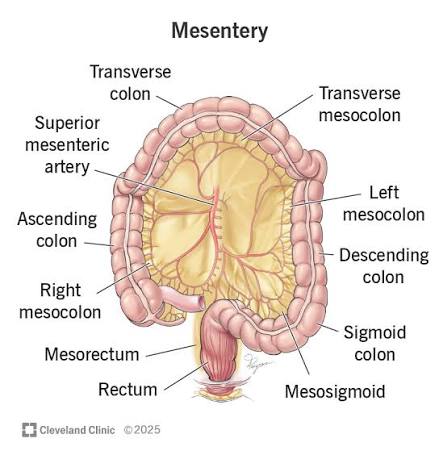
3.2.1 The Mesentery (of the Small Intestine)
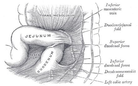
The mesentery of the small intestine (often simply called "the mesentery") is a broad, fan-shaped fold of peritoneum that suspends the jejunum and ileum from the posterior abdominal wall. It has a root (attachment to the posterior wall) that is approximately 15 cm long, extending from the duodenojejunal flexure (left of L2) to the ileocecal junction (right sacroiliac joint). The root of the mesentery crosses the following structures from above downward:
- Duodenum (third and fourth parts)
- Abdominal aorta
- Inferior vena cava
- Right ureter
- Right psoas major
- Right testicular/ovarian vessels
The free border of the mesentery is attached to the entire length of the jejunum and ileum and is approximately 6-7 meters long. The mesentery contains the superior mesenteric artery and vein, lymphatics, autonomic nerves, and variable amounts of fat. The mesenteric fat increases from the jejunum to the ileum.
3.2.2 Transverse Mesocolon
The transverse mesocolon is a broad peritoneal fold that suspends the transverse colon from the posterior abdominal wall. Its root crosses the duodenum, pancreas, and left kidney. The transverse mesocolon is continuous with the greater omentum inferiorly and the mesentery of the small intestine at the hepatic and splenic flexures. It contains the middle colic vessels (branches of the superior mesenteric artery and vein), lymphatics, and nerves.
The transverse mesocolon is an important surgical landmark because it divides the peritoneal cavity into the supracolic compartment (above) and the infracolic compartment (below). It also provides access to the pancreas and the retroperitoneal structures during surgery.
3.2.3 Sigmoid Mesocolon
The sigmoid mesocolon is an inverted V-shaped or S-shaped peritoneal fold that suspends the sigmoid colon from the pelvic wall. Its apex is located near the division of the left common iliac artery, and its limbs extend along the left pelvic brim. The sigmoid mesocolon contains the sigmoid vessels (branches of the inferior mesenteric artery and vein), lymphatics, and nerves. The length of the sigmoid mesocolon is variable, and a long, redundant mesocolon predisposes to sigmoid volvulus.
3.3 Omenta
Omenta are specific peritoneal folds that connect the stomach and proximal duodenum to other organs. There are two omenta: the greater omentum and the lesser omentum. Both are derived from the embryological mesenteries of the stomach and are specialized structures with important protective and metabolic functions.
3.3.1 Greater Omentum
The greater omentum is the largest peritoneal fold in the body. It is an apron-like structure that extends from the greater curvature of the stomach and the proximal duodenum, descends inferiorly over the transverse colon and small intestine, and then folds back upon itself to attach to the transverse mesocolon and posterior abdominal wall. It is composed of four layers of peritoneum (two layers descending and two layers ascending), between which are variable amounts of fat, connective tissue, lymphatic tissue, and blood vessels.
The greater omentum is highly mobile and is often called the "policeman of the abdomen" because it can wall off areas of peritonitis, localize infection, and prevent the spread of inflammation. It contains numerous omental milky spots (collections of macrophages and lymphocytes) that participate in immune surveillance and can trap bacteria and foreign particles.
The greater omentum is supplied by the gastro-omental (gastroepiploic) arteries, which run along the greater curvature of the stomach between the layers of the greater omentum. The right gastro-omental artery is a branch of the gastroduodenal artery, and the left gastro-omental artery is a branch of the splenic artery. They anastomose along the greater curvature.
3.3.2 Lesser Omentum
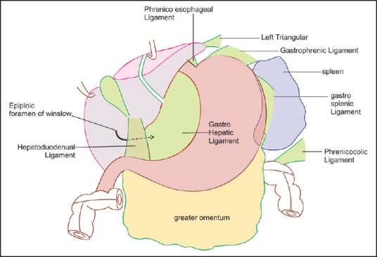
The lesser omentum is a smaller, two-layered peritoneal fold that extends from the lesser curvature of the stomach and the proximal duodenum to the visceral surface of the liver (specifically the porta hepatis and the fissure for the ligamentum venosum). It is divided into two parts:
- Hepatogastric ligament: The larger, medial portion that extends from the lesser curvature of the stomach to the liver.
- Hepatoduodenal ligament: The smaller, lateral portion that extends from the proximal duodenum to the liver.
The free edge of the lesser omentum (the right free margin of the hepatoduodenal ligament) contains the portal triad: the hepatic artery proper, the common bile duct, and the portal vein. These three structures run together within the hepatoduodenal ligament, with the bile duct anterior and to the right, the hepatic artery anterior and to the left, and the portal vein posterior to both. This free edge forms the anterior boundary of the foramen of Winslow.
3.4 Peritoneal Ligaments
Peritoneal ligaments are double-layered folds of peritoneum that connect organs to the abdominal wall or to other organs. They are essentially specialized mesenteries or omental extensions. Peritoneal ligaments serve to hold organs in position and provide conduits for neurovascular structures. They are classified based on the organs they connect:
3.4.1 Hepatic Ligaments
The liver is attached to the diaphragm and anterior abdominal wall by several ligaments:
- Falciform ligament: A sickle-shaped peritoneal fold that extends from the anterior abdominal wall (umbilicus and linea alba) to the liver, dividing the liver into right and left anatomical lobes. Its free inferior edge contains the ligamentum teres (round ligament of the liver), which is the remnant of the fetal umbilical vein.
- Coronary ligament: A peritoneal reflection that attaches the superior surface of the liver to the diaphragm. It encloses the bare area of the liver, which is not covered by peritoneum and is in direct contact with the diaphragm.
- Left and right triangular ligaments: Extensions of the coronary ligament at the lateral margins of the liver.
- Ligamentum venosum: The remnant of the fetal ductus venosus, located within a fissure on the visceral surface of the liver.
3.4.2 Gastric Ligaments
The stomach is connected to adjacent structures by several ligaments:
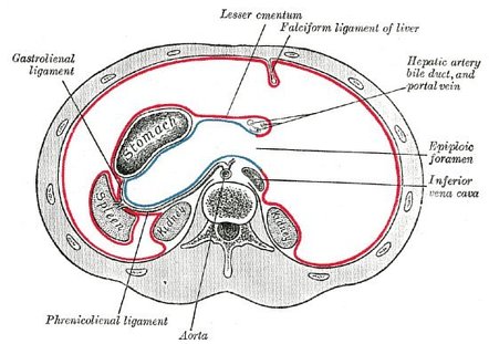
- Gastrophrenic ligament: Extends from the upper part of the greater curvature of the stomach to the diaphragm. It forms part of the left border of the lesser sac.
- Gastrosplenic (gastrolienal) ligament: Extends from the greater curvature of the stomach to the hilum of the spleen. It contains the short gastric vessels and the left gastro-omental vessels.
- Gastrocolic ligament: The superior part of the greater omentum that extends from the greater curvature of the stomach to the transverse colon.
3.4.3 Splenic Ligaments
The spleen is attached by several ligaments:
- Splenorenal (lienorenal) ligament: Extends from the hilum of the spleen to the anterior surface of the left kidney. It contains the splenic vessels and the tail of the pancreas.
- Gastrosplenic ligament: As described above, connecting stomach to spleen.
- Phrenicocolic ligament: Extends from the splenic flexure of the colon to the diaphragm. It supports the spleen and forms the left margin of the lesser sac.
CHAPTER 5: SUBDIVISIONS, SPACES, AND GUTTERS
5.1 Principal Compartments
- The Greater Sac: The larger anterior compartment of the peritoneal cavity, extending from the diaphragm superiorly to the pelvic cavity inferiorly. It contains most of the intraperitoneal organs and is the primary site of fluid accumulation in pathological conditions such as ascites.
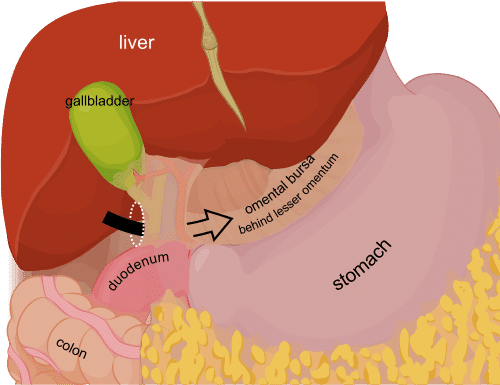
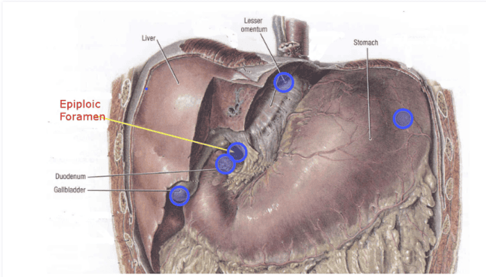
- The Lesser Sac (Omental Bursa): A smaller, posterior compartment situated behind the stomach and lesser omentum. It communicates with the greater sac through the epiploic foramen (foramen of Winslow).
The boundaries of the epiploic foramen are as follows:
- Superior: Caudate lobe of liver
- Anterior: Hepatoduodenal ligament (with portal triad)
- Inferior: First part of duodenum
- Posterior: Inferior Vena Cava (IVC)
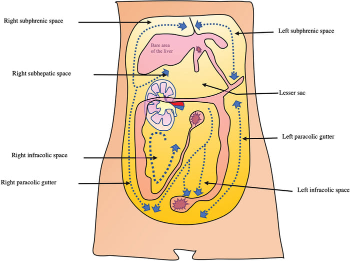
5.2 Subhepatic Space (Hepatorenal Recess / Morison's Pouch)
The right subhepatic space, also known as the hepatorenal recess or Morison's pouch, is a gravity-dependent space located between the right lobe of the liver and the right kidney. It is bounded superiorly by the inferior surface of the liver, posteriorly by the right kidney and suprarenal gland, and inferiorly by the right colic flexure and the transverse mesocolon. The hepatorenal recess is the most dependent part of the peritoneal cavity when the patient is in the supine position, making it the most common site for fluid accumulation in a supine patient.
The hepatorenal recess is named after the British surgeon James Rutherford Morison, who described it in 1894. It is a clinically important space because it is the first site to accumulate free fluid in the peritoneal cavity when the patient is lying supine. On ultrasound and CT imaging, even small amounts of free fluid (as little as 10-15 mL) can be detected in Morison's pouch, making it a sensitive indicator of intraperitoneal bleeding or fluid accumulation.
Subphrenic Abscess
Subphrenic abscess is a collection of pus in the subphrenic space, typically occurring as a complication of upper abdominal surgery (particularly after cholecystectomy, gastric surgery, or splenectomy) or as a result of perforated peptic ulcer or appendicitis. The right subphrenic space is more commonly affected than the left. Patients present with fever, upper abdominal pain, shoulder pain (referred via the phrenic nerve), and hiccups. Diagnosis is confirmed by CT scan or ultrasound. Treatment involves percutaneous drainage or surgical drainage.
The anatomical boundaries of the subphrenic spaces (particularly the coronary ligament and falciform ligament) can limit the spread of infection, causing it to remain localized.
5.3 Paracolic Gutters
The paracolic gutters are longitudinal depressions in the peritoneum located lateral to the ascending and descending colon. They are important channels that direct the flow of inflammatory exudates, pus, and free fluid within the peritoneal cavity. The paracolic gutters connect the upper and lower peritoneal spaces and allow the spread of infection from one region to another.
5.3.1 Right Paracolic Gutter
The right paracolic gutter is located lateral to the ascending colon and medial to the right abdominal wall. It is wider and more patent than the left paracolic gutter because the ascending colon is more laterally placed and has a shorter mesentery (or is retroperitoneal). The right paracolic gutter is continuous superiorly with the right subhepatic space (hepatorenal recess) and the right subphrenic space, and inferiorly with the right iliac fossa and the pelvic cavity. It provides a pathway for the spread of infection from the lower abdomen (e.g., appendicitis) to the upper abdomen and subphrenic spaces.
5.3.2 Left Paracolic Gutter
The left paracolic gutter is located lateral to the descending colon and medial to the left abdominal wall. It is narrower and less patent than the right paracolic gutter because the descending colon is more medially placed and is retroperitoneal (fixed to the posterior wall). The left paracolic gutter is partially interrupted superiorly by the phrenicocolic ligament, which limits the spread of infection from the left lower abdomen to the left subphrenic space. However, it is still a potential pathway for the spread of infection from the lower abdomen to the upper abdomen.
The Phrenicocolic Ligament as a Barrier
The phrenicocolic ligament is a peritoneal fold that extends from the splenic flexure of the colon to the diaphragm. It acts as a partial barrier to the spread of infection from the left lower abdomen to the left subphrenic space. This explains why left subphrenic abscesses are less common than right subphrenic abscesses, even though the left colon is a common source of infection. The phrenicocolic ligament is an important anatomical landmark that demonstrates how peritoneal reflections can influence the clinical behavior of peritoneal disease.
5.4 Infracolic Compartments
The infracolic compartment is the portion of the peritoneal cavity located below the transverse mesocolon. It is divided into right and left infracolic spaces by the root of the mesentery of the small intestine, which extends from the duodenojejunal flexure to the ileocecal junction.
5.4.1 Right Infracolic Space
The right infracolic space is located to the right of the root of the mesentery. It is bounded by the ascending colon laterally, the root of the mesentery medially, and the transverse mesocolon superiorly. The right infracolic space communicates with the right paracolic gutter laterally and the pelvic cavity inferiorly. It is a common site for fluid accumulation and abscess formation in the right lower abdomen.
5.4.2 Left Infracolic Space
The left infracolic space is located to the left of the root of the mesentery. It is bounded by the descending colon laterally, the root of the mesentery medially, and the transverse mesocolon superiorly. The left infracolic space is narrower than the right infracolic space because the descending colon is more medially placed. It communicates with the left paracolic gutter laterally and the pelvic cavity inferiorly.
5.5 Other Peritoneal Recesses
In addition to the major spaces described above, the peritoneal cavity contains numerous smaller recesses that are potential sites of fluid accumulation and internal herniation:
- Hepatorenal recess (Morison's pouch): As described above, the most dependent space in the supine patient.
- Retrocecal recess: Located posterior to the cecum; a common site for retrocecal appendicitis and retrocecal abscess.
- Intersigmoid recess: Located between the two layers of the sigmoid mesocolon; a potential site for internal herniation.
- Superior and inferior duodenal recesses: Small peritoneal folds near the duodenojejunal flexure; potential sites for internal herniation.
- Paraduodenal recesses: Located near the duodenum; potential sites for internal herniation (paraduodenal hernias).
- Retroduodenal recess: Located posterior to the duodenum; potential site for fluid accumulation.
CHAPTER 6: NEUROVASCULAR SUPPLY AND LYMPHATICS
6.1 Overview
The neurovascular supply of the peritoneum is of great clinical importance because it determines the patterns of pain perception, the response to injury, and the potential for collateral circulation. The parietal and visceral peritoneum have fundamentally different embryological origins, innervations, and pain characteristics, which has profound implications for the clinical presentation of peritoneal disease.
6.2 Parietal Peritoneum Supply
6.2.1 Vasculature
The parietal peritoneum is supplied by the somatic vessels of the abdominal wall, including the musculophrenic, epigastric, lumbar, and intercostal vessels. These vessels supply the parietal peritoneum as part of their course through the abdominal wall. The arterial supply is segmental, corresponding to the segmental innervation of the abdominal wall. The venous drainage follows the corresponding arteries and drains into the systemic venous circulation (inferior vena cava, azygos system, and portal system via the paraumbilical veins).
6.2.2 Innervation
The parietal peritoneum is innervated by the somatic nerves that supply the abdominal wall: the lower intercostal nerves (T7-T11), the subcostal nerve (T12), and the iliohypogastric and ilioinguinal nerves (L1). These are the same nerves that supply the skin and muscles of the abdominal wall. The parietal peritoneum is therefore highly sensitive to the same stimuli as the skin: localized mechanical trauma, pressure, hot and cold stimuli, and laceration.
When the parietal peritoneum is irritated or inflamed (e.g., in peritonitis), the pain is sharp, well-localized, and somatic in character. The patient can accurately point to the site of pain, and the pain is aggravated by movement, coughing, or pressure. This is because the parietal peritoneum is innervated by somatic afferent fibers that travel in the same nerves as the skin and muscles of the abdominal wall, and the brain can accurately localize the source of the pain.
Rebound Tenderness and Guarding
The sharp, localized pain of parietal peritoneal irritation is the basis for the clinical signs of rebound tenderness and guarding.
- Rebound tenderness (Blumberg's sign) is elicited by pressing deeply on the abdomen and then suddenly releasing the pressure; the sudden stretching of the inflamed parietal peritoneum causes a sharp, stabbing pain.
- Guarding is the involuntary contraction of the abdominal muscles in response to peritoneal irritation, which is a protective reflex to minimize movement of the inflamed peritoneum.
These signs are characteristic of peritonitis and indicate that the parietal peritoneum is involved in the inflammatory process.
6.3 Visceral Peritoneum Supply
6.3.1 Vasculature
The visceral peritoneum is supplied directly by the vessels of the underlying organs. The arterial supply is derived from the branches of the celiac trunk (for the foregut organs), the superior mesenteric artery (for the midgut organs), and the inferior mesenteric artery (for the hindgut organs). The venous drainage follows the corresponding arteries and drains into the portal venous system (for the gastrointestinal organs) or the systemic venous system (for the non-gastrointestinal organs).
6.3.2 Innervation
The visceral peritoneum is innervated by the visceral afferent nerves that travel with the autonomic nerves (sympathetic and parasympathetic) to the abdominal organs. These are the same nerves that innervate the underlying organs. The visceral peritoneum is therefore insensitive to localized cutting, burning, or crushing stimuli that would cause severe pain if applied to the skin or parietal peritoneum.
However, the visceral peritoneum is sensitive to stretching, distension, and chemical irritation (e.g., from gastric acid, bile, or pancreatic enzymes). When the visceral peritoneum is stimulated by these stimuli, the pain is dull, poorly localized, and referred to the dermatome corresponding to the embryological origin of the organ. For example:
- Stomach (foregut, T7-T9): Pain is referred to the epigastrium
- Small intestine (midgut, T9-T11): Pain is referred to the periumbilical region
- Colon (hindgut, T11-L1): Pain is referred to the suprapubic region
This pattern of referred visceral pain is the basis for the characteristic progression of pain in appendicitis: initially, the inflamed appendix causes visceral peritoneal irritation, producing a dull, poorly localized periumbilical pain (T10 dermatome). As the inflammation progresses to involve the parietal peritoneum of the right iliac fossa, the pain becomes sharp, well-localized, and shifts to McBurney's point in the right lower quadrant.
The Pain Progression in Appendicitis
The classic progression of pain in appendicitis illustrates the difference between visceral and parietal peritoneal pain:
- Stage 1 (Visceral pain): The inflamed appendix irritates the visceral peritoneum. Pain is dull, poorly localized, and referred to the periumbilical region (T10 dermatome, corresponding to the embryological midgut origin of the appendix).
- Stage 2 (Parietal pain): The inflammation spreads to the parietal peritoneum of the right iliac fossa. Pain becomes sharp, well-localized, and constant at McBurney's point. The patient can accurately point to the site of pain.
- Stage 3 (Generalized peritonitis): If the appendix perforates, the inflammation spreads throughout the peritoneal cavity, causing generalized peritonitis with diffuse abdominal pain, rebound tenderness, and guarding.
6.4 Lymphatic Drainage
The lymphatic drainage of the peritoneum follows two main pathways:
- Parietal peritoneal lymphatics: Drain along the somatic lymphatic vessels of the abdominal wall to the lumbar, external iliac, and superficial inguinal lymph nodes, depending on the region of the abdominal wall.
- Visceral peritoneal lymphatics: Drain along the lymphatic vessels of the underlying organs to the celiac, superior mesenteric, and inferior mesenteric lymph nodes, and then to the cisterna chyli and thoracic duct.
The peritoneum also contains numerous milky spots (small collections of macrophages and lymphocytes) that are particularly abundant in the greater omentum. These milky spots play an important role in immune surveillance and can trap bacteria, foreign particles, and tumor cells. They are also involved in the formation of peritoneal adhesions.
CHAPTER 7: CLINICAL AND APPLIED ANATOMY
7.1 Peritonitis
Peritonitis is the inflammation of the peritoneum, most commonly resulting from bacterial contamination (e.g., from a perforated peptic ulcer, ruptured appendix, or perforated diverticulum) or chemical irritation (e.g., from gastric acid, bile, pancreatic enzymes, or blood). The pathophysiology involves the interaction between the inflammatory stimulus and the mesothelium, which responds by producing mediators, increasing vascular permeability, and recruiting neutrophils.
The clinical presentation depends on the type of peritonitis and the extent of involvement:
- Primary peritonitis (spontaneous bacterial peritonitis): Occurs without an identifiable source of contamination, typically in patients with ascites (e.g., from liver cirrhosis). The bacteria presumably translocate from the intestine through the intestinal wall into the peritoneal cavity.
- Secondary peritonitis: Results from contamination of the peritoneal cavity from an intra-abdominal source, such as a perforated viscus, gangrenous bowel, or infected pancreatic necrosis. This is the most common type of peritonitis.
- Tertiary peritonitis: A persistent or recurrent peritonitis that occurs after treatment of secondary peritonitis, often due to fungal or resistant bacterial infection in immunocompromised patients.
The clinical signs include abdominal pain (initially visceral, then parietal), rebound tenderness, guarding, abdominal distension, paralytic ileus, fever, tachycardia, and signs of sepsis. Absent bowel sounds occur in severe cases.
7.2 Ascites and Paracentesis
Ascites is the abnormal accumulation of serous fluid within the peritoneal cavity. It is most commonly caused by liver cirrhosis (portal hypertension), but can also result from malignancy (peritoneal carcinomatosis), heart failure, nephrotic syndrome, tuberculosis, and pancreatic disease. The pathophysiology involves an imbalance between fluid production (increased hydrostatic pressure) and fluid absorption (decreased oncotic pressure, impaired lymphatic drainage).
The peritoneal cavity can accommodate large volumes of fluid (up to 10-20 liters in severe cases). The fluid accumulates in the most dependent parts, which vary with the patient's position:
- Supine position: Fluid accumulates in the hepatorenal recess (Morison's pouch), the pelvis, and the paracolic gutters.
- Upright position: Fluid accumulates in the pelvis (rectovesical or rectovaginal pouch) and the lower abdomen.
- Lateral decubitus position: Fluid accumulates in the dependent paracolic gutter and flank.
7.2.1 Paracentesis
Paracentesis is the needle drainage of ascitic fluid. It is performed for diagnostic purposes (to determine the cause) or therapeutic purposes (to relieve respiratory compromise). The anatomical approach utilizes specific landmarks to avoid injury to the inferior epigastric vessels. Standard sites include:
- Midline infraumbilical approach: 2 cm below the umbilicus in the midline, avoiding the linea alba vessels. This is the preferred site for small-volume paracentesis.
- Left lower quadrant approach: In the LLQ, 2-3 cm above and 2-3 cm medial to the ASIS. This avoids the cecum and appendix on the right side and the inferior epigastric vessels.
Avoiding the Inferior Epigastric Vessels
The inferior epigastric vessels arise from the external iliac vessels and ascend posterior to the rectus abdominis muscle within the rectus sheath. They are located along the lateral border of the rectus abdominis, approximately midway between the midline and the ASIS. Paracentesis should be performed lateral to the rectus abdominis (in the left or right lower quadrant) or in the midline (where the linea alba is relatively avascular) to avoid these vessels. Injury can cause significant hemorrhage, particularly in patients with coagulopathy or portal hypertension. Ultrasound guidance is increasingly used.
7.3 Peritoneal Dialysis
Peritoneal dialysis is a form of renal replacement therapy that utilizes the vast surface area and semi-permeable properties of the peritoneum for waste filtration. The peritoneum has a surface area of approximately 1-2 square meters (comparable to the surface area of the glomerular capillaries in both kidneys), making it an effective dialysis membrane.
The catheter is inserted typically in the midline below the umbilicus. Dialysis fluid (dialysate) is infused and remains for a dwell period (typically 4-6 hours). Waste products (urea, creatinine, potassium) and excess fluid diffuse from the blood vessels across the three layers: the mesothelium, the interstitium, and the capillary endothelium.
Complications: Peritonitis, catheter-related complications (malposition, leakage), hernia (due to increased intra-abdominal pressure), and encapsulating peritoneal sclerosis (rare thickening and fibrosis).
7.4 Internal Hernias
Internal hernias are herniations of bowel loops through congenital or acquired defects in the peritoneum or mesentery. They are much less common than external hernias (inguinal, femoral, umbilical) but can cause serious complications including bowel obstruction, strangulation, and ischemia. Internal hernias account for approximately 1-5% of all cases of small bowel obstruction.
Potential sites of internal herniation include:
- Paraduodenal hernias: Most common type; occurring through congenital fossae near the duodenum (e.g., fossa of Landzert on the left, fossa of Waldeyer on the right).
- Foramen of Winslow hernia: Herniation through the foramen into the lesser sac. Rare but can cause bowel strangulation.
- Intersigmoid hernia: Through the intersigmoid recess between sigmoid mesocolon layers.
- Transmesenteric hernia: Through a defect in the mesentery of the small intestine or colon.
- Retrocecal hernia: Into the retrocecal recess, posterior to the cecum.
7.5 Peritoneal Adhesions
Peritoneal adhesions are fibrous bands that form between damaged peritoneal surfaces. They are a common complication of abdominal surgery, occurring in up to 90% of patients after laparotomy. Adhesions represent the body's attempt to repair injury by depositing fibrin, which is then organized into fibrous tissue by fibroblasts.
Steps of pathophysiology: Peritoneal injury → Fibrin deposition → Fibrinolysis (dissolved by plasmin within 72 hours) vs. Organization (if fibrinolysis is impaired by ischemia or inflammation) → Adhesion maturation (developing blood vessels).
Clinical consequences: Small bowel obstruction (accounting for 60-70% of cases), chronic abdominal pain, female infertility (impairing ovum capture), and difficult reoperation.
CHAPTER 8: SUMMARY AND REVIEW
8.1 Key Anatomical Structures
| Structure | Description | Clinical Relevance |
|---|---|---|
| Parietal peritoneum | Lines wall; somatic mesoderm origin | Sensitive to pain/temp; sharp localized pain |
| Visceral peritoneum | Covers organs; splanchnic mesoderm | Insensitive to cutting; sensitive to stretch; dull referred pain |
| Peritoneal cavity | Space between layers | Closed in males; communicates with exterior in females; site of ascites |
| Greater omentum | 4-layered fold from greater curvature | "Policeman of abdomen"; wall-off infection |
| Lesser omentum | 2-layered fold; curvature to liver | Contains portal triad; edge at foramen of Winslow |
| Foramen of Winslow | Opening between greater/lesser sacs | Site of Pringle maneuver; potential internal hernia site |
| Lesser sac | Pocket posterior to stomach | Site of lesser sac abscess; internal herniation |
| Hepatorenal recess | Morison's pouch; liver to R kidney | First site of fluid accumulation in supine patient |
| Paracolic gutters | Depressions lateral to colon | Channels for spread of infection and fluid |
| Mesentery proper | Suspends jejunum and ileum | Contains superior mesenteric vessels; site of volvulus |
| Transverse mesocolon | Suspends transverse colon | Divides cavity into supra and infracolic compartments |
8.2 Organ Classification Summary
| Category | Organs | Embryological Origin |
|---|---|---|
| Intraperitoneal | Stomach, spleen, liver (mostly), jejunum, ileum, transverse colon, sigmoid colon, appendix, cecum, 1st part of duodenum | Developed with mesentery; suspended into peritoneal cavity |
| Primary retroperitoneal | Kidneys, ureters, suprarenal glands, aorta, IVC, esophagus (abdominal), rectum (lower 2/3) | Developed outside peritoneum; never had mesentery |
| Secondary retroperitoneal | Pancreas (except tail), duodenum (parts 2-4), ascending colon, descending colon | Developed with mesentery; lost mesentery during development |
8.3 Clinical Applications Summary
| Clinical Scenario | Anatomical Consideration | Key Structure |
|---|---|---|
| Peritonitis | Inflammation of parietal peritoneum causes sharp localized pain | Parietal vs. visceral peritoneal innervation |
| Ascites | Fluid accumulates in dependent spaces (Morison's pouch, pelvis) | Peritoneal cavity; gravity-dependent spaces |
| Paracentesis | Avoid inferior epigastric vessels; use LLQ or midline | Inferior epigastric vessels; rectus abdominis |
| Peritoneal dialysis | Utilizes peritoneal surface area (~1-2 m2) for waste filtration | Peritoneal membrane; mesothelium |
| Subphrenic abscess | Fluid accumulates in subphrenic spaces after surgery | Subphrenic spaces; coronary ligament |
| Internal hernia | Bowel herniates through peritoneal fossae or foramen of Winslow | Paraduodenal fossae; foramen of Winslow |
| Peritoneal adhesions | Fibrous bands form after peritoneal injury | Peritoneal healing; fibrin deposition |
| Pringle maneuver | Clamp hepatoduodenal ligament to control hepatic bleeding | Foramen of Winslow; portal triad |
