The Human Skeleton
A comprehensive anatomical guide to the structural framework of the human body, detailing the Axial and Appendicular divisions, bone morphology, and the joints that integrate the two systems.
1. Introduction to the Human Skeleton
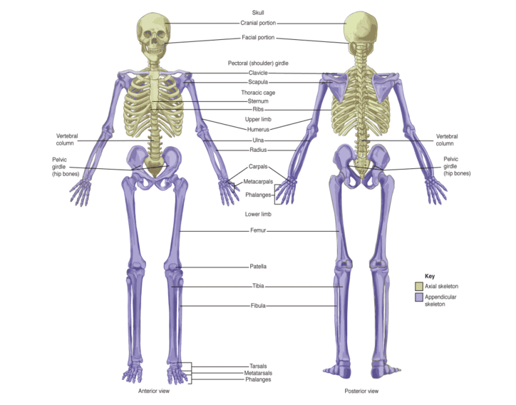
The adult human skeleton consists of 206 bones. This system is organized into two major divisions based on structural and functional roles: the Axial Skeleton and the Appendicular Skeleton.
Functional Overview
- Axial Skeleton (80 bones): Forms the longitudinal axis of the body. Its primary functions are to protect vital organs (brain, heart, lungs), support the head and trunk, and provide attachment points for muscles.
- Appendicular Skeleton (126 bones): Comprises the bones of the upper and lower limbs and the girdles that attach them to the axial skeleton. Its primary functions are locomotion and the manipulation of objects.
2. The Axial Skeleton
The axial skeleton includes the bones of the skull, vertebral column, and the thoracic cage. It forms the central supporting structure of the body.
2.1 Skull Bones
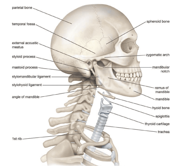
The skull consists of 22 bones divided into two groups: cranial bones (8) and facial bones (14).
Cranial Bones (8 total):
- Frontal bone (1): Forms the forehead and the superior part of the eye orbits.
- Parietal bones (2): Form the upper sides and roof of the skull.
- Temporal bones (2): Located on the lower sides; contain the mastoid process and styloid process.
- Occipital bone (1): Forms the posterior and base of the skull; contains the foramen magnum.
- Sphenoid bone (1): A "keystone" bone that joins almost every other skull bone; contains the sella turcica (pituitary gland seat).
- Ethmoid bone (1): Forms part of the nose, medial eye orbits, and the skull base.
Facial Bones (14 total):
These include the Maxillae (2), Zygomatic (2), Mandible (1), Nasal (2), Palatine (2), Nasal conchae (2), Lacrimal (2), and Vomer (1).
2.2 Vertebral Column
The vertebral column (spine) includes 26 bones in adults (24 vertebrae plus the sacrum and coccyx). It is grouped into five distinct regions:
| Region | Vertebral Level | Count | Function/Notes |
|---|---|---|---|
| Cervical | C1–C7 | 7 | Bones of the neck; supports the head. |
| Thoracic | T1–T12 | 12 | Articulate with ribs to form the posterior rib cage. |
| Lumbar | L1–L5 | 5 | Support most body weight; thick kidney-shaped bodies. |
| Sacrum | Fusion of 5 | 1 | Triangular bone; forms the posterior pelvic wall. |
| Coccyx | Fusion of 4 | 1 | Tailbone; attaches to pelvic floor muscles. |
2.3 Thoracic Cage (Rib Cage)
Protects the heart and lungs. It consists of:
- Sternum (breastbone): Divided into the manubrium (upper), body (middle), and xiphoid process (lower).
- Ribs: 12 pairs (24 total) connected posteriorly to thoracic vertebrae:
- True ribs (1–7): Attach directly to the sternum via costal cartilage.
- False ribs (8–10): Attach indirectly to the sternum.
- Floating ribs (11–12): No anterior attachment.
2.4 Auditory Ossicles and Hyoid Bone
- Auditory ossicles (6 total): The smallest bones in the body (malleus, incus, and stapes) located in each middle ear; they transfer sound vibrations.
- Hyoid bone (1): A horseshoe-shaped bone in the neck; supports tongue movement, swallowing, and speech.
3. The Appendicular Skeleton
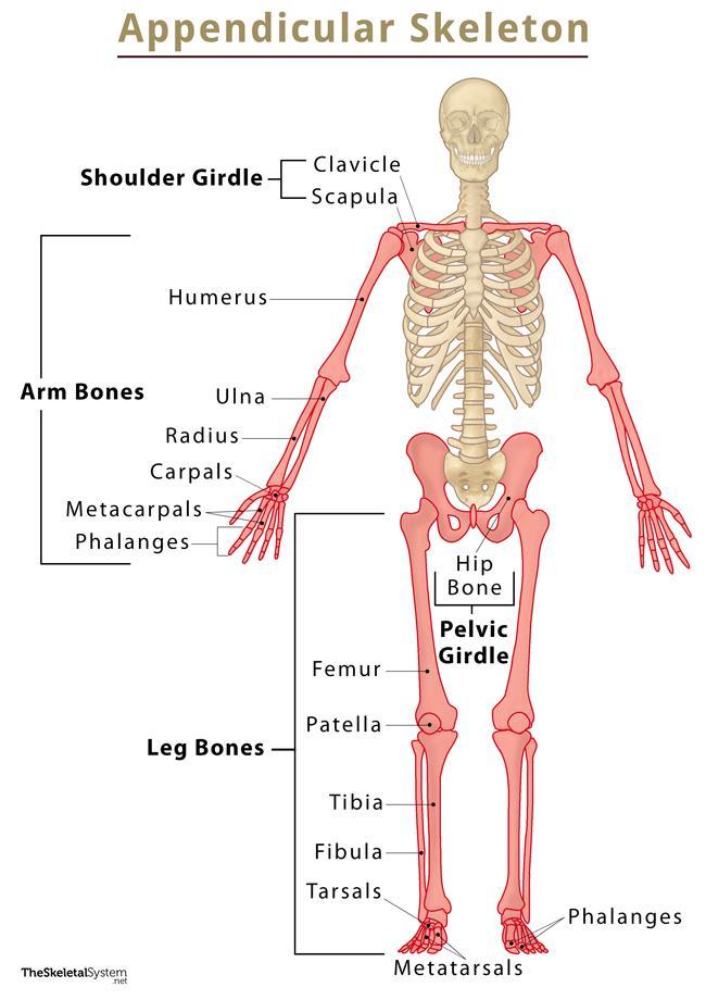
Comprises 126 bones, representing approximately 61% of all bones in the body. It is specialized for mobility and locomotion.
3.1 Pectoral Girdle and Upper Limb
Pectoral (Shoulder) Girdle (4 bones):
- Clavicle (collarbone): S-shaped bone; acts as a strut to maintain shoulder position.
- Scapula (shoulder blade): Triangular bone on the upper back; provides muscle attachment.
Upper Limb (60 bones total):
- Humerus (1): Longest bone of the upper limb; articulates with scapula and radius/ulna.
- Radius (1): Lateral forearm bone (thumb side).
- Ulna (1): Medial forearm bone; forms the distal radioulnar joint.
- Carpals (8): Wrist bones in two rows. Proximal: scaphoid, lunate, triquetrum, pisiform. Distal: trapezium, trapezoid, capitate, hamate.
- Metacarpals (5): Palm bones.
- Phalanges (14): Finger bones (2 in thumb, 3 in others).
3.2 Pelvic Girdle and Lower Limb
Pelvic Girdle (2 hip bones): Each hip bone is formed by the fusion of three bones: Ilium (superior), Ischium (inferoposterior), and Pubis (anterior). They unite at the pubic symphysis.
Lower Limb (60 bones total):
- Femur (1): Thigh bone; the longest and strongest bone in the body.
- Patella (1): Kneecap; the largest sesamoid bone.
- Tibia (1): Larger medial leg bone; bears body weight.
- Fibula (1): Thinner lateral leg bone; site for muscle attachment.
- Tarsals (7): Ankle bones: talus, calcaneus (heel), cuboid, navicular, and 3 cuneiforms.
- Metatarsals (5): Forefoot bones.
- Phalanges (14): Toe bones.
3.3 Sesamoid Bones
Sesamoid bones are small, round bones embedded within tendons or joint capsules. They protect tendons from excessive stress and wear. The patella is the largest, but others are found in the hands and feet.
4. Comparison: Axial vs. Appendicular
| Feature | Axial Skeleton | Appendicular Skeleton |
|---|---|---|
| Number of Bones | 80 bones | 126 bones |
| Percentage of Total | ~39% | ~61% |
| Main Components | Skull, vertebral column, ribs, sternum, hyoid, ossicles | Girdles (pectoral/pelvic), upper and lower limbs |
| Primary Function | Protection of vital organs; support of head/trunk | Locomotion; manipulation of objects |
| Mobility | Relatively immobile; protective structure | Highly mobile; designed for movement |
| Weight Bearing | Supports trunk weight via vertebral column | Bears and transmits body weight (lower limbs) |
5. Joints Connecting the Two Skeletons
The appendicular skeleton connects to the axial skeleton at two primary bilateral points:
- Sternoclavicular Joint: Where the sternum (axial) articulates with the clavicle (appendicular). This is a synovial joint allowing movement of the shoulder girdle.
- Sacroiliac Joint: Where the sacrum (axial) articulates with the ilium (appendicular). This is both a synovial joint and a syndesmosis, critical for transferring load to the lower extremities.
- Thoracoscapular (scapulothoracic) articulation: An indirect connection between the scapula and ribs 2–7, allowing smooth shoulder movement (not a true synovial joint).
