Posterior Abdominal Wall
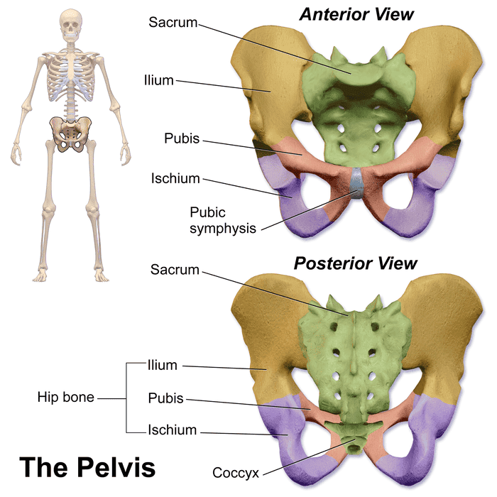
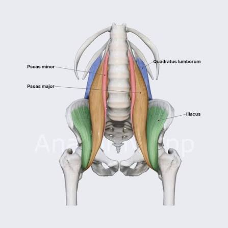
The Posterior Abdominal Wall A Comprehensive Anatomical Guide for Undergraduate Medical Students. Covering: Skeletal Framework, Musculature, Fasciae, Vascular Supply, Innervation, Lymphatic Drainage, and Clinical Anatomy. CHAPTER 1: Introduction and General Organization 1.1 Definition and Boundaries The posterior abdominal wall constitutes the posterior boundary of the abdominal cavity and represents a complex structural composite of bone, muscle, fascia, and neurovascular elements. It extends from the diaphragm superiorly to the pelvic brim inferiorly, and from the quadratus lumborum muscles laterally to the vertebral column medially. The wall serves as the primary structural support for abdominal viscera, the conduit for major vascular and neural pathways, and the anatomical foundation for the retroperitoneal space. 1.2 Topographical Regions The posterior abdominal wall may be subdivided into three principal topographical regions: Suprarenal region (T11–T12): Contains the suprarenal glands, celiac trunk, and superior mesenteric artery origin, bounded superiorly by the diaphragm and medially by the crura. Renal region (L1–L3): Contains the kidneys, ureters, gonadal vessels, and the origins of the renal arteries; the psoas major muscles form the lateral boundaries. Infrarenal region (L3–L5): Contains the inferior mesenteric artery, common iliac vessels, and the bifurcation of the abdominal aorta at the L4 vertebral level; the sacral promontory marks the inferior limit. CLINICAL CORRELATION Retroperitoneal Haemorrhage The posterior abdominal wall contains the major vascular trunks of the abdomen. Traumatic injury to the abdominal aorta, inferior vena cava, or lumbar vessels may result in massive retroperitoneal haemorrhage. Blood accumulates within the retroperitoneal space and may track along fascial planes, producing Grey Turner’s sign (flank ecchymosis) or Cullen’s sign (periumbilical ecchymosis) in severe pancreatitis or retroperitoneal bleeding. The psoas sheath may contain haematoma, causing femoral nerve compression and presenting with weakness of knee extension and sensory loss on the anterior thigh. CHAPTER 2: Skeletal Framework and Bony Landmarks 2.1 Vertebral Column The vertebral column provides the central osseous support of the posterior abdominal wall. Five lumbar vertebrae (L1–L5) constitute the principal bony elements, each characterised by large, kidney-shaped bodies adapted to bear substantial weight, and massive transverse and spinous processes that serve as attachment sites for the deep muscles of the back and abdominal wall. 2.1.1 Lumbar Vertebrae: Detailed Morphology Each lumbar vertebra exhibits the following distinctive features: Body: Large, kidney-shaped, and broader transversely than anteroposteriorly. The superior surface is concave with prominent epiphyseal rims; the inferior surface is convex. The L1 body measures approximately 30 mm in anteroposterior diameter and 45 mm in transverse diameter, increasing progressively to L5. Pedicles: Short, thick, and directed posterolaterally from the superolateral aspect of the vertebral body. The inferior vertebral notch is deeper than the superior notch, contributing to the intervertebral foramen. Laminae: Broad, thick plates that unite posteriorly to form the spinous process. The laminae are broader than those of thoracic vertebrae, providing extensive attachment for the erector spinae muscles. Transverse processes: Long, slender, and project laterally and slightly posteriorly. Each transverse process bears an accessory process posteriorly and a mammillary process on the posterior surface of the base, serving as attachment for the intertransversarii and multifidus muscles, respectively. Spinous process: Thick, broad, and quadrilateral, projecting almost horizontally posteriorly. It provides attachment for the supraspinous and interspinous ligaments and the thoracolumbar fascia. Superior articular processes: Bear concave articular facets that face medially (or posteromedially), articulating with the inferior articular facets of the vertebra above. Inferior articular processes: Bear convex articular facets that face laterally (or anterolaterally), articulating with the superior articular facets of the vertebra below. 2.1.2 Vertebral Levels of Clinical Significance Structure Vertebral Level Clinical Relevance Caval opening (diaphragm) T8 IVC passage; right phrenic nerve branches Oesophageal hiatus (diaphragm) T10 Oesophagus and vagus nerve passage Aortic hiatus (diaphragm) T12 Aorta, thoracic duct, azygos vein passage Celiac trunk origin T12–L1 Foregut arterial supply; landmark for CT imaging Superior mesenteric artery L1 Midgut arterial supply; nutcracker syndrome site Renal arteries L1–L2 (intervertebral disc) Kidney vascularisation; endovascular access Gonadal arteries L2 Testicular/ovarian supply; high ligation site Inferior mesenteric artery L3 Hindgut arterial supply Aortic bifurcation L4 Common iliac artery origin; umbilicus level Conus medullaris termination L1–L2 Spinal cord end; cauda equina begins below 2.2 Ribs and Costal Elements The eleventh and twelfth ribs are floating ribs that articulate only with their corresponding vertebral bodies and do not reach the sternum. The twelfth rib is of particular anatomical significance: It articulates with the body of T12 via a single costovertebral joint (no costotransverse joint). It is shorter, more horizontal, and more deeply situated than the eleventh rib. It is crossed anteriorly by the subcostal nerve (T12) and vessels, and the iliohypogastric nerve (L1). The quadratus lumborum muscle inserts onto its inferior border, stabilising it during inspiration. The kidney lies posterior to the 11th and 12th ribs on the left, and the 12th rib on the right. 2.3 Pelvic Bones The iliac bones contribute to the inferior aspect of the posterior abdominal wall. The iliac fossa, a large concavity on the medial surface of the ilium, provides origin for the iliacus muscle. The iliac crest extends from the anterior superior iliac spine to the posterior superior iliac spine, serving as an important surface anatomy landmark. The sacrum articulates with L5 via the lumbosacral joint and with the ilia at the sacroiliac joints, forming the posterior pelvic wall. 2.4 Surface Anatomy and Palpable Landmarks The following surface anatomy landmarks are essential for clinical examination and procedural access: Tuffier’s line: A horizontal line connecting the highest points of both iliac crests (intercristal line), which reliably crosses the L4–L5 intervertebral space. This is the standard landmark for lumbar puncture and spinal anaesthesia. McBurney’s point: Located one-third of the distance from the anterior superior iliac spine to the umbilicus (typically at the junction of the lateral and middle thirds), corresponding to the base of the appendix. While anterior, its relationship to the posterior abdominal wall muscles is relevant for appendicitis diagnosis. Costovertebral angle: The angle formed by the 12th rib and the vertebral column, located posteriorly. Percussion tenderness at this angle (Murphy’s percussion sign) indicates renal inflammation or infection. Posterior