Key Terms in Skeletal System
Anatomy notes covering Bone Morphology, Articulation, and Ossification.
1. BONE (Os / Osteon)
Definition: Bone is a specialized, mineralized connective tissue that forms the rigid framework of the body — the skeleton. It is a living, dynamic tissue capable of growth, repair, and remodeling throughout life. Bone is composed of an organic matrix (primarily Type I collagen) and an inorganic mineral phase (hydroxyapatite crystals, mainly calcium phosphate).
Bone Structure Overview
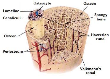
Key Structural Components
| Component | Description | Clinical Significance |
|---|---|---|
| Compact (Cortical) Bone | Dense outer layer; composed of osteons (Haversian systems) with concentric lamellae around central Haversian canals containing blood vessels and nerves. | Accounts for ~80% of skeletal mass. Site of resistance to compressive forces. Radiographically radiopaque (white). |
| Cancellous (Spongy) Bone | Porous inner layer with trabeculae arranged along lines of mechanical stress. Contains red bone marrow in adults. | Site of hematopoiesis. More metabolically active than compact bone. First affected in osteoporosis. Radiolucent on X-ray. |
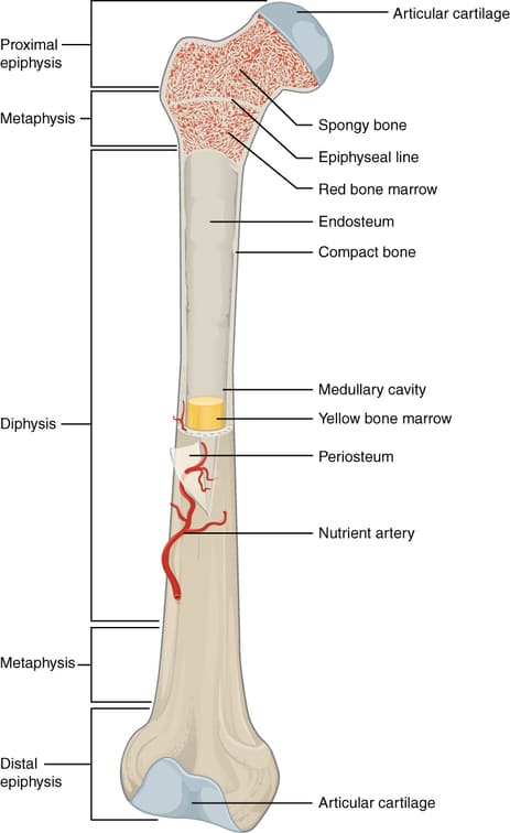
| Periosteum | Double-layered membrane covering external bone surface. Outer fibrous layer + inner cambium (osteogenic) layer. | Essential for bone growth, repair, and nutrition. Rich innervation → primary source of bone pain. Must be preserved during orthopedic surgery. |
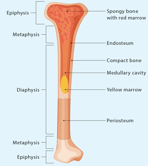
| Endosteum | Thin cellular membrane lining the medullary cavity and trabecular surfaces. | Contains osteoprogenitor cells. Active in bone remodeling and fracture repair. |
| Medullary Cavity | Central hollow shaft containing bone marrow (yellow in adults, red in children). | Site of fat storage (yellow marrow) and blood cell production (red marrow). Target for bone marrow biopsy. |
| Articular Cartilage | Hyaline cartilage covering articulating bone ends. Avascular and aneural. | Provides smooth, low-friction joint surfaces. Degeneration leads to osteoarthritis. Cannot self-repair effectively. |
Long Bone Anatomy
Bone Cells (Osteogenic Cells)
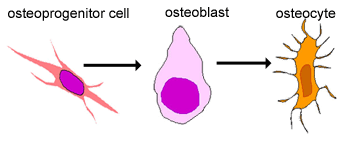
- Osteoprogenitor Cells: Mesenchymal stem cells capable of differentiating into osteoblasts. Found in periosteum, endosteum, and bone marrow. Clinical: Critical for fracture healing and bone regeneration.
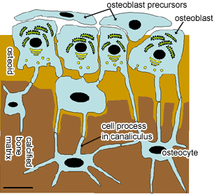
- Osteoblasts: Bone-forming cells that synthesize and secrete osteoid (unmineralized bone matrix). Express alkaline phosphatase. Become trapped in matrix → differentiate into osteocytes. Clinical: Target of anabolic osteoporosis therapies (e.g., teriparatide).
- Osteocytes: Mature bone cells residing in lacunae, connected via canaliculi. Mechanosensory cells that regulate bone remodeling by signaling osteoblasts and osteoclasts. Clinical: Dysfunction implicated in osteoporosis and osteopetrosis.
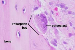
- Osteoclasts: Large multinucleated cells derived from hematopoietic monocyte/macrophage lineage. Secrete HCl and cathepsin K to resorb bone. Ruffled border increases surface area. Clinical: Target of bisphosphonates and denosumab.
- Bone Lining Cells: Inactive osteoblasts on bone surfaces. Maintain ionic homeostasis and serve as barrier between bone fluid and extracellular fluid.
Bone Cell Types — Histology
Bone Matrix Composition
The bone matrix consists of:
- Organic components (35%): Primarily Type I collagen (90% of organic), osteocalcin, osteonectin, and osteopontin. Collagen provides tensile strength and flexibility.
- Inorganic/mineral components (65%): Mainly hydroxyapatite [Ca₁₀(PO₄)₆(OH)₂], with calcium carbonate and magnesium phosphate. Mineral provides compressive strength.
In osteogenesis imperfecta, defective Type I collagen leads to brittle bones despite normal mineralization. In osteomalacia, defective mineralization of normal collagen matrix leads to soft, bendable bones.
2. ARTICULATION (Joint)
Definition: An articulation (joint) is the site where two or more bones meet, permitting varying degrees of movement. The structure of a joint reflects its functional demands — stability versus mobility.
Classification of Joints
| Type | Structural Basis | Movement | Clinical Relevance |
|---|---|---|---|
| Fibrous (Synarthrosis) | Bones united by dense fibrous connective tissue (sutures, syndesmoses, gomphoses) | Immovable or slightly movable | Craniosynostosis (premature suture fusion); high ankle sprain (syndesmotic injury) |
| Cartilaginous (Amphiarthrosis) | Bones united by cartilage (synchondroses, symphyses) | Slightly movable | Epiphyseal plate fracture can stunt growth; disc herniation; pubic symphysis diastasis in childbirth |
| Synovial (Diarthrosis) | Bones separated by joint cavity with articular cartilage, synovial membrane, and fluid | Freely movable (varies by subtype) | Most common site of arthritis, dislocations, and sports injuries |
Synovial Joint Structure — Detailed
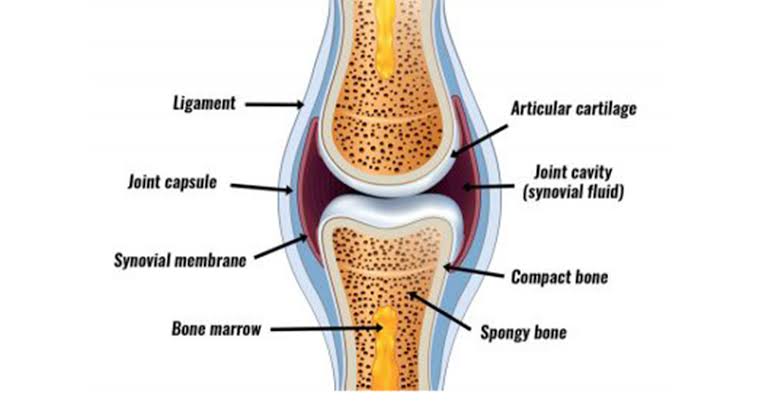
- Articular Cartilage: Hyaline cartilage (2–4 mm thick) covering articulating bone ends. Avascular, aneural, alymphatic — relies on synovial fluid diffusion for nutrition. Composed of chondrocytes in extracellular matrix of Type II collagen and proteoglycans (aggrecan). Clinical: Focal defects do not heal; full-thickness loss leads to osteoarthritis.
- Joint Cavity: Potential space containing synovial fluid (0.5–4 mL in major joints). Under negative pressure, which contributes to joint stability.
- Articular Capsule: Two layers — outer fibrous capsule (dense irregular CT, continuous with periosteum) and inner synovial membrane (highly vascularized, produces synovial fluid).
- Synovial Fluid: Viscous, straw-colored ultrafiltrate of plasma plus hyaluronic acid and lubricin. Functions: lubrication, nutrition of avascular cartilage, shock absorption, phagocytosis of debris. Clinical: Joint effusion indicates pathology.
- Accessory Structures: Menisci (fibrocartilage discs), fat pads (infrapatellar fat pad), bursae (fluid-filled sacs reducing friction), and ligaments (intracapsular: ACL, PCL; extracapsular: MCL, LCL).
Types of Synovial Joints (by Movement)
| Type | Movement Axes | Movements Allowed | Examples | Common Pathologies |
|---|---|---|---|---|
| Plane (Gliding) | Non-axial / Multi-axial | Gliding/sliding | Intercarpal, intertarsal, sternoclavicular | Carpal instability, AC joint separation |
| Hinge (Ginglymus) | Uniaxial | Flexion, extension only | Elbow (humeroulnar), knee, ankle | Tennis elbow, ACL tear, ankle sprain |
| Pivot (Trochoid) | Uniaxial | Rotation only | Atlantoaxial (C1-C2), proximal radioulnar | Atlantoaxial subluxation (RA), nursemaid's elbow |
| Condyloid (Ellipsoid) | Biaxial | Flexion, extension, abduction, adduction (no rotation) | Radiocarpal (wrist), MCP joint | Colles' fracture, rheumatoid arthritis |
| Saddle (Sellar) | Biaxial | Flexion, extension, abduction, adduction, circumduction | Carpometacarpal of thumb, sternoclavicular | Basal joint arthritis (thumb CMC) |
| Ball-and-Socket | Multiaxial (Triaxial) | Flexion, extension, abduction, adduction, rotation, circumduction | Shoulder (glenohumeral), hip (coxal) | Shoulder dislocation (anterior >95%), hip fracture, avascular necrosis |
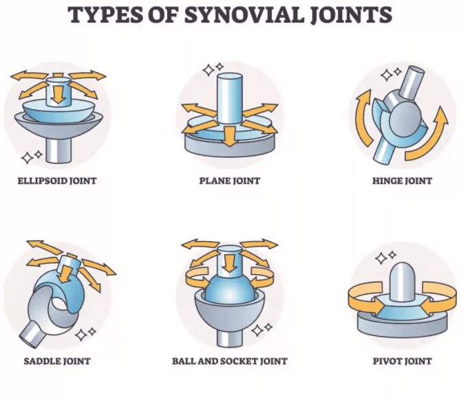
The shoulder (glenohumeral joint) sacrifices stability for mobility — it is the most commonly dislocated joint. The hip, conversely, is highly stable due to its deep acetabular socket and strong ligaments, making dislocation rare but fractures more common in elderly patients.
3. OSSIFICATION (Osteogenesis)
Definition: Ossification is the process of bone formation. It occurs through two distinct mechanisms: intramembranous ossification (direct formation from mesenchyme) and endochondral ossification (replacement of a hyaline cartilage model). Both processes produce identical bone tissue but differ in their embryological origin.
A. Intramembranous Ossification
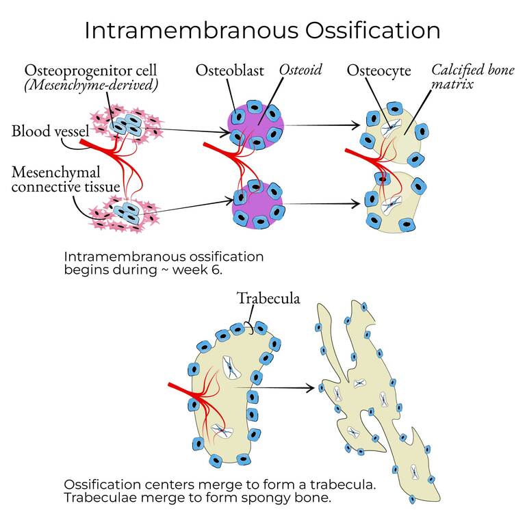
Definition: Direct bone formation within mesenchymal connective tissue membranes, without a cartilage precursor. This process forms flat bones of the skull, the mandible, the maxilla, and the clavicles.
Steps of Intramembranous Ossification:
- Step 1 — Mesenchymal Condensation: Mesenchymal stem cells cluster at the site of future bone, forming a highly vascularized membrane with dense capillary networks.
- Step 2 — Differentiation of Osteoblasts: Some mesenchymal cells differentiate into osteoprogenitor cells, then into osteoblasts. Osteoblasts begin secreting osteoid (unmineralized matrix of Type I collagen and ground substance).
- Step 3 — Formation of Ossification Centers: Osteoblasts aggregate into clusters called ossification centers. As osteoid is secreted, it surrounds osteoblasts, which become trapped and differentiate into osteocytes.
- Step 4 — Mineralization: Calcium and phosphate ions deposit into the osteoid, converting it to mineralized bone matrix (hydroxyapatite formation). This occurs within days of osteoid secretion.
- Step 5 — Formation of Trabeculae and Compact Bone: Interconnecting bony spicules (trabeculae) form a network of spongy bone. Blood vessels become trapped within the bone, forming red bone marrow. On the outer surface, mesenchymal cells form the periosteum, and osteoblasts beneath it secrete layers of compact bone via appositional growth.
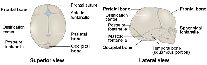
At birth, the skull bones are not fully ossified — fontanelles (soft spots) remain between cranial bones, allowing skull deformation during vaginal delivery and brain growth. The anterior fontanelle closes at 12–18 months; posterior at 2–3 months. Delayed closure suggests hypothyroidism, rickets, or hydrocephalus. Premature closure (craniosynostosis) causes abnormal head shape and increased intracranial pressure.
B. Endochondral Ossification
Definition: Bone formation by replacement of a hyaline cartilage model. This is the mechanism for formation of long bones, short bones, and the vertebrae. It is more complex than intramembranous ossification and involves both cartilage degradation and bone deposition.
Steps of Endochondral Ossification:
- Step 1 — Cartilage Model Formation: In the 6–8 week embryo, mesenchymal cells differentiate into chondroblasts, which secrete cartilage matrix. Chondroblasts become chondrocytes trapped in lacunae. The perichondrium surrounds the model.
- Step 2 — Primary Ossification Center (Diaphysis): Chondrocytes in the center hypertrophy and accumulate glycogen. The matrix calcifies, blocking nutrient diffusion → chondrocyte apoptosis → lacunae merge into cavities. Blood vessels invade, bringing osteoprogenitor cells and osteoclasts. The perichondrium becomes periosteum. Osteoblasts form a bone collar around the diaphysis. Osteoclasts break down calcified cartilage; osteoblasts deposit bone on remaining cartilage spicules → primary spongiosa.
- Step 3 — Secondary Ossification Centers (Epiphyses): After birth, secondary centers appear at the bone ends (epiphyses). The process is similar but lacks a bone collar. Cartilage at the articular surfaces and the epiphyseal plate remains.
- Step 4 — Epiphyseal (Growth) Plate: The cartilage between primary and secondary centers becomes the epiphyseal plate — the site of longitudinal bone growth.
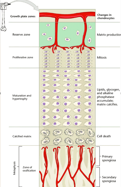
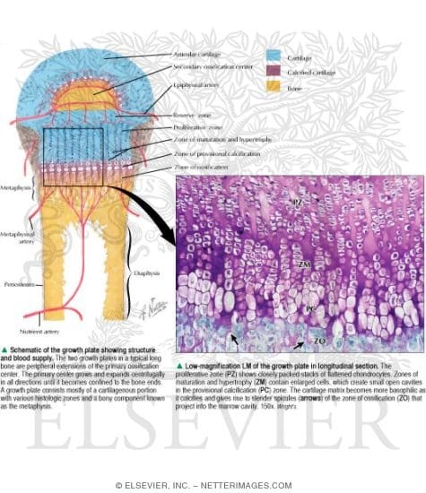
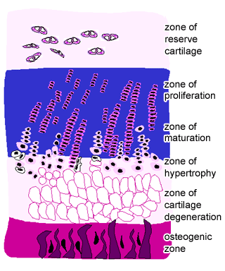
Zones of the Epiphyseal Growth Plate
| Zone | Histological Features | Function | Clinical Relevance |
|---|---|---|---|
| 1. Reserve (Resting) Zone | Small, scattered chondrocytes; abundant matrix; no active division | Anchors plate to epiphysis; reservoir of chondrocytes | Salter-Harris Type V (crush injury) damages this zone → growth arrest |
| 2. Proliferative Zone | Flattened chondrocytes arranged in longitudinal columns; active mitosis | Rapid cell division pushes epiphysis away from diaphysis → longitudinal growth | Most radiosensitive zone; radiation/chemotherapy can impair growth |
| 3. Hypertrophic Zone | Chondrocytes enlarge 5–10×; accumulate glycogen; matrix begins to calcify | Cell enlargement contributes to bone lengthening; matrix prepares for calcification | Rickets: defective mineralization here → widened, frayed metaphysis |
| 4. Calcification Zone | Matrix heavily calcified; chondrocytes die (apoptosis); lacunae empty | Creates scaffold for osteoblast bone deposition | Osteopetrosis: defective osteoclast resorption → dense, brittle bones |
| 5. Ossification Zone | Osteoblasts deposit bone on calcified cartilage; osteoclasts resorb cartilage | Replacement of cartilage by bone; formation of metaphyseal trabeculae | Site of metaphyseal fracture in children (weakest mechanical point) |
Classification of Growth Plate Fractures:
- Type I: Straight separation through the plate (slipped epiphysis). Good prognosis.
- Type II: Fracture through plate + metaphyseal fragment (most common, ~75%). Good prognosis.
- Type III: Fracture through plate + epiphyseal fragment. Intra-articular; ORIF required.
- Type IV: Fracture crosses plate, metaphysis, and epiphysis. High risk of growth arrest.
- Type V: Crush injury to the plate. Worst prognosis; almost always causes growth disturbance.
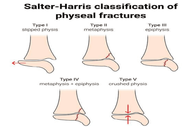
Salter-Harris Mnemonic (S-A-L-T-ER)
- S — Straight across
- A — Above the plate
- L — Lower (below the plate)
- T — Through (Two or both sides)
- ER — ERasure (Crush injury)
C. Bone Remodeling
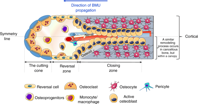
Bone remodeling is a lifelong process of coupled bone resorption and formation that maintains skeletal integrity, repairs microdamage, and regulates calcium homeostasis. It is carried out by the Basic Multicellular Unit (BMU):
- Activation: Osteocytes detect mechanical strain or microdamage and signal lining cells to retract, exposing bone surface.
- Resorption: Osteoclasts attach to bone via integrins, form a sealed compartment (ruffled border), and secrete HCl (pH ~4.5) and cathepsin K to dissolve mineral and digest collagen. Takes ~2–3 weeks.
- Reversal: Osteoclasts undergo apoptosis; mononuclear cells prepare the surface for osteoblasts.
- Formation: Osteoblasts lay down new osteoid in layers (lamellae). Mineralization lags ~10 days behind deposition. Takes ~3–4 months.
- Quiescence: Osteoblasts become lining cells or osteocytes; the cycle completes.
Postmenopausal Osteoporosis
In postmenopausal osteoporosis, estrogen deficiency increases osteoclast activity and lifespan, creating a negative bone balance with each remodeling cycle. Bisphosphonates inhibit osteoclast function; teriparatide stimulates osteoblast activity.
4. Key Terminology Summary for Clinical Officers
| Term | Definition | Clinical Context |
|---|---|---|
| Diaphysis | The shaft of a long bone; composed mainly of compact bone surrounding the medullary cavity | Site of primary ossification; common fracture location in children (greenstick fractures) |
| Epiphysis | The end of a long bone, covered with articular cartilage; contains spongy bone | Secondary ossification center; avascular necrosis risk in femoral head (Legg-Calvé-Perthes disease) |
| Metaphysis | The neck region between diaphysis and epiphysis; contains the growth plate in children | Common site of osteosarcoma in adolescents; metaphyseal fractures are common in children |
| Epiphyseal Plate (Physis) | Hyaline cartilage disc responsible for longitudinal bone growth | Fractures here (Salter-Harris) can cause growth arrest; widened in rickets |
| Periosteum | Fibrous membrane covering bone; outer fibrous + inner osteogenic layer | Essential for fracture healing (callus formation); elevated in osteosarcoma (Codman's triangle) |
| Osteoid | Unmineralized organic bone matrix secreted by osteoblasts | Excess osteoid in osteomalacia; decreased in osteoporosis |
| Appositional Growth | Increase in bone width/diameter by deposition on outer (periosteal) surface and resorption on inner (endosteal) surface | How bones widen during growth; periosteal reaction seen in infection or trauma |
