Peritoneum
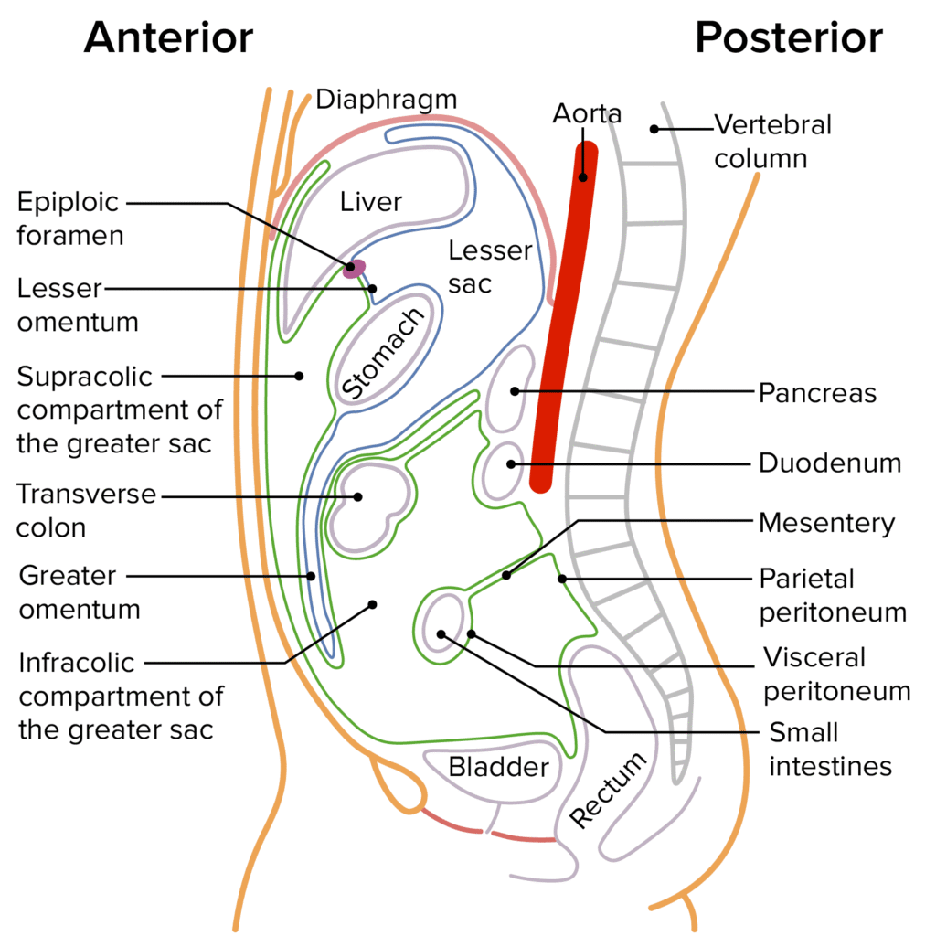
The Peritoneum A Comprehensive Anatomical Notes Covering Structural Organization, Peritoneal Reflections and Folds, Subdivisions of the Peritoneal Cavity, Spaces and Gutters, Neurovascular Supply, and Clinical Applied Anatomy. CHAPTER 1: INTRODUCTION AND FUNCTIONAL OVERVIEW 1.1 Functional Significance of the Peritoneum The peritoneum is a continuous, glistening serous membrane that lines the abdominal cavity and invests the viscera contained within it. It represents the largest and most complexly arranged serous membrane in the human body. The peritoneum acts to minimize friction between moving organs, transmit neurovascular structures, and serve as a dynamic defensive immunological barrier within the abdominopelvic cavity. The peritoneum is not merely a passive lining; it is an active, dynamic organ with significant physiological and immunological functions. It secretes serous fluid that lubricates the surfaces of the abdominal organs, allowing them to move freely against one another during peristalsis, respiration, and changes in body position. The peritoneum also plays a critical role in immune defense, with mesothelial cells capable of phagocytosis and the production of inflammatory mediators. The peritoneal cavity serves as a potential space that can accommodate pathological processes such as infection, hemorrhage, and malignancy, making understanding of its anatomy essential for surgical and diagnostic procedures. The peritoneum is ultimately one continuous sheet, but for descriptive purposes, it is divided into two layers: the parietal peritoneum that lines the abdominal wall and the visceral peritoneum that covers the abdominal organs. Between these two layers lies the peritoneal cavity, a potential space that normally contains only a thin film of serous fluid (approximately 50-100 mL) that lubricates the surfaces and reduces friction. Key Concept The Peritoneum as a Functional Unit The peritoneum operates not as a passive membrane but as an active biological interface. The mesothelial cells that line the peritoneum secrete lubricating fluid, participate in immune surveillance, and can undergo metaplasia in response to chronic irritation. The peritoneal cavity, though normally a potential space, can expand significantly to accommodate pathological fluid collections (ascites), air (pneumoperitoneum), or blood (hemoperitoneum). Understanding the three-dimensional anatomy of the peritoneum and its reflections is therefore fundamental to abdominal surgery, interventional radiology, and emergency medicine. CHAPTER 2: STRUCTURAL ORGANIZATION AND LAYERS 2.1 Parietal Peritoneum The parietal peritoneum is the portion of the peritoneum that lines the internal surface of the abdominopelvic wall. It is derived embryologically from the somatic mesoderm, which also gives rise to the body wall muscles, bones, and connective tissue. This embryological origin determines its innervation by somatic nerves and its sensitivity to the same stimuli as the skin: pain, pressure, temperature, and laceration. The parietal peritoneum is firmly attached to the transversalis fascia of the abdominal wall by loose areolar tissue. It lines the anterior and lateral abdominal walls, the pelvic walls, the inferior surface of the diaphragm, and the anterior surface of the retroperitoneal organs. The parietal peritoneum reflects onto the viscera at various points, becoming continuous with the visceral peritoneum. 2.2 Visceral Peritoneum The visceral peritoneum is the portion of the peritoneum that directly invests the abdominal organs. It is derived embryologically from the splanchnic mesoderm, which also gives rise to the smooth muscle and connective tissue of the gastrointestinal tract. This embryological origin determines its innervation by autonomic (visceral) nerves and its relative insensitivity to most stimuli. The visceral peritoneum covers the organs almost completely (in the case of intraperitoneal organs) or only on their anterior surface (in the case of retroperitoneal organs). At the points where organs are attached to the body wall or to other organs by mesenteries, omenta, or ligaments, the visceral peritoneum is continuous with the parietal peritoneum or with the visceral peritoneum of adjacent organs. 2.3 The Peritoneal Cavity The peritoneal cavity is the potential space between the parietal and visceral layers of the peritoneum. It is not a true “cavity” in the sense of being empty; rather, it is a potential space that normally contains only a thin film of serous fluid (approximately 50-100 mL) that lubricates the peritoneal surfaces and allows the organs to move freely against one another. The peritoneal cavity is completely closed in males, forming a sealed potential space. In females, however, the peritoneal cavity communicates with the external environment via the uterine tubes (fallopian tubes), uterus, and vagina. This communication pathway is clinically significant because it provides a potential route for the spread of infection from the exterior to the peritoneal cavity (e.g., ascending pelvic inflammatory disease) and explains why pneumoperitoneum can occur in females after sexual intercourse or vaginal procedures without necessarily having a perforated viscus. Clinical Correlation Sexual Dimorphism of the Peritoneal Cavity The open communication between the peritoneal cavity and the exterior in females (via the uterine tubes, uterus, and vagina) has several clinical implications: It provides a route for ascending infection, which is why pelvic inflammatory disease (PID) can lead to peritonitis. It explains why females can develop pneumoperitoneum after sexual intercourse or vaginal procedures without necessarily having a perforated viscus. It provides a potential route for the spread of malignancy from the genital tract to the peritoneal cavity. In contrast, the sealed peritoneal cavity in males means that any pneumoperitoneum is highly suggestive of a perforated viscus or recent surgery. 2.4 Organ Relationship Classifications Abdominal organs are classified based on their relationship to the peritoneum into three categories: intraperitoneal, primarily retroperitoneal, and secondarily retroperitoneal. This classification has profound implications for surgical approach, organ mobility, and the patterns of disease spread. 2.4.1 Intraperitoneal Organs Intraperitoneal organs are almost entirely covered by visceral peritoneum and are suspended into the peritoneal cavity by mesenteries. They are mobile and have a greater range of movement than retroperitoneal organs. Intraperitoneal organs include: Organ Mesentery Notes Stomach Lesser and greater omenta Highly mobile; attached at lesser and greater curvatures Spleen Gastrosplenic and splenorenal ligaments Intraperitoneal but relatively fixed by ligaments Liver Falciform, coronary, triangular ligaments Mostly intraperitoneal except bare area Jejunum and Ileum Mesentery of small intestine Highly mobile; long mesentery allows wide movement Transverse colon Transverse mesocolon Mobile; suspended from posterior abdominal wall Sigmoid colon Sigmoid