Comprehensive Anatomy of the Abdominal Gastrointestinal Tract
A systematic study covering general organization, regional structures (foregut, midgut, hindgut), associated organs, neurovascular corridors, and clinical applications.
CHAPTER 1: General Organization and the Peritoneum
The abdominal gastrointestinal tract (GIT) constitutes a continuous muscular tube extending from the distal esophagus to the anal canal, along with its associated accessory digestive organs. The tract is organized anatomically and embryologically into three distinct regions: the foregut, midgut, and hindgut. Each region possesses distinct vascular, lymphatic, and autonomic nerve profiles that reflect its embryological origin.
1.1 Surface Anatomy and Abdominal Quadrants
The abdomen is divided into nine regions by two horizontal and two vertical planes to facilitate accurate localization of intra-abdominal viscera:
Horizontal Planes:
- Transpyloric plane: Passes through the pylorus of the stomach, approximately at the level of the first lumbar vertebra (L1). It lies midway between the jugular notch (suprasternal notch) and the pubic symphysis.
- Transtubercular plane: Passes through the tubercles of the iliac crests, approximately at the level of the fifth lumbar vertebra (L5).
Vertical Planes:
- Right and left lateral (midclavicular) planes: Pass vertically through the midclavicular lines from the costal margin to the inguinal ligaments.
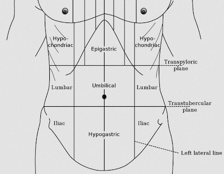
The Nine Abdominal Regions:
| Region | Boundaries | Key Contents |
|---|---|---|
| Right Hypochondrium | Above transpyloric plane, right of right lateral plane | Right lobe of liver, gallbladder, right kidney, right suprarenal gland, hepatic flexure of colon |
| Epigastrium | Above transpyloric plane, between lateral planes | Stomach, liver (left lobe), pancreas, duodenum (proximal), spleen (partial) |
| Left Hypochondrium | Above transpyloric plane, left of left lateral plane | Spleen, stomach (fundus), left lobe of liver, left kidney, left suprarenal gland, splenic flexure of colon, tail of pancreas |
| Right Lumbar | Between transpyloric and transtubercular planes, right of right lateral plane | Ascending colon, right kidney, duodenum (descending part), head of pancreas |
| Umbilical | Between transpyloric and transtubercular planes, between lateral planes | Transverse colon, small intestine (jejunum and ileum), aorta, inferior vena cava |
| Left Lumbar | Between transpyloric and transtubercular planes, left of left lateral plane | Descending colon, left kidney, small intestine, duodenum (horizontal and ascending parts) |
| Right Iliac (Inguinal) | Below transtubercular plane, right of right lateral plane | Cecum, appendix, terminal ileum, right ureter, right ovary/testis |
| Hypogastrium (Pubic) | Below transtubercular plane, between lateral planes | Urinary bladder (when distended), uterus (in females), rectum, sigmoid colon, small intestine |
| Left Iliac (Inguinal) | Below transtubercular plane, left of left lateral plane | Sigmoid colon, left ureter, left ovary/testis, small intestine |
The Four Quadrants: A simpler division using a vertical line through the umbilicus and a horizontal line through the umbilicus produces the right upper quadrant (RUQ), left upper quadrant (LUQ), right lower quadrant (RLQ), and left lower quadrant (LLQ). This system is commonly used in clinical practice for rapid localization of abdominal pain and pathology.
The RUQ contains the liver, gallbladder, and duodenum; the LUQ contains the stomach, spleen, and splenic flexure; the RLQ contains the appendix, cecum, and terminal ileum; the LLQ contains the sigmoid colon. Understanding these relationships is essential for clinical examination and differential diagnosis.
1.2 Peritoneal Cavity Dynamics
The peritoneum is a continuous serous membrane lining the abdominal cavity and investing the abdominal viscera. It consists of two continuous layers:
- Parietal Peritoneum: Lines the internal surface of the abdominal wall. It is supplied by the same somatic vessels and nerves that supply the overlying body wall. Consequently, pain from the parietal peritoneum is sharp, well-localized, and transmitted by somatic afferent fibers.
- Visceral Peritoneum: Covers the surfaces of abdominal organs. It is supplied by autonomic nerves and is insensitive to pain from cutting, crushing, or burning. Distension of the visceral peritoneum, however, produces poorly localized, dull, cramping pain referred to the dermatome of the organ's embryological origin.
The Peritoneal Cavity:
The potential space between the parietal and visceral layers of the peritoneum is termed the peritoneal cavity. In the male, this cavity is completely closed. In the female, it communicates with the exterior via the fallopian tubes, uterus, and vagina. The peritoneal cavity is divided into two principal compartments:
- The Greater Sac: The larger anterior compartment of the peritoneal cavity, extending from the diaphragm superiorly to the pelvic cavity inferiorly. It contains most of the intraperitoneal organs and is the primary site of fluid accumulation in pathological conditions such as ascites.
- The Lesser Sac (Omental Bursa): A smaller, posterior compartment situated behind the stomach and lesser omentum. It communicates with the greater sac through the epiploic foramen (foramen of Winslow), an opening bounded anteriorly by the hepatoduodenal ligament, posteriorly by the inferior vena cava, superiorly by the caudate lobe of the liver, and inferiorly by the first part of the duodenum.
The Epiploic Foramen (Foramen of Winslow): This is the only natural communication between the greater and lesser sacs. The portal triad (hepatic portal vein, hepatic artery proper, and common bile duct) runs within the free edge of the lesser omentum anterior to the foramen.
1.3 Peritoneal Formations
Peritoneal formations are double-layered folds of peritoneum that connect organs to each other or to the abdominal wall.
A. Mesenteries
- Mesentery Proper: A broad, fan-shaped fold suspending the jejunum and ileum from the posterior abdominal wall. Its root extends obliquely from the duodenojejunal flexure (left of L2) to the ileocecal junction (right sacroiliac joint), approximately 15 cm. The intestinal border is approximately 6 meters long. Contains the superior mesenteric artery and vein, lymphatic vessels, autonomic nerve plexuses, and abundant fat.
- Transverse Mesocolon: A broad peritoneal fold suspending the transverse colon from the posterior abdominal wall. Contains the middle colic vessels.
- Sigmoid Mesocolon: An inverted V-shaped peritoneal fold suspending the sigmoid colon. The apex lies at the division of the left common iliac artery.
B. Omenta
- Greater Omentum: A large, apron-like fold of peritoneum that descends from the greater curvature of the stomach, folds back upon itself, and ascends to the transverse colon. It consists of four layers of peritoneum and contains variable amounts of fat, blood vessels, lymphatic vessels, and macrophages. It migrates to sites of inflammation and has been termed the "abdominal policeman."
- Lesser Omentum: A double-layered peritoneal fold extending from the lesser curvature of the stomach and the first part of the duodenum to the liver. Divided into the hepatogastric ligament and the hepatoduodenal ligament. The free right margin contains the portal triad.
C. Ligaments
- Falciform Ligament: A sickle-shaped peritoneal fold from the anterior abdominal wall to the anterior surface of the liver. Its free margin contains the ligamentum teres hepatis (remnant of the left umbilical vein).
- Splenorenal (Lienorenal) Ligament: From splenic hilum to left kidney; contains splenic vessels and pancreatic tail.
- Gastrosplenic Ligament: From greater curvature to splenic hilum; contains short gastric vessels.
1.4 Intraperitoneal and Retroperitoneal Organs
Abdominal organs are classified according to their relationship to the peritoneum. This classification has significant implications for surgical approach, mobility, and patterns of disease spread.
| Category | Definition | Examples |
|---|---|---|
| Intraperitoneal | Almost completely covered by peritoneum; suspended by mesentery; highly mobile | Stomach, jejunum, ileum, cecum, appendix, transverse colon, sigmoid colon, liver (mostly), spleen |
| Primarily Retroperitoneal | Develop and remain behind peritoneum; only anterior surface covered | Kidneys, ureters, suprarenal glands, pancreas (except tail), duodenum (parts 2-4), ascending colon, descending colon, rectum (upper 2/3), aorta, IVC |
| Secondarily Retroperitoneal | Were intraperitoneal during development but later fused to posterior abdominal wall | Duodenum (parts 2-4), ascending colon, descending colon, pancreas |
Intraperitoneal organs are mobile and accessible for surgical manipulation. Retroperitoneal organs are relatively fixed and may be injured during retroperitoneal surgery or trauma. Fluid collections in the retroperitoneal space may be difficult to detect clinically.
CHAPTER 2: Foregut Structures
The foregut extends from the distal esophagus to the proximal half of the duodenum, terminating at the entry of the bile duct into the duodenal lumen. It is supplied by the celiac trunk and receives parasympathetic innervation from the vagus nerve (via the celiac plexus) and sympathetic innervation from the greater splanchnic nerve (T5-T9). Pain from foregut structures is referred to the epigastric region (T7-T9 dermatomes).
2.1 The Abdominal Esophagus
The esophagus enters the abdomen through the esophageal hiatus of the diaphragm at the level of the tenth thoracic vertebra (T10). The phrenicoesophageal ligament anchors the esophagus to the diaphragm, permitting movement during respiration and swallowing.
- Course and Relations: The abdominal esophagus is approximately 1-2 cm in length. It descends slightly to the left and terminates at the cardiac orifice of the stomach at the level of T11, to the left of the midline. Anteriorly related to the left lobe of the liver; posteriorly to the right crus of the diaphragm and the aorta.
- Arterial Supply: Primarily the left gastric artery, a branch of the celiac trunk. The lower esophagus also receives branches from the left inferior phrenic artery.
- Venous Drainage: Via the left gastric vein (coronary vein) into the hepatic portal vein. This represents one of the principal sites of portosystemic anastomosis. In portal hypertension, blood may flow retrograde through the left gastric vein into the esophageal venous plexus, producing esophageal varices.
- The Lower Esophageal Sphincter (LES): A physiological high-pressure zone maintained by intrinsic smooth muscle, the cricopharyngeus-like muscular arrangement at the gastroesophageal junction, and the diaphragmatic crura. Failure results in gastroesophageal reflux disease (GERD).
2.2 The Stomach (Gaster)
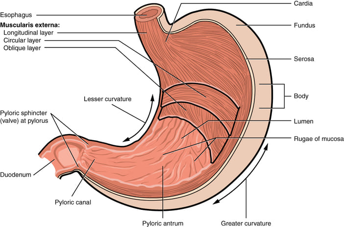
The stomach is a J-shaped, dilated portion of the gastrointestinal tract situated in the left upper quadrant, primarily in the epigastrium and left hypochondrium. It serves as a reservoir for ingested food and initiates digestion through mechanical churning and secretion of gastric acid and enzymes.
A. Macroscopic Anatomy
- Cardia: Region surrounding the cardiac orifice at T11, to the left of the midline. The cardiac notch (incisura cardiaca) is the acute angle between the esophagus and the fundus.
- Fundus: Dome-shaped portion superior and to the left of the cardia; usually filled with gas.
- Body (Corpus): Largest portion; contains gastric glands secreting hydrochloric acid, pepsinogen, and intrinsic factor.
- Pyloric Antrum: Wider portion of the pyloric region; mixing chamber.
- Pyloric Canal: Narrow tubular portion leading to the pylorus.
- Pylorus: Distal portion containing the pyloric sphincter at L1, to the right of the midline.
Curvatures: The lesser curvature (concave, right border) extends from the cardiac notch to the pylorus, bearing the angular incisure. The greater curvature (convex, left border) extends from the cardiac notch along the fundus and body to the pylorus.
B. Anatomical Relations
- Anterior: Left lobe of the liver, anterior abdominal wall, diaphragm, greater omentum.
- Posterior (The Stomach Bed): Separated from the following structures by the lesser sac:
- Pancreas: Body and neck of pancreas. Pancreatitis may produce epigastric pain radiating to the back.
- Splenic Artery: Courses along superior border of pancreas. A posterior gastric ulcer may erode into it, causing severe hemorrhage.
- Left Kidney and Left Suprarenal Gland: Posterior to the fundus and upper body.
- Diaphragm: Left crus posterior to the cardia.
- Transverse Mesocolon and Transverse Colon: Cross the lower posterior stomach surface.
- Spleen: Gastric surface related to the fundus.
C. Vascularity
Arteries of the Lesser Curvature:
- Left Gastric Artery: From celiac trunk; descends along lesser curvature, anastomosing with the right gastric artery at the incisura angularis.
- Right Gastric Artery: From hepatic artery proper; runs along lesser curvature.
Arteries of the Greater Curvature:
- Left Gastro-omental Artery: Terminal branch of splenic artery; courses along greater curvature within greater omentum.
- Right Gastro-omental Artery: From gastroduodenal artery; courses along greater curvature.
Arteries of the Fundus:
- Short Gastric Arteries: 4-6 small vessels from terminal splenic artery; pass through gastrosplenic ligament. Vulnerable during splenectomy.
Venous Drainage: Into hepatic portal system. Left and right gastric veins → portal vein. Left gastro-omental → splenic vein. Right gastro-omental → superior mesenteric vein.
Lymphatic Drainage: Superior gastric nodes → celiac nodes. The celiac nodes are the principal group for gastric carcinoma staging.
The extensive anastomotic network means ligation of a single gastric artery does not typically produce ischemia. However, the short gastric arteries are fragile and may be avulsed during splenectomy. The left gastric vein is a critical component of the portosystemic anastomosis at the lower esophagus.
2.3 The Duodenum
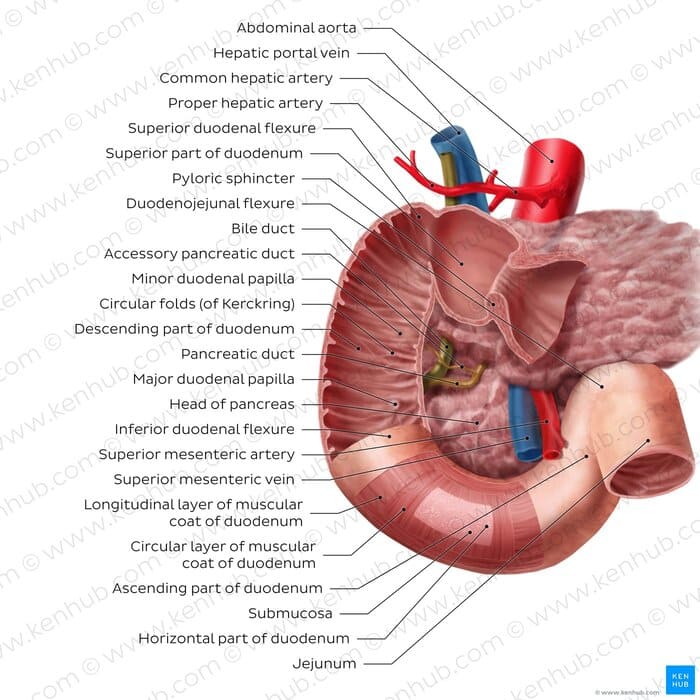
The duodenum is the first and shortest part of the small intestine, approximately 25-30 cm in length. It is C-shaped and wraps around the head of the pancreas. The proximal half (parts 1-2) is foregut; the distal half (parts 3-4) is midgut.
A. First (Superior) Part
Extends from the pylorus to the superior duodenal flexure. Approximately 5 cm long at L1. Its proximal 2 cm (the ampulla or duodenal cap) is intraperitoneal and mobile; the remainder is retroperitoneal. Related anteriorly to the liver and gallbladder, and posteriorly to the portal vein, common bile duct, and gastroduodenal artery. It is the most common site of duodenal ulceration.
B. Second (Descending) Part
Descends from the superior duodenal flexure to the inferior duodenal flexure, along the right side of the vertebral column from L1 to L3. Entirely retroperitoneal. The major duodenal papilla (of Vater) is located on the posteromedial wall, approximately 8-10 cm from the pylorus. This is the common opening for the bile duct and the main pancreatic duct, surrounded by the sphincter of Oddi. The minor duodenal papilla, approximately 2 cm proximal, is the opening of the accessory pancreatic duct (duct of Santorini).
The proximity of the gastroduodenal artery to the posterior wall of the first part means that a penetrating duodenal ulcer may erode into this artery, producing massive upper gastrointestinal hemorrhage. The major duodenal papilla is the target for endoscopic retrograde cholangiopancreatography (ERCP).
CHAPTER 3: Midgut Structures
The midgut extends from the distal half of the duodenum to the proximal two-thirds of the transverse colon. It is supplied by the superior mesenteric artery (SMA) and receives parasympathetic innervation from the vagus nerve and sympathetic innervation from the lesser splanchnic nerve (T10-T11). Pain from midgut structures is referred to the periumbilical region (T10 dermatome).
3.1 Distal Duodenum (Third and Fourth Parts)
A. Third (Horizontal) Part
Extends horizontally from the inferior duodenal flexure to the left, crossing the midline anterior to the abdominal aorta and inferior vena cava at L3. Entirely retroperitoneal. The superior mesenteric artery and vein cross anterior to this part.
B. Fourth (Ascending) Part
Ascends from the left side of L3 to the duodenojejunal flexure at L2. Retroperitoneal and suspended by the suspensory muscle of the duodenum (ligament of Treitz). This muscle anchors the duodenojejunal flexure and marks the boundary between duodenum and jejunum.
The third part may be compressed between the abdominal aorta posteriorly and the superior mesenteric artery anteriorly. This superior mesenteric artery (SMA) syndrome occurs when the aortomesenteric angle is acutely narrowed, causing postprandial epigastric pain, nausea, vomiting, and weight loss.
3.2 The Jejunum and Ileum
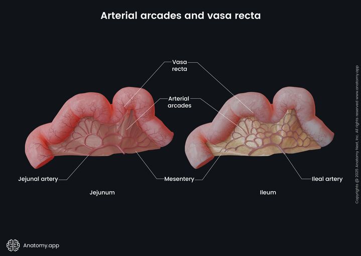
The jejunum and ileum constitute the mobile, coiled portion of the small intestine, suspended by the mesentery proper. Together they measure approximately 6-7 meters. The jejunum occupies the upper left abdomen; the ileum occupies the lower right abdomen and pelvis.
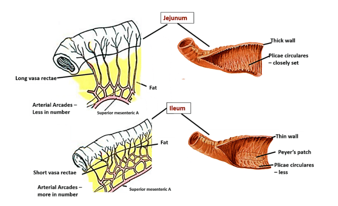
A. Comparative Anatomy
| Feature | Jejunum | Ileum |
|---|---|---|
| Location | Upper left abdomen | Lower right abdomen and pelvis |
| Diameter | Wider (~4 cm) | Narrower (~3 cm) |
| Wall Thickness | Thicker | Thinner |
| Plicae Circulares | Tall, numerous, closely packed | Low, sparse, absent distally |
| Arterial Arcades | Simple (1-2 tiers) | Complex (3-4+ tiers) |
| Vasa Recta | Long, straight | Short, numerous |
| Mesenteric Fat | Less fat | More abundant fat |
| Lymphoid Tissue | Scattered solitary nodules | Aggregated nodules (Peyer's patches) |
| Vascularity | More vascular (redder) | Less vascular (pinker) |
B. The Ileocecal Valve
Located at the junction of the terminal ileum and the cecum. Consists of two horizontal, crescentic mucosal folds projecting into the cecal lumen. Regulates passage from ileum to cecum and prevents retrograde reflux. Blood supply from ileal branches and the ileocolic artery (SMA branch).
3.3 The Large Intestine: Proximal Components
The large intestine extends from the ileocecal junction to the anus (~1.5 meters). It is characterized by teniae coli, haustra, and omental appendices (appendices epiploicae).
A. The Cecum
Blind-ended pouch in the right iliac fossa; ~6 cm long. Usually intraperitoneal with variable mesentery (mesocecum). The ileocecal orifice opens into the medial aspect, and the vermiform appendix arises from the posteromedial wall, approximately 2 cm below the ileocecal junction.
B. The Vermiform Appendix
Narrow, blind-ended tube from posteromedial cecal wall; 8-10 cm long. Possesses own mesentery, the mesoappendix, containing the appendicular artery (branch of ileocolic artery).
- Positions: Retrocecal (65-70%), pelvic (30%), subcecal, preileal, postileal, paracecal. The retrocecal position may produce minimal anterior signs; the pelvic position may cause urinary symptoms.
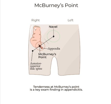
- McBurney's Point: One-third of the distance from the right anterior superior iliac spine to the umbilicus. Tenderness here is a classic sign of acute appendicitis.
The appendicular artery is an end artery within the mesoappendix; obstruction by a fecalith or lymphoid hyperplasia leads to ischemia, necrosis, and perforation of the appendix.
C. The Ascending Colon
From cecum to hepatic flexure; ~15 cm; retroperitoneal. Related anteriorly to anterior abdominal wall and small intestine coils; posteriorly to iliacus, quadratus lumborum, and transversus abdominis.
D. The Hepatic Flexure
Bend between ascending and transverse colon, beneath the right lobe of the liver. Connected to liver by the hepatocolic ligament.
E. The Transverse Colon (Proximal Two-Thirds)
From hepatic to splenic flexure, suspended by transverse mesocolon. Supplied by the middle colic artery (SMA branch) — midgut derivative. Intraperitoneal and highly mobile.
CHAPTER 4: Hindgut Structures
The hindgut extends from the distal one-third of the transverse colon to the upper anal canal. It is supplied by the inferior mesenteric artery (IMA) and receives parasympathetic innervation from pelvic splanchnic nerves (S2-S4). Pain is referred to the pubic/hypogastric region (L1-L2 dermatomes).
4.1 The Distal Colon
- A. The Splenic (Left Colic) Flexure: Bend between transverse and descending colon, beneath the spleen and anterior to the left kidney. Connected to diaphragm by the phrenicocolic ligament. Marks the midgut-hindgut transition.
- B. The Descending Colon: From splenic flexure to sigmoid colon at left iliac crest; ~25 cm; entirely retroperitoneal. Related anteriorly to small intestine coils and anterior abdominal wall; posteriorly to left kidney, quadratus lumborum, and transversus abdominis.
- C. The Sigmoid Colon: From descending colon to rectum at S3; ~40 cm; S-shaped loop in pelvis. Suspended by sigmoid mesocolon (inverted V-shaped). Highly mobile.
4.2 The Rectum
From sigmoid colon at S3 to anorectal junction; ~12 cm. Not straight — follows sacral and coccygeal curvature. Retroperitoneal along upper two-thirds; subperitoneal along lower third.
- Course and Relations: Three lateral flexures (upper and lower to right, middle to left). Anteriorly: rectovesical pouch/seminal vesicles/prostate/bladder (male) or rectouterine pouch/cervix/vagina (female). Posteriorly: sacrum, coccyx, pelvic autonomic plexuses.
- The Rectal Ampulla: Lower dilated portion; temporary fecal reservoir. Supported by puborectalis sling forming anorectal angle (~90°), critical for continence.
- Peritoneal Reflections: Upper third covered anteriorly and laterally, forming rectovesical pouch (male) or rectouterine pouch (pouch of Douglas) (female). The pouch of Douglas is the most dependent portion of the peritoneal cavity in females. Middle third covered only anteriorly. Lower third entirely below peritoneal reflection.
- Transverse Rectal Folds (Valves of Houston): Three semilunar folds: superior on right at S3, middle on left (largest), inferior on right. Support fecal material and may be mistaken for pathology during endoscopy.
4.3 The Anal Canal
The anal canal is the terminal 3-4 cm of the gastrointestinal tract, extending from the anorectal junction to the anus. It is surrounded by the internal and external anal sphincters.
A. The Pectinate (Dentate) Line
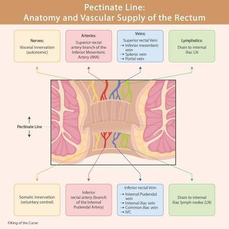
A critical embryological boundary at the level of the anal valves, approximately 2 cm above the anus. Represents the junction between endoderm-derived upper anal canal and ectoderm-derived lower anal canal.
| Feature | Above Pectinate Line | Below Pectinate Line |
|---|---|---|
| Embryological Origin | Endoderm (hindgut) | Ectoderm (proctodeum) |
| Epithelium | Columnar (mucosa) | Stratified squamous (skin) |
| Arterial Supply | Superior rectal artery (IMA branch) | Inferior rectal artery (internal pudendal branch) |
| Venous Drainage | Superior rectal vein → portal system | Inferior rectal vein → systemic (IVC) |
| Lymphatic Drainage | Internal iliac → para-aortic nodes | Superficial inguinal nodes |
| Innervation | Autonomic (visceral) — painless | Somatic (pudendal nerve) — highly sensitive |
| Hemorrhoids | Internal (painless bleeding) | External (painful, thrombosis) |
B. The Sphincteric Complex
- Internal Anal Sphincter: Thickened circular smooth muscle; involuntary control; 70-85% of resting anal pressure. Relaxes during defecation.
- External Anal Sphincter: Skeletal muscle; voluntary control via pudendal nerve (S2-S4); deep, superficial, and subcutaneous parts.
The pectinate line is the most important landmark in the anal canal. Internal hemorrhoids are painless (autonomic innervation); external hemorrhoids are exquisitely painful (somatic innervation). The portosystemic anastomosis at the anorectal junction explains internal hemorrhoids in portal hypertension.
CHAPTER 5: Associated Hepatobiliary Organs and Spleen
The accessory digestive organs of the abdomen are intimately related to the gastrointestinal tract both anatomically and functionally. They are derived from the foregut (except the spleen, a lymphoid organ of mesodermal origin) and share vascular, lymphatic, and innervational pathways with foregut structures.
5.1 The Liver (Hepar)
The largest gland in the body (~1.5 kg), situated in the right upper quadrant, extending across the epigastrium to the left hypochondrium.
- A. Gross Lobes:
- Right Lobe: Largest; right hypochondrium and right epigastrium; separated from left lobe by falciform ligament.
- Left Lobe: Smaller; left hypochondrium and left epigastrium.
- Caudate Lobe: Posterior, between IVC and ligamentum venosum.
- Quadrate Lobe: Inferior surface, between gallbladder fossa and ligamentum teres.
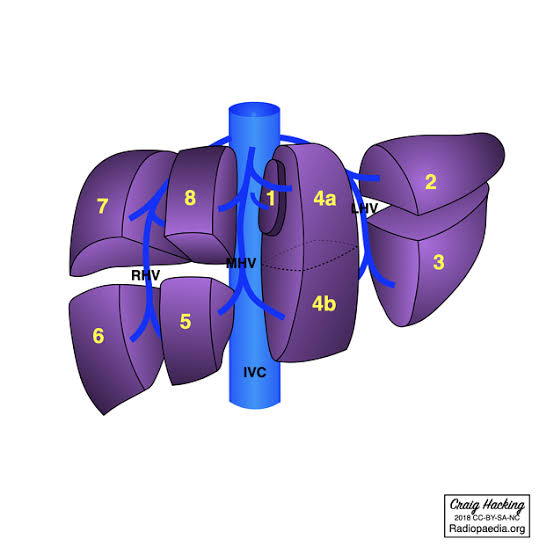
- B. Functional Segmentation (Couinaud Classification): Eight functional segments based on portal triad distribution and hepatic veins. The principal plane (Cantlie's line) from gallbladder fossa to IVC divides right and left hemilivers. The middle hepatic vein runs within this plane. The right hepatic vein divides the right hemiliver into anterior and posterior sections; the left hepatic vein divides the left hemiliver into medial and lateral sections.
- C. The Porta Hepatis: The hilum on the inferior surface between caudate and quadrate lobes. Contains the portal triad enclosed within the hepatoduodenal ligament:
- Hepatic Portal Vein (posterior) — from splenic and SMV confluence.
- Hepatic Artery Proper (anterior, left) — from common hepatic artery.
- Common Hepatic Duct (anterior, right) — from union of right and left hepatic ducts.
5.2 The Gallbladder and Biliary Tree
The gallbladder is a pear-shaped reservoir for bile on the inferior surface of the liver.
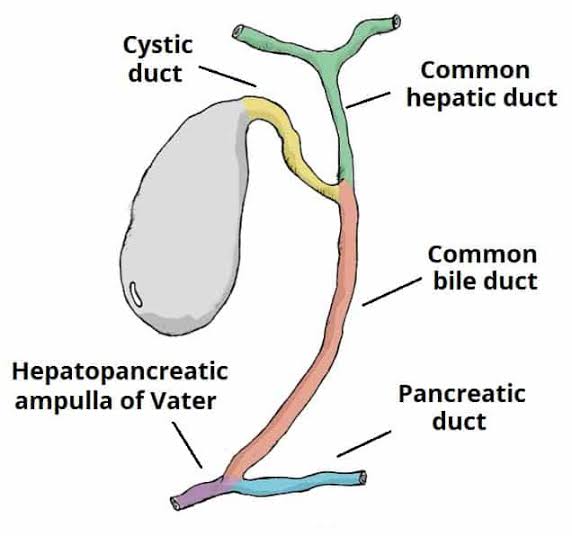
Bile Flow Pathway:
- Hepatic ducts (right and left) from liver segments.
- Common Hepatic Duct (union of right and left hepatic ducts).
- Cystic Duct (from gallbladder neck).
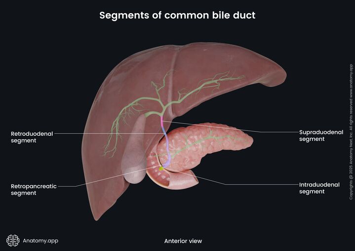
- Common Bile Duct (choledochus) — union of common hepatic duct and cystic duct; ~8 cm.
- Descends posterior to first part of duodenum, then in groove between duodenum and pancreatic head.
- Joins main pancreatic duct to form hepatopancreatic ampulla (ampulla of Vater).
- Opens on major duodenal papilla, surrounded by sphincter of Oddi.
5.3 The Pancreas
A retroperitoneal gland extending from the C-loop of the duodenum to the splenic hilum.
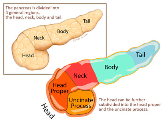
- Head: Within C-loop of duodenum; related to IVC, renal vessels, and SMA posteriorly.
- Uncinate Process: Projection from inferior head, posterior to SMA and SMV.
- Neck: Anterior to SMA/SMV confluence; portal vein forms posteriorly.
- Body: Crosses midline anterior to aorta at L1; related to stomach anteriorly (separated by lesser sac).
- Tail: Extends to splenic hilum within splenorenal ligament.
- Ducts:
- Main Pancreatic Duct (of Wirsung): From tail to head; joins common bile duct at hepatopancreatic ampulla.
- Accessory Pancreatic Duct (of Santorini): From superior part of head; opens at minor duodenal papilla.
5.4 The Spleen (Lien)
A lymphoid organ in the left hypochondrium under ribs 9-11.
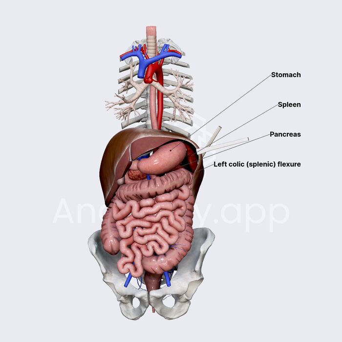
- Surfaces: Diaphragmatic (convex; related to diaphragm and ribs 9-11) and Visceral (contains hilum and impressions for stomach, kidney, and colon).
- Relations: Anterior — stomach (fundus); Posterior — left kidney, left suprarenal gland; Inferior — splenic flexure of colon, tail of pancreas.
- Vascularity: Splenic artery (tortuous branch of celiac trunk) along superior pancreatic border → splenorenal ligament → spleen. Splenic vein (formed by tributaries at hilum) runs posterior to pancreas, joins SMV to form portal vein.
Splenomegaly may cause early satiety by compressing the stomach. Splenic vein thrombosis may cause left-sided portal hypertension with gastric varices.
CHAPTER 6: Neurovascular Corridors and Lymphatics
Arterial Trunks, Collateral Circulation, Portal System, Autonomic Innervation, and Lymphatic Drainage.
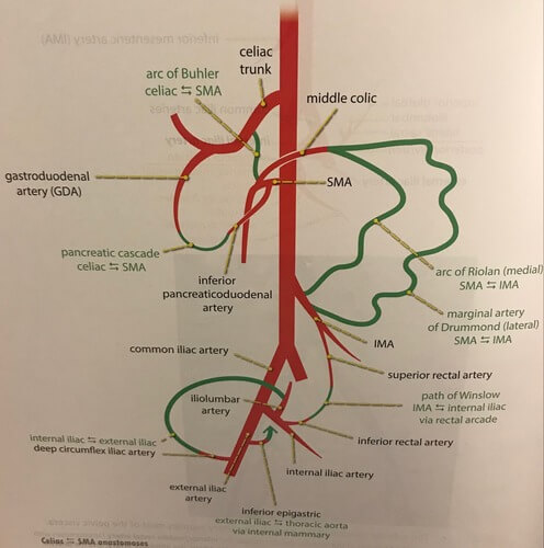
6.1 The Three Unpaired Branches of the Abdominal Aorta
| Artery | Origin | Level | Territory | Major Branches |
|---|---|---|---|---|
| Celiac Trunk | Anterior aorta | T12-L1 | Foregut | Left gastric, splenic, common hepatic |
| SMA | Anterior aorta | L1 | Midgut | Inferior pancreaticoduodenal, middle colic, right colic, ileocolic, intestinal branches |
| IMA | Anterior aorta | L3 | Hindgut | Left colic, sigmoid branches, superior rectal |
6.2 Collateral Circulation
- Marginal Artery of Drummond: Anastomotic arcade parallel to the mesenteric border of the colon, connecting ileal branch of SMA to sigmoid branch of IMA.
- Arc of Riolan (Central Anastomotic Mesenteric Artery): Proximal connection between middle colic branch of SMA and left colic branch of IMA, near the splenic flexure. Provides collateral flow in SMA or IMA occlusion.
6.3 The Hepatic Portal Venous System
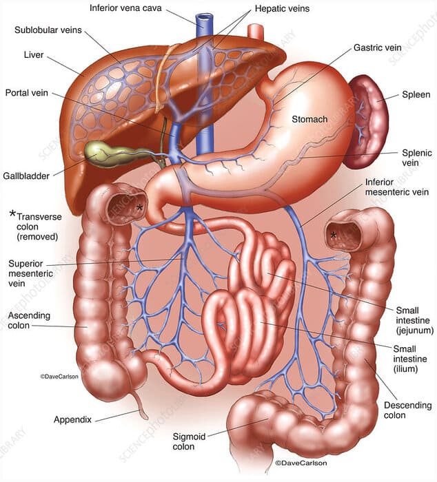
The hepatic portal vein is formed by the confluence of the splenic vein and superior mesenteric vein behind the neck of the pancreas. It carries nutrient-rich, deoxygenated blood from the GIT, spleen, and pancreas to the liver.
Major Tributaries: Splenic vein (with inferior mesenteric vein joining it), superior mesenteric vein, left gastric (coronary) vein, right gastric vein, cystic vein, paraumbilical veins.
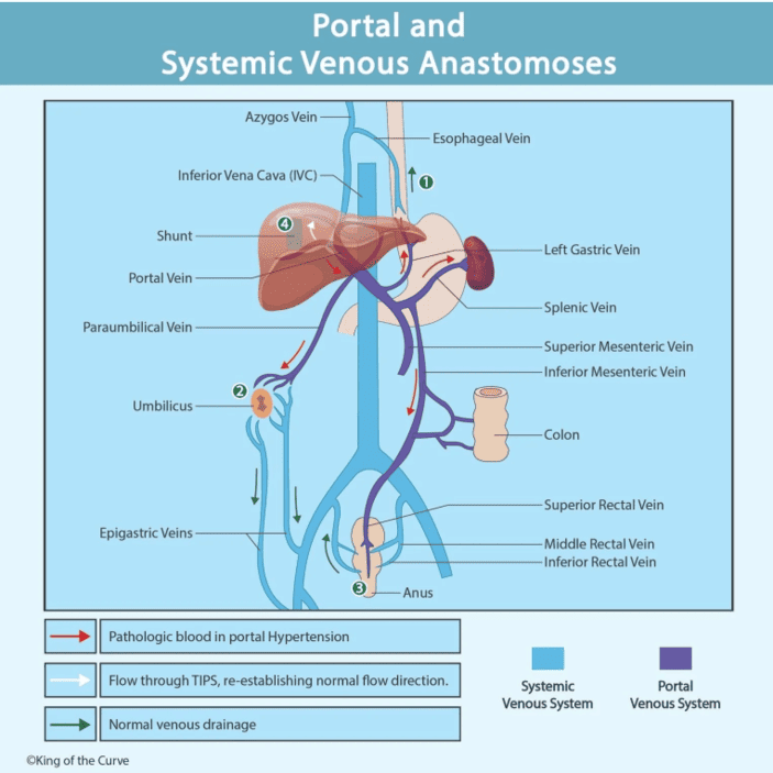
Portosystemic (Portacaval) Anastomoses
| Site | Portal System | Systemic System | Clinical Manifestation |
|---|---|---|---|
| Lower esophagus | Left gastric vein | Azygos vein | Esophageal varices |
| Umbilicus | Paraumbilical veins | Superficial epigastric veins | Caput medusae |
| Anorectal junction | Superior rectal vein | Middle and inferior rectal veins | Internal hemorrhoids |
| Retroperitoneal | Colic veins | Lumbar veins | Ascites component |
6.4 Autonomic Innervation
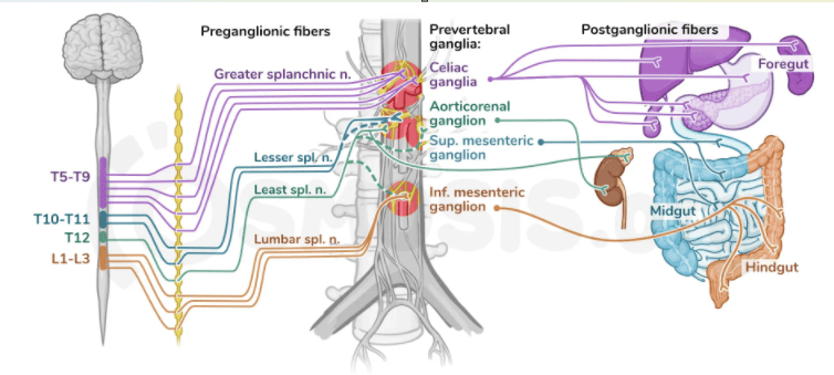
Sympathetic Pathways:
- Foregut: Greater splanchnic nerve (T5-T9) → celiac ganglia.
- Midgut: Lesser splanchnic nerve (T10-T11) → superior mesenteric ganglion.
- Hindgut: Lumbar splanchnic nerves (L1-L3) → inferior mesenteric ganglion.
- Sympathetic fibers cause vasoconstriction, decreased motility, and increased sphincter tone.
Parasympathetic Pathways:
- Foregut and Midgut: Vagus nerve → celiac and superior mesenteric plexuses.
- Hindgut: Pelvic splanchnic nerves (S2-S4) → inferior hypogastric plexus.
- Parasympathetic fibers increase motility and secretory activity and relax sphincters.
6.5 Lymphatic Drainage
Lymph from the GIT follows the arterial supply in a retrograde direction:
Pathway: Mucosal plexuses → submucosal plexuses → epicolic nodes (on colon wall) → paracolic nodes (along marginal artery) → intermediate nodes (along main colic arteries) → mesenteric nodes (celiac, superior mesenteric, inferior mesenteric) → intestinal lymph trunk → cisterna chyli (at L1-L2, posterior to aorta, right of midline) → thoracic duct.
CHAPTER 7: Clinical and Applied Anatomy
Correlation of Anatomical Knowledge with Clinical Pathology.
7.1 Portal Hypertension
Portal hypertension is elevated pressure in the portal venous system (typically >5 mmHg or hepatic venous pressure gradient >10 mmHg). The most common cause is cirrhosis with increased hepatic vascular resistance.
Clinical Manifestations at Portosystemic Anastomoses:
- Esophageal Varices: Dilated submucosal veins at lower esophagus from reversed flow through left gastric vein. Prone to rupture causing life-threatening hematemesis.
- Caput Medusae: Dilated periumbilical veins creating a radiating pattern around the umbilicus.
- Internal Hemorrhoids: Dilated superior rectal venous plexus from reversed portal flow.
- Ascites: Fluid accumulation in peritoneal cavity from portal hypertension and hypoalbuminemia.
7.2 Referred Pain Pathways
Visceral pain from abdominal organs is referred to somatic dermatomes corresponding to the embryological origin:
| Embryological Division | Pain Location | Dermatome | Representative Conditions |
|---|---|---|---|
| Foregut | Epigastric region | T7-T9 | Gastric ulcer, pancreatitis, biliary colic |
| Midgut | Periumbilical region | T10 | Appendicitis (early), small bowel obstruction, SMA syndrome |
| Hindgut | Pubic/hypogastric region | L1-L2 | Diverticulitis, sigmoid colitis, uterine conditions |
7.3 Appendicitis Progression
- Early Stage: Vague periumbilical pain from distension of the appendix and visceral afferent fibers traveling with sympathetic fibers to T10 spinal cord. Pain is dull, poorly localized, accompanied by nausea and vomiting.
- Late Stage: As inflammation progresses to involve the parietal peritoneum, pain shifts to the right iliac fossa (McBurney's point). The pain becomes sharp, constant, and well-localized due to somatic innervation of the parietal peritoneum. Rebound tenderness and guarding develop.
The transition from visceral to somatic pain reflects the transition from autonomic to somatic innervation of the involved peritoneal surfaces.
7.4 Peptic Ulcer Perforation
Gastric Ulcer Perforation:
- Anterior ulcer: Perforation into greater sac → sudden severe epigastric pain with rigid abdomen (acute peritonitis); free air under diaphragm (pneumoperitoneum) on upright chest X-ray.
- Posterior ulcer: Perforation into lesser sac or erosion into splenic artery → severe upper GI hemorrhage.
Duodenal Ulcer Perforation:
- Anterior perforation: Free air under diaphragm; bile-stained peritonitis.
- Posterior perforation: Erosion into gastroduodenal artery → massive hemorrhage.
7.5 Superior Mesenteric Artery (SMA) Syndrome
Compression of the third part of the duodenum between the abdominal aorta posteriorly and the SMA anteriorly. The normal aortomesenteric angle is 38-65°; in SMA syndrome, this angle is acutely narrowed (<25°).
- Predisposing Factors: Rapid weight loss, prolonged supine positioning, corrective spinal surgery, severe burns, prolonged bed rest.
- Symptoms: Postprandial epigastric pain, nausea, vomiting (often bilious), early satiety, weight loss. Symptoms are relieved by prone or left lateral decubitus positioning.
7.6 Ischemic Colitis
Results from insufficient blood supply to the colon, most commonly at watershed areas between arterial territories:
- Griffiths' Point: At the splenic flexure, the watershed area between the SMA territory (middle colic artery) and IMA territory (left colic artery). This is the most common site of ischemic colitis.
- Sudeck's Point: At the rectosigmoid junction, the watershed area between the IMA territory (superior rectal artery) and internal iliac territory (middle and inferior rectal arteries).
- Pathophysiology: Low flow states (shock, heart failure, arrhythmias) or arterial occlusion reduce perfusion to these watershed areas, causing mucosal ischemia, ulceration, and potentially full-thickness necrosis.
