Doctors Revision

Author name: doctorsrevision@gmail.com

Cranial Nerves
Anatomy

Cranial Nerves

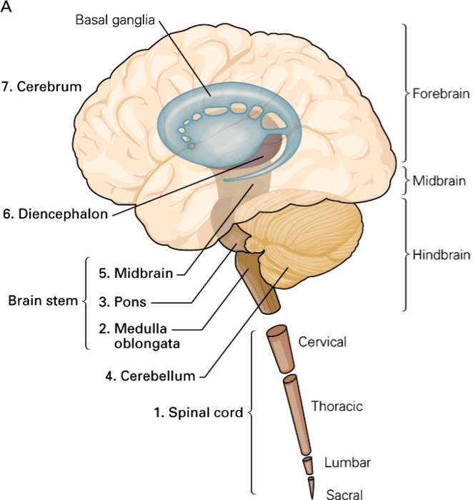
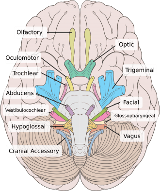
Cranial Nerves Comprehensive Notes on Neuroanatomy. Module Overview This exhaustive master guide covers the neuroanatomy of the 12 Cranial Nerves, integrating their functional components, brainstem organization, exit points, and high-yield clinical pathophysiology. By the end of this guide, you will master: The 7 Functional Components (Modalities) of cranial nerves. The Brainstem Organization and the rule of the Sulcus Limitans. Detailed anatomical pathways and clinical lesions for Cranial Nerves I through XII. How to differentiate Upper Motor Neuron (UMN) vs. Lower Motor Neuron (LMN) cranial nerve lesions. Part 1: Foundations of Cranial Nerve Anatomy Before studying individual nerves, we must understand the overarching rules that govern how they are organized, what type of information they carry, and where they originate in the brainstem. 1.1 The 7 Functional Components (Modalities) Every cranial nerve fiber acts as a specific type of wire, carrying a specific type of signal. We classify these fibers into one of seven functional categories: Abbreviation Full Name Direction What It Carries Example GSA General Somatic Afferent Sensory (→ CNS) Touch, pain, temperature, pressure from skin & mucosa. Facial sensation (CN V). SSA Special Somatic Afferent Sensory (→ CNS) Special senses of vision, hearing, and balance. Optic nerve (CN II), Vestibulocochlear (CN VIII). GVA General Visceral Afferent Sensory (→ CNS) Sensation from internal organs (stretch, chemoreception). Carotid sinus baroreceptors (CN IX). SVA Special Visceral Afferent Sensory (→ CNS) Special chemical senses of taste & smell. Taste from tongue (CN VII, IX, X). GSE General Somatic Efferent Motor (← CNS) Motor to skeletal muscles derived from embryonic somites. Extraocular muscles (CN III, IV, VI), tongue (CN XII). GVE General Visceral Efferent Motor (← CNS) Parasympathetic (autonomic) fibers to glands & smooth muscle. Pupil constriction (CN III), salivation (CN VII, IX). SVE / BME Special Visceral Efferent / Branchial Motor Motor (← CNS) Motor to skeletal muscles derived from pharyngeal (branchial) arches. Facial expression (CN VII), mastication (CN V), pharynx/larynx (CN IX, X, XI). Memory Trick “Some Say Marry Money, But My Brother Says Big Brains Matter More” This classic mnemonic helps you remember the primary function of Cranial Nerves I to XII in order: Some = Sensory (CN I, II, VIII) Say = Sensory + Motor (CN V, VII, IX, X) Marry = Motor (CN III, IV, VI, XI, XII) Money = Motor + Parasympathetic (CN III, VII, IX, X) 1.2 The Sulcus Limitans & Brainstem Organization The sulcus limitans is a crucial anatomical groove found on the floor of the fourth ventricle. It serves as a strict dividing line that organizes the brainstem into two distinct functional zones during embryological development. Key Rule: The “M-S RULE” Medial = Motor (Nuclei located medial to the sulcus limitans control motor functions). Lateral = Sensory (Nuclei located lateral to the sulcus limitans process sensory information). Note: This is the exact same organization as the spinal cord (where anterior horn = motor, posterior horn = sensory), except the neural tube has been “unzipped” and rotated 90 degrees in the brainstem, laying it flat. Columnar Organization of Nuclei (From Medial to Lateral) The cranial nerve nuclei are perfectly organized in longitudinal columns: Most medial: GSE nuclei (somatic motor) — CN III, IV, VI, XII. Next: GVE nuclei (parasympathetic) — Edinger-Westphal, superior/inferior salivatory, dorsal motor nucleus of vagus. Next: SVE nuclei (branchial motor) — motor nucleus of V, facial motor nucleus, nucleus ambiguus, spinal accessory nucleus. At sulcus limitans: SVA (taste) and GVA (visceral sensory) — nucleus of the solitary tract. Lateral: GSA (general sensation) — main and spinal trigeminal nuclei. Most lateral: SSA (special sensation) — cochlear & vestibular nuclei. 1.3 Exit Points from the Brainstem — “Factor in 4’s” A simple way to memorize where the cranial nerves exit the brainstem is the “Factor in 4’s” rule: Above the Pons (Supratentorial/Midbrain): CN I–IV (Olfactory, Optic, Oculomotor, Trochlear). At the Pons: CN V–VIII (Trigeminal, Abducens, Facial, Vestibulocochlear). Below the Pons (Medulla): CN IX–XII (Glossopharyngeal, Vagus, Accessory, Hypoglossal). 1.4 Brainstem Exits and Skull Foramina Cranial Nerve Brainstem Exit Skull Foramen Brainstem Level CN I Olfactory Forebrain (not true brainstem) Cribriform plate of ethmoid Supratentorial CN II Optic Diencephalon Optic canal Supratentorial CN III Oculomotor Interpeduncular fossa of midbrain Superior orbital fissure Midbrain CN IV Trochlear Dorsal midbrain (posterior!) Superior orbital fissure Midbrain CN V Trigeminal Lateral pons Sup. orbital fissure (V1), For. rotundum (V2), For. ovale (V3) Pons CN VI Abducens Pontomedullary junction Superior orbital fissure Pons CN VII Facial Cerebellopontine angle Internal acoustic meatus → stylomastoid foramen Pons CN VIII Vestibulocochlear Cerebellopontine angle Internal acoustic meatus Pons CN IX Glossopharyngeal Post-olivary sulcus of medulla Jugular foramen Medulla CN X Vagus Post-olivary sulcus of medulla Jugular foramen Medulla CN XI Accessory Post-olivary sulcus + C1–C5 spinal cord Jugular foramen Medulla/Spinal CN XII Hypoglossal Pre-olivary sulcus of medulla Hypoglossal canal Medulla 1.5 Parasympathetic Fibers in Cranial Nerves (GVE) It is vital to remember that only 4 cranial nerves carry parasympathetic (GVE) fibers. They dictate rest, digestion, and glandular secretion in the head, neck, and viscera. Nerve Preganglionic Nucleus Ganglion Target Effect CN III Oculomotor Edinger-Westphal nucleus (midbrain) Ciliary ganglion Sphincter pupillae + Ciliary muscle Pupil constriction + Lens accommodation CN VII Facial Superior salivatory nucleus (pons) Pterygopalatine + Submandibular ganglia Lacrimal, submandibular & sublingual glands Tearing + Salivation CN IX Glossopharyngeal Inferior salivatory nucleus (medulla) Otic ganglion Parotid gland Salivation CN X Vagus Dorsal motor nucleus of vagus (medulla) Terminal ganglia in/near target organs Thoracic & abdominal viscera “Rest & digest” functions (decreased HR, increased digestion) 1.6 UMN vs LMN Lesions: The Clinical Divide Determining whether a nerve lesion is “Upper” (in the brain) or “Lower” (at or after the nucleus) is a fundamental clinical skill. Feature Supranuclear (UMN) Lesion Nuclear/Infranuclear (LMN) Lesion Location Above the cranial nerve nucleus (e.g., motor cortex, internal capsule, upper brainstem). At or below the nucleus (the nerve root, the peripheral nerve itself, or the skull base). Muscle Tone Increased (spasticity). Decreased (flaccidity). Reflexes Hyperreflexia. Hyporeflexia / Areflexia. Fasciculations Absent. May be prominently present (twitching). Atrophy Absent or very mild (disuse).
Blood Supply Of The CNS
Anatomy

Blood Supply Of The CNS

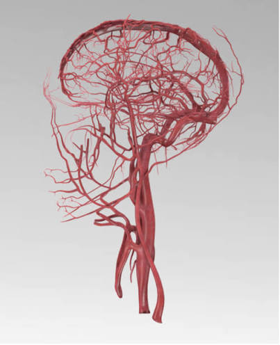
Blood supply of the CNS Comprehensive Notes on the Blood Supply of the Central Nervous System Module Learning Objectives By the end of this comprehensive guide, you will be deeply conversant with: The complete Arterial Inflow pathways to the brain, including the Internal Carotid and Vertebrobasilar systems. The anatomical layout and functional importance of the Circle of Willis as a collateral network. The specific Cerebral Cortical Territories (ACA, MCA, PCA) and their functional correlates, including watershed zones. The intricate Deep Structure & Brainstem Perfusion, including the vulnerable lenticulostriate arteries. The layout of Spinal Cord Blood Supply and the critical Artery of Adamkiewicz. The Venous Drainage & Dural Sinuses, highlighting the anatomy and vulnerability of the cavernous sinus. Key Clinical Anatomy & Pathophysiology, distinguishing between stroke syndromes, lacunar infarcts, and types of intracranial hemorrhages (Epidural, Subdural, Subarachnoid). 1. Arterial Inflow to the Brain & The Circle of Willis The brain is a highly metabolically active organ. Despite accounting for only about 2% of total body weight, it receives 15-20% of the body’s cardiac output and consumes 20% of its oxygen. This massive demand is met by two major arterial systems that converge at the base of the brain. A. The Anterior Circulation (Internal Carotid System) The anterior circulation supplies the majority of the cerebral hemispheres, specifically the frontal, parietal, and lateral temporal lobes, as well as deep structures like the basal ganglia. Pathway from the Heart: Aortic Arch → Brachiocephalic trunk (on the Right) / Left Common Carotid (on the Left) → Common Carotid Artery (CCA). Bifurcation: The CCA bifurcates at the C3-C4 vertebral level into the External Carotid (supplying the face/neck) and the Internal Carotid Artery (ICA). Distribution: The ICA enters the skull and ultimately bifurcates into the Anterior Cerebral Artery (ACA) (supplying the medial hemisphere) and the Middle Cerebral Artery (MCA) (supplying the lateral hemisphere). The Four Segments of the Internal Carotid Artery (ICA) Segment Course and Anatomical Significance 1. Cervical Runs from the CCA bifurcation to the skull base. Contains NO branches in the neck. This is the primary site for atherosclerotic plaque buildup (evaluated via auscultation for bruits and treated with carotid endarterectomy). 2. Petrous Enters the carotid canal within the petrous portion of the temporal bone. It runs anterior to the cochlea. A fracture here can lead to massive epistaxis (nosebleeds) or hearing issues. 3. Cavernous Passes directly through the cavernous venous sinus, forming an S-shaped curve known as the “carotid siphon.” It is intimately surrounded by cranial nerves (CN III, IV, V1, V2, VI). Trauma here can cause a carotid-cavernous fistula. 4. Cerebral (Supraclinoid) Pierces the dura mater to become intradural. It gives off important branches: Ophthalmic artery, Posterior Communicating Artery (PCoA), and Anterior Choroidal Artery (AChA), before finally bifurcating into the ACA and MCA. B. The Posterior Circulation (Vertebrobasilar System) The posterior circulation supplies the brainstem, cerebellum, occipital lobes, and inferior temporal lobes. Pathway: Subclavian Artery → Vertebral Artery → Ascends through the transverse foramina of cervical vertebrae (C6-C1) → Enters the skull via the Foramen Magnum. The Basilar Artery: The two vertebral arteries merge at the pontomedullary junction to form the single Basilar Artery, which runs up the front of the pons. Distribution: The basilar artery bifurcates at the midbrain into the two Posterior Cerebral Arteries (PCA). These join the Circle of Willis via the Posterior Communicating Arteries (PCoA). C. The Circle of Willis The Circle of Willis is a ring of vessels at the base of the brain that connects the anterior and posterior circulations, as well as the left and right sides of the brain. Functional Importance It serves as a critical pressure-equalizing anastomotic network. If one major artery slowly occludes (e.g., gradual ICA stenosis), blood can reroute across the communicating arteries to maintain perfusion to the deprived area, preventing a stroke. Interestingly, a complete, anatomically perfect circle is present in only ~50% of individuals (many have hypoplastic or missing segments, reducing this collateral capacity). Components of the Circle: Anterior Communicating Artery (AComm): Connects the left and right ACAs. This is the most common site for berry aneurysms. Anterior Cerebral Artery (ACA): Supplies the medial cerebral hemispheres. Internal Carotid Artery (ICA): The main input for the anterior system. Posterior Communicating Artery (PComm): The vital bridge connecting the ICA (anterior) to the PCA (posterior). Posterior Cerebral Artery (PCA): Formed from the basilar artery; supplies the occipital lobe. Basilar Artery: (While technically the input, it is the foundation of the posterior portion of the circle). 2. Cerebral Cortical Territories & Watershed Zones Understanding which artery supplies which part of the brain cortex is essential for diagnosing strokes based on a patient’s physical symptoms. This relies heavily on the “motor and sensory homunculus”—the map of the body on the brain’s surface. Arterial Territories & Functional Correlates Artery Cortical Surface Supplied Key Functional Areas Affected Classic Stroke Deficits ACA (Anterior Cerebral) Medial Surface: Medial frontal and parietal lobes, cingulate gyrus, anterior corpus callosum. Motor & sensory cortex for the LEG and FOOT, prefrontal cortex (executive function), micturition center. Contralateral leg weakness > arm weakness. Urinary incontinence. Personality changes, abulia (lack of will), akinetic mutism. Grasp reflex return. MCA (Middle Cerebral) Lateral Surface: Most of the lateral convexity (precentral, postcentral, inferior frontal, superior temporal, angular gyri), insula. Motor & sensory cortex for the FACE and ARM. Broca’s area (speech production – dominant side). Wernicke’s area (speech comprehension). Contralateral face/arm weakness > leg weakness. Aphasia (if dominant hemisphere, usually left). Hemineglect (if non-dominant, usually right). Gaze deviation toward the lesion. PCA (Posterior Cerebral) Medial Occipital & Inferior Temporal: Lingual gyrus, calcarine sulcus, cuneus, thalamus (deep). Primary Visual Cortex (calcarine fissure), visual association areas, memory formation. Contralateral homonymous hemianopia (loss of half the visual field) with macular sparing. Alexia without agraphia (can write but can’t read). Thalamic pain syndromes. Watershed Zones (Border Zones) Watershed zones are the regions at the very periphery of arterial territories where the most distal branches of two different major arteries meet. Why are they vulnerable? Because they represent the absolute end of

Internal structures of the CNS (ventricles)
Anatomy

Internal structures of the CNS (ventricles)

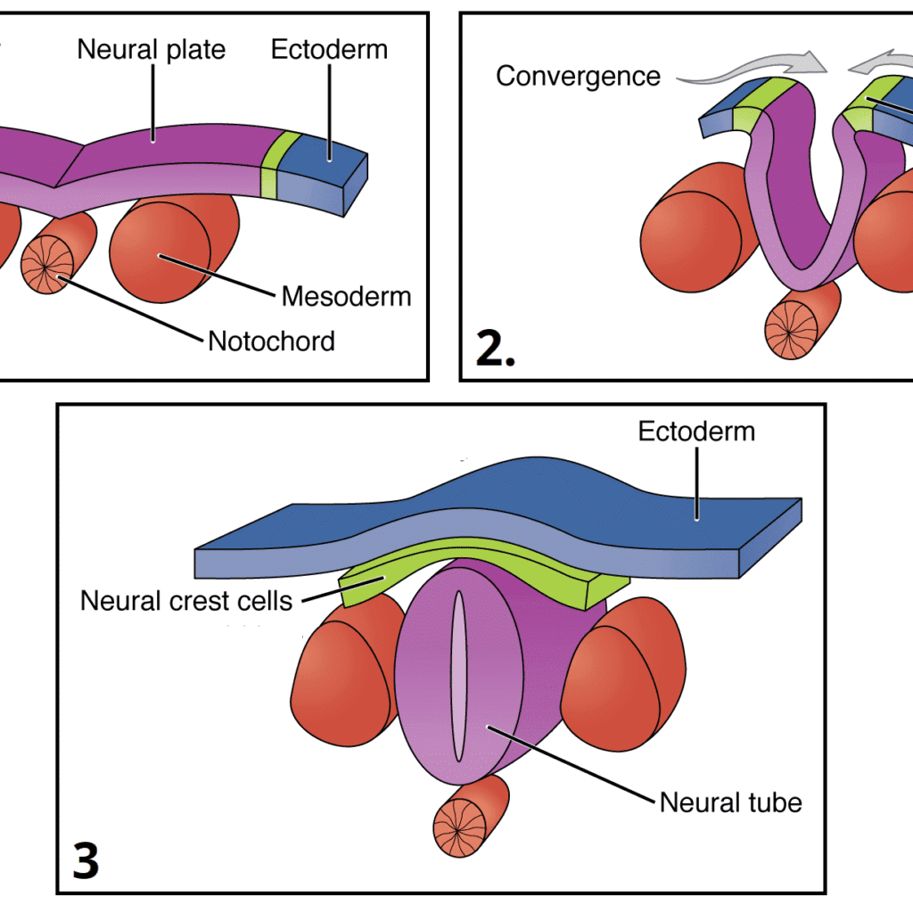
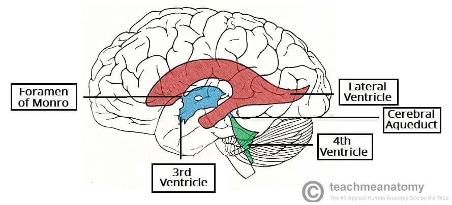
Internal Structures (Ventricles) of tha CNS & Cerebrospinal Fluid (CSF) Dynamics Module Learning Objectives By the conclusion of this exhaustive master guide, you will be deeply conversant with the internal structures of the Central Nervous System, specifically focusing on: The Embryological Origins of the neural tube and how it develops into the adult ventricular system. The intricate Structural Morphology and Boundaries of the lateral, third, and fourth ventricles, as well as the cerebral aqueduct. The comprehensive CSF Hydrodynamics and Flow Kinetics, from production to resorption. The specific Foramina and Communications that act as doorways between the ventricular chambers. The Neuroradiological Correlations for identifying these structures on CT and MRI scans. The Clinical Pathophysiology of Hydrocephalus and raised intracranial pressure. 1. Embryological Origins: From Neural Tube to Adult Ventricles To truly understand the complex, winding shapes of the adult ventricles, we must first look at how they develop. The entire ventricular system is simply the adult remnant of the original hollow center of the embryonic neural tube. The Neural Tube Timeline Week 3: The neural plate forms from the ectoderm on the dorsal surface of the embryo. Week 4: The neural plate folds inward to form the neural groove, and the edges fuse to create the neural tube. It closes from the middle outward (like a zipper). Failure of this closure leads to neural tube defects (e.g., Spina Bifida). The Core Concept: The hollow center of this neural tube becomes the fluid-filled ventricular system of the brain and the central canal of the spinal cord. Think of it like a simple garden hose that eventually gets pinched, bent, and ballooned out into different shapes. Primary Brain Vesicles (Weeks 4-5) The anterior (head) end of the neural tube swells into three primary fluid-filled chambers: 1. Prosencephalon (Forebrain) The most anterior portion. The hollow space inside will eventually become the Lateral and 3rd Ventricles. 2. Mesencephalon (Midbrain) The middle portion. The hollow space here narrows to become the Cerebral Aqueduct. 3. Rhombencephalon (Hindbrain) The posterior portion. The hollow space becomes the 4th Ventricle. Note: The rest of the neural tube extending down the back becomes the spinal cord, and its hollow center becomes the extremely narrow Central Canal. Secondary Vesicles: The Forebrain Splits As development continues, the primary vesicles divide further to form the mature structures of the brain. The Prosencephalon splits into: Telencephalon: This forms the massive cerebral hemispheres, basal ganglia, and hippocampus. Because it grows so large and splits into two hemispheres, its internal cavity also splits to form the paired, C-shaped Lateral Ventricles. Diencephalon: This forms the central core (thalamus, hypothalamus, epithalamus, subthalamus). Its internal cavity remains in the exact midline and forms the slit-like Third Ventricle. The Rhombencephalon splits into: Metencephalon: Forms the pons and cerebellum. Myelencephalon: Forms the medulla oblongata. Both of these structures share the diamond-shaped Fourth Ventricle. Key Memory Points for Embryology PRO = FORE = FRONT: The lateral and 3rd ventricles are in the FRONT of the brain. MESO = MIDDLE: The cerebral aqueduct runs through the MIDDLE of the brain (midbrain). RHOMBO = DIAMOND: The 4th ventricle is DIAMOND-shaped. Lateral ventricles are PAIRED because the telencephalon forms two distinct cerebral hemispheres. The 3rd ventricle is MIDLINE because the diencephalon is a single central structure. The cerebral aqueduct is the NARROWEST part—it is essentially a squeezed, unexpanded portion of the original neural tube lumen. 2. Structural Morphology: Boundaries, Recesses, and Relations The ventricles are not empty voids; they are anatomically precise rooms bordered by specific brain structures. Knowing these borders is essential for neurosurgery and reading brain scans. A. The Lateral Ventricles (The C-Shaped Chambers) These are paired structures, one buried deep within each cerebral hemisphere. Each lateral ventricle is massive and has four distinct parts (a central body with three horn-like extensions): Region Roof Floor Medial Wall Lateral Wall Frontal (Anterior) HornExtends forward into the frontal lobe Corpus callosum (genu) Head of the caudate nucleus Septum pellucidum Corpus callosum Body (Central Part)The main horizontal portion Corpus callosum (body) Thalamus + body of caudate nucleus Septum pellucidum + fornix Tapetum of the corpus callosum Atrium (Trigone)The wide junction where the body, temporal, and occipital horns meet Corpus callosum Collateral trigone Crus of the fornix Tapetum + optic radiation Temporal (Inferior) HornCurves downward and forward into the temporal lobe Tapetum + tail of caudate nucleus Hippocampus (Very important landmark!) Stria terminalis Tapetum Occipital (Posterior) HornExtends backward into the occipital lobe Splenium of the corpus callosum Collateral trigone Crus of fornix + splenium Tapetum B. The Third Ventricle (The Midline Slit) The third ventricle is a narrow, vertical, slit-like cavity located exactly in the midline, sandwiched between the left and right halves of the diencephalon. Roof: Formed by the tela choroidea and the body of the fornix. The tela choroidea is a thin, two-layered membrane containing the highly vascular choroid plexus. Floor: Formed by the structures of the hypothalamus (including the mammillary bodies, tuber cinereum, and the optic chiasm). Anterior Wall: Formed by the lamina terminalis (a thin sheet of gray matter that marks the anterior limit of the original neural tube) and the anterior commissure. Posterior Wall: The pineal gland and the posterior commissure. Lateral Walls: Formed predominantly by the medial surfaces of the two thalami. Massa Intermedia (Interthalamic Adhesion): In about 70% of people, a small bridge of gray matter crosses directly through the center of the third ventricle to connect the left and right thalami. Recesses of the 3rd Ventricle: These are small, blind-ended extensions of the fluid space pushing into surrounding structures. They include the Optic recess (above the optic chiasm), Infundibular recess (funneling down into the pituitary stalk), Pineal recess (pushing into the pineal gland), and Suprapineal recess. C. The Cerebral Aqueduct (of Sylvius) — The Bottleneck This is a narrow channel running through the center of the midbrain. It connects the 3rd ventricle above to the 4th ventricle below. Dimensions: Approximately 15 mm long and only 1-2 mm in diameter. It is the NARROWEST part of the entire ventricular system,

Major divisions of the CNS
Anatomy

Major divisions of the CNS

Major divisions of the Central Nervous System A comprehensive note about the major divisions of the CNS Module Learning Objectives & Overview By the conclusion of this exhaustive master guide, you will be deeply conversant with: The Prosencephalon (Forebrain): Detailed anatomy of the Telencephalon (cortex, white matter tracts, basal ganglia) and the Diencephalon (thalamus, hypothalamus, epithalamus). The Mesencephalon (Midbrain): Structural division into the Tectum, Tegmentum, and Crus Cerebri, including cranial nerve exits. The Rhombencephalon (Hindbrain): In-depth exploration of the Metencephalon (Pons & Cerebellum) and Myelencephalon (Medulla Oblongata). The Spinal Cord: Gross external boundaries, internal gray/white matter organization, and ascending/descending tracts. The Ventricular System & CSF: Fluid production, circulation pathways, and clinical pathologies (Hydrocephalus). I. PROSENCEPHALON (Forebrain) The prosencephalon, or forebrain, is the most rostral (forward) division of the developing neural tube. It is the seat of the highest level of neural processing in the human body. It gives rise to the telencephalon (which becomes the massive cerebral hemispheres and deep gray matter structures) and the diencephalon (the central core containing the thalamus, hypothalamus, and associated structures). Together, these regions process sophisticated sensory information, regulate primitive emotions, control fine voluntary movement, and coordinate higher cognitive functions like language, memory, and executive thought. A. Telencephalon (Cerebral Hemispheres & Deep Structures) The telencephalon forms the absolute largest part of the human brain. It consists of the two large cerebral hemispheres, highly convoluted surface cortex (gray matter), underlying massively connected white matter tracts, and deep gray matter nuclei (the basal ganglia and limbic structures). 1. Surface Anatomy & Lobes To fit the massive surface area of the cerebral cortex into the rigid skull, the brain folds into ridges called gyri and grooves called sulci (or deeper fissures). The cerebral cortex is divided into major lobes by three key anatomical landmarks: Central Sulcus (of Rolando): A prominent, continuous vertical groove separating the frontal lobe (anterior) from the parietal lobe (posterior). Clinical Importance: It is your absolute primary landmark for locating the primary motor and primary sensory cortices. Lateral Fissure (of Sylvius): A deep, sweeping horizontal fissure separating the temporal lobe (inferior) from the frontal and parietal lobes (superior). If you pull the lips (opercula) of this fissure apart, the hidden insular cortex lies buried deep within. Parieto-occipital Sulcus: Found primarily on the medial surface of the hemisphere, marking the definitive boundary between the parietal and occipital lobes. Key Gyri to Master Precentral Gyrus: Located immediately anterior to the central sulcus. This is the primary motor cortex (Brodmann area 4). It houses the upper motor neurons that control voluntary movement of the contralateral (opposite) side of the body. Postcentral Gyrus: Located immediately posterior to the central sulcus. This is the primary somatosensory cortex (Brodmann areas 3, 1, 2). It receives all tactile, pain, temperature, and proprioceptive information from the contralateral body. Insular Cortex: A triangular-shaped lobe buried deep in the lateral fissure. It is highly involved in interoception (awareness of the body’s internal state), taste processing (gustatory cortex), and primitive emotion/empathy. EXAM TIP: On practical exams, always identify the central sulcus first to orient yourself. Precentral = anterior = motor (Evolutionarily, action comes FIRST). Postcentral = posterior = sensory (Perception of that action comes AFTER). 2. White Matter Tracts White matter consists of heavily myelinated axon bundles connecting different brain regions. They are categorized into three types: Commissural fibers (connect hemispheres), Projection fibers (connect cortex to lower structures), and Association fibers (connect areas within the same hemisphere). Corpus Callosum: The absolute largest commissure in the brain, containing hundreds of millions of axons connecting the left and right cerebral hemispheres to ensure they act as a unified whole. It is divided into four parts (from anterior to posterior): Rostrum: The thin, beak-like anterior portion pointing downward. Genu: The sharp “knee” or bend at the anterior end. Body (Trunk): The long, massive central portion covering the lateral ventricles. Splenium: The thickened, bulbous posterior end (splenium means “bandage”). Internal Capsule: A massive, V-shaped band of projection white matter containing almost all ascending (sensory) and descending (motor) fibers traveling between the cerebral cortex and subcortical structures (brainstem/spinal cord). It has three highly distinct parts: Anterior Limb: Located between the head of the caudate nucleus and the lentiform nucleus. Contains frontopontine and thalamocortical fibers. Genu: The sharp bend; critically contains corticobulbar fibers (the motor fibers descending to innervate the cranial nerve nuclei in the brainstem for face/jaw/swallow movement). Posterior Limb: Located between the thalamus and the lentiform nucleus. Highly critical as it contains the corticospinal tract (motor to the body) and major ascending somatosensory fibers from the thalamus to the cortex. Anterior Commissure: A smaller, secondary commissure connecting the temporal lobes and olfactory bulbs across the midline. It plays a role in pain sensation and smell. Fornix: A prominent C-shaped bundle of white matter that serves as the major output tract of the hippocampus, connecting it to the mammillary bodies of the hypothalamus. It is a critical highway for the limbic system and memory formation. Clinical Pearl Internal Capsule Stroke (Lacunar Infarct) The internal capsule is incredibly HIGH-YIELD. Because millions of descending motor fibers are packed tightly into the tiny space of the posterior limb, a very small stroke here (a lacunar infarct caused by occlusion of the tiny lenticulostriate arteries) will cause massive, devastating pure motor hemiparesis (paralysis) on the contralateral side of the entire body. Rule: One small capsule, big consequences. 3. Deep Gray Matter (Basal Ganglia & Limbic Structures) Deep within the white matter of the cerebral hemispheres lie clusters of neuronal cell bodies (gray matter) that regulate sophisticated aspects of movement, primitive emotion, and long-term memory. Corpus Striatum: The largest and most functionally critical component of the basal ganglia motor circuit. It consists of: Caudate Nucleus: A large, C-shaped structure that perfectly follows the curve of the lateral ventricle. It has three parts: the head (a large bulbous anterior portion bulging into the anterior horn of the lateral ventricle), the body (central portion), and the tail (tapering down and back into the temporal lobe). Lentiform Nucleus:

Development of the CNS
Anatomy

Development of the CNS

Development of the Central Nervous System Learning Objectives & Overview By the end of this highly detailed, exhaustive master guide on neuroembryology, you will be deeply conversant with: The molecular inductions and primary germ layer origins orchestrating the Central Nervous System (CNS) formation. The complex Sonic Hedgehog (SHH) and Bone Morphogenetic Protein (BMP) signaling pathways governing dorsal-ventral patterning. The chronological evolution of primary and secondary brain vesicles and their precise adult derivatives (structures and ventricles). The intricate processes of cellular proliferation, radial glial scaffolding, and the Epithelial-to-Mesenchymal Transition (EMT) of the Neural Crest. High-yield clinical embryology, including Neural Tube Defects (NTDs), posterior fossa malformations, and migration defects. SECTION 1: MOLECULAR & EARLY EMBRYONIC INDUCTIONS The development of the Central Nervous System (CNS) is arguably the most complex, highly orchestrated embryological process in human development. It requires exact spatial and temporal precision, relying heavily on molecular cross-talk between different embryonic tissues. 1.1 Primary Germ Layer Origin & Neurulation Before the CNS can form, the embryo must undergo Gastrulation (Week 3), converting the bilaminar disc into a trilaminar disc consisting of three primary germ layers: Ectoderm, Mesoderm, and Endoderm. The entire nervous system traces its lineage back to the outermost layer. The Origin: The Central Nervous System, Peripheral Nervous System, and the skin epidermis are all derived from the Ectoderm. The Induction Process: The ectoderm does not become neural tissue on its own. It requires a chemical command. A specialized rod of mesodermal tissue called the Notochord (the primitive axial skeleton) develops directly beneath the dorsal ectoderm. The Notochord’s Role: The notochord acts as the primary embryonic organizer. It secretes powerful signaling molecules (like Noggin, Chordin, and Sonic Hedgehog) that diffuse upward into the overlying ectoderm. These signals block inhibitory bone morphogenetic proteins (BMPs), commanding the ectoderm to thicken and transform into the Neural Plate. This specialized tissue is now officially committed to becoming the nervous system (Neuroectoderm). Neurulation: The neural plate folds inward to form the neural groove, and the edges (neural folds) fuse together to create the hollow Neural Tube. This closure begins in the cervical (neck) region and zips up towards the head (rostral neuropore closes day 25) and down towards the tail (caudal neuropore closes day 27). 1.2 Sonic Hedgehog (SHH) Signaling Pathway Sonic Hedgehog (SHH) is one of the most critical morphogens in developmental biology. In the CNS, it is responsible for ventral patterning (making the bottom half of the brain and spinal cord) and the separation of the single primitive brain vesicle into two distinct right and left cerebral hemispheres. The Complete Signaling Cascade (Step-by-Step): Secretion & Gradient: The Notochord secretes SHH protein. The SHH diffuses upward, creating a concentration gradient (highest concentration is ventral, lowest is dorsal). This SHH induces the ventralmost part of the neural tube to become the Floor Plate, which then also begins secreting SHH. Receptor Binding: SHH binds to a specific inhibitory cell-surface receptor called Patched-1 (PTCH1). Release of Inhibition: Normally, PTCH1 constantly inhibits another transmembrane protein called Smoothened (SMO). When SHH binds PTCH1, the inhibition on SMO is immediately lifted. Activation of Transcription: Activated SMO prevents the cleavage of GLI complex proteins. The full, intact GLI zinc-finger transcription factors translocate into the nucleus. Gene Expression: GLI activates the expression of specific ventralizing genes, turning the local neural stem cells into motor neurons and interneurons. Clinical Correlation Holoprosencephaly (HPE) Mutations affecting the SHH signaling pathway (e.g., mutations in the SHH gene itself, or maternal exposure to cholesterol synthesis inhibitors, or teratogens like cyclopamine) prevent the embryonic forebrain from properly dividing into two separate hemispheres. Result: A spectrum of severe midline defects. The brain remains a single fused lobe (alobar HPE). Phenotype: Presents with severe facial abnormalities including cyclopia (a single central eye), proboscis (a tube-like nose above the eye), cleft lip/palate, and a single fused central incisor. 1.3 Dorsal-Ventral Patterning: Alar vs. Basal Plates As the neural tube develops, it forms a longitudinal groove along its inner lateral walls called the Sulcus Limitans. This acts as a definitive boundary line, splitting the neural tube into a dorsal (top) half and a ventral (bottom) half. This separation dictates the entire sensory and motor organization of the adult CNS. Feature Alar Plate (Dorsal) Basal Plate (Ventral) Location Dorsal to the Sulcus Limitans. Ventral to the Sulcus Limitans. Function SENSORY (Afferent) processing. MOTOR (Efferent) processing. Molecular Inducer Induced by BMPs and Wnt secreted by the Roof Plate and overlying surface ectoderm. Induced by Sonic Hedgehog (SHH) secreted by the Notochord and Floor Plate. Adult Spinal Cord Derivative Becomes the Dorsal Horn (receives sensory input from dorsal root ganglia). Becomes the Ventral Horn (contains lower motor neurons projecting to muscles) and Lateral Horn (autonomics). Adult Brainstem Derivative Migrates laterally. Forms the sensory cranial nerve nuclei (e.g., Trigeminal sensory, Vestibulocochlear). Remains medially located. Forms the motor cranial nerve nuclei (e.g., Oculomotor, Hypoglossal). Master Mnemonic – “SAME DAVE”: Sensory = Afferent, Motor = Efferent. Dorsal = Afferent, Ventral = Efferent. SECTION 2: PRIMARY AND SECONDARY VESICULATION Once the neural tube forms, its cranial (head) end undergoes a rapid series of swellings and expansions to form the complex structures of the brain. This is known as vesiculation, occurring in two distinct chronological phases. 2.1 The Chronological Evolution of the Neural Tube Week 4: The 3 Primary Vesicles At approximately day 28 of gestation, the cranial end of the neural tube expands to form three distinct swellings (primary vesicles): Prosencephalon (Forebrain): The most rostral portion. Mesencephalon (Midbrain): The middle portion. Rhombencephalon (Hindbrain): The caudal portion, continuous with the spinal cord. Week 5: The 5 Secondary Vesicles By day 35, the primary vesicles undergo further division. The Prosencephalon and Rhombencephalon split, while the Mesencephalon remains a single vesicle. This creates five secondary vesicles that give rise to the definitive adult brain structures. From PROSENCEPHALON Telencephalon: Rapidly outgrows all other structures, folding over them. It becomes the Cerebral Hemispheres (Cortex, White Matter, Basal Ganglia, Hippocampus, Amygdala, and Olfactory bulbs). Diencephalon: The central core. It becomes the Thalamus, Hypothalamus, Epithalamus (Pineal

Embryology (Pharyngeal Arches)
Anatomy

Embryology (Pharyngeal Arches)

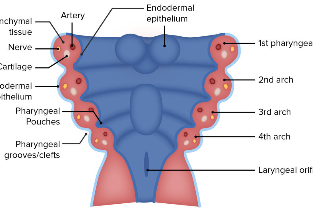
Embryology of the Head & Neck: Pharyngeal Arches An exhaustive, highly detailed anatomical and clinical master guide covering the structure, derivatives, special organs, and clinical conditions of the Pharyngeal Arches. Module Learning Objectives By the conclusion of this comprehensive guide, you will be deeply conversant with: The Basic Structure & Organization of the pharyngeal apparatus (Arches, Clefts, and Pouches) and their embryonic timeline. The exhaustive list of Arch Derivatives, perfectly correlating each arch with its specific Cranial Nerve, Skeletal/Cartilaginous elements, Muscles, and Aortic Blood Vessels. The intricate embryonic development of Special Organs, specifically the Tongue, Face, Thyroid Gland, and Pharyngeal Pouch derivatives. The pathophysiology and presentation of major Clinical Conditions and Congenital Anomalies linked to developmental failures in this region. SECTION 1: Basic Structure & Organization 1.1 When Do Pharyngeal Arches Develop? (The Embryonic Timeline) The pharyngeal (branchial) arches are the fundamental building blocks of the head and neck. The term “branchial” is historically derived from the Greek word branchia, meaning “gill,” because during early embryonic development, these human structures strongly resemble the gill slits seen in fish and amphibian embryos. The development occurs rapidly during a highly vulnerable window of gestation: Week 4 (First Arch Appears): The head and neck region begins to take shape. The first pharyngeal arch appears as a distinct bar of mesoderm covered by ectoderm on the outside and endoderm on the inside. Weeks 4 to 5 (All Arches Form): All five human arches (labeled I, II, III, IV, and VI) form in a strict craniocaudal sequence (from top/head to bottom/tail). Crucial Embryological Note: There is NO 5th Pharyngeal Arch in humans. In human embryos, the 5th arch either never forms at all, or it appears as a tiny, transient structure that regresses and disappears almost instantly without leaving any adult derivatives. Weeks 5 to 6 (Derivatives Begin): Each arch acts as an independent factory, beginning to produce its own highly specific structures: a cranial nerve, an artery, muscle tissue, and a cartilage bar. Weeks 6 to 8 (Major Structures Formed): The arches remodel, fuse, and migrate. Their initial primitive derivatives morph into easily recognizable adult structures in the head and neck (e.g., the jaw, the bones of the middle ear, the larynx). 1.2 The Four Main Parts of Each Arch Think of a pharyngeal arch as an independently pre-packaged “starter kit” for a segment of the neck. Every single arch consists of a core of mesodermal and neural crest tissue, and contains exactly four fundamental components: 1. The Nerve Cranial Nerve Branch: Each arch is supplied by one specific cranial nerve that grows into it from the developing brainstem. This nerve will exclusively control the muscles that develop from that specific arch, and will provide sensory innervation to the skin/mucosa derived from it. 2. The Artery Aortic Arch Branch: Each arch has its own arterial blood supply, known as an aortic arch. These vessels arise from the primitive heart tube (aortic sac) and course through the arches. They eventually remodel to become the major arteries of the adult chest, neck, and head. 3. The Muscle Skeletal Muscle: The muscle component is derived from the Mesoderm (specifically paraxial and lateral plate mesoderm). These muscle precursor cells migrate into the arch and differentiate into the skeletal muscles of facial expression, mastication, swallowing, and vocalization. 4. The Cartilage Skeletal Element: The cartilage and bone of the arches are derived primarily from Neural Crest Cells (neuroectoderm). These highly migratory cells travel into the arches to form the structural skeleton of the face, jaw, and neck. 1.3 Arches vs. Clefts vs. Pouches (The Three Layers) The pharyngeal apparatus is not just a solid block; it is corrugated. It consists of three distinct anatomical and embryological layers. Misunderstanding these layers is the leading cause of confusion in head and neck embryology. Structure Anatomical Location Embryonic Germ Layer Origin What It Becomes (General Fate) Pharyngeal Cleft (Groove) The indentations on the OUTSIDE of the embryo neck. Ectoderm Only the 1st Cleft forms a permanent adult structure: the External Auditory Meatus (Ear Canal). Clefts 2, 3, and 4 are normally overgrown by Arch 2 and disappear. Pharyngeal Arch The bulging tissue masses between the cleft and pouch. Mesoderm + Neural Crest Forms the core structures: Muscles, bones, cartilage, specific cranial nerves, and arteries of the face and neck. Pharyngeal Pouch The indentations on the INSIDE of the primitive pharynx. Endoderm Forms crucial internal cavities and endocrine glands: Middle ear cavity, Palatine Tonsils, Thymus, and Parathyroid glands. SECTION 2: Arch Derivatives (What the Arches Become) 2.1 Cranial Nerves of the Pharyngeal Arches The cranial nerves are the wiring of the head and neck. As muscles migrate away from their original arch during development, they drag their specific nerve with them. Therefore, knowing a muscle’s nerve supply instantly tells you which pharyngeal arch it originated from. Arch Cranial Nerve Number Nerve Name Main Function / Territory Arch I CN V Trigeminal Nerve(Specifically V3 – Mandibular Division) Chewing (Muscles of Mastication), general face and jaw sensation. Arch II CN VII Facial Nerve Facial expression (smiling, frowning, blinking), and taste to the anterior 2/3 of the tongue. Arch III CN IX Glossopharyngeal Nerve Swallowing (Stylopharyngeus muscle), and general sensation + taste to the posterior 1/3 of the tongue. Arch IV CN X Vagus Nerve(Superior Laryngeal Branch) Sensation to the larynx ABOVE the vocal cords, and swallowing (pharyngeal constrictors). Arch VI CN X Vagus Nerve(Recurrent Laryngeal Branch) Motor control to all intrinsic muscles of the larynx (voice production) BELOW the vocal cords. 2.2 Bones & Cartilages (Skeletal Derivatives) The cartilaginous rods within each arch give rise to the rigid structures of the jaw, middle ear, and voice box. Arch I Cartilage Meckel’s Cartilage This acts as the primary cartilage model for the lower face. However, most of Meckel’s cartilage actually disappears (degenerates) and is replaced by bone via intramembranous ossification. Bones Formed: Mandible (lower jaw – forms around the cartilage, not from it), Maxilla (upper jaw), Zygomatic bone (cheekbone). Middle Ear Bones: The proximal ends of

Special Senses (Eye & Ear)
Anatomy

Special Senses (Eye & Ear)

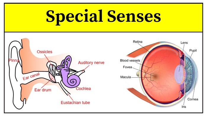
Eye & Ear Anatomy: Comprehensive Master Guide Module Learning Objectives By the conclusion of this exhaustive master guide, you will be deeply conversant with: The complex bony framework, foramina, and neurovascular contents of the orbit. The precise origins, insertions, and actions of the extraocular muscles, along with their associated cranial nerve palsies. The intricate multi-layered anatomy of the eyeball (fibrous, vascular, and nervous tunics), including fluid dynamics and glaucoma. The autonomic pathways governing pupillary responses (mydriasis and miosis) and the complete visual pathway. The anatomical subdivisions of the temporal bone and ear (external, middle, inner), including the mechanical amplification of the auditory ossicles. The physiological mechanics of sound transduction and vestibular equilibrium. The complete, segment-by-segment course of the Facial Nerve (CN VII). I. The Orbit: Overview & Bony Framework The orbit is a bilateral, pyramidal bony cavity situated in the upper facial skeleton. It serves to house, protect, and support the eyeball, extraocular muscles, highly delicate vessels, cranial nerves, the lacrimal apparatus, and a protective cushion of orbital adipose (fat) tissue. Shape & Volume: It is pyramidal, with the apex directed posteriorly (pointing towards the optic canal) and the base facing anteriorly (forming the orbital margin). The total volume of the orbit is approximately ~30 mL, of which the eyeball occupies only about ~7 mL (the rest is fat, muscles, and neurovasculature). The Seven Bones of the Orbit The orbital walls are constructed by contributions from seven different bones. (Mnemonic: Many Friendly Zebras Enjoy Lazy Summer Picnics → Maxilla, Frontal, Zygomatic, Ethmoid, Lacrimal, Sphenoid, Palatine). Orbital Wall Bony Composition Clinical & Anatomical Relations Roof (Superior) Frontal bone (anteriorly) + Lesser wing of sphenoid (posteriorly). Directly separates the orbit from the frontal sinus (inferiorly) and the anterior cranial fossa (superiorly). Floor (Inferior) Maxilla (main portion) + Zygomatic (anterolateral) + Palatine (tiny posteromedial contribution). Separates the orbit from the underlying maxillary sinus. Highly prone to “blowout” fractures. Medial Wall Maxilla (anterior lacrimal crest), Lacrimal bone (lacrimal sac fossa), Ethmoid (the paper-thin lamina papyracea), Sphenoid body (posteriorly). THE THINNEST WALL (0.2–0.4 mm thick). Most susceptible to fracture and provides the easiest, most dangerous route for sinus infections (ethmoid sinusitis) to spread directly into the orbit, causing orbital cellulitis. Lateral Wall Zygomatic (anterior) + Greater wing of sphenoid (posterior). THE THICKEST WALL. Separates the orbit from the temporal fossa and the middle cranial fossa. II. Orbital Openings, Foramina & Neurovasculature The structural integrity of the orbit is perforated by specific gaps and canals that allow critical nerves and blood vessels to communicate between the brain, the face, and the eye. Optic Canal Location: Located exclusively within the lesser wing of the sphenoid bone, at the extreme orbital apex. Length is ~8-12 mm. Transmits: The OPTIC NERVE (CN II) and the OPHTHALMIC ARTERY. Function: Connects the orbit directly to the middle cranial fossa. Superior Orbital Fissure (SOF) Location: A dramatic, comma-shaped gap between the lesser wing (above) and greater wing (below) of the sphenoid. It is wider medially and narrows laterally. Transmits: CN III (Oculomotor), CN IV (Trochlear), CN V1 (Ophthalmic division of trigeminal—specifically its Frontal, Lacrimal, and Nasociliary branches), CN VI (Abducens), Superior Ophthalmic Vein, and Sympathetic fibers. Inferior Orbital Fissure (IOF) Location: Between the maxilla (anterior/medial) and the greater wing of the sphenoid (posterior/lateral). Transmits: Infraorbital nerve (branch of V2/maxillary), Infraorbital vessels, Zygomatic nerve (branch of V2), and the Inferior Ophthalmic Vein (which notably connects to the pterygoid venous plexus). Other Foramina Supraorbital Notch/Foramen: In the frontal bone. Transmits the supraorbital nerve (V1 branch) and vessels to the forehead. Infraorbital Foramen: In the maxilla. Transmits the infraorbital nerve (V2 branch) and vessels to the cheek. Nasolacrimal Canal: In the maxilla, medial wall. Transmits the nasolacrimal duct, dumping tears into the inferior nasal meatus. Clinical Correlates: SOFS vs. Orbital Apex Syndrome Superior Orbital Fissure Syndrome (SOFS): Compression or injury (due to skull base fractures, tumors, or cavernous sinus thrombosis) of the structures passing purely through the SOF. Symptoms: Complete ophthalmoplegia (paralysis of eye movement due to CN III, IV, VI palsies), a fixed dilated pupil (parasympathetic fibers of CN III lost), loss of corneal sensation (V1), and proptosis (bulging eye due to blocked venous drainage). Vision is SPARED. Orbital Apex Syndrome: This occurs when the pathology extends just slightly deeper to involve the Optic Canal as well. Key Distinction: Orbital Apex Syndrome presents exactly like SOFS, but WITH profound vision loss because the Optic Nerve (CN II) is now involved. Orbital Blood Supply Overview The eye is supplied primarily by the Ophthalmic Artery, which is the very first branch of the Internal Carotid Artery (arising just after the ICA exits the cavernous sinus). It enters the orbit via the optic canal, positioned inferolaterally to the optic nerve, and then typically crosses over the optic nerve from lateral to medial, giving off multiple branches. Central Retinal Artery: The most critical branch. Enters the optic nerve ~10-12 mm behind the globe, running perfectly within its dural sheath. It enters the eye at the optic disc and bifurcates to supply the inner retina. Lacrimal Artery: Supplies the lacrimal gland and lateral rectus. Posterior Ciliary Arteries: Short ones supply the choroid; long ones supply the iris and ciliary body. Ophthalmologic Emergency Central Retinal Artery Occlusion (CRAO) If an embolus blocks the Central Retinal Artery, it causes acute, painless, profound monocular vision loss. Upon fundoscopic examination, the retina is pale and swollen, with a classic “Cherry-red spot” visible at the macula (because the extremely thin macula is supplied by the underlying choroid circulation, which shines red against the pale dead retina). Retinal Ischemia Time Window: You have approximately 90-100 minutes to restore blood flow before irreversible, permanent blindness occurs! III. Extraocular Muscles & Detailed Actions There are exactly SIX extraocular muscles that control the precise movement of the globe: Four recti (superior, inferior, medial, lateral) and two obliques (superior, inferior). Common Tendinous Ring (Annulus of Zinn): A fibrous ring at the orbital apex that surrounds the optic canal and part of the superior orbital fissure. It is the

Cavities & Passageways
Anatomy

Cavities & Passageways

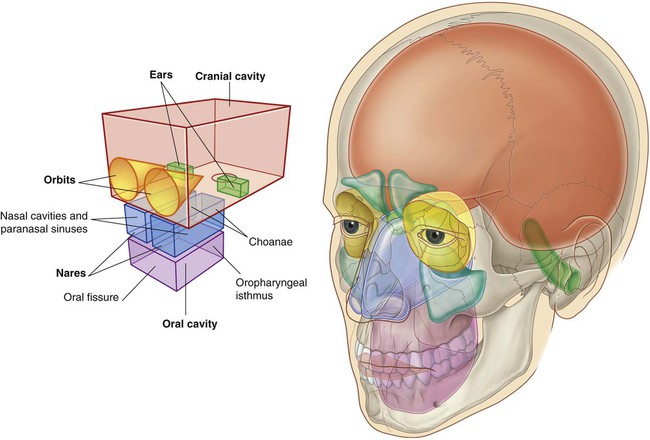
Cavities and Passageways of the Head and Neck A Comprehensive Master Guide for Medical Students & Clinical Practice Module Learning Objectives This exhaustive guide covers the essential anatomy, neurovascular supply, and clinical pathology of the aerodigestive tracts, skull base fossae, and fascial spaces of the head and neck. By mastering these regions, you will be deeply conversant with: The Nasal Cavity & Paranasal Sinuses and the pathways of deadly intracranial infections. The Pharyngeal & Laryngeal structures crucial for airway management and swallowing. The 3D boundaries and contents of the Cranial Fossae, Infratemporal Fossa, and Pterygopalatine Fossa. The catastrophic spread of Deep Neck Space Infections. The complex neurovascular intersections of the Orbit, Ear, and Cavernous Sinus. SECTION 1: THE NASAL CAVITY AND PARANASAL SINUSES 1.1 Overview and Clinical Significance The nasal cavity is the proximal, uppermost portion of the respiratory tract. It extends from the external nostrils (anterior nares) to the choanae (posterior nares), where it directly communicates with the nasopharynx. It is divided symmetrically into right and left halves by the nasal septum. Crucial Functions: Warming and humidifying inspired air via its extensive, highly vascular mucosal surface. Filtering particulate matter and trapping microbes using vibrissae (hairs) and the mucociliary escalator. Providing the special sense of olfaction (smell). Acting as a resonating chamber to give the voice its characteristic tone. The paranasal sinuses are four paired, air-filled cavities entirely encased within the bones of the skull. They communicate directly with the nasal cavity. They serve to reduce the overall weight of the skull, provide voice resonance, act as shock absorbers for the brain during facial trauma, and produce immunologic mucus. CLINICAL PEARL: The nasal cavity possesses a rich vascularity and a direct anatomical connection to the cranial vault via the perforated cribriform plate. Severe nasal trauma can result in cerebrospinal fluid (CSF) rhinorrhea. Because of this direct highway, aggressive nasal infections can spread intracranially, resulting in devastating, fatal meningitis or brain abscesses. 1.2 Boundaries and Bony Framework The nasal cavity is a pyramidal space governed by four distinct walls: ROOF: Narrow and delicate. Formed primarily by the cribriform plate of the ethmoid bone. This is the thinnest and most clinically dangerous boundary, heavily perforated by small foramina transmitting the olfactory nerve filaments (CN I). The sphenoid sinus lies directly posterior to it, inferior to the sella turcica and pituitary gland. FLOOR: Smooth and concave. Formed by the palatine process of the maxilla (anterior 2/3) and the horizontal plate of the palatine bone (posterior 1/3). Together, these form the hard palate. MEDIAL WALL: The nasal septum (discussed below). LATERAL WALL: The most complex, convoluted wall. It features bony scroll-like projections called conchae (turbinates) and the air channels beneath them called meatuses. The inferior nasal concha is an independent bone, while the superior and middle conchae are mere projections of the massive ethmoid bone. 1.3 The Nasal Septum and Kiesselbach’s Plexus The nasal septum acts as the central dividing pillar. It comprises both bony and cartilaginous elements. Cartilaginous Component The septal nasal cartilage (quadrangular cartilage) forms the anterior and inferior portion. It provides the flexible structure of the nose, articulating superiorly with the perpendicular plate of the ethmoid. Bony Components The perpendicular plate of the ethmoid forms the superior-posterior aspect. The vomer forms the posterior-inferior foundation. The sphenoid crest and palatine processes lock the septum into place. Kiesselbach’s Plexus (Little’s Area) This is a massive, highly superficial vascular anastomosis located on the anterior-inferior nasal septum. It is the anatomical site for 90% of all epistaxis (nosebleeds), frequently triggered by dry air, digital trauma (nose-picking), or hypertension. Five contributing arteries anastomose here: Sphenopalatine artery (Terminal branch of Maxillary a.) Anterior ethmoidal artery (Branch of Ophthalmic a.) Posterior ethmoidal artery (Branch of Ophthalmic a.) Superior labial artery (Branch of Facial a.) Greater palatine artery (Branch of Maxillary a.) Clinical Pearl: Because this plexus receives high-pressure blood from BOTH the internal carotid system (ethmoidal arteries) and the external carotid system (facial/maxillary arteries), epistaxis can be shockingly profuse. First-line management is firm, direct mechanical pressure against the septum for 10-15 uninterrupted minutes. 1.4 Lateral Nasal Wall and Meatuses The lateral wall maximizes surface area. The spaces beneath the conchae are the receiving drains for the sinuses and eyes. Inferior Concha & Meatus: The largest concha. The inferior meatus receives the nasolacrimal duct. (This is why your nose runs heavily when you cry). Middle Concha & Meatus: The most clinically vital space. It receives drainage from the frontal sinus, maxillary sinus, and anterior/middle ethmoidal air cells. Superior Concha & Meatus: Receives drainage strictly from the posterior ethmoidal air cells. The Ostiomeatal Complex (OMC): This is the functional “choke point” of the lateral wall. It comprises the maxillary ostium, hiatus semilunaris, ethmoid bulla, and uncinate process. Because so many sinuses drain through this narrow corridor, mucosal swelling here (from allergies or colds) causes OMC obstruction—the leading cause of acute bacterial rhinosinusitis. 1.5 Paranasal Sinuses: Anatomy and Drainage Sinus Anatomy & Drainage Pathway Clinical Significance Frontal Sinuses Located in the frontal bone. Drains via the frontonasal duct into the middle meatus. Absent at birth; develops fully by puberty. Infection can erode the anterior bone causing Pott’s puffy tumor (frontal osteomyelitis), or erode posteriorly causing epidural/brain abscesses. Maxillary Sinuses The largest sinuses (~15 mL). Drains via the maxillary ostium into the hiatus semilunaris (middle meatus). The drainage ostium is located HIGH on the medial wall, forcing it to drain completely against gravity. The floor shares a border with upper molar roots; dental infections frequently punch through to cause massive odontogenic sinusitis. Ethmoid Sinuses 3-18 honeycomb-like cells per side. Separated from the orbit only by the paper-thin lamina papyracea. Anterior drains to middle meatus; posterior to superior meatus. Because the bone is so thin, ethmoid sinusitis is the most common cause of devastating, vision-threatening orbital cellulitis in children. Sphenoid Sinuses Within the sphenoid bone body. Drains into the sphenoethmoidal recess. Sits directly beneath the pituitary gland and beside the cavernous sinus. Neurosurgeons use this sinus as the primary, minimally invasive surgical

Deep Structures of the Head
Anatomy

Deep Structures of the Head

Deep Structures of the Head: High-Yield Anatomy & Clinical Correlations Module Learning Objectives By the conclusion of this exhaustive master guide, you will be deeply conversant with: The osteology, compartmentalization, and major foramina of the cranial fossae, including their exact neurovascular contents. The complex 3D boundaries, contents, and clinical significance of the Infratemporal Fossa (ITF) and Pterygopalatine Fossa (PPF). The intricate pathways of cranial nerves, specifically the “hitchhiking” routes of parasympathetic autonomic fibers. The anatomical basis for differentiating Central (UMN) vs. Peripheral (LMN) nerve lesions. The deep vascular networks, focusing on the Cavernous Sinus, its connections, and the fatal progression of Cavernous Sinus Thrombosis (CST). SECTION 1: Osteology & Foramina of the Skull Base The base of the skull is the foundational floor upon which the brain rests. It is a highly complex, perforated bony landscape that serves as the gateway between the central nervous system and the rest of the body. Pathologies here—such as fractures, tumors, or infections—have profound, often immediate life-threatening consequences. The Three Cranial Fossae The cranial floor is organized into three distinct “terraces” or fossae, descending like steps from front to back. 1. Anterior Fossa Houses the frontal lobes and olfactory bulbs. Boundaries: Posteriorly bounded by the lesser wing of the sphenoid bone. Anteriorly bounded by the frontal bone. The midline floor is formed by the delicate, perforated cribriform plate of the ethmoid bone. Clinical Correlation: Severe head trauma can shear the fragile olfactory filaments passing through the cribriform plate, resulting in permanent anosmia (loss of smell). A fracture here tears the meninges, leading to CSF rhinorrhea (cerebrospinal fluid leaking from the nose). 2. Middle Fossa Houses the temporal lobes and the pituitary gland. Boundaries: Anteriorly bounded by the lesser wing of the sphenoid. Posteriorly bounded by the thick, bony petrous ridge of the temporal bone. The floor consists of the greater wing of the sphenoid and the squamous temporal bone. Clinical Correlation: The pituitary gland sits squarely in the midline (sella turcica). Pituitary macroadenomas usually expand upward, compressing the optic chiasm (causing bitemporal hemianopsia/tunnel vision). Massive pressure in this fossa can cause the temporal lobe to undergo uncal herniation down through the tentorial incisura, compressing the brainstem. 3. Posterior Fossa Houses the cerebellum, pons, and medulla oblongata. Boundaries: Anteriorly bounded by the petrous ridge and the dorsum sellae. Posteriorly bounded by the occipital bone. The center features the massive opening, the foramen magnum. Clinical Correlation: Due to the rigid boundaries, any expanding mass (like a cerebellar tumor or hemorrhage) will cause catastrophic fatal mass effect. The pressure forces the cerebellar tonsils down through the foramen magnum (Tonsillar herniation), crushing the respiratory and cardiac centers in the medulla. Major Foramina: Exact Contents Mastering the exact contents of these “holes” (foramina) is crucial, as a tumor growing in a specific foramen will present with predictable deficits. Foramen / Opening Location Exact Neurovascular Contents Cribriform Plate Anterior Fossa Olfactory nerve filaments (CN I) from the nasal mucosa. Optic Canal Middle Fossa Optic nerve (CN II) + Ophthalmic artery (first branch of ICA). Superior Orbital Fissure (SOF) Middle Fossa CN III (Oculomotor), CN IV (Trochlear), CN V1 (Ophthalmic division of Trigeminal), CN VI (Abducens) + sympathetic fibers + Superior ophthalmic vein. Foramen Rotundum Middle Fossa Maxillary nerve (CN V2) passing to the Pterygopalatine Fossa. Foramen Ovale Middle Fossa Mandibular nerve (CN V3) + Accessory meningeal artery + emissary veins. Foramen Spinosum Middle Fossa Recurrent meningeal branch of V3 + Middle meningeal artery & vein. (Epidural hematomas originate here). Internal Acoustic Meatus (IAM) Posterior Fossa CN VII (Facial) + CN VIII (Vestibulocochlear) + Labyrinthine artery. Jugular Foramen Posterior Fossa Pars nervosa: CN IX (Glossopharyngeal), X (Vagus), XI (Spinal Accessory).Pars vascularis: CN X, XI + Internal jugular vein. Hypoglossal Canal Posterior Fossa CN XII (Hypoglossal nerve). Carotid Canal Middle Fossa Internal carotid artery (ICA – petrous and lacerum segments) + sympathetic plexus. CLINICAL PEARL: The Foramen Lacerum Illusion In a dried skull, the foramen lacerum looks like a massive hole. However, in a living human, it is completely plugged and filled with cartilage. The Internal Carotid Artery (ICA) passes OVER it, sliding across the cartilage bed; it does not travel THROUGH it. Mnemonic for Middle Fossa (Lateral → Medial): Stupid Old Rats Frequently Spin = SOF, Optic canal, foramen Rotundum, foramen Ovale, foramen Spinosum. Skull Base Fractures & CSF Leakage Basilar skull fractures are notoriously difficult to see on standard X-rays. Diagnosis relies heavily on highly specific clinical signs. Anterior Fossa Fracture (Frontal/Ethmoid bones): CSF Rhinorrhea: Tearing of the meninges over the cribriform plate allows cerebral spinal fluid to drip directly into the nasal cavity. ‘Halo Sign’: If bloody fluid from the nose is dripped onto filter paper or bed linen, the rapidly spreading CSF forms a clear, yellowish ring (halo) around a central dot of blood. Raccoon Eyes (Bilateral periorbital ecchymosis): Blood tracks down into the soft tissue around the eyes. Because the nasal cavity is heavily colonized with bacteria, these patients are at massive risk for ascending bacterial meningitis. Middle Fossa Fracture (Temporal bone): CSF Otorrhea: A fracture through the tegmen tympani (roof of the middle ear) combined with a perforated tympanic membrane (eardrum) allows CSF to leak out of the ear. Battle’s Sign: Ecchymosis (bruising) over the mastoid process behind the ear. This is a delayed sign, taking 24–48 hours to appear. Nerve Deficits: The facial canal and IAM are housed in the temporal bone. Fractures here frequently severe or compress the Facial nerve (causing CN VII palsy/facial droop) and the Vestibulocochlear nerve (causing CN VIII dysfunction: extreme vertigo, tinnitus, and sensorineural hearing loss). Posterior Fossa Fracture: Rare, but associated with extremely high mortality due to direct brainstem compression and massive deficits in lower cranial nerves (CN IX–XII). SECTION 2: The Infratemporal Fossa (ITF) The Infratemporal Fossa is an irregularly shaped, completely hidden space located deep and inferior to the zygomatic arch, and deep to the ramus of the mandible. It acts as a massive distribution center for the neurovasculature of the lower jaw, chewing muscles, and

Superficial structures
Anatomy

Superficial structures

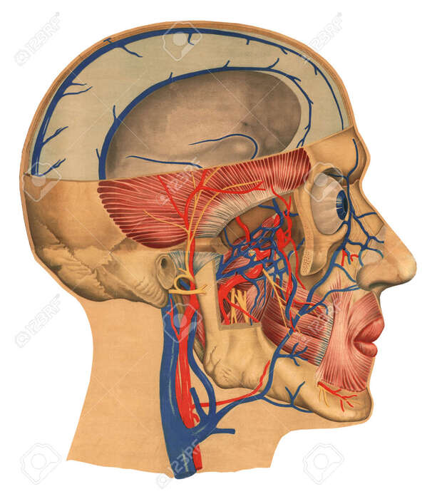
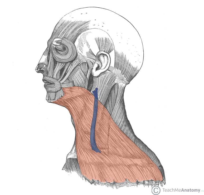
Superficial Structures of the Head and Neck A Comprehensive Anatomical Master Guide for Clinical Practice Module Learning Objectives By the conclusion of this exhaustive anatomical guide, you will be deeply conversant with: The complex layering, neurovascular supply, and clinical implications of the Scalp. The origins, insertions, actions, and innervation of all major Muscles of Facial Expression. The precise sensory and motor distributions of the Superficial Nerves (Trigeminal, Facial, and Cervical Plexus). The intricate Superficial Vascular Supply, focusing on the External Carotid branches and the critical venous “Danger Triangle”. The topographical regionalization of the Neck Triangles, their borders, deep contents, and pivotal surface landmarks for clinical procedures. I. The Scalp: Layers, Blood Supply, and Innervation The scalp is the soft tissue envelope that covers the cranial vault. Extending from the superior nuchal lines and occipital protuberances posteriorly, to the supraorbital margins anteriorly, and laterally down to the zygomatic arches, it plays a vital role in protecting the neurocranium and regulating temperature. The Five Layers of the Scalp (Mnemonic: S.C.A.L.P.) The scalp consists of five distinct layers. The first three layers are tightly bound together and move as a single functional unit. S — Skin: Typically thick and hair-bearing. It contains an abundant supply of sebaceous (oil) glands and hair follicles that extend deeply into the connective tissue below. (High density of sebaceous glands makes the scalp prone to sebaceous cysts). C — Connective Tissue (Dense): A dense, highly vascularized, and innervated fibro-fatty layer. Because the collagen fibers tightly tether the blood vessels, vessels here cannot retract and constrict when cut, leading to the characteristic profuse bleeding seen in superficial head wounds. A — Aponeurosis (Galea Aponeurotica): A strong, immense, tendinous sheet connecting the frontal and occipital bellies of the occipitofrontalis muscle. It is immobile and prevents the scalp from stretching. Suturing this layer is critical in deep scalp lacerations to prevent the wound from gaping wide open. L — Loose Areolar Tissue (The “Danger Area”): A sponge-like, easily separable layer that allows the upper three layers (the scalp proper) to glide smoothly over the skull. It is termed the “Danger Area” because it contains Emissary Veins—valveless veins that directly connect the superficial scalp veins to the deep intracranial dural venous sinuses. P — Pericranium: The deepest layer. This is the dense periosteum covering the outer surface of the calvarium (skull bones). It is tightly adherent to the suture lines of the skull and contains the vascular networks vital for bone support and repair. Clinical Significance of Scalp Layers The “Danger Area” & Infection: Pus or blood accumulating in the loose areolar layer can spread widely across the entire dome of the skull. Worse, infections here can track directly down the valveless emissary veins into the brain, causing lethal Meningitis or Cavernous Sinus Thrombosis. Scalp Avulsion: In horrific industrial or machinery accidents where hair is caught and ripped, the scalp peels off exactly at the plane of the loose areolar tissue. The first three layers (S-C-A) detach cleanly as a single unit away from the pericranium. Profuse Bleeding: Because the dense connective tissue holds arteries open, even small scalp cuts bleed dramatically. Bleeding is best controlled by applying direct, firm pressure against the hard underlying skull bone. II. Muscles of Facial Expression Approximately 20 flat, thin skeletal muscles lie immediately beneath the skin of the face and scalp. These muscles are biologically unique compared to other skeletal muscles. They originate from facial bones or fibrous structures and insert directly into the dermis of the skin, allowing them to pull the skin to create expressions. Embryological and Neurological Rule: All muscles of facial expression lack deep fascia (with the exception of the buccinator), are derived from the Second Pharyngeal Arch, and are universally innervated by the Facial Nerve (CN VII). 1. Orbital Group (Muscles Around the Eye) Orbicularis Oculi The sphincter muscle of the eyelids. Origin: Medial orbital margin, medial palpebral ligament, lacrimal bone. Insertion: Skin around the orbital margin, tarsal plates. Action: Palpebral part: Gently closes eyelids (blinking to spread tears). Orbital part: Forcefully, tightly closes eyelids (squinting against bright light/dust). Corrugator Supercilii The “frowning” muscle of the brow. Origin: Medial side of the superciliary arch. Insertion: Skin superior to the supraorbital area (mid-eyebrow). Action: Pulls the eyebrows inferomedially (down and in). Creates the vertical forehead wrinkles associated with a “worried” or “angry” expression. 2. Nasal Group (Muscles of the Nose) Involved in respiration and conveying anger or disgust. Muscle Origin / Insertion Action & Expression Nasalis (Transverse Part) Origin: Maxilla, lateral to nose. Insertion: Aponeurosis across dorsum of nose. Compresses the nasal aperture (closes nostrils). Nasalis (Alar Part) Origin: Maxilla over lateral incisor. Insertion: Alar cartilage. Dilates the nostrils (“flaring” during anger or heavy breathing). Procerus Origin: Nasal bone/cartilage. Insertion: Skin over glabella (between eyebrows). Depresses medial eyebrows, wrinkling the skin over the bridge of the nose. Expression of “Disgust” or “Disdain”. Depressor Septi Nasi Origin: Maxilla above medial incisor. Insertion: Nasal septum. Pulls the nasal septum inferiorly to widen the nasal opening. Assists alar nasalis in deep inspiration. 3. Oral Group (Muscles Around the Mouth) The mouth is highly dynamic, surrounded by elevators, depressors, and a main sphincter. Crucial Landmark: The Modiolus The Modiolus is a dense, fibromuscular hub located just lateral to the angle of the mouth. It acts as the functional center of facial expression. Multiple muscles converge and anchor directly into this dense nodule, including the Zygomaticus major, Risorius, Buccinator, Levator anguli oris, and Depressor anguli oris. Orbicularis Oris Origin: Maxilla, mandible, mouth angle. Insertion: Mucous membrane of lips. Action: Closes the oral fissure, compresses and protrudes the lips (The “Kissing” or whistling muscle). Buccinator Origin: Maxilla, mandible alveolar processes, pterygomandibular raphe. Insertion: Orbicularis oris, modiolus. Action: Compresses cheek tightly against the molars to keep food on the teeth while chewing. (The “Trumpeter’s” muscle). Zygomaticus Major & Minor Origin: Zygomatic bone. Insertion: Modiolus (Major) and upper lip (Minor). Action: Major elevates the labial commissure (The “Smiling” muscle). Minor elevates and everts the upper lip (Sadness). Risorius

Scroll to Top