Theoretical Models in Nursing Informatics : Theories
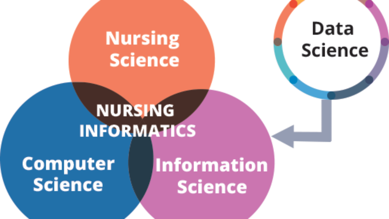
Theoretical Models in Nursing Informatics
Theoretical models are like maps or blueprints that help us understand concepts. In nursing informatics, these models provide a framework for understanding and applying informatics principles, guiding how we think about data, manage change, and implement technology effectively in healthcare.
1. The DIKW Model: From Data to Wisdom
This is a foundational model, often depicted as a pyramid, illustrating how raw, unprocessed facts evolve into profound understanding and expert judgment. It's crucial for understanding the value proposition of nursing informatics – transforming simple observations into actionable wisdom for patient care.
Data
Raw, isolated, and unprocessed facts without context or meaning. By itself, it doesn't tell a story or answer a question. Simplified: Just numbers, words, or observations.
Expanded Example:
- A single blood pressure reading: "150/95 mmHg".
- A patient's temperature: "39°C".
- A lab result: "White Blood Cell count: 15,000".
- A patient's complaint: "I have a headache".
- In a Ugandan clinic: A register entry showing "Patient John Doe, Age 45, Malaria test positive".
Information
Data that has been organized, structured, processed, or interpreted within a specific context. It answers "who," "what," "where," and "when." Data with meaning.
Expanded Example:
- A series of blood pressure readings over 24 hours (e.g., 150/95, 148/92, 155/98) showing a consistently high trend, which the EHR flags as "hypertension" based on predefined ranges. The 39°C temperature is flagged as a "fever" by comparing it to normal body temperature ranges. This gives context.
- A patient's medication list, their history of allergies, and current lab results, all presented together in their EHR profile.
- In a Ugandan clinic: Seeing that "John Doe, Age 45" (data points) tested positive for malaria after visiting a specific village where there's a known outbreak (context), and correlating this with his symptoms of fever and chills (more context). This provides actionable information about his condition and potential exposure.
Knowledge
The synthesis of information, often through experience, education, and research, to identify relationships, patterns, and principles. It answers "how" to apply information and understand its implications. Understanding why something is happening and what it means.
Expanded Example:
- The nurse combines the information (consistently high blood pressure, persistent fever, high WBC count) with their clinical knowledge (nursing science). They recognize that high blood pressure increases cardiovascular risk, that a fever and high WBC count could indicate an infection (e.g., bacterial), and that the patient's complaint of headache might be related to these findings.
- Knowing that patients on certain medications are more prone to falls or that a particular cough pattern is indicative of a specific respiratory illness.
- In a Ugandan context: A nurse knowing that a positive malaria test in a patient from a high-transmission area, combined with a persistent fever, means they need specific antimalarial treatment and patient education on prevention.
Wisdom
The ability to apply knowledge, experience, and intuition with judgment to manage and solve problems effectively and ethically, especially in complex or novel situations. It involves understanding "why" to do something and "when" to do it, considering values and societal implications. Expert judgment and decision-making that leads to the best outcome.
Expanded Example:
- Knowing the patient's history of sepsis and considering the current high fever and elevated WBCs, the seasoned nurse uses their wisdom not just to treat the fever symptomatically, but to immediately initiate the sepsis protocol. This involves drawing blood cultures before administering antibiotics, administering broad-spectrum antibiotics promptly, monitoring vital signs intensely, alerting the physician with a specific concern for sepsis, and educating the family on the gravity of the situation. This proactive, expert decision-making significantly improves the patient's outcome by acting rapidly and holistically.
- A nurse informaticist, using their wisdom, might recommend designing an EHR alert system to be subtle for common benign interactions but highly prominent for life-threatening situations, balancing user experience with patient safety.
- In a Ugandan context: A community health nurse, observing a pattern of increasing malaria cases after a specific rainfall period in their region (knowledge), uses their wisdom to mobilize community leaders for a mass bed net distribution campaign and initiate an immediate health education drive, rather than just treating individual cases as they present.
2. Graves & Corcoran's Model (1989)
This early and influential model provided a crucial conceptual framework for nursing informatics. It's often visualized as three overlapping circles (nursing science, computer science, information science) with data, information, and knowledge flowing through them, all directed towards supporting nursing practice. It was groundbreaking because it shifted the focus from merely using technology to understanding the purpose of information processing in nursing care.
Core Idea: Nursing informatics integrates the three core sciences to manage and process data, information, and knowledge effectively for the benefit of nursing practice.
Aims of the Model
The model was designed to provide a clear roadmap for nursing informatics with three primary goals:
- Identify the information needs in nursing: To figure out exactly what information nurses need to do their jobs effectively, whether they are at the bedside, in a classroom, or managing a clinic. _ Specify the sources and systems that provide information: To pinpoint where this necessary information comes from (e.g., the patient, lab results, other departments) and what technological systems (like EHRs) are needed to deliver it.
- Show how informatics can help nurses achieve their goals: To demonstrate how technology can be a powerful tool to help nurses accomplish their objectives in all areas, including patient care, education, research, and management.
Main Components
- Users: The people who need and use the information. This isn't just nurses; it includes doctors, administrators, technicians, and even patients and their families who interact with health information.
- Roles: The specific functions or jobs these users perform. A person's role determines what kind of information they need. For example, a clinician needs patient data, an educator needs learning resources, a researcher needs aggregated data, and an administrator needs operational data.
- Settings: Where the nursing activities take place. The setting heavily influences the technology and information needed. A nurse in a high-tech urban hospital has different resources and needs than a nurse in a remote community clinic or a patient's home for home care.
- Goals: The desired outcomes or what the user is trying to accomplish. Goals can be clinical (improve patient safety), educational (enhance student learning), research-focused (discover new knowledge), or administrative (increase workflow efficiency).
- Knowledge Base: The foundation of professional knowledge required to perform the role. For a nurse, this includes clinical guidelines, nursing theory, pharmacology, and evidence-based practice, as well as knowledge from other fields like ethics and leadership.
- Information Technologies: The specific tools and systems used to achieve the goals. This includes hardware (computers, tablets) and software (EHRs, telehealth platforms, clinical decision support systems, virtual simulation labs, scheduling software).
Flow of the Model
The model works by systematically connecting the components. The following table shows a practical example of how to apply the model step-by-step:
| Steps | Examples in an Educational Context |
|---|---|
| Step 1: Identify the user and role. | User: Nurse Educator Role: Teaching |
| Step 2: Define the setting. | Setting: Nursing school or a university's skills lab. |
| Step 3: Clarify the goal. | Goal: Improve students’ skills and confidence in performing complex patient assessments. |
| Step 4: Apply knowledge & select technology. | Knowledge Base: Educational theory (e.g., experiential learning), clinical assessment guidelines, and best practices in simulation. Information Technologies: Select virtual simulation labs and interactive online case study platforms. |
| Step 5: Achieve the nursing outcome. | Outcome: Students practice assessments in a safe, repeatable virtual environment, leading to increased competency and better preparedness for real clinical settings. |
3. Theories of Change: Implementing New Technology
Implementing new technology, like a new EHR system or a telehealth platform, is a significant undertaking that requires careful management of human behavior and organizational processes. Understanding change theories is essential for successful adoption and minimizing resistance.
a) Lewin's Change Theory (Unfreeze-Change-Refreeze)
Kurt Lewin's classic model provides a simple yet powerful three-step process for managing planned change within an organization.
Step 1: Unfreezing
Preparing the organization or individuals for change by creating awareness of why the old way of doing things is no longer sufficient and establishing a felt need for change. It involves breaking down old habits and assumptions. Convincing everyone that the old way isn't working and a new way is needed.
Expanded Example: A hospital management team presents compelling data on the high rates of medication errors, documentation inefficiencies, and patient complaints linked to the current paper-based charting system. They hold town hall meetings and workshops, actively involving nursing staff, to discuss the critical need for a new EHR system to improve patient safety, streamline workflows, and enhance overall quality of care. They highlight the financial and reputational costs of not changing.
Step 2: Moving (or Changing)
The actual implementation phase where the change occurs. This stage involves significant training, communication, support, and adaptation as people learn new processes and tools. Rolling out the new system and teaching everyone how to use it.
Expanded Example: The new EHR system is rolled out systematically, perhaps unit by unit or department by department. Intensive, hands-on training sessions are conducted for all nursing staff. Specially trained "super-users" (nurses proficient in the new system) are deployed on the floors to provide immediate, peer-to-peer support. The IT department establishes a dedicated 24/7 help desk. Regular feedback sessions are held to identify and quickly address any technical glitches or workflow issues.
Step 3: Refreezing
Stabilizing the change and making it the new standard practice. This involves integrating the new methods into the organizational culture, updating policies, and reinforcing the benefits of the change. Making the new way the permanent, normal way of doing things.
Expanded Example: New hospital policies and procedures are formally established that mandate the exclusive use of the EHR for all patient documentation and order entry. Leadership publicly celebrates early successes (e.g., reduction in medication errors, improved documentation compliance). Ongoing refresher training is provided for new hires and to address advanced features. Audits are performed to ensure compliance, and the use of the EHR becomes deeply embedded in the daily workflow, becoming the "new normal."
b) Rogers' Diffusion of Innovations Theory
Everett Rogers' theory examines how new ideas, technologies, or practices spread through a social system over time. It's particularly useful for understanding how different groups of people adopt innovations at different rates, allowing for tailored communication and implementation strategies.
Adopter Categories:
Innovators (2.5%)
These are the first to adopt new ideas, often risk-takers and enthusiasts. They are eager to experiment.
Role: Often seek out new technology and are willing to try it even if it's not perfect.
Example: When a new telehealth platform is introduced in a Ugandan district, an innovator nurse might be the first to volunteer for the pilot program, eager to test its capabilities and provide early feedback, even if internet connectivity is sometimes challenging.
Early Adopters (13.5%)
Respected opinion leaders within the community or profession. They adopt new ideas early but with more thought and evaluation than innovators. They are crucial for influencing the broader group.
Role: Act as role models and champions, legitimizing the innovation.
Example: Other experienced and well-respected nurses in the district watch the innovators. Seeing the benefits, an early adopter nurse begins using the telehealth platform and actively champions its use to their peers, sharing their positive experiences and insights during staff meetings.
Early Majority (34%)
Deliberate individuals who adopt new ideas just before the average person. They need to see evidence that the innovation works and is useful.
Role: They make the innovation mainstream.
Example: After seeing positive results and hearing positive feedback from the early adopters, the majority of nurses in the district begin to adopt the telehealth platform, recognizing its practical benefits for patient care and convenience.
Late Majority (34%)
Skeptical individuals who will only adopt an innovation after a majority of people have tried it and it has become widely accepted. They are often influenced by peer pressure or economic necessity.
Role: Their adoption signals widespread acceptance.
Example: Some nurses are hesitant and prefer traditional methods. They only begin to use the telehealth platform when it becomes an established and expected part of routine practice, perhaps after a mandate or when training and support are widely available.
Laggards (16%)
Traditionalists who are the last to adopt an innovation. They are often resistant to change, prefer traditional methods, and have little to no opinion leadership.
Role: May only adopt when older options are no longer available.
Example: A few nurses may resist using the telehealth platform until there's virtually no other option for certain consultations or if their traditional methods become unsustainable. They might require significant individual support and encouragement.
Application: Understanding these categories helps implementers tailor their communication, training, and support strategies to each group to maximize adoption.
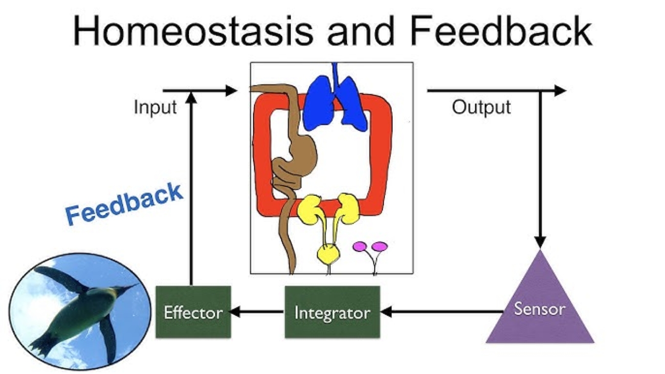
4. General Systems Theory
This theory views any organization, like a hospital, a clinic, or even a patient's body, as a complex system. It posits that a system is made up of many interconnected parts (subsystems) that work together to achieve a common goal. A key tenet is that a change in one part of the system will inevitably affect all other parts, highlighting the importance of a holistic perspective.
Core Idea: Everything is connected. When you change one thing in a system, it impacts everything else.
Key Concepts:
Input
Resources, information, or energy entering the system from its environment.
Example: Patient demographic data, lab results, nurse staffing levels, available IT infrastructure (computers, internet connectivity), medical supplies.
Throughput (Process)
The activities or work done within the system to transform the input.
Example: Nurses and doctors entering and processing patient data within the EHR, administering medications, performing patient assessments, collaborating with other departments, and making clinical decisions.
Output
The products, services, or outcomes that result from the system's processes.
Example: A complete and accurate patient health record, clinical decision support alerts, patient discharge summaries, billing information, improved patient outcomes (e.g., reduced readmission rates).
Feedback
Information about the system's output that is fed back into the system to make adjustments, correct errors, and improve future performance.
Example: Nurses report that a specific screen in the EHR is confusing or takes too long to complete, leading to a system modification by the IT team. Patient satisfaction surveys, infection rates, or medication error reports provide feedback on the quality of care delivered.
Environment
External factors, conditions, or influences that interact with and affect the system but are largely outside its direct control.
Example: Government regulations (e.g., data protection laws like Uganda's Data Protection and Privacy Act, or international standards like HIPAA), economic conditions, technological advancements, patient demographics, public health crises (like an epidemic), and relationships with technology vendors.
Expanded Example (EHR Implementation)
When implementing a new EHR in a large hospital through a systems lens:
- Input: The system requires patient demographic data, past medical histories (often from old paper charts), new lab results, skilled IT staff, adequate budget, and training materials.
- Throughput: Nurses, doctors, and other healthcare professionals spend time learning the new system, entering data, navigating interfaces, and adapting their workflows. Data flows between different modules (e.g., admissions, lab, pharmacy).
- Output: A comprehensive, digital patient record, automated clinical decision support alerts, streamlined billing, and eventually, potentially improved patient outcomes and reduced errors.
- Feedback: Nurses complain about too many clicks to perform a common task. This feedback prompts the informatics team to optimize the workflow. Analytics show a reduction in medication errors, reinforcing the system's value.
- Environment: The national Ministry of Health mandates certain data reporting standards, requiring the EHR to be updated. A power outage (environmental factor) can bring the entire system down, highlighting the need for robust backup systems and generators.
Summary Table
| Concepts | Example: Implementation of an EHR |
|---|---|
| System | Hospital’s health information infrastructure. |
| Input | Patient data (demographics, vitals, lab results), staff expertise, IT resources. |
| Throughput (Process) | Data entry, storage, processing, and analysis within the EHR system. |
| Output | Patient charts, clinical decision support alerts, discharge summaries. |
| Feedback | User satisfaction surveys, system usage reports, error rates. |
| Environment | Government regulations, technology vendors, patient needs, and funding. |
| Open vs. Closed Systems |
Open Systems:
|
The Impact and Practice of Nursing Informatics
This module explores the tangible benefits that nursing informatics brings to healthcare and introduces the crucial role of the Nurse Informaticist, the specialist who drives many of these advancements.
Overall Benefits of Nursing Informatics
Nursing informatics isn't just about using computers; it's about fundamentally transforming healthcare for the better. Here are some of its most significant impacts:
1. Making Previously Buried Data Usable & Actionable
Historically, vast amounts of clinical data were locked away in paper charts, making it incredibly difficult to analyze or learn from. Informatics systems (like EHRs) digitize this data, allowing it to be easily searched, aggregated, and analyzed. Turns piles of paper notes into smart insights.
Expanded Example: Instead of manually sifting through hundreds of patient files to find out how many patients with malaria responded to a specific treatment, an informaticist can query the EHR. This data can then be used for quality improvement projects (e.g., "Are we giving the right malaria treatment?") or for research. In a broader sense, this data can inform public health strategies in Uganda by showing patterns of disease outbreaks or the effectiveness of vaccination campaigns across different districts.
2. Improving Patient Safety & Reducing Errors
Informatics systems are designed with patient safety at their core, significantly reducing the potential for human error. Built-in safeguards prevent mistakes.
Expanded Examples: Barcode Medication Administration (BCMA) provides a crucial safety net. Clinical Decision Support (CDS) Alerts can automatically flag a severe drug allergy or a dangerous drug-drug interaction. Eliminating illegible handwriting ensures all care providers are working with accurate information.
3. Providing Data That Healthcare Buyers Demand (Quality Reporting)
Informatics systems capture the data needed to demonstrate quality outcomes and cost-effectiveness to insurance companies, government agencies, and donors. Shows proof of good care and efficient spending.
Expanded Example: A hospital can easily generate reports showing its infection rates or patient readmission rates. In Uganda, this could mean demonstrating to the Ministry of Health that specific health interventions are effective and resources are being used wisely, leading to continued funding and support.
4. Easier Storage and Retrieval of Records
The digital nature of EHRs eliminates the physical space, security risks, and retrieval delays associated with paper charts. Information can be accessed instantly from multiple locations simultaneously. No more lost charts or endless searching; everything is a click away.
Expanded Example: Instead of searching through dusty paper archives, a nurse can access a patient's medical history instantly from a workstation or mobile device, even if the patient was last seen years ago. This is highly beneficial where maintaining a continuous paper record is challenging.
5. Saving Time and Money
By streamlining workflows, reducing redundant tests, preventing errors, and improving efficiency, informatics contributes significantly to cost savings and better resource utilization. More efficient care, fewer wasted resources.
Expanded Example: Reduced time spent on documentation means nurses have more time for direct patient care. Preventing a medication error avoids the costs associated with extended hospital stays and additional treatments. Electronic ordering of tests and medications reduces errors and speeds up processes.
The Role of the Nurse Informaticist
A Nurse Informaticist (NI) is a highly specialized registered nurse who possesses a deep understanding of both clinical nursing practice and information technology. They are critical to successful technology integration in healthcare, acting as the indispensable link between the worlds of nursing and IT.
- Who they are: A registered nurse with advanced training or experience in informatics.
- Their unique value: They speak the language of both clinicians and IT professionals, ensuring that technology solutions truly meet the needs of patient care.
Common Roles and Responsibilities:
System Implementation
Play a central role in bringing new clinical information systems into an organization, from evaluation and customization to rollout.
Example: When a hospital in Kampala upgrades its EHR, the NI leads requirements gathering from nurses, works with vendors to configure the system for local practices, designs workflows, and oversees testing and go-live.
Workflow Optimization
Analyze existing clinical processes and identify areas where technology can make them more efficient, safer, and user-friendly.
Example: An NI might observe nurses spending too much time navigating the EHR to document vitals. They would then work with IT to streamline the process by creating a consolidated "daily care" screen, reducing charting burden.
Training and Support
Develop and deliver comprehensive training programs for all nursing staff on how to use technology effectively and serve as a primary resource for troubleshooting.
Example: After a new patient monitoring system is installed, the NI will design and lead hands-on training, create user guides, and be on-site during go-live to provide immediate support.
Data Analysis
Extract, interpret, and present data from clinical systems to identify trends, monitor quality metrics, and support research.
Example: An NI might analyze EHR data to identify a correlation between a specific staffing model and patient fall rates, providing evidence for practice changes. In public health, they might analyze vaccination rates against disease incidence.
Policy and Procedure Development
Develop and update organizational policies related to the secure and ethical use of clinical information systems, including data privacy and security.
Example: The NI will ensure the hospital's policies for accessing EHR data comply with national data privacy laws, developing clear guidelines on what information nurses can share and how to handle sensitive data.
Competencies and Ethical Responsibilities
As technology becomes more ingrained in nursing, every nurse needs a foundational understanding of informatics. This module highlights the essential competencies and the critical ethical responsibilities that come with managing patient data.
Developing Your Informatics Competencies
It's not just Nurse Informaticists who need informatics skills; all nurses require a baseline level of competency. The TIGER (Technology Informatics Guiding Education Reform) Initiative provides a widely recognized framework:
Basic Computer Skills
The fundamental ability to use computers and software. Knowing how to use a computer. This includes using a keyboard, navigating operating systems, sending emails, and basic file management.
Information Literacy
The ability to find, evaluate, and use relevant information. Knowing how to find good information and tell if it's trustworthy. This includes searching databases like PubMed and critically evaluating online sources.
Information Management
The ability to use clinical information systems like the EHR. Knowing how to use patient record systems effectively. This includes documenting care, retrieving data, and using decision support tools.
Ethical Considerations in Nursing Informatics
The power of digital health information comes with significant ethical responsibilities. Nurses are trusted custodians of patient data.
Patient Privacy & Confidentiality
Protecting sensitive patient information from unauthorized access or misuse. Keeping patient information secret. This means never sharing data without consent and being mindful of who can see or hear information.
Data Security
Implementing safeguards to protect electronic health information from being lost, stolen, or corrupted. Protecting patient data from hackers and mistakes. This includes using strong passwords and logging out of systems.
Data Integrity
Ensuring that the data being entered, stored, and retrieved is accurate, complete, and reliable. Making sure patient records are always correct. Inaccurate data can lead to serious patient harm.
Reflection and Future Outlook
This module provides an opportunity for personal reflection on the material covered and looks ahead to the exciting and rapidly evolving future of nursing informatics.
Reflection & Takeaways: Connecting Concepts to Your Practice
Take a moment to pause and consider how the concepts we've discussed apply to your own experiences and aspirations in nursing.
- Which nursing informatics model resonates most with you and why? Consider the DIKW model, Lewin's Change Theory, or Rogers' Diffusion of Innovations. How do they explain experiences you've had in your workplace?
- Identify two areas you would like to improve in your technology competence. Based on the TIGER competencies, where do you see opportunities for personal growth?
- Consider how ICT (Information and Communication Technology) will shape your future nursing practice. Think about emerging technologies like AI, telehealth, and genomics. How might these tools change how you deliver care?
The Future of Nursing Informatics
The field of nursing informatics is dynamic and constantly evolving. Here are some key trends that will shape its future:
Telehealth and Virtual Care Expansion
The use of technology to deliver healthcare remotely. Expect a continued surge in virtual appointments and remote patient monitoring, which is transformative for extending healthcare access to underserved rural populations, a significant benefit in Uganda.
Big Data and Predictive Analytics
Analyzing enormous volumes of healthcare data to identify patterns, predict outcomes, and guide interventions. Nurses will use this to identify high-risk patients for events like sepsis or falls before they occur, allowing for proactive, personalized care.
Artificial Intelligence (AI) and Machine Learning (ML)
AI will integrate into clinical decision support, offering sophisticated guidance on diagnoses and treatment plans. It may also automate routine documentation, freeing up nurses for more direct patient interaction.
Patient Engagement Technologies
Empowering patients to take a more active role in their health through advanced patient portals, mobile health apps (mHealth), and wearable devices. Nurses will be key in educating patients on how to use these tools effectively.
Interoperability and Seamless Data Exchange
The ability of different healthcare systems to communicate and exchange data seamlessly. The goal is for a patient's health information to follow them effortlessly across all providers, reducing redundant tests and improving care coordination.
Test Your Knowledge
A quiz on Theoretical Models in Nursing Informatics.
1. In the DIKW Pyramid, what is defined as "raw facts without context"?
- Information
- Knowledge
- Data
- Wisdom
Correct (c): "Data" is explicitly defined as "Raw facts without context."
Incorrect (a): Information is organized data with meaning.
Incorrect (b): Knowledge is synthesized information for decision-making.
Incorrect (d): Wisdom is applying knowledge effectively.
2. According to the DIKW Model, when is information combined with nursing experience and evidence?
- To form Data
- To become Wisdom
- To create Knowledge
- To generate new Information
Correct (c): Knowledge is formed when information is combined with nursing experience and evidence (e.g., knowing high BP increases cardiovascular risk).
Incorrect (b): Wisdom is the application of knowledge.
3. Which of the following would be an example of 'Wisdom' in the DIKW Pyramid?
- A patient's temperature of 39°C.
- The EHR flagging 39°C as a "fever."
- Knowing that a fever could indicate infection.
- Creating a personalized care plan based on a patient's risks.
Correct (d): Wisdom is applying knowledge to make a sound decision, such as creating a personalized care plan based on identified risks.
Incorrect (a): This is Data.
Incorrect (b): This is Information.
Incorrect (c): This is Knowledge.
4. The Graves & Corcoran Model (1989) describes the key components for nurses to effectively use what?
- Individual patient charts
- Manual documentation systems
- Information systems
- Only hardware tools
Correct (c): The model explicitly "describes the key components and relationships needed for nurses to effectively use information systems in healthcare."
5. Which of the following is NOT listed as a main component of the Graves & Corcoran Model?
- Users
- Goals
- Funding
- Settings
Correct (c): The main components listed are Users, Roles, Settings, Goals, Knowledge Base, and Information Technologies. Funding is not a core component of the model itself.
6. In the Graves & Corcoran Model, which step involves applying the knowledge base and selecting technologies?
- Step 1: Identify the user and role.
- Step 2: Define the setting.
- Step 3: Clarify the goal.
- Step 4: Apply knowledge base and select technologies.
Correct (d): The model's flow clearly outlines Step 4 as "Apply the knowledge base and select the right information technologies."
7. Roger's Diffusion of Innovation Theory is categorized as which type of change?
- Planned
- Unplanned
- Static
- Revolutionary
Correct (b): Roger's theory describes how innovations spread naturally through a social system, which is considered an "unplanned" pattern of change.
Incorrect (a): Lewin's Change Theory is an example of a "planned" change model.
8. According to Roger's theory, which category of adopters are opinion leaders and promoters?
- Innovators
- Early adopters
- Early majority
- Laggards
Correct (b): Early adopters are described as "opinion leaders who function as promoters of innovation."
Incorrect (a): Innovators are the very first to adopt but are a smaller group.
Incorrect (c): The early majority are averse to risks.
Incorrect (d): Laggards are suspicious and resistant to change.
9. The "Late majority" in Roger's theory typically needs what before adopting an innovation?
- To be convinced it's cutting-edge.
- To be sure the innovation is beneficial.
- To be the first ones to try it.
- No convincing, they adopt quickly.
Correct (b): The late majority are skeptical and require proof that the innovation is beneficial and has been adopted by many others before they will consider it.
10. Which step in Lewin's Change Theory involves preparing people and explaining why change is needed?
- Unfreezing
- Moving (changing)
- Refreezing
- Innovating
Correct (a): "Unfreezing" is the initial step where awareness is created, and people are prepared to move away from the current state.
Incorrect (b): Moving is the implementation phase.
Incorrect (c): Refreezing is about sustaining the change.
11. Providing hands-on training and user support during a new system implementation falls under which step of Lewin's theory?
- Unfreezing
- Moving (changing)
- Refreezing
- Evaluating
Correct (b): The "Changing (Moving)" step is the transition phase where the new system is rolled out, and users are provided with training and support.
12. What is the goal of the 'Refreezing' step in Lewin's Change Theory?
- To identify the need for change.
- To implement the new system.
- To ensure the new way becomes part of everyday practice.
- To revert to the old methods if resistance is high.
Correct (c): The goal of "Refreezing" is to stabilize the change and integrate it into the normal workflow to ensure it becomes permanent.
Incorrect (a): This is the goal of Unfreezing.
Incorrect (b): This is the goal of Moving.
13. In General Systems Theory, what is "Resources, information, or energy entering the system"?
- Output
- Throughput
- Feedback
- Input
Correct (d): "Input" is defined as "Resources, information, or energy entering the system."
Incorrect (a): Output is what is generated by the system.
Incorrect (b): Throughput is how inputs are transformed.
Incorrect (c): Feedback is information about performance.
14. User satisfaction surveys and system usage reports are examples of what concept in General Systems Theory?
- Input
- Throughput
- Feedback
- Environment
Correct (c): "Feedback" is "Information about the system’s performance used to make adjustments," which perfectly describes surveys and reports.
15. An EHR connecting with labs, pharmacies, and insurance is an example of what type of system?
- Closed system
- Linear system
- Open system
- Static system
Correct (c): An open system interacts with its environment. The EHR interacting with external entities like labs and pharmacies is a prime example.
Incorrect (a): Closed systems are self-contained and do not interact with their environment.
16. In the DIKW Pyramid, _______ is defined as synthesized information for decision-making.
17. The Graves & Corcoran Model shows how informatics helps nurses achieve goals in patient care, education, research, and _________.
18. Adopters in Roger's theory who are suspicious of innovation and very intractable are known as _________.
19. Lewin's Change Theory involves a three-step process: Unfreezing, Moving (changing), and _________.
20. In General Systems Theory, external factors like government regulations are referred to as the _________.
Quiz Complete!
Your Score:
0%
0 / 0 correct


 Starling Forces - The Drivers of Capillary Exchange:


 Superficial Layer