The Posterior Abdominal Wall
A Comprehensive Anatomical Guide for Undergraduate Medical Students. Covering: Skeletal Framework, Musculature, Fasciae, Vascular Supply, Innervation, Lymphatic Drainage, and Clinical Anatomy.
CHAPTER 1: Introduction and General Organization
1.1 Definition and Boundaries
The posterior abdominal wall constitutes the posterior boundary of the abdominal cavity and represents a complex structural composite of bone, muscle, fascia, and neurovascular elements. It extends from the diaphragm superiorly to the pelvic brim inferiorly, and from the quadratus lumborum muscles laterally to the vertebral column medially. The wall serves as the primary structural support for abdominal viscera, the conduit for major vascular and neural pathways, and the anatomical foundation for the retroperitoneal space.
1.2 Topographical Regions
The posterior abdominal wall may be subdivided into three principal topographical regions:
- Suprarenal region (T11–T12): Contains the suprarenal glands, celiac trunk, and superior mesenteric artery origin, bounded superiorly by the diaphragm and medially by the crura.
- Renal region (L1–L3): Contains the kidneys, ureters, gonadal vessels, and the origins of the renal arteries; the psoas major muscles form the lateral boundaries.
- Infrarenal region (L3–L5): Contains the inferior mesenteric artery, common iliac vessels, and the bifurcation of the abdominal aorta at the L4 vertebral level; the sacral promontory marks the inferior limit.
Retroperitoneal Haemorrhage
The posterior abdominal wall contains the major vascular trunks of the abdomen. Traumatic injury to the abdominal aorta, inferior vena cava, or lumbar vessels may result in massive retroperitoneal haemorrhage. Blood accumulates within the retroperitoneal space and may track along fascial planes, producing Grey Turner's sign (flank ecchymosis) or Cullen's sign (periumbilical ecchymosis) in severe pancreatitis or retroperitoneal bleeding. The psoas sheath may contain haematoma, causing femoral nerve compression and presenting with weakness of knee extension and sensory loss on the anterior thigh.
CHAPTER 2: Skeletal Framework and Bony Landmarks
2.1 Vertebral Column
The vertebral column provides the central osseous support of the posterior abdominal wall. Five lumbar vertebrae (L1–L5) constitute the principal bony elements, each characterised by large, kidney-shaped bodies adapted to bear substantial weight, and massive transverse and spinous processes that serve as attachment sites for the deep muscles of the back and abdominal wall.
2.1.1 Lumbar Vertebrae: Detailed Morphology
Each lumbar vertebra exhibits the following distinctive features:
- Body: Large, kidney-shaped, and broader transversely than anteroposteriorly. The superior surface is concave with prominent epiphyseal rims; the inferior surface is convex. The L1 body measures approximately 30 mm in anteroposterior diameter and 45 mm in transverse diameter, increasing progressively to L5.
- Pedicles: Short, thick, and directed posterolaterally from the superolateral aspect of the vertebral body. The inferior vertebral notch is deeper than the superior notch, contributing to the intervertebral foramen.
- Laminae: Broad, thick plates that unite posteriorly to form the spinous process. The laminae are broader than those of thoracic vertebrae, providing extensive attachment for the erector spinae muscles.
- Transverse processes: Long, slender, and project laterally and slightly posteriorly. Each transverse process bears an accessory process posteriorly and a mammillary process on the posterior surface of the base, serving as attachment for the intertransversarii and multifidus muscles, respectively.
- Spinous process: Thick, broad, and quadrilateral, projecting almost horizontally posteriorly. It provides attachment for the supraspinous and interspinous ligaments and the thoracolumbar fascia.
- Superior articular processes: Bear concave articular facets that face medially (or posteromedially), articulating with the inferior articular facets of the vertebra above.
- Inferior articular processes: Bear convex articular facets that face laterally (or anterolaterally), articulating with the superior articular facets of the vertebra below.
2.1.2 Vertebral Levels of Clinical Significance
| Structure | Vertebral Level | Clinical Relevance |
|---|---|---|
| Caval opening (diaphragm) | T8 | IVC passage; right phrenic nerve branches |
| Oesophageal hiatus (diaphragm) | T10 | Oesophagus and vagus nerve passage |
| Aortic hiatus (diaphragm) | T12 | Aorta, thoracic duct, azygos vein passage |
| Celiac trunk origin | T12–L1 | Foregut arterial supply; landmark for CT imaging |
| Superior mesenteric artery | L1 | Midgut arterial supply; nutcracker syndrome site |
| Renal arteries | L1–L2 (intervertebral disc) | Kidney vascularisation; endovascular access |
| Gonadal arteries | L2 | Testicular/ovarian supply; high ligation site |
| Inferior mesenteric artery | L3 | Hindgut arterial supply |
| Aortic bifurcation | L4 | Common iliac artery origin; umbilicus level |
| Conus medullaris termination | L1–L2 | Spinal cord end; cauda equina begins below |
2.2 Ribs and Costal Elements
The eleventh and twelfth ribs are floating ribs that articulate only with their corresponding vertebral bodies and do not reach the sternum. The twelfth rib is of particular anatomical significance: It articulates with the body of T12 via a single costovertebral joint (no costotransverse joint). It is shorter, more horizontal, and more deeply situated than the eleventh rib. It is crossed anteriorly by the subcostal nerve (T12) and vessels, and the iliohypogastric nerve (L1). The quadratus lumborum muscle inserts onto its inferior border, stabilising it during inspiration. The kidney lies posterior to the 11th and 12th ribs on the left, and the 12th rib on the right.
2.3 Pelvic Bones
The iliac bones contribute to the inferior aspect of the posterior abdominal wall. The iliac fossa, a large concavity on the medial surface of the ilium, provides origin for the iliacus muscle. The iliac crest extends from the anterior superior iliac spine to the posterior superior iliac spine, serving as an important surface anatomy landmark. The sacrum articulates with L5 via the lumbosacral joint and with the ilia at the sacroiliac joints, forming the posterior pelvic wall.
2.4 Surface Anatomy and Palpable Landmarks
The following surface anatomy landmarks are essential for clinical examination and procedural access:
- Tuffier's line: A horizontal line connecting the highest points of both iliac crests (intercristal line), which reliably crosses the L4–L5 intervertebral space. This is the standard landmark for lumbar puncture and spinal anaesthesia.
- McBurney's point: Located one-third of the distance from the anterior superior iliac spine to the umbilicus (typically at the junction of the lateral and middle thirds), corresponding to the base of the appendix. While anterior, its relationship to the posterior abdominal wall muscles is relevant for appendicitis diagnosis.
- Costovertebral angle: The angle formed by the 12th rib and the vertebral column, located posteriorly. Percussion tenderness at this angle (Murphy's percussion sign) indicates renal inflammation or infection.
- Posterior superior iliac spine (PSIS): Palpable 3–4 cm lateral to the midline at the level of S2. It serves as a landmark for sacroiliac joint assessment and caudal epidural access.
Spondylolisthesis and Spondylolysis
The lumbosacral junction (L5–S1) is the most common site of spondylolisthesis, where the L5 vertebral body slips anteriorly over the sacral promontory. This may compress the L5 nerve root within the intervertebral foramen, producing pain radiating along the distribution of the superior gluteal nerve. The pars interarticularis defect (spondylolysis) is best visualised on oblique radiographs as the 'Scotty dog' sign. Surgical stabilisation requires thorough knowledge of the posterior abdominal wall musculature and the course of the lumbar plexus nerves to avoid iatrogenic injury.
CHAPTER 3: Muscles of the Posterior Abdominal Wall
3.1 Overview
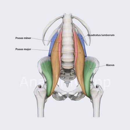
The posterior abdominal wall is composed of five principal muscles: the diaphragm, psoas major, psoas minor, iliacus, and quadratus lumborum. These muscles are arranged in a layered fashion from medial to lateral, with the psoas major occupying the most medial position adjacent to the vertebral column, and the quadratus lumborum forming the lateral muscular boundary. The diaphragm constitutes the superior boundary.
3.2 Psoas Major
3.2.1 Origin and Insertion
The psoas major is a long, thick, fusiform muscle that lies in the groove between the transverse processes and the bodies of the lumbar vertebrae. It originates from:
- Vertebral bodies: The lateral aspects and intervertebral discs of T12 through L5. The origin from T12 is variable and may be absent.
- Transverse processes: The anterior aspects of the transverse processes of all lumbar vertebrae (L1–L5).
The muscle fibres converge inferiorly, forming a tendon that passes deep to the inguinal ligament and inserts onto the lesser trochanter of the femur. The tendon is joined by the tendon of the iliacus muscle, forming the iliopsoas tendon.
3.2.2 Relations
- Anteriorly: Related to the kidney (left psoas to left kidney, right psoas to right kidney), ureter, gonadal vessels, lumbar sympathetic trunk, and the common iliac vessels (inferiorly). The genitofemoral nerve pierces the anterior surface of the psoas major at the L3–L4 level.
- Posteriorly: The lumbar vertebrae and intervertebral discs, lumbar vessels, and the lumbar plexus (which forms within the substance of the psoas major).
- Laterally: The quadratus lumborum muscle (separated by the lumbar plexus nerves emerging from the lateral border of the psoas), and the iliacus muscle (inferiorly).
- Medially: The vertebral column and the lumbar sympathetic trunk.
3.2.3 Action
The psoas major is the principal flexor of the hip joint. When acting from its femoral insertion, it flexes the thigh and, with the assistance of the iliacus, laterally rotates the femur. When acting from its vertebral origin, it produces lateral flexion of the lumbar vertebral column to the ipsilateral side. Bilateral contraction produces flexion of the trunk upon the pelvis, as in the sit-up movement.
3.2.4 Innervation
The psoas major receives segmental innervation from the anterior rami of L1, L2, and L3 spinal nerves. These fibers pass directly into the muscle from the lumbar plexus roots. Additionally, the femoral nerve (L2–L4) supplies a branch to the psoas major as it descends through the muscle.
3.3 Psoas Minor
The psoas minor is a long, slender muscle that lies on the anterior surface of the psoas major. It is present in only approximately 60% of the population and is more frequently absent on the right side. It originates from the vertebral bodies of T12 and L1 and the intervertebral disc between them, and inserts onto the pectineal line of the pubis (iliopectineal eminence). Its action is weak flexion of the lumbar vertebral column. It is innervated by the anterior ramus of L1.
3.4 Iliacus
3.4.1 Origin and Insertion
The iliacus is a large, fan-shaped (or triangular) muscle that fills the iliac fossa. It originates from:
- The superior two-thirds of the iliac fossa.
- The ala of the sacrum.
- The anterior sacroiliac ligaments.
Its fibres converge inferiorly and medially, joining the tendon of the psoas major to form the iliopsoas muscle, which inserts onto the lesser trochanter of the femur.
3.4.2 Action
The iliacus, in conjunction with the psoas major, forms the iliopsoas muscle—the most powerful flexor of the hip joint. It also stabilises the hip joint and, when the lower limb is fixed, assists in flexing the trunk toward the thigh (as in rising from a seated position).
3.4.3 Innervation
The iliacus is innervated by the femoral nerve (L2–L4), specifically by branches arising from the nerve as it passes between the psoas major and iliacus muscles.
3.5 Quadratus Lumborum
3.5.1 Origin and Insertion
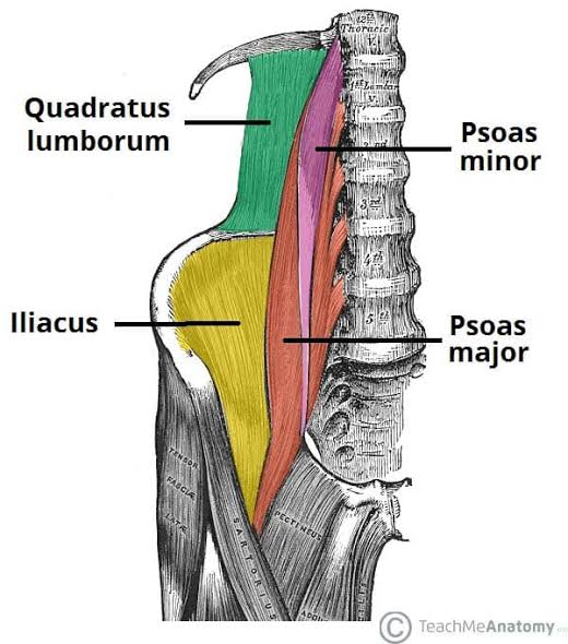
The quadratus lumborum is a thick, quadrilateral muscular sheet located in the lateral portion of the posterior abdominal wall, between the medial psoas major and the lateral transversus abdominis. It originates from:
- The posterior aspect of the iliac crest (medial half).
- The iliolumbar ligament.
- The transverse process of L5 (occasionally).
It inserts onto:
- The medial half of the inferior border of the 12th rib.
- The tips of the transverse processes of L1–L4 vertebrae.
3.5.2 Relations
- Anteriorly: The colon (ascending on the right, descending on the left), kidney, and the subcostal and iliohypogastric nerves.
- Posteriorly: The erector spinae muscles and the thoracolumbar fascia.
- Laterally: The transversus abdominis muscle and the internal oblique (separated by the transversalis fascia).
- Medially: The psoas major muscle, separated by the lumbar plexus nerves.
3.5.3 Action and Innervation
Action: Extends the lumbar vertebral column when both sides contract. Unilateral contraction produces lateral flexion of the vertebral column to the ipsilateral side. It also fixes the 12th rib during inspiration, preventing its elevation and thereby stabilising the origin of the diaphragm for more efficient contraction.
Innervation: Innervation from the subcostal nerve (T12) and the anterior rami of L1–L4 spinal nerves (lumbar intercostal nerves). These nerves pass across the anterior surface of the muscle.
3.6 Diaphragm
3.6.1 Structure and Attachments
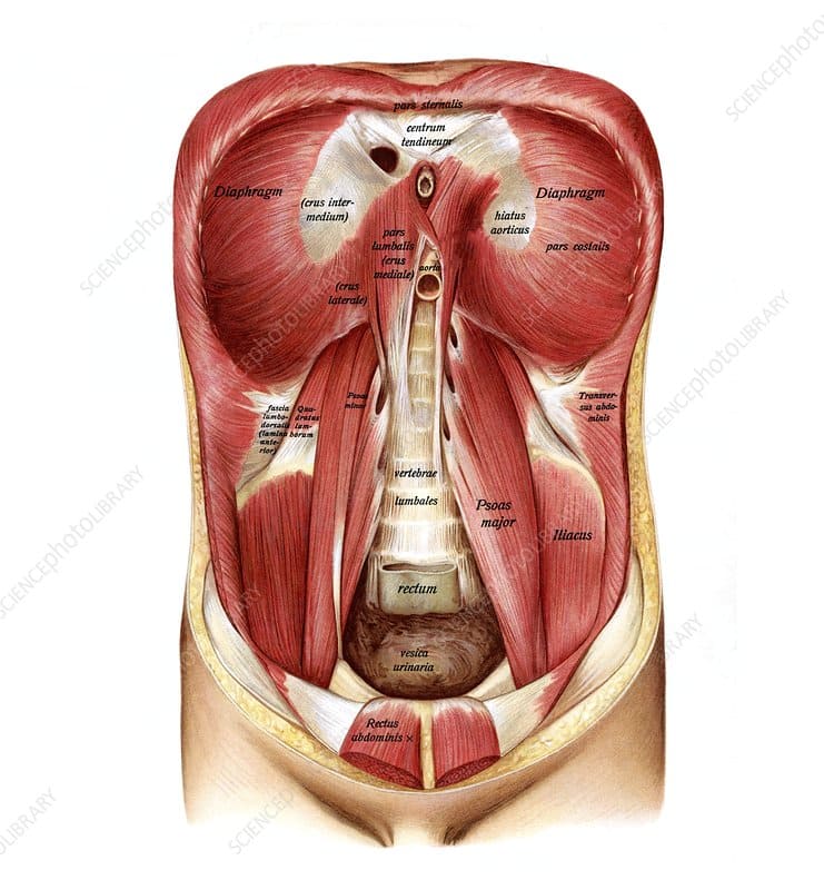
The diaphragm is a dome-shaped musculotendinous partition separating the thoracic and abdominal cavities. It consists of a peripheral muscular portion and a central aponeurotic tendon. The muscle fibres converge from three peripheral attachments:
- Sternal part: Two muscular slips arising from the posterior surface of the xiphoid process.
- Costal part: Arising from the inner surfaces of the lower six costal cartilages (ribs 7–12) and interdigitating with the transversus thoracis muscle.
- Vertebral (lumbar) part: Comprising the right and left crura and three arcuate ligaments:
— Right crus: Arises from the anterior aspects of the bodies and intervertebral discs of L1–L3. Some fibres loop around the oesophageal hiatus, forming a physiological sphincter.
— Left crus: Arises from the anterior aspects of the bodies and intervertebral discs of L1–L2.
— Median arcuate ligament: A fibrous arch connecting the medial borders of the two crura, crossing anterior to the aorta at the aortic hiatus (T12).
— Medial arcuate ligament: Extends from the side of the body of L1 (or L2) to the transverse process of L1, crossing anterior to the psoas major muscle.
— Lateral arcuate ligament: Extends from the transverse process of L1 to the 12th rib, crossing anterior to the quadratus lumborum muscle.
3.6.2 Apertures of the Diaphragm
| Aperture | Vertebral Level | Structures Passing Through | Clinical Note |
|---|---|---|---|
| Caval opening | T8 (central tendon) | Inferior vena cava; terminal branches of right phrenic nerve | IVC wall fused with central tendon; no muscle contraction affects it |
| Oesophageal hiatus | T10 (muscular sling of right crus) | Oesophagus; anterior and posterior vagal trunks; oesophageal branches of left gastric vessels | Hiatal hernia common; physiological sphincter formed by right crus fibres |
| Aortic hiatus | T12 (behind diaphragm, between crura) | Descending aorta; thoracic duct; azygos vein | Not a true aperture; structures pass behind, not through, diaphragm |
| Lesser apertures | Variable | Greater, lesser, and least splanchnic nerves (through crura); sympathetic trunk (under medial arcuate ligament); subcostal nerve/vessels (under lateral arcuate ligament) | Sites of potential nerve compression or herniation |
3.6.3 Innervation and Action
Innervation: Motor supply from the phrenic nerve (C3, C4, C5), with the right phrenic nerve supplying the right hemidiaphragm and the left phrenic nerve supplying the left. Sensory innervation to the parietal pleura and peritoneum covering the diaphragm is also provided by the phrenic nerve (central portion) and the lower intercostal nerves (peripheral portion). The phrenic nerve pierces the central tendon to reach the inferior surface.
Action: The diaphragm is the principal muscle of inspiration. Upon contraction, the domes descend and flatten, increasing the vertical diameter of the thoracic cavity and producing negative intrathoracic pressure that draws air into the lungs. During expiration, the diaphragm relaxes and returns to its domed position, assisted by elastic recoil of the lungs and contraction of the abdominal wall muscles.
3.7 Comparative Anatomy Table: PAW Muscles
| Muscle | Origin | Insertion | Action | Innervation |
|---|---|---|---|---|
| Psoas major | Bodies, discs, and transverse processes of T12–L5 | Lesser trochanter of femur (with iliacus) | Hip flexion; lateral flexion of lumbar spine | L1–L3 (direct); femoral nerve |
| Psoas minor | Bodies and disc of T12–L1 | Pectineal line of pubis | Weak flexion of lumbar spine | L1 |
| Iliacus | Superior 2/3 of iliac fossa; ala of sacrum; anterior sacroiliac ligaments | Lesser trochanter of femur (with psoas major) | Hip flexion; hip stabilisation | Femoral nerve (L2–L4) |
| Quadratus lumborum | Iliac crest; iliolumbar ligament; transverse process of L5 | 12th rib; transverse processes of L1–L4 | Extension and lateral flexion of lumbar spine; fixes 12th rib | Subcostal (T12); L1–L4 |
| Diaphragm | Xiphoid; ribs 7–12; L1–L3 (crura) | Central tendon | Principal muscle of inspiration | Phrenic nerve (C3–C5) |
Psoas Abscess and Haematoma
The psoas major muscle is a common site for abscess formation, particularly in tuberculosis of the spine (Pott's disease). Infection from the vertebral body tracks along the psoas sheath, producing a cold abscess that may present as a swelling in the groin (as the abscess tracks along the psoas tendon beneath the inguinal ligament) or in the lumbar triangle. The psoas sign—pain on passive extension of the hip—is a classic clinical test for appendicitis, as the retrocaecal appendix lies in contact with the right psoas major. A positive psoas sign indicates retrocaecal appendicitis. Similarly, psoas haematoma may occur in patients receiving anticoagulation therapy, compressing the femoral nerve and producing weakness of knee extension (quadriceps paralysis) and sensory loss over the anterior thigh and medial leg.
CHAPTER 4: Fasciae and Connective Tissue Planes
4.1 Overview of Fascial Layers
The fascial system of the posterior abdominal wall is organised into distinct layers that compartmentalise muscles, provide pathways for neurovascular structures, and define the boundaries of the retroperitoneal space. The principal fascial layers include the psoas fascia, iliac fascia, quadratus lumborum fascia, transversalis fascia, and the thoracolumbar fascia proper.
4.2 Psoas Fascia (Psoas Sheath)
The psoas fascia is a strong fibrous sheath that encloses the psoas major muscle. It is attached medially to the bodies and intervertebral discs of the lumbar vertebrae and to the pelvic brim inferiorly. Superiorly, the psoas fascia thickens to form the medial arcuate ligament of the diaphragm, which arches over the psoas major muscle from the body of L1 (or L2) to the transverse process of L1. The psoas sheath is continuous inferiorly with the iliac fascia that covers the iliacus muscle. The psoas sheath is of considerable clinical importance: it may contain haematoma (e.g., from ruptured lumbar vessels or in anticoagulated patients) or pus (psoas abscess), and provides a pathway for the spread of infection from the spine to the groin.
4.3 Iliac Fascia
The iliac fascia covers the iliacus muscle within the iliac fossa. It is continuous superiorly with the psoas fascia and inferiorly with the fascia lata of the thigh. The iliac fascia is attached laterally to the inner lip of the iliac crest and medially to the pelvic brim. It fuses with the psoas fascia at the inguinal ligament, forming a common fascial sheath (iliopsoas fascia) that encloses the iliopsoas muscle as it passes deep to the inguinal ligament into the thigh. The femoral nerve lies in the groove between the psoas major and iliacus muscles, covered by the iliac fascia. Swelling within this fascial compartment (e.g., iliacus haematoma) may compress the femoral nerve.
4.4 Quadratus Lumborum Fascia
The quadratus lumborum fascia is the anterior layer of the thoracolumbar fascia. It covers the anterior surface of the quadratus lumborum muscle and is continuous medially with the psoas fascia. Superiorly, it thickens to form the lateral arcuate ligament of the diaphragm, which arches over the quadratus lumborum from the transverse process of L1 to the 12th rib. The quadratus lumborum fascia is pierced by the subcostal, iliohypogastric, and ilioinguinal nerves as they course across the anterior surface of the muscle.
4.5 Thoracolumbar Fascia
The thoracolumbar fascia (lumbodorsal fascia) is a complex, three-layered fascial structure in the lumbar region that encloses the deep muscles of the back (erector spinae and transversospinalis groups) and provides attachment for the latissimus dorsi, internal oblique, and transversus abdominis muscles. It consists of three layers:
- Posterior layer: The thickest layer, attached medially to the spinous processes and supraspinous ligaments, and laterally to the angles of the ribs and the iliac crest. It gives origin to the latissimus dorsi.
- Middle layer: Attached medially to the tips of the lumbar transverse processes and laterally to the posterior layer at the lateral border of the erector spinae. It separates the erector spinae from the quadratus lumborum.
- Anterior layer (quadratus lumborum fascia): Attached medially to the transverse processes and laterally to the posterior layer at the lateral border of the quadratus lumborum. It is continuous with the transversalis fascia.
4.6 Transversalis Fascia
The transversalis fascia is the deep fascial layer lining the inner surface of the transversus abdominis muscle. In the posterior abdominal wall, it is continuous with the anterior layer of the thoracolumbar fascia and the quadratus lumborum fascia. It extends inferiorly into the pelvis, where it contributes to the fascial coverings of the pelvic viscera. The transversalis fascia is of particular importance in hernia surgery, as it forms the posterior wall of the inguinal canal and must be reinforced during hernia repair.
4.7 Retroperitoneal Fascial Planes and Spaces
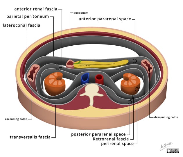
The retroperitoneal space is the anatomical region between the parietal peritoneum anteriorly and the transversalis fascia posteriorly. It is subdivided into distinct compartments:
- Anterior pararenal space: Located between the parietal peritoneum and the anterior renal fascia (Gerota's fascia). It contains the ascending colon, descending colon, duodenum, and pancreas.
- Perirenal space: Enclosed by the anterior and posterior renal fasciae (Gerota's and Zuckerkandl's fasciae). It contains the kidney, renal pelvis, proximal ureter, renal vessels, suprarenal gland, and perirenal fat.
- Posterior pararenal space: Located between the posterior renal fascia and the transversalis fascia (or quadratus lumborum fascia). It contains only fat and vessels and communicates inferiorly with the extraperitoneal pelvic space.
The fascial layers separating these spaces are not complete, allowing pathological processes (infection, haemorrhage, tumour spread) to track between compartments.
4.8 Lumbar Triangles
The lumbar triangles are potential sites of weakness in the posterior abdominal wall where herniation may occur:
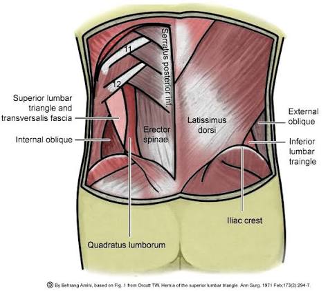
- Superior lumbar triangle (Grynfeltt-Lesshaft triangle): Bounded superiorly by the 12th rib and serratus posterior inferior, inferiorly by the internal oblique muscle, and medially by the erector spinae (sacrospinalis). The floor is formed by the transversalis fascia and the aponeurosis of the transversus abdominis. The base is directed medially toward the vertebral column.
- Inferior lumbar triangle (Petit's triangle): Bounded anteriorly by the external oblique muscle, posteriorly by the latissimus dorsi, and inferiorly by the iliac crest. The floor is formed by the internal oblique muscle and the transversalis fascia. It is more commonly involved in lumbar hernias than the superior triangle.
Lumbar Hernias
Lumbar hernias occur through the superior or inferior lumbar triangles and account for less than 2% of all abdominal hernias. They may be congenital (Grynfeltt hernia) or acquired (Petit's hernia), the latter often following trauma, surgery, or infection. The contents typically include extraperitoneal fat, colon, or small bowel. Diagnosis is often delayed due to the subtle presentation. CT imaging is the gold standard for diagnosis and surgical planning. Repair may be performed via open or laparoscopic approaches, with mesh reinforcement recommended for acquired hernias. The surgeon must be aware of the subcostal and iliohypogastric nerves coursing through the region to avoid iatrogenic injury and subsequent chronic neuropathic pain.
CHAPTER 5: Vascular Supply
5.1 Arterial Supply
5.1.1 Abdominal Aorta
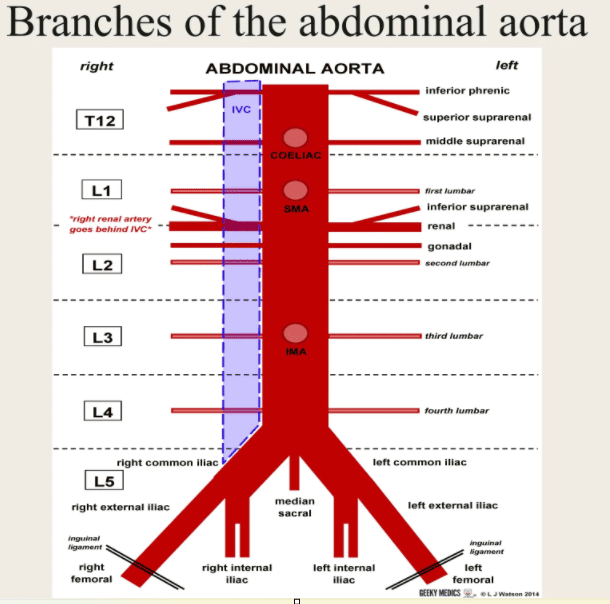
The abdominal aorta is the continuation of the thoracic aorta, beginning at the aortic hiatus of the diaphragm at the level of the T12 vertebra. It descends anterior to the bodies of the lumbar vertebrae, slightly to the left of the midline, and terminates by bifurcating into the right and left common iliac arteries at the level of the L4 vertebra (approximately 2 cm below and to the left of the umbilicus). The abdominal aorta measures approximately 13 cm in length and 2 cm in diameter at its origin, tapering to approximately 1.5 cm at its bifurcation.
The abdominal aorta gives off three sets of branches:
- Unpaired visceral branches: Coeliac trunk, superior mesenteric artery, inferior mesenteric artery.
- Paired visceral branches: Middle suprarenal, renal, gonadal arteries.
- Paired parietal branches: Inferior phrenic, lumbar arteries.
5.1.2 Parietal Branches
- Inferior Phrenic Arteries: The first branches of the abdominal aorta, arising immediately below the diaphragm. They ascend to supply the inferior surface of the diaphragm and the suprarenal glands. The right inferior phrenic artery passes posterior to the inferior vena cava. Each artery gives off a superior suprarenal branch to the suprarenal gland.
- Lumbar Arteries: Typically four pairs (occasionally five) arising from the posterolateral aspect of the aorta at the level of L1–L4. Each lumbar artery passes posterior to the sympathetic trunk, then divides into a dorsal branch (supplying the skin and muscles of the back) and a spinal branch (entering the vertebral canal to supply the spinal cord, meninges, and vertebral bodies). The lumbar arteries anastomose with each other and with the subcostal, iliolumbar, and deep circumflex iliac arteries, forming a rich collateral network.
- Median Sacral Artery: A small, unpaired artery arising from the posterior aspect of the aorta just above its bifurcation (at L4). It descends over the anterior surface of the sacrum to the coccyx, supplying the sacrum, coccyx, rectum, and anorectal junction. It anastomoses with the lateral sacral arteries.
5.1.3 Visceral Branches
- Coeliac Trunk: Arises from the anterior aspect of the aorta at the T12–L1 level, immediately below the aortic hiatus. It is 1.5–2 cm long and divides into the left gastric, splenic, and common hepatic arteries, supplying the foregut derivatives (stomach, liver, gallbladder, pancreas, duodenum proximal to the ampulla of Vater, and spleen).
- Superior Mesenteric Artery (SMA): Arises from the anterior aspect of the aorta at the L1 level, approximately 1 cm below the coeliac trunk. It descends anterior to the left renal vein and the uncinate process of the pancreas, then crosses anterior to the third part of the duodenum and the aorta. It supplies the midgut derivatives (distal duodenum, jejunum, ileum, caecum, appendix, ascending colon, and proximal two-thirds of the transverse colon).
- Inferior Mesenteric Artery (IMA): Arises from the anterior aspect of the aorta at the L3 level, approximately 3–4 cm above the aortic bifurcation. It descends to the left, crossing the left common iliac vessels, and divides into the left colic, sigmoid, and superior rectal arteries. It supplies the hindgut derivatives (distal one-third of the transverse colon, descending colon, sigmoid colon, and rectum).
- Middle Suprarenal Arteries: Paired vessels arising from the lateral aspect of the aorta at the L1 level, between the coeliac trunk and the renal arteries. Each passes laterally to the suprarenal gland, where it anastomoses with the superior suprarenal (from the inferior phrenic) and inferior suprarenal (from the renal) arteries.
- Renal Arteries: Large paired vessels arising from the lateral aspect of the aorta at the L1–L2 intervertebral disc level, approximately 1.5 cm below the SMA. The right renal artery is longer than the left and passes posterior to the inferior vena cava. Each artery divides into segmental branches (typically four or five) before entering the renal hilum.
- Gonadal Arteries: Paired vessels arising from the anterior aspect of the aorta at the L2 level, approximately 2 cm below the renal arteries. The testicular arteries are longer and more tortuous than the ovarian arteries. They descend obliquely over the psoas major muscle, cross the ureter anteriorly, and pass through the inguinal canal (testicular) or the suspensory ligament of the ovary (ovarian) to reach the gonads.
5.1.4 Arterial Anastomoses
| Anastomotic Network | Participating Arteries | Clinical Significance |
|---|---|---|
| Suprarenal arterial circle | Superior (phrenic), middle (aortic), inferior (renal) suprarenal arteries | Maintains gland perfusion if one vessel is occluded |
| Lumbar arterial arcade | Lumbar aa. (L1–L4), subcostal, iliolumbar, deep circumflex iliac | Collateral supply to posterior abdominal wall and spinal cord |
| Coeliac–SMA–IMA marginal artery of Drummond | Left colic, sigmoid, superior rectal, middle colic, right colic, ileocolic | Critical collateral in IMA or SMA occlusion |
| Renal capsular anastomosis | Renal capsular, suprarenal, ureteric, gonadal, lumbar aa. | Maintains kidney perfusion in renal artery stenosis |
| Epigastric anastomosis | Superior epigastric (internal thoracic), inferior epigastric (external iliac) | Collateral pathway in aortic or iliac occlusion |
5.2 Venous Drainage
5.2.1 Inferior Vena Cava (IVC)
The inferior vena cava is the largest vein in the body, formed by the union of the right and left common iliac veins at the L5 level (posterior to the right common iliac artery and anterior to the sacral promontory). It ascends on the right side of the aorta, embedded in the areolar tissue of the retroperitoneum, and passes through the caval opening of the diaphragm at the T8 level to drain into the right atrium. The IVC measures approximately 25 cm in length and 2.5 cm in diameter.
Unlike the aorta, the IVC has no unpaired visceral tributaries. Its principal tributaries include:
- Paired visceral tributaries: Renal veins (L1–L2), suprarenal veins, gonadal veins.
- Parietal tributaries: Lumbar veins (L1–L4), inferior phrenic veins.
- Hepatic veins: Three major hepatic veins (right, middle, left) draining directly into the IVC within the liver, just below the diaphragm.
5.2.2 Important Venous Asymmetries
Several asymmetries in the venous drainage of the posterior abdominal wall are of clinical significance:
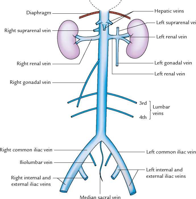
- Renal veins: The left renal vein is longer (approximately 7–8 cm) than the right (approximately 2–3 cm) because the IVC is positioned to the right of the aorta. The left renal vein passes anterior to the aorta and posterior to the SMA (the 'nutcracker' configuration).
- Gonadal veins: The right gonadal vein drains directly into the IVC, while the left gonadal vein drains into the left renal vein. This asymmetry explains the higher incidence of left-sided varicocele in males, as the left testicular vein must drain against gravity into the renal vein before reaching the IVC.
- Suprarenal veins: The right suprarenal vein drains into the IVC, while the left suprarenal vein drains into the left renal vein.
5.2.3 Lumbar Veins and 5.2.4 Ascending Lumbar Veins
Typically four pairs of lumbar veins drain the posterior abdominal wall and spinal canal. The upper two pairs (L1–L2) usually drain directly into the IVC, while the lower two pairs (L3–L4) may drain into the ascending lumbar veins or directly into the IVC. The lumbar veins accompany the lumbar arteries and receive dorsal branches from the skin and muscles of the back, as well as spinal branches from the vertebral canal.
The ascending lumbar veins are longitudinal channels that connect the lumbar veins and communicate superiorly with the azygos/hemiazygos system and inferiorly with the common iliac veins. They lie on the anterior surface of the vertebral bodies, medial to the psoas major muscles. These veins serve as important collateral pathways in IVC obstruction and may become engorged in portal hypertension.
Nutcracker Syndrome
Nutcracker syndrome occurs when the left renal vein is compressed between the aorta posteriorly and the superior mesenteric artery anteriorly (the 'nutcracker' configuration at the L1 level). This compression results in left renal venous hypertension, producing haematuria, left flank pain, and left-sided varicocele in males. The syndrome may be diagnosed by Doppler ultrasound showing elevated left renal vein velocity and by CT angiography demonstrating the aortomesenteric angle (normally 45–60 degrees; reduced to less than 35 degrees in nutcracker syndrome). Treatment options include endovascular stenting, left renal vein transposition, or gonadal vein ligation.
CHAPTER 6: Innervation
6.1 Overview
The posterior abdominal wall receives innervation from somatic, autonomic (sympathetic and parasympathetic), and visceral afferent sources. The somatic innervation is provided by the subcostal nerve (T12) and the lumbar plexus (L1–L4), which supply the muscles and skin of the posterior abdominal wall and lower limb. The autonomic innervation is provided by the lumbar sympathetic trunk and the parasympathetic fibres from the vagus nerve (via the coeliac and mesenteric plexuses).
6.2 Somatic Innervation: The Lumbar Plexus
6.2.1 Formation and Location
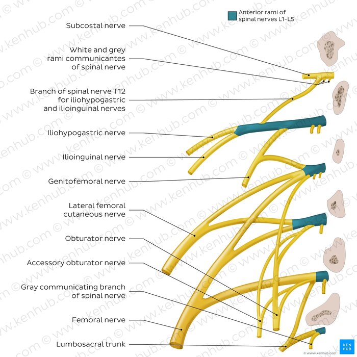
The lumbar plexus is formed within the posterior portion of the psoas major muscle, anterior to the transverse processes of the lumbar vertebrae. It is composed of the anterior rami of spinal nerves L1–L4, with a contribution from the subcostal nerve (T12). The plexus is located in the retroperitoneal space, embedded within the substance of the psoas major muscle. The anterior rami divide into anterior (ventral) and posterior (dorsal) divisions, which then combine to form the peripheral nerves of the plexus.
6.2.2 Branches of the Lumbar Plexus
The branches of the lumbar plexus emerge from the lateral, anterior, or medial borders of the psoas major muscle. They are organised as follows:
- Iliohypogastric Nerve (L1, with contribution from T12): Emerges from the lateral border of the psoas major and descends obliquely across the anterior surface of the quadratus lumborum toward the iliac crest. It pierces the transversus abdominis near the ASIS and divides into lateral and anterior cutaneous branches. It supplies motor innervation to the internal oblique and transversus abdominis muscles, and sensory innervation to the skin of the posterolateral gluteal region and the suprapubic region.
- Ilioinguinal Nerve (L1): Emerges from the lateral border of the psoas major, inferior to the iliohypogastric nerve. It follows a similar course, pierces the transversus abdominis and internal oblique, and enters the inguinal canal via the deep inguinal ring. It supplies motor innervation to the internal oblique and transversus abdominis, and sensory innervation to the skin of the upper medial thigh and the external genitalia (root of penis and anterior scrotum in males; mons pubis and labium majus in females).
- Genitofemoral Nerve (L1, L2): Formed within the substance of the psoas major. It pierces the anterior surface of the psoas major at the L3–L4 level and descends on the anterior surface of the muscle. It divides into:
— Genital branch: Passes through the inguinal canal (within the spermatic cord in males) to supply the cremasteric muscle and the skin of the upper anterior scrotum/mons pubis. It is responsible for the cremasteric reflex.
— Femoral branch: Passes posterior to the inguinal ligament to supply the skin of the upper anterior thigh. - Lateral Femoral Cutaneous Nerve (L2, L3): Formed from the posterior divisions of L2 and L3. It emerges from the lateral border of the psoas major, crosses the iliacus muscle obliquely toward the ASIS, and passes posterior to the inguinal ligament approximately 1–2 cm medial to the ASIS. It supplies sensory innervation to the skin of the lateral thigh along the iliotibial tract, extending to the knee.
- Femoral Nerve (L2, L3, L4): The largest branch of the lumbar plexus, formed from the posterior divisions of L2–L4. It emerges from the lower lateral border of the psoas major, descends in the groove between the psoas major and iliacus muscles (covered by the iliac fascia), and passes deep to the inguinal ligament lateral to the femoral vessels. Motor supply: Iliacus, pectineus, sartorius, quadriceps femoris. Sensory supply: Skin of the anteromedial thigh, and the medial leg and foot (saphenous nerve). The femoral nerve also supplies the hip and knee joints.
- Obturator Nerve (L2, L3, L4): Formed from the anterior divisions of L2–L4. It emerges from the medial border of the psoas major, descends along the pelvic wall, and exits the pelvis through the obturator canal. Motor supply: Obturator externus, adductor longus, adductor brevis, adductor magnus (anterior part), gracilis, and pectineus. Sensory supply: Skin of the medial thigh.
- Accessory Obturator Nerve (L3, L4): Present in approximately 20–30% of individuals. It emerges from the medial border of the psoas major, descends over the superior pubic ramus, and supplies the pectineus and hip joint.
- Lumbosacral Trunk (L4, L5): Formed by the descending branch of L4 and the entire anterior ramus of L5. It descends over the ala of the sacrum, medial to the psoas major, and joins the sacral plexus (S1–S4) in the pelvis. It carries fibres that contribute to the sciatic nerve.
Mnemonic for Lumbar Plexus Branches
The major branches of the lumbar plexus may be remembered by the mnemonic: 'Interested In Getting Lunch On Friday?'
- I — Iliohypogastric
- I — Ilioinguinal
- G — Genitofemoral
- L — Lateral femoral cutaneous
- O — Obturator
- F — Femoral
6.3 Autonomic Innervation
6.3.1 Lumbar Sympathetic Trunk
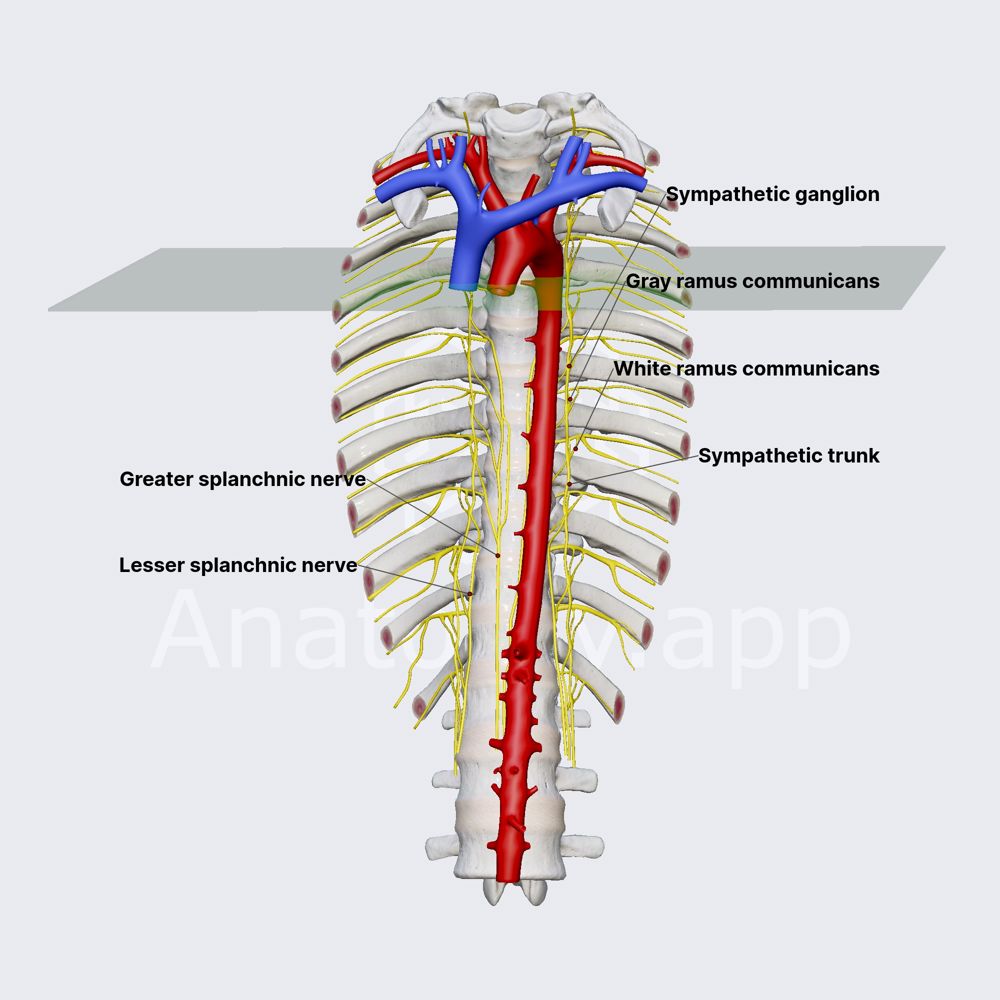
The lumbar sympathetic trunk is the continuation of the thoracic sympathetic trunk, which crosses the medial arcuate ligament to enter the abdomen. It descends along the anterolateral aspect of the lumbar vertebral bodies, medial to the psoas major muscle and anterior to the lumbar vessels. The trunk contains four ganglia (L1–L4). Each ganglion receives white rami communicantes from the corresponding spinal nerve and gives off grey rami communicantes that return to the spinal nerve. The lumbar sympathetic trunk also gives off branches that form the lumbar splanchnic nerves, which contribute to the coeliac, aortic, and superior hypogastric plexuses.
6.3.2 Coeliac Plexus and Mesenteric Plexuses
The coeliac plexus (solar plexus) is the largest autonomic plexus in the abdomen, located at the level of L1, surrounding the coeliac trunk and superior mesenteric artery origins. It receives sympathetic fibres from the greater and lesser splanchnic nerves (T5–T12) and parasympathetic fibres from the vagus nerve (via the posterior vagal trunk). The coeliac plexus gives rise to subsidiary plexuses (renal, hepatic, etc.) that supply the viscera. The aortic plexus lies on the anterior aspect of the abdominal aorta.
6.3.3 Parasympathetic Innervation
The posterior abdominal wall receives parasympathetic innervation via the vagus nerve (cranial nerve X), which enters the abdomen through the oesophageal hiatus (T10). The anterior and posterior vagal trunks descend along the oesophagus and stomach, contributing to the coeliac and mesenteric plexuses. The pelvic splanchnic nerves (S2–S4) provide parasympathetic innervation to the distal colon and pelvic viscera, but do not directly supply the posterior abdominal wall.
6.4 Comparative Table: Nerves of the PAW
| Nerve | Spinal Roots | Course | Motor Supply | Sensory Distribution |
|---|---|---|---|---|
| Subcostal | T12 | Crosses quadratus lumborum; pierces abdominal wall muscles | Transversus abdominis, internal oblique | Skin of lower abdominal wall and lateral gluteal region |
| Iliohypogastric | L1 (T12) | Lateral border of psoas; across quadratus lumborum to iliac crest | Transversus abdominis, internal oblique | Posterolateral gluteal skin; suprapubic skin |
| Ilioinguinal | L1 | Lateral border of psoas; through inguinal canal | Transversus abdominis, internal oblique | Upper medial thigh; external genitalia |
| Genitofemoral | L1, L2 | Pierces anterior psoas; divides into genital and femoral branches | Cremaster muscle (genital branch) | Upper anterior scrotum/mons pubis; upper anterior thigh |
| Lateral femoral cutaneous | L2, L3 | Lateral border of psoas; crosses iliacus; under inguinal ligament | None (purely sensory) | Lateral thigh to knee |
| Femoral | L2, L3, L4 | Between psoas and iliacus; deep to inguinal ligament | Iliopsoas, pectineus, sartorius, quadriceps | Anteromedial thigh; medial leg and foot (saphenous) |
| Obturator | L2, L3, L4 | Medial border of psoas; obturator canal | Obturator externus, adductors, gracilis, pectineus | Medial thigh |
Meralgia Paraesthetica
Meralgia paraesthetica is a condition characterised by tingling, numbness, and burning pain in the anterolateral thigh, caused by compression of the lateral femoral cutaneous nerve as it passes under the inguinal ligament. It is commonly associated with tight clothing, obesity, pregnancy, or prolonged prone positioning. The nerve may also be injured during pelvic surgery or iliac crest bone graft harvesting. Diagnosis is clinical, confirmed by nerve conduction studies. Treatment includes weight loss, avoidance of compressive garments, local anaesthetic and corticosteroid injection, and, in refractory cases, surgical decompression or neurectomy.
CHAPTER 7: Lymphatic Drainage
7.1 Overview and 7.2 Lumbar Lymph Nodes
The lymphatic drainage of the posterior abdominal wall is organised into two principal systems: the lumbar lymph nodes and the cisterna chyli. The lumbar lymph nodes are arranged in three principal groups, located around the abdominal aorta and inferior vena cava:
- Lateral aortic (lumbar) nodes: Located lateral to the aorta, along the course of the lumbar vessels. They receive lymph from the kidneys, suprarenal glands, gonads, ureters, and the posterior abdominal wall (including the deep muscles and fascia). Efferent vessels from these nodes drain to the pre-aortic nodes.
- Pre-aortic nodes: Located anterior to the aorta, clustered around the origins of the coeliac trunk, superior mesenteric artery, and inferior mesenteric artery. They are subdivided into coeliac, superior mesenteric, and inferior mesenteric nodes. They receive lymph from the gastrointestinal tract, liver, gallbladder, pancreas, and spleen. Efferent vessels form the intestinal lymphatic trunks.
- Post-aortic (retrocaval) nodes: Located posterior to the aorta and inferior vena cava, along the lumbar vessels. They receive lymph from the posterior abdominal wall muscles, vertebral bodies, and meninges.
7.3 Cisterna Chyli
The cisterna chyli is a dilated sac-like structure located at the L1–L2 level, anterior to the bodies of the vertebrae and posterior to the right crus of the diaphragm. It is formed by the union of the right and left lumbar lymphatic trunks and the intestinal lymphatic trunk. The cisterna chyli is the origin of the thoracic duct, which ascends through the aortic hiatus (T12) to drain lymph into the left venous angle (junction of the left subclavian and internal jugular veins). The cisterna chyli receives lymph from the entire body below the diaphragm, with the exception of the right upper quadrant (which drains via the right lymphatic duct).
7.4 Lymphatic Drainage of Specific Structures
- Kidneys: The renal lymphatic vessels follow the renal veins and drain to the lateral aortic (lumbar) nodes. The left kidney drains predominantly to the left lateral aortic nodes, while the right kidney drains to the right lateral aortic nodes.
- Suprarenal Glands: The suprarenal lymphatic vessels drain to the lateral aortic nodes and the coeliac nodes.
- Gonads: The testicular lymphatic vessels ascend with the testicular arteries, pass through the inguinal canal, and drain to the lateral aortic nodes at the L2 level. The ovarian lymphatic vessels follow the ovarian vessels and drain to the lateral aortic nodes.
- PAW Muscles: The deep lymphatic vessels drain to the lateral aortic and post-aortic nodes. Superficial lymphatic vessels drain to the superficial inguinal nodes.
Enlargement of the lumbar lymph nodes may occur in lymphoma, metastatic carcinoma (particularly from the kidneys, testes, ovaries, and gastrointestinal tract), and tuberculosis. Retroperitoneal lymphadenopathy may compress the ureters (hydronephrosis) or the IVC (lower limb oedema). A lymph node diameter greater than 1 cm in the short axis is generally considered pathological.
Retroperitoneal Fibrosis (Ormond's Disease)
Retroperitoneal fibrosis (Ormond's disease) is a rare condition characterised by the proliferation of fibrous tissue in the retroperitoneum, typically encasing the abdominal aorta, inferior vena cava, and ureters at the L4–L5 level. The fibrous tissue may extend superiorly to the renal hila and inferiorly to the pelvic brim. It produces bilateral ureteric obstruction, leading to hydronephrosis and renal failure. The condition may be idiopathic (associated with IgG4-related disease) or secondary to malignancy or drugs (e.g., methysergide). Treatment includes corticosteroids or surgical ureterolysis.
CHAPTER 8: Clinical Anatomy and Surgical Considerations
8.1 Surgical Approaches to the PAW
The posterior abdominal wall is accessed through several surgical approaches:
- Retroperitoneal approach: The retroperitoneal space is accessed via a flank incision, typically extending from the 12th rib to the iliac crest. The external oblique, internal oblique, and transversus abdominis muscles are incised in layers, and the peritoneum is swept medially to expose the kidney, ureter, aorta, and IVC. The surgeon must identify and preserve the subcostal, iliohypogastric, and ilioinguinal nerves, which course across the quadratus lumborum.
- Anterior transperitoneal approach: The abdomen is opened through a midline or paramedian incision. The posterior parietal peritoneum is incised over the aorta to expose retroperitoneal vessels. This approach provides excellent exposure but carries a higher risk of postoperative ileus and adhesions.
- Laparoscopic approach: Minimally invasive techniques. The retroperitoneal approach (posterior lumbar approach) is particularly useful for adrenalectomy and nephrectomy, as it avoids peritoneal violation.
8.2 Common Pathologies
8.2.1 Psoas Abscess
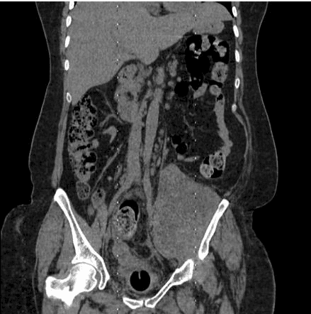
A collection of pus within the psoas muscle sheath, most commonly secondary to spinal tuberculosis (Pott's disease), pyogenic vertebral osteomyelitis, or spread from adjacent abdominal or pelvic infections. The abscess may present with fever, back pain, hip flexion contracture (to reduce tension on the psoas), and a palpable mass in the groin. CT is the diagnostic modality of choice, demonstrating a fluid collection within the psoas muscle with peripheral rim enhancement.
8.2.2 Retroperitoneal Haemorrhage
May result from traumatic injury to the aorta, IVC, lumbar, or renal vessels; from rupture of an abdominal aortic aneurysm; or from anticoagulation. Clinical features include abdominal or flank pain, hypotension, tachycardia, and signs of shock. CT with intravenous contrast demonstrates a hyperdense collection within the retroperitoneum.
8.2.3 Abdominal Aortic Aneurysm (AAA)
An abdominal aortic aneurysm is defined as a focal dilation of the abdominal aorta exceeding 3 cm in diameter (or 1.5 times the normal diameter). The vast majority (95%) are infrarenal, with the aneurysm originating below the renal arteries. The aneurysm expands at an average rate of 0.3–0.5 cm per year. Rupture risk increases exponentially with diameter:
- 1% per year for aneurysms 4–5 cm.
- 5–10% per year for aneurysms 5–6 cm.
- Greater than 25% per year for aneurysms exceeding 7 cm.
Clinical presentation ranges from asymptomatic to sudden, catastrophic rupture. Elective repair is recommended for aneurysms exceeding 5.5 cm in diameter (or 5.0 cm in women) or those expanding at greater than 0.5 cm per year.
8.4 Summary Table: Clinical Conditions and Anatomical Basis
| Condition | Anatomical Basis | Key Clinical Features | Diagnostic Modality |
|---|---|---|---|
| Psoas abscess | Infection tracks along psoas sheath from spine to groin | Back pain, hip flexion contracture, groin mass, fever | CT with contrast |
| Retroperitoneal haemorrhage | Rupture of aorta, IVC, or lumbar vessels; blood tracks in fascial planes | Flank pain, hypotension, Grey Turner's sign, shock | CT angiography |
| AAA rupture | Degeneration of aortic wall media; infrarenal segment most common | Sudden abdominal/back pain, pulsatile mass, hypotension | CT angiography; bedside ultrasound |
| Nutcracker syndrome | Left renal vein compressed between aorta and SMA | Haematuria, left flank pain, left varicocele | Doppler ultrasound; CT angiography |
| Lumbar hernia | Defect in superior or inferior lumbar triangle | Flank mass, reducible, enlarges with Valsalva | CT abdomen/pelvis |
| Psoas haematoma | Bleeding into psoas sheath (trauma, anticoagulation) | Groin mass, femoral nerve palsy, hip pain | CT or MRI |
| Meralgia paraesthetica | Lateral femoral cutaneous nerve compressed under inguinal ligament | Burning pain, numbness in lateral thigh | Clinical; nerve conduction studies |
| Retroperitoneal fibrosis | Fibrous tissue encases aorta, IVC, and ureters | Bilateral ureteric obstruction, renal failure, back pain | CT or MRI |
Psoas Sign and Obturator Sign in Appendicitis
The psoas sign is a clinical test for retrocaecal appendicitis. The patient lies on their left side, and the examiner passively extends the right hip. Stretching of the right psoas major muscle produces pain if the retrocaecal appendix is inflamed and in contact with the psoas. A positive psoas sign strongly suggests retrocaecal appendicitis and may influence the surgical approach. The obturator sign (passive internal rotation of the flexed right hip) tests for pelvic appendicitis, where the inflamed appendix lies in contact with the obturator internus muscle.
APPENDIX: KEY LEARNING POINTS SUMMARY
Muscles Quick Reference
- Psoas major: Origin T12–L5; insertion lesser trochanter; action hip flexion; innervation L1–L3, femoral nerve.
- Iliacus: Origin iliac fossa, ala of sacrum; insertion lesser trochanter; action hip flexion; innervation femoral nerve (L2–L4).
- Quadratus lumborum: Origin iliac crest, iliolumbar ligament; action extension and lateral flexion of lumbar spine; innervation subcostal (T12), L1–L4.
- Diaphragm: Origin xiphoid, ribs 7–12, L1–L3; insertion central tendon; principal muscle of inspiration; innervation phrenic nerve (C3–C5).
Clinical Red Flags
- Positive psoas sign → retrocaecal appendicitis.
- Grey Turner's sign → retroperitoneal haemorrhage.
- Left varicocele + haematuria → nutcracker syndrome.
- Pulsatile abdominal mass + hypotension → ruptured AAA.
- Burning lateral thigh pain → meralgia paraesthetica.
