Doctors Revision

Author name: doctorsrevision@gmail.com

Definition of medical psychology
Medical Psychology

Definition of medical psychology

Medical Psychology A comprehensive study of the application of psychological principles to the understanding, prevention, and treatment of physical illness, emphasizing the interaction between biological, psychological, and social factors. 1.1 Definition of Medical Psychology 1.1.1 What is Medical Psychology? Medical psychology is the branch of psychology that applies psychological principles, theories, and techniques to the understanding, prevention, and treatment of physical illness, and to the promotion of health and well-being. It examines how biological, psychological, and social factors interact to influence health and disease. Simple Definition Medical psychology is the study of how the mind and behavior affect the body, health, and illness — and how illness affects the mind. 1.1.2 Alternative Terms While used in similar contexts, these terms have specific relationships to the broader field of medical psychology: Term Relationship to Medical Psychology Health Psychology Often used interchangeably; focuses specifically on health promotion and illness prevention. Behavioral Medicine An interdisciplinary field combining psychology, medicine, and behavioral sciences. Psychosomatic Medicine Focuses on physical diseases caused or worsened by psychological factors. 1.1.3 Scope of Medical Psychology The scope of medical psychology is broad, covering every aspect of the patient’s journey and the healthcare delivery system: Area What It Involves Patient Behavior Understanding why patients seek or avoid medical care. Illness Behavior How people perceive, evaluate, and respond to symptoms. Doctor-Patient Relationship Communication, trust, empathy, and rapport. Stress and Disease How stress contributes to physical illness (e.g., hypertension, ulcers). Pain Management Psychological techniques for managing chronic pain. Patient Compliance Why patients do or do not follow medical advice (adherence). Health Beliefs Cultural and personal beliefs about illness and treatment. Coping with Illness Psychological adjustment to chronic disease, disability, or terminal illness. Lifestyle and Health Smoking, alcohol, diet, exercise, and risky behaviors. 1.1.4 Medical Psychology vs. Clinical Psychology It is essential to distinguish between these two branches, as they differ in primary focus, patient types, and clinical goals. Feature Medical Psychology Clinical Psychology Primary Focus Psychological aspects of physical illness and health Mental disorders and abnormal behavior Setting Hospitals, clinics, community health centers Psychiatric hospitals, counseling centers Approach Biopsychosocial model Primarily psychological/therapeutic Patient Type Patients with physical illness + psychological issues Patients with mental illness Goal Improve health outcomes and quality of life Treat and manage mental disorders Clinical Correlation The Biopsychosocial Model in Uganda A clinical officer in Uganda treating a diabetic patient must understand medical psychology to address: Compliance: Why the patient skips insulin doses. Adjustment: Feelings of depression regarding the chronic diagnosis. Health Beliefs: Whether the patient believes traditional herbs are superior to or sufficient compared to medical treatment. Points for Attention Remember that Medical Psychology is not just about mental illness; it is the application of psychology to physical health. A key exam distinction is that Medical Psychology uses the Biopsychosocial model to bridge the gap between medicine and the mind. Applied Clinical Scenario Case Study Hypertension and Health Beliefs Scenario: A 45-year-old patient in a rural Ugandan clinic refuses to start hypertensive medication, stating that “herbal tea from the village elder is more powerful than tablets.” Question: Which area of the scope of Medical Psychology must the Clinical Officer address to improve this patient’s outcome? Answer: The Clinical Officer must address Health Beliefs and Patient Compliance. By understanding the patient’s cultural and personal beliefs regarding illness and treatment, the provider can better communicate the necessity of the medication in a way that respects the patient’s context.
Characteristics of urine
Anatomy

Characteristics of urine

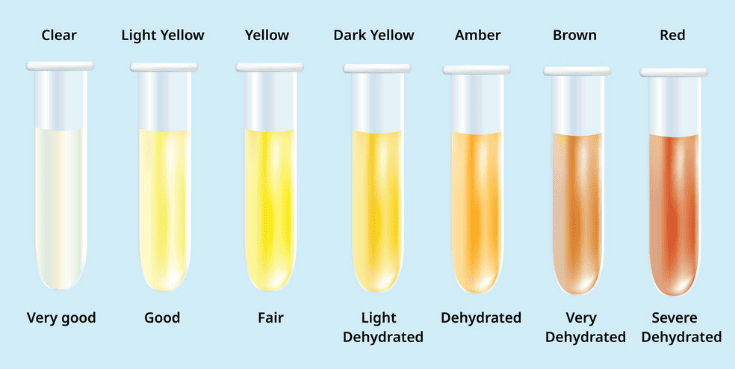
Characteristics of Urine 1. INTRODUCTION AND DEFINITION OF URINE Definition: Urine is a liquid excretory product formed by the kidneys through the processes of glomerular filtration, tubular reabsorption, and tubular secretion. It serves as the primary mechanism for eliminating metabolic waste products, excess water, electrolytes, and foreign substances from the body. Composition: Urine is approximately 95% water and 5% dissolved solids. The solid components include: Nitrogenous wastes: Urea (the largest component), creatinine, uric acid, and ammonia. Electrolytes: Sodium, potassium, chloride, bicarbonate, calcium, magnesium, and phosphate. Pigments: Urochrome (gives urine its yellow color), urobilinogen, and bilirubin (when present). Other substances: Hormones, enzymes, vitamins, drugs, and toxins in trace amounts. Clinical Significance Examination of urine characteristics provides critical information about renal function, metabolic status, hydration, and the presence of infection or systemic disease. Abnormal urine findings often represent the earliest detectable signs of conditions such as diabetes mellitus, urinary tract infection, glomerular disease, and hepatic dysfunction. 2. ANATOMY AND PHYSIOLOGY OF URINE FORMATION 2.1 Overview of the Urinary System The urinary system consists of two kidneys, two ureters, the urinary bladder, and the urethra. The kidneys are paired, bean-shaped organs located retroperitoneally on either side of the vertebral column, extending from T12 to L3. Each kidney weighs approximately 150 grams in adults and measures about 11 cm in length, 6 cm in width, and 3 cm in thickness. The functional unit of the kidney is the nephron. Each kidney contains approximately 1 to 1.5 million nephrons. The nephron consists of two main components: the renal corpuscle (comprising the glomerulus and Bowman’s capsule) and the renal tubule (including the proximal convoluted tubule, loop of Henle, distal convoluted tubule, and collecting duct). 2.2 Three Processes of Urine Formation A. Glomerular Filtration: Blood enters the glomerulus through the afferent arteriole. High hydrostatic pressure (~55 mmHg) forces water and small solutes through the filtration membrane into Bowman’s capsule. The filtration barrier consists of three layers: fenestrated endothelium, basement membrane, and podocyte slit diaphragm. It prevents the passage of blood cells and large proteins (molecular weight >70,000 Daltons). B. Tubular Reabsorption: As filtrate passes through the tubules, essential substances are returned to the bloodstream. The PCT reabsorbs approximately 65% of filtered water, sodium, glucose, and amino acids. The loop of Henle establishes a concentration gradient via the countercurrent multiplier system. C. Tubular Secretion: Active transport of substances (H+, K+, creatinine, drugs, and toxins) from peritubular capillaries into the lumen. This is particularly important for acid-base homeostasis. 2.3 Regulation of Urine Formation ADH (vasopressin): Released from posterior pituitary; increases water reabsorption in collecting ducts, producing concentrated urine. Aldosterone: Secreted by adrenal cortex; promotes sodium reabsorption and potassium secretion. ANP: Released from cardiac atria; inhibits sodium reabsorption and promotes diuresis. PTH: Regulates calcium and phosphate reabsorption. 3. PHYSICAL CHARACTERISTICS OF URINE The physical examination is the first step in urinalysis. Characteristics evaluated include color, clarity, odor, volume, specific gravity, pH, and foam. 3.1 Color Normal urine color ranges from pale yellow to deep amber. The pigment responsible is urochrome (urobilin), a breakdown product of hemoglobin metabolism. Abnormal Colors and Clinical Significance: Red or Pink: May indicate hematuria, hemoglobinuria, or myoglobinuria. Causes include UTI, renal calculi, glomerulonephritis, trauma, and strenuous exercise. Medications like rifampin and phenazopyridine also produce red urine. Orange: Associated with dehydration, bilirubinuria (liver disease), or medications like warfarin. Green or Blue: Rare. Caused by Pseudomonas aeruginosa UTI, biliverdin (biliary stasis), or propofol. Brown or Tea-Colored: Suggests bile pigments (bilirubin), old blood, or myoglobin. Seen in acute viral hepatitis, cirrhosis, and rhabdomyolysis. Cloudy or Milky: Results from pyuria (WBCs), bacteriuria, chyluria (lymphatic fluid), or lipiduria. In Uganda, filariasis is an important cause of chyluria. 3.2 Clarity and Odor Clarity: Normal urine is clear or translucent. Turbidity results from cellular material, microorganisms, mucus, or crystals. Clinical Note: If cloudiness clears with acetic acid, precipitation of phosphates is likely. Odor: Normal urine has a faint aromatic odor. — Ammoniacal: Bacterial decomposition of urea (UTI). — Sweet/Fruity: Presence of ketone bodies (DKA). — Musty/Mousy: Characteristic of phenylketonuria (PKU). — Maple Syrup: MSUD. 3.4 Volume and Specific Gravity Normal daily output: 600 to 2,000 mL (average 1,000-1,600 mL). Minimum required: 400-500 mL/day. Polyuria (>2,500-3,000 mL/day): Diabetes mellitus, diabetes insipidus, excessive intake. Oliguria (<400-500 mL/day): Dehydration, shock, heart failure, ATN, obstruction. Anuria (<100 mL/day): Medical emergency; bilateral obstruction or renal cortical necrosis. Specific Gravity: Normal 1.002 to 1.035. High (>1.025) in dehydration; low (<1.010) in DI or ATN. A fixed gravity of ~1.010 indicates loss of tubular concentrating ability. 4. CHEMICAL CHARACTERISTICS OF URINE Commonly performed using reagent-impregnated dipsticks. 4.1 Proteins Normal excretion is ≤150 mg/day. — Glomerular proteinuria: Albumin leaks through damaged barrier (Nephrotic syndrome >3.5 g/day). — Tubular proteinuria: Failure of reabsorption (e.g., ATN, Fanconi syndrome). — Overflow proteinuria: Bence Jones proteins in multiple myeloma. — Functional: Transient after exercise or fever. 4.2 Glucose and Ketones Glycosuria: Occurs when glucose exceeds renal threshold (~180 mg/dL). Common in Diabetes Mellitus. Ketonuria: Indicates increased lipolysis. Seen in DKA, starvation, and severe diarrhea in children. 4.4 Bilirubin and Urobilinogen Bilirubin: Only conjugated (direct) bilirubin appears in urine. Presence indicates hepatocellular damage or biliary obstruction. Urobilinogen: Elevated in hemolysis (hemolytic anemia, sickle cell) and hepatic dysfunction. Decreased in complete biliary obstruction. Diagnostic Rule — Bilirubin (+) / Urobilinogen (-): Obstructive jaundice. — Bilirubin (-) / Urobilinogen (+): Hemolysis. 5. MICROSCOPIC EXAMINATION OF URINE Timely examination (within 1-2 hours) is critical for identifying cellular elements and casts. 5.1 Cells Red Blood Cells: Normal 0–2/HPF. Dysmorphic RBCs suggest glomerular origin; isomorphic suggest lower tract bleeding. White Blood Cells: Normal 0–5/HPF. Pyuria (>5/HPF) indicates infection or inflammation. Epithelial Cells: Renal tubular epithelial cells are normally absent; their presence indicates tubular damage (ATN). 5.2 Casts Hyaline Casts: Composed of Tamm-Horsfall protein; seen in concentrated urine or fever. Red Blood Cell Casts: Diagnostic of glomerular bleeding (Acute glomerulonephritis). White Blood Cell Casts: Indicate renal parenchymal infection (pyelonephritis). Waxy and Broad Casts: Indicate advanced chronic kidney disease and severe tubular atrophy. Fatty Casts: “Maltese cross” pattern; associated with Nephrotic Syndrome.

Obstruction, Renal failure, Renal stones, UTIs
Anatomy

Obstruction, Renal failure, Renal stones, UTIs

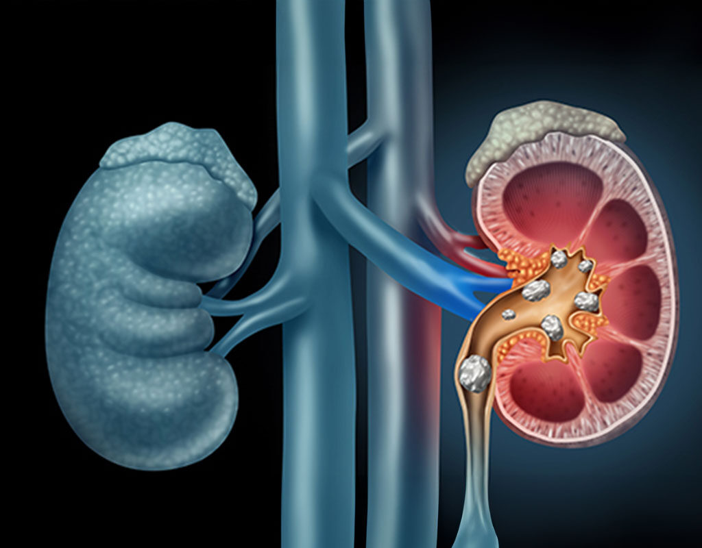
Common Disorders of the Urinary System A comprehensive clinical resource covering the epidemiology, pathophysiology, classification, and management of major renal and urological conditions, including infections, calculi, and systemic syndromes. 1. Urinary Tract Infections (UTIs) 1.1 Definition and Epidemiology Urinary tract infections (UTIs) are among the most common bacterial infections encountered in clinical practice, affecting any part of the urinary tract from the urethra to the kidneys. Escherichia coli accounts for approximately 80–90% of uncomplicated UTIs. Key Anatomical Rule UTIs are significantly more common in females due to the shorter urethra (approximately 4 cm compared to 20 cm in males), which allows easier bacterial ascent from the perineum into the bladder. 1.2 Classification by Anatomical Location Lower UTI — Cystitis Cystitis is the infection and inflammation of the bladder mucosa. It is characterized by: Dysuria — painful or burning urination Frequency — frequent voiding of small amounts Urgency — sudden compelling need to urinate Suprapubic pain or discomfort Hematuria — blood in the urine (occasional) Absence of fever (this distinguishes cystitis from pyelonephritis) Cloudy or foul-smelling urine Upper UTI — Pyelonephritis Pyelonephritis is the infection of the renal pelvis and renal parenchyma. It usually results from ascending infection but may occur via hematogenous spread. Clinical features include: Fever and chills Flank pain — tenderness at the costovertebral angle Nausea and vomiting Dysuria, frequency, and urgency Systemic symptoms — malaise, tachycardia Cloudy urine with possible pus 1.3 Risk Factors Female sex, sexual activity, use of spermicides, pregnancy, urinary tract obstruction, vesicoureteral reflux, diabetes mellitus, immunosuppression, indwelling urinary catheters, incomplete bladder emptying, and anatomical abnormalities. 1.4 Diagnosis and Management Diagnosis: Urinalysis showing Pyuria (WBCs), bacteriuria, positive nitrites, and positive leukocyte esterase. Microscopic examination may reveal WBC casts in pyelonephritis. Urine Culture is the gold standard (Colony count ≥10⁵ CFU/mL is diagnostic). Management of Uncomplicated Cystitis: TMP-SMX for 3 days Nitrofurantoin for 5 days Fosfomycin as a single dose Adequate hydration (2–3 liters daily) Management of Pyelonephritis: Fluoroquinolones (Ciprofloxacin, Levofloxacin) for 7–14 days Cephalosporins (Ceftriaxone) if fluoroquinolones are contraindicated 2. Kidney Stones (Urolithiasis / Nephrolithiasis) Kidney stones are hard mineral and salt deposits. Prevalence is higher in hot climates due to dehydration. Men are affected more often, with peak incidence between 20 and 50 years. 2.2 Types of Renal Calculi Type Composition Frequency Key Associations Calcium Oxalate Calcium + Oxalate ~70–80% Hypercalciuria, hyperoxaluria, low citrate Calcium Phosphate Calcium + Phosphate ~10–15% Renal tubular acidosis, hyperparathyroidism Uric Acid Uric Acid ~5–10% Gout, acidic urine (pH < 5.5), high purine diet Struvite Magnesium ammonium phosphate ~10% Infection with urease-producing bacteria (Proteus) Cystine Cystine < 1% Autosomal recessive cystinuria 2.4 Clinical Presentation Renal Colic: Severe, colicky flank pain radiating to the groin (testis/labia) caused by ureteral spasm. Restlessness: The patient cannot find a comfortable position (distinguishes from peritonitis where the patient lies still). Hematuria, nausea/vomiting, and urinary frequency. 2.5 Management Conservative: Hydration, NSAIDs (Analgesia), and alpha-blockers (Tamsulosin) as medical expulsive therapy. Interventional: ESWL (Stones < 2 cm), Ureteroscopy (Endoscopic removal), or PCNL (For large stones > 2 cm or staghorn calculi). 3. Glomerulonephritis (GN) Glomerulonephritis is the inflammation of the glomeruli, classified as Primary (kidney-limited) or Secondary (systemic diseases like SLE or vasculitis). 3.2 Pathophysiology Immune Complex Deposition: Antigen-antibody complexes activate complement (Post-streptococcal GN, Lupus). Anti-GBM Antibodies: Antibodies attack the basement membrane; seen in Goodpasture syndrome. Pauci-Immune: Neutrophil-mediated injury without significant deposits (ANCA-positive vasculitis). 3.3 Nephritic Syndrome The classic presentation of GN: Hematuria — smoky or coca-cola colored urine; dysmorphic RBCs and RBC casts. Oliguria — decreased urine output (< 400 mL/day). Hypertension — due to sodium and water retention. Mild Proteinuria, Azotemia, and Periorbital Edema. Common Types Acute Post-Streptococcal GN: Follows Group A strep infection; subepithelial “hump” deposits on EM. RPGN (Crescentic GN): Severe form with crescent formation in Bowman’s space; rapid renal failure. IgA Nephropathy (Berger Disease): Most common worldwide; mesangial IgA deposits. MPGN: “Tram-track” double basement membrane appearance. 4. Nephrotic Syndrome Clinical constellation caused by heavy glomerular protein loss resulting from damage to the glomerular filtration barrier (podocytes and slit diaphragms). 4.2 Diagnostic Criteria (The Big Four) Heavy Proteinuria: >3.5 g/day (Adults) or >50 mg/kg/day (Children). Hypoalbuminemia: Serum albumin < 30 g/L (3 g/dL). Edema: Pitting edema (periorbital and dependent). Hyperlipidemia and Lipiduria: Elevated cholesterol; “oval fat bodies” in urine. Complications Loss of antithrombin III and increased clotting factor synthesis lead to a Hypercoagulable state (Renal vein thrombosis). Loss of IgG increases Infection risk. 5. Chronic Kidney Disease (CKD) Defined as abnormalities of kidney structure or function present for > 3 months. 5.2 Staging by GFR Stage GFR (mL/min/1.73m²) Description G1 ≥90 Normal or high G2 60–89 Mildly decreased G3a 45–59 Mildly to moderately decreased G3b 30–44 Moderately to severely decreased G4 15–29 Severely decreased G5 3 RBCs per HPF). Classification by origin: Origin Microscopy Findings Common Causes Glomerular (Medical) Dysmorphic RBCs, RBC casts, Proteinuria Glomerulonephritis, IgA Nephropathy Non-Glomerular (Surgical) Isomorphic (Normal) RBCs, No casts UTI, Stones, Tumors, Trauma, BPH Evaluation Rule Urine dipstick detects heme but cannot distinguish RBCs from free hemoglobin or myoglobin. Formal microscopy is essential. For adults >40 with unexplained hematuria, cystoscopy is mandatory to exclude malignancy.

elimination of urine
Anatomy

Elimination of urine

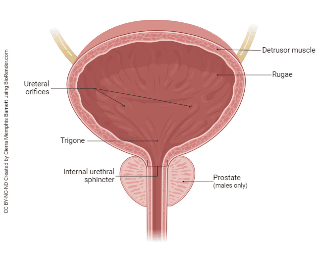
Elimination of Urine (Micturition) Comprehensive anatomical and physiological overview of the micturition process, neural control mechanisms, and clinical pathophysiology of the lower urinary tract. 1. Definition Elimination of urine, also called micturition or voiding, is the process by which urine is expelled from the urinary bladder through the urethra to the exterior of the body. It is the final step in urine handling, following filtration by the kidneys and transport through the ureters. 2. Anatomy of the Lower Urinary Tract The Urinary Bladder The bladder is a hollow, muscular, distensible organ located in the anterior pelvis. In adults, it typically holds 400–600 mL of urine. The physiological desire to void typically begins when the volume reaches 200–300 mL. Layers of the bladder wall (inner to outer): Mucosa: Transitional epithelium (urothelium) that stretches without damage; forms folds called rugae when the bladder is empty. Submucosa: Connective tissue layer containing blood vessels and nerves. Muscularis (Detrusor Muscle): Smooth muscle arranged in three interlacing layers; its contraction is responsible for expelling urine. Adventitia/Serosa: The outermost protective covering. Key Anatomical Regions: Apex: The anterior tip, connected to the umbilicus by the median umbilical ligament. Body: The main central portion of the bladder. Fundus (Base): The posterior surface containing the trigone—a smooth, triangular area bounded by the two ureteric orifices and the internal urethral orifice. Neck: The inferior narrowing where the bladder joins the urethra. Key Concept The trigone is highly sensitive to stretch. Because it is embryologically distinct from the rest of the bladder (mesodermal origin), it lacks rugae and remains smooth regardless of the bladder’s distension state. The Urethra Female urethra: Approximately 3–4 cm long. Its short length increases susceptibility to urinary tract infections (UTIs) as bacteria can ascend more easily. Male urethra: Approximately 18–20 cm long, divided into three main portions: prostatic, membranous, and spongy (penile). The Urethral Sphincters Sphincter Location Muscle Type Control Function Internal urethral sphincter Bladder neck Smooth muscle Involuntary (autonomic) Closes during filling; relaxes during voiding. External urethral sphincter Deep perineal pouch / pelvic floor Skeletal muscle Voluntary (somatic) Consciously prevents or permits urination. 3. Physiology of Micturition Micturition involves two alternating phases controlled by a combination of reflex (involuntary) and voluntary mechanisms. Phase 1: Storage (Filling) Urine flows continuously from the kidneys via the ureters into the bladder. The detrusor muscle remains relaxed to allow for high compliance. Both sphincters remain contracted to maintain continence. Sympathetic (Hypogastric) and somatic (Pudendal) nerves are active. Parasympathetic activity is inhibited. Stretch receptors in the bladder wall send signals to the spinal cord. At low volumes, these are suppressed by higher brain centres. Phase 2: Voiding (Emptying) Triggered consciously when the setting is appropriate through the following sequence: Voluntary relaxation of the external urethral sphincter (cortical inhibition of the pudendal nerve). Sympathetic inhibition → internal sphincter relaxes. Parasympathetic activation (pelvic splanchnic nerves, S2–S4) → detrusor muscle contracts strongly. Intravesical pressure rises; the bladder neck opens. Urine flows out through the urethra. Sensory feedback from the urethra reinforces detrusor contraction until the bladder is completely empty. 4. Neural Control Afferent (Sensory) Pathways Stretch receptors in the bladder wall and trigone detect distension. Signals travel via the pelvic splanchnic nerves to the sacral spinal cord (S2–S4), then ascend to the pontine micturition centre (PMC) and the cerebral cortex. Efferent (Motor) Pathways Pathway Nerve Spinal Origin Neurotransmitter Effect Parasympathetic Pelvic splanchnic nerves S2–S4 Acetylcholine (M3 receptors) Detrusor contraction; internal sphincter relaxation. Sympathetic Hypogastric nerve T11–L2 Noradrenaline (α1 and β3 receptors) Detrusor relaxation (β3); internal sphincter contraction (α1). Somatic Pudendal nerve S2–S4 Acetylcholine (nicotinic) External sphincter contraction (voluntary control). Higher Control Centres Sacral Micturition Centre (S2–S4): Contains the basic reflex arc. In infants, this produces involuntary voiding. Pontine Micturition Centre (PMC): Coordinates the switch between storage and voiding; ensures detrusor contraction and sphincter relaxation occur together. Cerebral Cortex: Provides conscious, voluntary control. Allows for the delay of voiding or initiation at will. 5. Pathophysiology Urinary Retention Inability to empty the bladder. Causes include: Obstructive: Benign prostatic hyperplasia (BPH), urethral stricture, bladder stones. Neurogenic: Spinal cord injury, cauda equina syndrome, diabetic neuropathy. Pharmacological: Anticholinergics, opioids, tricyclic antidepressants. Postoperative: Occurs frequently after pelvic or spinal surgery. Urinary Incontinence The involuntary leakage of urine, classified by mechanism: Type Mechanism Key Features Stress Weak pelvic floor / sphincter Leakage with cough, sneeze, or exertion. Urge Detrusor overactivity Sudden urgency, large-volume loss. Overflow Chronic retention with passive overflow Dribbling, palpable bladder, weak stream. Functional Physical/cognitive barriers Inability to reach the toilet in time despite normal tract function. Special Pathological Conditions Neurogenic Bladder (Spastic): Lesion above sacral cord → detrusor overactivity, small capacity, and reflux risk. Neurogenic Bladder (Flaccid): Lesion at/below sacral cord → detrusor areflexia, large capacity, and high residual urine. Vesicoureteral Reflux: Backflow of urine to ureters due to failure of the ureterovesical valvular mechanism. Predisposes to pyelonephritis. 6. Clinical Relevance Common Local Scenarios Acute retention in elderly males: BPH is the leading cause; requires urgent catheterisation. UTI: Extremely common, especially in females; diagnosed by urinalysis and culture. Obstetric fistula: Resulting from prolonged obstructed labour; causes continuous incontinence and social stigmatization. Spinal cord injury: Requires intermittent catheterisation to prevent secondary renal damage. Patient Assessment History: Voiding pattern, hematuria, and neurological symptoms. Examination: Abdominal palpation for bladder distension, digital rectal examination (males). Investigations: Urine culture, bladder scan, renal ultrasound, and urodynamics. 7. Summary: Storage Phase vs. Voiding Phase Feature Storage Phase Voiding Phase Detrusor muscle Relaxed Contracted Internal sphincter Closed Open External sphincter Closed (voluntary) Open (voluntary relaxation) Parasympathetic Inhibited Active Sympathetic Active Inhibited Intravesical pressure Low and stable Elevated 9. Key Points Micturition is a reflex act modulated by voluntary cortical control. Parasympathetic nerves (S2–S4, pelvic nerve) promote voiding. Sympathetic nerves (T11–L2, hypogastric nerve) promote storage. The pontine micturition centre coordinates the synergy between detrusor contraction and sphincter relaxation. In Uganda, BPH, UTIs, and Obstetric fistula are primary drivers of lower urinary tract clinical visits.

Urine formation
Anatomy

Urine formation

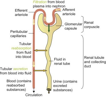
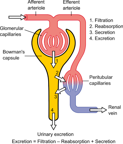
Urine Formation Complete Study Notes covering the coordinated mechanisms of filtration, reabsorption, and secretion. 1. OVERVIEW Urine formation occurs in the nephron, the functional unit of the kidney. Each human kidney contains approximately 1–1.5 million nephrons. The process involves three coordinated physiological mechanisms: Glomerular Filtration: Passive filtration of blood plasma into Bowman’s capsule. Tubular Reabsorption: The return of useful substances from the tubular fluid back to the blood. Tubular Secretion: Active transport of wastes and excess substances from the blood into the tubular fluid. Key Numbers Filtrate formed: ~180 L/day (men) / ~150 L/day (women) Urine excreted: ~1–2 L/day Reabsorbed: ~99% of total filtrate GFR (Glomerular Filtration Rate): ~125 mL/min (men) / ~105 mL/min (women) 2. GLOMERULAR FILTRATION 2.1 The Process Glomerular filtration is a passive process in which hydrostatic pressure forces water and small solutes from glomerular capillaries across the filtration membrane into Bowman’s space. The resulting fluid is called filtrate. Blood enters the glomerulus via the afferent arteriole and exits via the efferent arteriole. 2.2 The Filtration Barrier (Three Layers) Layer 1: Fenestrated Endothelium: The capillary endothelium contains large pores (fenestrae, 70–100 nm) that allow plasma to pass but block blood cells. Layer 2: Glomerular Basement Membrane: A gel-like layer of type IV collagen and negatively charged proteoglycans. It blocks large proteins and repels negatively charged molecules. Layer 3: Podocyte Slit Diaphragms: Specialized epithelial cells with foot-like processes (pedicels) that wrap around capillaries. Gaps between adjacent foot processes (filtration slits, ~25 nm) are bridged by thin slit diaphragms acting as the final molecular sieve. 2.3 What Gets Filtered? Passes Freely Blocked (Retained in Blood) Water, Na+, K+, Cl– Blood cells (RBCs, WBCs) Glucose, Amino acids Albumin, Globulins Urea, Creatinine Platelets 2.4 Starling Forces (Net Filtration Pressure) The movement of fluid is determined by Net Filtration Pressure (NFP), calculated as: NFP = PG − PB − πG Glomerular hydrostatic pressure (PG): ~55 mmHg — promotes filtration. Bowman’s capsule pressure (PB): ~15 mmHg — opposes filtration. Plasma colloid osmotic pressure (πG): ~30 mmHg — opposes filtration. Net Filtration Pressure: ~10 mmHg. 3. TUBULAR REABSORPTION Approximately 99% of the filtrate is returned to the blood. Reabsorption occurs along the entire length of the renal tubule. 3.1 Proximal Convoluted Tubule (PCT) The PCT reabsorbs ~65% of filtered water, sodium, and chloride, and virtually 100% of glucose and amino acids. Sodium (Na+): Actively pumped out by Na+/K+-ATPase on the basolateral membrane. Glucose & Amino Acids: Reabsorbed by Na+-cotransporters (SGLT) on the apical membrane. Water: Follows solutes by osmosis via aquaporin-1 channels. Bicarbonate (HCO3–): Reabsorbed indirectly. H+ is secreted into the lumen, combines with HCO3– to form H2CO3, which is converted to CO2 + H2O by carbonic anhydrase. CO2 diffuses into the cell and is reconverted to HCO3–. Clinical Note Transport Maximum (Tm) Each carrier has a maximum rate. For glucose, Tm ≈ 375 mg/min. When blood glucose exceeds the renal threshold (~180 mg/dL), glucose appears in the urine (glucosuria). 3.2 Loop of Henle Reabsorbs ~25% of filtered solutes and establishes the medullary osmotic gradient. Segment Permeability Mechanism Thin Descending Limb Highly permeable to H2O; impermeable to solutes. Water leaves by osmosis; tubular fluid becomes concentrated. Thin Ascending Limb Impermeable to H2O; permeable to Na+/Cl–. Passive salt reabsorption into the interstitium. Thick Ascending Limb (TAL) Impermeable to H2O; highly permeable to ions. Active Na+-K+-2Cl– cotransport (NKCC2); dilutes urine. 3.3 Distal Convoluted Tubule (DCT) & 3.4 Collecting Duct DCT: Reabsorbs 5–10% of filtered Na+ and water. Features the Na+-Cl– cotransporter (NCC) and is the site for PTH-stimulated Ca2+ reabsorption. Site of action for thiazide diuretics. Collecting Duct: Final site of modification. — Principal cells: Reabsorb Na+ (aldosterone-dependent), secrete K+, reabsorb water (ADH-dependent via aquaporin-2). — Intercalated cells: Type A secretes H+; Type B secretes HCO3–. 4. TUBULAR SECRETION The active transport of substances from blood into tubular fluid, complementing filtration. Substance Site Mechanism Importance H+ PCT, DCT, Collecting duct Na+/H+ antiporter; H+-ATPase pump Acid-base balance; HCO3– reabsorption K+ Principal cells (CD) Aldosterone-stimulated secretion Potassium homeostasis Creatinine PCT Passive and active transport Waste product; used to estimate GFR Uric acid PCT Active transport End product of purine metabolism Drugs/Toxins PCT Specific organic acid/base transporters Elimination of Penicillin, morphine, aspirin 5. URINE CONCENTRATION AND DILUTION The kidneys can produce urine ranging from 50 mOsm/L (very dilute) to 1200 mOsm/L (highly concentrated). 5.1 Countercurrent Multiplication Single Effect: The TAL pumps Na+, K+, and 2Cl– into the interstitium via NKCC2. Interstitium becomes hyperosmotic. Equilibration: Descending limb loses water to the hyperosmotic medulla, concentrating the fluid. Multiplication: Concentrated fluid enters the ascending limb, more solutes are pumped out, progressively increasing osmolarity from 300 mOsm/L (cortex) to 1200 mOsm/L (tip). 5.2 Role of Antidiuretic Hormone (ADH) Dehydration (High ADH): ADH binds V2 receptors on principal cells. Aquaporin-2 channels are inserted. Water is reabsorbed. Result: concentrated urine (~1200 mOsm/L), small volume (~0.5 L/day). Well-hydrated (Low ADH): Little ADH released. No aquaporin-2 inserted. CD remains water-impermeable. Result: dilute urine (~50–100 mOsm/L), large volume. 5.3 Vasa Recta & 5.4 Urea Recycling The Vasa Recta are hairpin capillaries that act as countercurrent exchangers, preserving the osmotic gradient by preventing “washout.” Urea Recycling: The inner medullary collecting duct is permeable to urea in the presence of ADH. Urea diffuses into the interstitium, contributing 40–50% of medullary osmolarity. Some urea re-enters the loop of Henle, creating a recycling loop.

Structure and functions of the lower urinary tract (Bladder, Urethra)
Anatomy

Structure and functions of the lower urinary tract (Bladder, Urethra)

Structure and Function of the Lower Urinary Tract Comprehensive exhaustive notes on the anatomy, histology, and physiology of the lower urinary tract (LUT), focusing on the storage and expulsive functions of the bladder and urethra. 1. Introduction The lower urinary tract (LUT) comprises the urinary bladder and the urethra. Its two principal functions are to act as a low-pressure reservoir for the storage of urine and to expel urine at high pressure at an appropriate time and under voluntary control. Urine is produced continuously by the kidneys (~1–2 L/day). The bladder must accommodate this volume without a significant rise in pressure (compliance), then switch rapidly to an expulsive mode during micturition. 2. Gross Anatomy 2.1 The Urinary Bladder The bladder is a hollow, muscular, distensible organ located in the anterior pelvis, posterior to the pubic symphysis. Adult capacity is typically 400–500 mL, though it can distend to over 600 mL. Position & Surface Anatomy: Empty: Entirely within the pelvis, posterior to the pubic symphysis. Full: The dome rises into the abdomen, pushing the peritoneum upward. Clinical Importance A full bladder allows for suprapubic aspiration or catheterization. Because the distended bladder pushes the peritoneum superiorly, a needle can be inserted just above the pubic symphysis into the bladder without entering the peritoneal cavity. Anatomical Parts & Relations: Parts: Apex, Base (Fundus), Body, Neck, Dome, and Trigone. Superior: Peritoneum. Anterior: Pubic symphysis, retropubic space (Space of Retzius). Posterior (Male): Rectum, seminal vesicles, vas deferens. Posterior (Female): Uterus (vesicouterine pouch), upper vagina. Inferolateral: Pelvic side wall, obturator internus, levator ani. 2.2 The Urethra The urethra is the only urinary tract organ with significant sexual dimorphism. Female Urethra: Length: ~3–4 cm (short). Course: Runs inferiorly from the bladder neck, embedded in the anterior vaginal wall. Clinical significance: Its short length and proximity to the anus/vagina predispose females to ascending UTIs. Male Urethra: Length: ~18–20 cm. Divided into four parts: Pre-prostatic: Above the prostate. Prostatic (3–4 cm): Passes through the prostate; contains the urethral crest and seminal colliculus (verumontanum). Membranous (1–2 cm): Passes through the deep perineal pouch; surrounded by the external urethral sphincter. This is the narrowest and least distensible part. Spongy (penile) (~15 cm): Passes through the corpus spongiosum; receives ducts of bulbourethral (Cowper’s) glands. Clinical Pearl The membranous urethra is the most susceptible to injury in pelvic fractures. The prostatic urethra is the primary site of obstruction in Benign Prostatic Hyperplasia (BPH). 2.3 The Prostate Gland (Male) A fibromuscular (30%) and glandular (70%) organ. It is walnut-sized (~20 g) and located inferior to the bladder neck. McNeal Zonal Anatomy: Peripheral Zone (70%): Site where most carcinomas arise. Central Zone (25%): Surrounds ejaculatory ducts. Transitional Zone (5%): Site where BPH typically arises. 3. Microscopic Structure / Histology The bladder wall consists of four layers from inside out: 3.1 Mucosa (Urothelium + Lamina Propria) Urothelium (Transitional Epithelium): Stratified epithelium (5–7 layers relaxed; 2–3 layers distended). Features umbrella cells that provide a permeability barrier. Lamina Propria: Loose connective tissue with myofibroblasts and afferent nerve endings. 3.2 Muscularis (Detrusor Muscle) Three indistinct layers: inner longitudinal, middle circular, outer longitudinal. The middle circular layer is thickest at the neck, forming the internal urethral sphincter (smooth muscle, involuntary). Sensory Hub The Trigone The trigone is histologically and functionally distinct. It is derived from the mesonephric duct (not the urogenital sinus). Its mucosa is smooth (no rugae) and contains a dense network of afferent fibers (rich in P2X3 and substance P), acting as a critical sensor for bladder filling and pain. 4. Neurovascular Supply 4.1 Arterial Supply Structure Arterial Supply Bladder (superior) Superior vesical arteries (from internal iliac/umbilical) Bladder (inferior) Inferior vesical (male) / vaginal arteries (female) Urethra Internal pudendal branches Prostate Inferior vesical, middle rectal arteries 4.4 Nerve Supply The LUT receives input from three nervous systems: Nerve Origin Type Function Pelvic nerve S2–S4 Parasympathetic Motor: Detrusor contraction (voiding). Sensory: Fullness/pain. Hypogastric nerve T12–L2 Sympathetic Motor: Detrusor relaxation & internal sphincter contraction (storage). Pudendal nerve S2–S4 Somatic Motor: External sphincter contraction (voluntary continence). 5. Physiology of the Lower Urinary Tract The LUT operates in two mutually exclusive phases: storage and voiding. 5.1 Urine Storage Phase Events: Bladder filling (50–400 mL). Detrusor relaxation: Mediated by sympathetic stimulation via β3-adrenoceptors. Internal sphincter contraction: Sympathetic α-adrenergic stimulation. External sphincter contraction: Somatic tonic contraction via pudendal nerve. Guarding reflex: Spinal reflexes increase external sphincter tone as volume increases. 5.2 The Micturition Reflex Trigger: Bladder volume reaches ~300–400 mL; stretch receptor firing increases dramatically. The Reflex Arc: Afferent: Stretch receptors (Aδ) → Pelvic nerve → Sacral cord (S2-S4) → Ascends to periaqueductal gray (PAG). Central Integration: PAG relays to pontine micturition center (PMC) (Barrington’s nucleus). The Cerebral Cortex provides voluntary inhibition until socially appropriate. Efferent: PMC activates Pelvic nerve (Detrusor contraction) and inhibits Sympathetic/Somatic tone (Sphincter relaxation). 6. Clinical Correlations 6.1 Urinary Tract Infections (UTIs) Pathophysiology: Ascending infection by coliforms (*E. coli*). High risk in females due to short urethra and proximity to the anus. 6.2 Urinary Incontinence Stress Incontinence: Leakage with increased intra-abdominal pressure (e.g., coughing) due to weak pelvic floor. Urge Incontinence: Detrusor overactivity; involuntary contraction. Overflow Incontinence: Chronic retention with dribbling (e.g., BPH or stricture). 6.4 Neurogenic Bladder Lesion Level Bladder Type Clinical Features Suprapontine (Stroke) Uninhibited Urgency, frequency, urge incontinence. Spinal Cord (Above S2) Reflex / Automatic Detrusor-sphincter dyssynergia (DSD); risk of upper tract damage. Conus / Cauda Equina Acontractile Flaccid bladder, overflow incontinence, painless retention. Clinical Summary Urethral Stricture Narrowing of the lumen due to scarring. Common causes in Uganda: Post-gonococcal infection and trauma (pelvic fractures or traumatic catheterization). Leads to obstructive voiding symptoms and chronic retention.

Structure and functions of the upper urinary tract (Kidneys, Ureters)
Anatomy

Structure and functions of the upper urinary tract (Kidneys, Ureters)

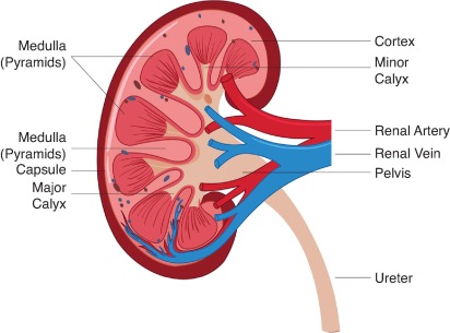
Structure & Function of the Upper Urinary Tract Comprehensive coverage of kidney and ureter anatomy, nephron physiology, renal vasculature, and detailed clinical correlations. 1. OVERVIEW The upper urinary tract comprises the kidneys and ureters — the organs responsible for filtering blood, forming urine, and conveying it to the bladder. The kidneys are paired retroperitoneal organs that perform excretion, electrolyte regulation, acid-base balance, blood pressure control, and endocrine functions. The ureters are muscular conduits that propel urine from the renal pelvis to the urinary bladder via peristalsis. 2. THE KIDNEYS — GROSS ANATOMY Location & Position Position: Paired retroperitoneal organs located between the T12 and L3 vertebrae. Orientation: The left kidney sits slightly more superior than the right (due to the liver). Shape: Bean-shaped (reniform); medial concavity = hilum; lateral convexity. Size: ~4–5 inches (10–12 cm) long; ~150 g in adults (size of a fist). Covering: Enclosed by renal fascia (Gerota’s fascia) — a dense elastic connective tissue sheath; surrounded by perirenal fat. External Features Feature Description Renal Capsule Three layers of connective tissue/fat covering the kidney; protects and stabilizes the organ. Hilum Medial indentation where renal artery, renal vein, and ureter enter/exit. Renal Pelvis Funnel-shaped structure collecting urine; continuous with the ureter. Adrenal Gland Sits atop each kidney; produces cortisol and other hormones. Renal Hilum Contents Anterior to posterior: Renal vein → Renal artery → Ureter. Mnemonic Very Angry Unicorn (Vein, Artery, Ureter). The renal sinus is the cavity within the hilum containing fat, vessels, and the collecting system. 3. THE KIDNEYS — INTERNAL STRUCTURE The kidney is divided into two primary regions: the renal parenchyma and the collecting system (pyelocalyceal system). A. Renal Parenchyma Renal Cortex (Outer Layer) Contains most nephron components: renal corpuscles, proximal & distal convoluted tubules, cortical collecting ducts. Renal Mantle: Peripheral cortical tissue covering the base of each renal pyramid. Renal Columns (of Bertin): Extensions of cortical tissue projecting inward between pyramids toward the renal sinus. Renal Medulla (Inner Layer) Organized into renal pyramids (~9 per kidney). Contains medullary rays (descending/ascending limbs of Loop of Henle + medullary collecting ducts). Each pyramid + overlying cortex = renal lobe (anatomical unit). Renal Papilla: Apex of each pyramid; urine drains through the area cribrosa into minor calyces. B. Renal Collecting System Structure Description Minor Calyces 7–9 per kidney; each encloses a renal papilla. May be simple (1 papilla) or compound (2–3 papillae). Major Calyces Formed by convergence of 2–3 minor calyces; 2–3 per kidney. Renal Pelvis Funnel-shaped structure formed by union of major calyces; marks transition to the proximal ureter. 4. THE NEPHRON — FUNCTIONAL UNIT The nephron is the structural and functional unit of the kidney. Each adult kidney contains 1 to 1.5 million nephrons (occasionally >2.5 million). Types of Nephrons Type Location Loop of Henle Function Cortical (Superficial) Near cortical surface Short Bulk filtration; ~80% of nephrons Juxtamedullary Near corticomedullary junction Long (deep into medulla) Concentrating urine; countercurrent multiplication A. Renal Corpuscle (Filtration Unit) The renal corpuscle consists of the glomerulus + Bowman’s capsule. The Glomerulus A tuft of fenestrated capillaries supplied by the afferent arteriole and drained by the efferent arteriole. Filtration Barrier (3 layers): Fenestrated Endothelium: Pores 70–100 nm; provides size selectivity. Glomerular Basement Membrane (GBM): Composed of type IV collagen, laminin, fibronectin, proteoglycans; provides charge selectivity (repels negatively charged proteins). Podocytes (Visceral Epithelium): Specialized cells with interdigitating foot processes forming slit diaphragms — the final filtration barrier. Bowman’s Capsule Double-walled cup surrounding the glomerulus. Parietal layer: Simple squamous epithelium. Visceral layer: Podocytes. Bowman’s (Capsular) Space: Collects the ultrafiltrate. Vascular pole: Where afferent/efferent arterioles enter/exit. Urinary pole: Where filtrate exits into the PCT.

Functions of a joint
Anatomy

Functions of a joint

Functions of Joints Joints act like biomechanical and biological systems that enable movement, maintain stability, nourish tissues, sense position, and protect vital structures. 4.1 Mechanical Functions A. Mobility Joints allow the skeleton to move in multiple planes — sagittal, frontal, and transverse. The range of motion (ROM) is determined by the specific articular geometry and individual anatomical variations. Types of Movement by Joint Classification: Joint Type Movement Allowed Examples Uniaxial Movement in one plane Hinge (elbow), Pivot (atlantoaxial) Biaxial Movement in two planes Condyloid (knuckles), Saddle (thumb) Multiaxial Movement in three planes + rotation Ball-and-socket (shoulder, hip) Ball-and-socket joints provide the greatest range of motion, allowing for flexion/extension, abduction/adduction, rotation, and circumduction. Clinical Application Goniometry Measuring ROM with a goniometer is the gold standard in orthopedic and physiotherapy assessment. It quantifies joint angles in degrees, enabling objective tracking of rehabilitation progress and surgical outcomes. B. Stability Joints must balance mobility with stability — a fundamental trade-off in biomechanics. Stability is maintained through several integrated mechanisms: Stabilizer Mechanism Example Bony architecture Deep sockets and congruent surfaces Deep acetabulum of the hip Ligaments Passive restraint; limits excessive motion ACL and MCL in the knee Muscles/Tendons Dynamic stabilization via active contraction Rotator cuff muscles in the shoulder Joint capsule Fibrous enclosure maintaining negative pressure Glenohumeral (shoulder) capsule Negative intra-articular pressure Suction effect drawing surfaces together All major synovial joints Labrum/Meniscus Deepens the socket and improves congruency Glenoid labrum, acetabular labrum The Mobility-Stability Continuum The shoulder sacrifices stability for mobility; its shallow glenoid fossa allows extensive ROM but makes it the most frequently dislocated major joint. Conversely, the hip prioritizes stability for weight-bearing via a deep acetabulum, resulting in reduced mobility. Clinical Application Rotator Cuff Dynamics Shoulder dislocations account for up to 50% of all major joint dislocations. The rotator cuff muscles (supraspinatus, infraspinatus, teres minor, subscapularis) act as essential dynamic stabilizers by actively compressing the humeral head into the glenoid fossa during movement. C. Shock Absorption Articular cartilage and menisci distribute compressive and shear forces across joint surfaces. Synovial fluid acts as a viscous dampener, reducing friction to near-zero levels and dissipating energy. Key Structures in Shock Absorption: Articular (hyaline) cartilage: Covers bone ends; matrix of type II collagen and proteoglycans. It is avascular and aneural. Fibrocartilage menisci: C-shaped discs that increase joint congruency and absorb 30–50% of compressive forces. Synovial fluid: Viscous fluid with a coefficient of friction of ~0.002–0.04. Clinical Application Meniscectomy Risks Loss of shock absorption following meniscectomy leads to accelerated osteoarthritis. Partial meniscal removal can increase contact pressures on articular cartilage by 200–300%, significantly elevating degenerative risk. D. Force Transmission Joints transmit mechanical forces from muscles to produce movement. Weight-bearing joints transmit multiples of body weight (BW) during various activities: Activity Force Relative to Body Weight Standing 1x BW Walking 3–4x BW Running 5–7x BW Jumping/Landing 10–12x BW Pathophysiological Driver Abnormal force transmission — due to malalignment, muscle weakness, or joint instability — is a primary driver of osteoarthritis. The knee joint, for instance, transmits forces exceeding 3,000 N during normal gait. 4.2 Nutritional & Metabolic Functions Because articular cartilage is avascular, it relies entirely on diffusion for nutrient delivery and waste removal. Synovial fluid is the primary medium for this exchange. Mechanism of Cartilage Nutrition: Synovial fluid production: Secreted by the synovial membrane as a blood plasma ultrafiltrate with added hyaluronic acid. Diffusion: Small solutes (glucose, oxygen) diffuse from fluid into the cartilage matrix. “Milking” action: Joint movement creates cyclic loading/unloading, generating a pumping mechanism that enhances fluid exchange. Subchondral bone contribution: Medullary cavities of underlying epiphyseal bone also contribute to nutrition in loaded joints. Clinical Application Immobilization Effects Prolonged bed rest or casting leads to cartilage degeneration. Without the “milking” action of movement, nutrient exchange diminishes, causing chondrocyte death and matrix breakdown. Early mobilization is critical post-surgery. 4.3 Proprioceptive Function Proprioception is the body’s ability to perceive its position and movement in space. Specialized mechanoreceptors provide critical sensory feedback. Types of Joint Receptors: Receptor Location Response Function Type I (Ruffini) Superficial capsule Slow-adapting Static joint position; sustained stretch Type II (Pacinian) Deep capsule Rapidly-adapting Dynamic movement; acceleration Type III (Golgi) Ligaments High-threshold Tension monitoring; protective inhibition Type IV (Free Nerve) Capsule/Ligaments Nociceptive Pain signaling Ascending Pathways: Information travels via the dorsal column-medial lemniscal (DCML) pathway to the somatosensory cortex and via spinocerebellar tracts to the cerebellum. Clinical Application Joint injury (e.g., ACL tear) disrupts proprioception, increasing re-injury risk by 40–70%. Rehabilitation must include balance boards and perturbation exercises to restore neuromuscular control. 4.4 Protection Joints protect underlying vital structures through various mechanical arrangements: Skull sutures: Rigid fibrous joints absorb impact; fontanelles allow cranial molding during birth. Vertebral joints: Intervertebral discs and facet joints cushion and align the vertebral canal (protecting the spinal cord). Rib cage joints: Provide a flexible but protective enclosure for the heart and lungs. Pelvic joints: Maintain the integrity of the pelvic ring to protect pelvic viscera. Summary Table: Functions of a Joint Function Key Structures Clinical Relevance Mobility Articular surfaces, capsule, fluid Goniometry; ROM restoration Stability Ligaments, muscles, labrum ACL reconstruction; Rotator cuff repairs Shock Absorption Cartilage, menisci, synovial fluid Meniscectomy -> Osteoarthritis Nutrition Synovial fluid and membrane Immobilization -> Degeneration Proprioception Ruffini, Pacinian, Golgi receptors Injury -> High re-injury risk Protection Sutures, discs, rib cartilage Trauma protection; CNS safety

Common joint disorders
Anatomy

Common joint disorders

Common Joint Disorders A Comprehensive Clinical Reference Guide covering Dislocation, Bursitis, Arthritis, Ligament & Meniscal Injuries, Tendon Disorders, and Clinical Red Flags. 5.1 DISLOCATION (LUXATION) Definition: Complete loss of contact between articulating bone surfaces of a joint. Subluxation refers to a partial dislocation where some articular contact is maintained. Classification by Direction (Shoulder Focus): Anterior: Most common (>90% of shoulder dislocations); the humeral head is displaced anteriorly. Posterior: Less common; often associated with seizures or electrocution. Inferior (Luxatio Erecta): Rare; the arm is held in a fixed overhead position. Common Sites & Key Features: Joint Most Common Direction Key Associations Special Notes Shoulder Anterior (>90%) Bankart lesion, Hill-Sachs lesion Recurrence rate 50-90% in young patients. Elbow Posterior Coronoid fracture, radial head fracture Ulnar nerve injury risk. Hip Posterior Sciatic nerve injury Associated with dashboard injury (MVA). Patella Lateral MPFL tear Common in adolescents; often recurrent. Finger (PIP) Dorsal Volar plate injury Common sports injury. Causes: Trauma: Direct blow, fall on outstretched hand (FOOSH), motor vehicle accidents. Congenital Laxity: Developmental dysplasia of the hip (DDH), Marfan syndrome. Connective Tissue Disorders: Ehlers-Danlos syndrome, osteogenesis imperfecta. Recurrent/Pathological: Seizures (posterior shoulder), ligamentous insufficiency. Clinical Features: Visible deformity with loss of normal joint contour. Loss of function: Inability to move the joint actively or passively. Severe pain at rest and with any attempted movement. Possible neurovascular compromise: MUST assess distal pulses, sensation, and motor function. CRITICAL Always perform neurovascular examination before and after reduction. Document axillary nerve function (shoulder), ulnar nerve (elbow), sciatic nerve (hip), and popliteal artery (knee). Management: Reduction: Closed reduction (first-line) vs. open reduction (indicated for failed closed reduction, associated fractures, or neurovascular compromise). Imaging: Pre-reduction X-rays (AP, lateral, axillary/scapular Y for shoulder); post-reduction films to confirm concentric reduction. Immobilization: Sling-and-swath (shoulder), posterior splint (elbow), abduction brace (hip). Rehabilitation: Early range of motion (ROM) to prevent stiffness; rotator cuff strengthening for the shoulder. Surgical Consideration: Recurrent dislocators, young athletes, Bankart lesions — arthroscopic stabilization. Associated Injuries to Remember: Bankart lesion: Anteroinferior glenoid labrum tear — increases recurrence risk. Hill-Sachs lesion: Compression fracture of posterolateral humeral head. Coronoid fracture: Part of the “Terrible triad of the elbow” (dislocation + radial head fracture + coronoid fracture). Sciatic nerve palsy: Occurs in 10-20% of posterior hip dislocations — check for foot drop. 5.2 BURSITIS Definition: Inflammation of a bursa — a small, fluid-filled sac that reduces friction between tissues (bone, muscle, tendon, skin). Bursae are strategically located at sites of potential friction throughout the body. Common Sites & Eponyms: Site Bursa Name Common Cause Key Clinical Feature Shoulder Subacromial / Subdeltoid Impingement, overhead activity Painful arc (60-120° abduction) Elbow Olecranon Prolonged pressure, trauma “Student’s elbow” or “Miner’s elbow” Knee Prepatellar Kneeling (carpet layers) “Housemaid’s knee” Knee Pes Anserine Obesity, OA, overuse Medial knee pain, tender 5cm below joint line Hip Trochanteric IT band friction Lateral hip pain, worse lying on side Causes and Clinical Features: Repetitive trauma / Overuse: Occupational or sports-related. Infection (Septic Bursitis): Staphylococcus aureus is most common; presents with erythema, warmth, fever. Crystal Deposition: Gout (monosodium urate) or pseudogout (CPPD). Clinical Features: Localized swelling (may be fluctuant), tenderness to palpation, and pain with movement of the adjacent joint (typically worse with specific motions). Management: RICE Protocol: Rest, Ice (15-20 min, 3-4x/day), Compression, Elevation. NSAIDs: Ibuprofen or naproxen to reduce inflammation. Aspiration: If septic etiology is suspected — send for Gram stain, culture, cell count, and crystal analysis. Corticosteroid Injection: Only after excluding infection; inject into the bursa, never the tendon. Physical Therapy: Stretching and ergonomic adjustments. Clinical Pearl Septic bursitis (especially olecranon and prepatellar) is more common than septic arthritis. Because the bursa is superficial, erythema and warmth are more prominent. Aspiration is both diagnostic and therapeutic. 5.3 ARTHRITIS (GENERAL) Arthritis refers to joint inflammation. While over 100 types exist, Osteoarthritis and Rheumatoid Arthritis are the most common. 5.3.1 OSTEOARTHRITIS (OA) Definition: Degenerative joint disease characterized by progressive loss of articular cartilage, subchondral bone sclerosis, and osteophyte formation. Pathophysiology: Cartilage degradation: Loss of proteoglycans leads to decreased resilience. Subchondral sclerosis: Bone thickening beneath cartilage. Osteophytes: Bony outgrowths at joint margins. Radiographic Features (Kellgren-Lawrence Grading): Grades 0 (None) to 4 (Large osteophytes, severe JSN, definite deformity). 5.3.2 RHEUMATOID ARTHRITIS (RA) Definition: Chronic autoimmune inflammatory arthritis characterized by symmetric synovitis, pannus formation, and progressive joint destruction. Key Features: Symmetric involvement: Hands, wrists, and feet (usually spares the DIP joints). Morning stiffness >30 minutes: Improves with movement throughout the day. Rheumatoid nodules: Subcutaneous nodules over extensor surfaces. Treatment: DMARDs (Methotrexate first-line), biologics (TNF inhibitors), and corticosteroids for flares. 5.3.3 SEPTIC ARTHRITIS Definition: Bacterial infection of a joint space — a MEDICAL EMERGENCY. Joint destruction can occur within 24-48 hours. Presentation: Acute monoarthritis (80-90% of cases), severe pain with minimal movement (patient resists any joint motion), and a hot, swollen joint. Management: Urgent joint aspiration for analysis. Start empiric IV antibiotics immediately after aspiration. 5.3.4 GOUT & PSEUDOGOUT (CRYSTAL DISEASES) Gout: Monosodium urate (MSU) crystals; needle-shaped, negatively birefringent. Classically affects the first MTP joint (podagra). Pseudogout (CPPD): Calcium pyrophosphate dihydrate crystals; rhomboid, weakly positively birefringent. Often affects the knee or wrist. 5.4 LIGAMENT INJURIES Definition: A sprain is a partial or complete tear of a ligament — the fibrous tissue connecting bone to bone. Grading System: Grade Pathology Clinical Features Stability I Stretching / Microtears Mild pain, minimal swelling Stable II Partial tear Moderate pain, swelling, bruising Mild laxity III Complete rupture Severe pain, significant swelling Gross instability 1. ACL Tear (Knee): Mechanism: Non-contact pivoting injury, deceleration with valgus stress. Signs: “Pop” sound, immediate swelling (hemarthrosis within 2 hours). Tests: Lachman test (most sensitive: 85-95%), anterior drawer test, and pivot shift test. O’Donoghue’s Unhappy Triad: ACL tear + MCL tear + medial meniscus tear. 5.5 MENISCAL INJURIES Definition: Tear of the meniscus — C-shaped fibrocartilaginous structures that provides shock absorption and stability. Anatomy: Medial meniscus is less mobile and more commonly torn. The outer third (red-red zone) is vascularized and can heal; the inner two-thirds (white-white zone) is avascular and cannot heal. Clinical Features: Joint

Types of joints
Anatomy

Types of joints

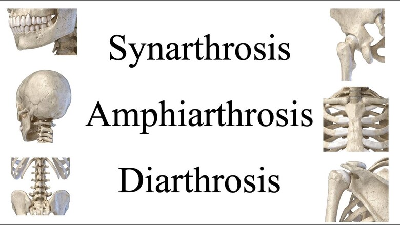
Types of Joints Comprehensive anatomy notes covering functional and structural classifications of joints. Quick Reference: Mobility vs. Structure Functional (Mobility) Structural (Tissue) Synarthrosis — Immovable Fibrous — Dense CT Amphiarthrosis — Slightly movable Cartilaginous — Cartilage Diarthrosis — Freely movable Synovial — Synovial cavity + fluid 1. Introduction to Articulations A joint (articulation) is any place where adjacent bones or bone and cartilage come together to form a connection. The 206 bones of the human body provide structural scaffolding, protect internal organs, and facilitate locomotion—but none of this is possible without joints allowing bones to articulate with one another. Joints are classified using two complementary systems: Functional Classification: Based on the degree of movement permitted (synarthrosis, amphiarthrosis, and diarthrosis). Structural Classification: Based on the type of connective tissue binding the bones (fibrous, cartilaginous, and synovial). Clinical Pearl When examining a patient, first determine if the joint is supposed to move (diarthrosis) or not (synarthrosis). Pathology in synarthroses presents very differently from diarthroses. For example, suture separation in infants indicates craniosynostosis—a condition fundamentally different from a synovial joint dislocation. 2. Functional Classification Functional classification is determined by the amount of mobility found between adjacent bones. These categories form a mobility spectrum from immobile to freely movable. A. Synarthrosis (Immovable Joint) A synarthrosis is an immobile or nearly immobile joint. This provides for a strong union between articulating bones—critical at locations where bones protect internal organs. Structural Basis: Fibrous or cartilaginous connections with no joint cavity. Key Examples: Sutures: Fibrous joints between skull bones (e.g., sagittal, coronal, lambdoid). They protect the brain and form the face. Gomphosis: Specialized fibrous joint anchoring a tooth into its alveolar socket via the periodontal ligament. This is unique to the dentition. Synchondrosis: Cartilaginous joint where bones are joined by hyaline cartilage. The epiphyseal (growth) plate is a temporary synchondrosis. The first sternocostal joint is permanent. Clinical Pearl Craniosynostosis Craniosynostosis is the premature fusion of one or more cranial sutures in infants. It can cause abnormal head shape (plagiocephaly, scaphocephaly) and increased intracranial pressure. Early surgical intervention is often required to allow normal brain growth. B. Amphiarthrosis (Slightly Movable Joint) An amphiarthrosis is a joint that permits limited mobility. These joints strike a balance between stability and flexibility—strong enough to maintain structural integrity, yet flexible enough to permit shock absorption. Structural Basis: Cartilaginous (symphysis) or fibrous (syndesmosis). Key Examples: Symphysis: Bones are joined by a fibrocartilage pad or disc. The pubic symphysis unites the hip bones. The intervertebral discs unite vertebrae; while each moves only slightly, they sum together for a large range of motion. Syndesmosis: Two parallel bones united by fibrous tissue (ligaments or interosseous membrane). Key example: the distal tibiofibular joint. Clinical Pearl Symphysis Pubis Dysfunction (SPD) affects up to 1 in 5 pregnant women. Increased relaxin hormone causes excessive mobility, leading to pelvic pain and difficulty walking. Intervertebral disc herniation occurs when the nucleus pulposus protrudes through the annulus fibrosus, most commonly at L4-L5 and L5-S1. C. Diarthrosis (Freely Movable Joint) A diarthrosis is a freely mobile joint. All synovial joints are functionally classified as diarthroses. They are found predominantly in the appendicular skeleton. Structural Basis: Feature articular cartilage, a joint cavity filled with synovial fluid, and a fibrous articular capsule. Axes of Movement: Uniaxial: Movement in a single plane (one axis). Example: elbow joint (flexion/extension). Biaxial: Movement in two planes (two axes). Example: metacarpophalangeal (knuckle) joints. Multiaxial (Polyaxial/Triaxial): Movement in all three anatomical planes. Example: shoulder and hip joints. Design Principle Joint mobility is inversely related to joint strength. Synarthroses provide maximum protection but zero movement. Diarthroses allow extensive movement but are the most frequently injured due to their looser articulations. 3. Structural Classification Based on how the bones are held together and the type of connective tissue binding the articulating surfaces. Structural Type Connective Tissue Mobility Examples Fibrous Dense regular CT (collagen-rich) Immovable / Slight Skull sutures, syndesmoses Cartilaginous Hyaline or fibrocartilage Immovable / Slight Synchondroses, symphyses Synovial Articular capsule + Synovial fluid Freely movable Shoulder, Hip, Knee, Ankle A. Fibrous Joints Adjacent bones are directly united by dense fibrous connective tissue. There is no joint cavity. Suture: Short fibers unite skull bones. With age, these may ossify completely (synostosis). Syndesmosis: Bones united by a ligament or membrane. More mobile than sutures. Example: distal tibiofibular joint. Gomphosis: A “peg-in-socket” joint where the tooth root is held by the periodontal ligament. Clinical Pearl Fontanelles (“soft spots”) on an infant’s skull are membranous gaps between cranial bones at sutures. The anterior fontanelle closes at 12–18 months; the posterior at 2–3 months. Delayed closure may indicate hydrocephalus, hypothyroidism, or rickets. B. Cartilaginous Joints Bones are united by cartilage; there is no joint cavity. Synchondrosis: Bones joined by hyaline cartilage. Most are temporary (epiphyseal plate). Symphysis: Bones covered by hyaline cartilage but united by a fibrocartilage pad. These are amphiarthroses. Key Difference Synchondroses use hyaline cartilage and are typically immobile. Symphyses use fibrocartilage (containing thick collagen bundles) which provides greater resistance to pulling and bending forces and permits slight movement. C. Synovial Joint Features Articular Capsule: A two-layered structure: (1) outer fibrous capsule of dense irregular CT for stability, and (2) inner synovial membrane that secretes fluid. Synovial Fluid: A viscous, egg-white-like fluid that lubricates the joint, nourishes avascular articular cartilage, and absorbs shock. Bursae & Tendon Sheaths: Fluid-filled sacs that reduce friction between adjacent structures. Clinical Pearl Synovial Fluid Analysis Normal fluid is clear, viscous, and pale yellow. Inflammatory arthritis (RA) produces cloudy fluid with elevated WBCs. Septic arthritis produces purulent fluid. Crystal analysis identifies needle-shaped urate (gout) or rhombic calcium pyrophosphate (pseudogout). 4. Synovial Joint Subtypes Classified into six subtypes based on the shape of articulating surfaces and axes of movement. A. Plane (Gliding) Joint Movement: Sliding/gliding only; non-axial. Articular Surface: Flat or slightly curved surfaces of equal size. Examples: Intercarpal joints (wrist), facet (zygapophyseal) joints of the spine. B. Hinge Joint Movement: Flexion/extension only; uniaxial. Articular Surface: Convex end of one bone fits into concave end of another. Examples: Elbow (humeroulnar), knee

Scroll to Top