Common Disorders of the Urinary System
A comprehensive clinical resource covering the epidemiology, pathophysiology, classification, and management of major renal and urological conditions, including infections, calculi, and systemic syndromes.
1. Urinary Tract Infections (UTIs)
1.1 Definition and Epidemiology
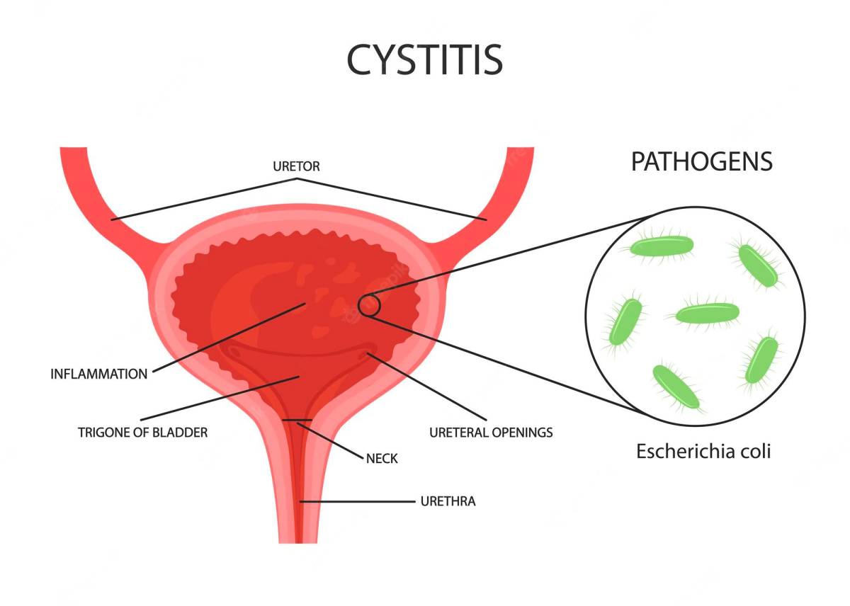
Urinary tract infections (UTIs) are among the most common bacterial infections encountered in clinical practice, affecting any part of the urinary tract from the urethra to the kidneys. Escherichia coli accounts for approximately 80–90% of uncomplicated UTIs.
UTIs are significantly more common in females due to the shorter urethra (approximately 4 cm compared to 20 cm in males), which allows easier bacterial ascent from the perineum into the bladder.
1.2 Classification by Anatomical Location
Lower UTI — Cystitis
Cystitis is the infection and inflammation of the bladder mucosa. It is characterized by:
- Dysuria — painful or burning urination
- Frequency — frequent voiding of small amounts
- Urgency — sudden compelling need to urinate
- Suprapubic pain or discomfort
- Hematuria — blood in the urine (occasional)
- Absence of fever (this distinguishes cystitis from pyelonephritis)
- Cloudy or foul-smelling urine
Upper UTI — Pyelonephritis
Pyelonephritis is the infection of the renal pelvis and renal parenchyma. It usually results from ascending infection but may occur via hematogenous spread. Clinical features include:
- Fever and chills
- Flank pain — tenderness at the costovertebral angle
- Nausea and vomiting
- Dysuria, frequency, and urgency
- Systemic symptoms — malaise, tachycardia
- Cloudy urine with possible pus
1.3 Risk Factors
Female sex, sexual activity, use of spermicides, pregnancy, urinary tract obstruction, vesicoureteral reflux, diabetes mellitus, immunosuppression, indwelling urinary catheters, incomplete bladder emptying, and anatomical abnormalities.
1.4 Diagnosis and Management
Diagnosis: Urinalysis showing Pyuria (WBCs), bacteriuria, positive nitrites, and positive leukocyte esterase. Microscopic examination may reveal WBC casts in pyelonephritis. Urine Culture is the gold standard (Colony count ≥10⁵ CFU/mL is diagnostic).
Management of Uncomplicated Cystitis:
- TMP-SMX for 3 days
- Nitrofurantoin for 5 days
- Fosfomycin as a single dose
- Adequate hydration (2–3 liters daily)
Management of Pyelonephritis:
- Fluoroquinolones (Ciprofloxacin, Levofloxacin) for 7–14 days
- Cephalosporins (Ceftriaxone) if fluoroquinolones are contraindicated
2. Kidney Stones (Urolithiasis / Nephrolithiasis)
Kidney stones are hard mineral and salt deposits. Prevalence is higher in hot climates due to dehydration. Men are affected more often, with peak incidence between 20 and 50 years.
2.2 Types of Renal Calculi
| Type | Composition | Frequency | Key Associations |
|---|---|---|---|
| Calcium Oxalate | Calcium + Oxalate | ~70–80% | Hypercalciuria, hyperoxaluria, low citrate |
| Calcium Phosphate | Calcium + Phosphate | ~10–15% | Renal tubular acidosis, hyperparathyroidism |
| Uric Acid | Uric Acid | ~5–10% | Gout, acidic urine (pH < 5.5), high purine diet |
| Struvite | Magnesium ammonium phosphate | ~10% | Infection with urease-producing bacteria (Proteus) |
| Cystine | Cystine | < 1% | Autosomal recessive cystinuria |
2.4 Clinical Presentation
- Renal Colic: Severe, colicky flank pain radiating to the groin (testis/labia) caused by ureteral spasm.
- Restlessness: The patient cannot find a comfortable position (distinguishes from peritonitis where the patient lies still).
- Hematuria, nausea/vomiting, and urinary frequency.
2.5 Management
- Conservative: Hydration, NSAIDs (Analgesia), and alpha-blockers (Tamsulosin) as medical expulsive therapy.
- Interventional: ESWL (Stones < 2 cm), Ureteroscopy (Endoscopic removal), or PCNL (For large stones > 2 cm or staghorn calculi).
3. Glomerulonephritis (GN)
Glomerulonephritis is the inflammation of the glomeruli, classified as Primary (kidney-limited) or Secondary (systemic diseases like SLE or vasculitis).
3.2 Pathophysiology
- Immune Complex Deposition: Antigen-antibody complexes activate complement (Post-streptococcal GN, Lupus).
- Anti-GBM Antibodies: Antibodies attack the basement membrane; seen in Goodpasture syndrome.
- Pauci-Immune: Neutrophil-mediated injury without significant deposits (ANCA-positive vasculitis).
3.3 Nephritic Syndrome
The classic presentation of GN:
- Hematuria — smoky or coca-cola colored urine; dysmorphic RBCs and RBC casts.
- Oliguria — decreased urine output (< 400 mL/day).
- Hypertension — due to sodium and water retention.
- Mild Proteinuria, Azotemia, and Periorbital Edema.
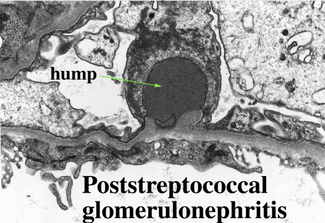
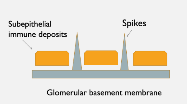
Acute Post-Streptococcal GN: Follows Group A strep infection; subepithelial "hump" deposits on EM.
RPGN (Crescentic GN): Severe form with crescent formation in Bowman's space; rapid renal failure.
IgA Nephropathy (Berger Disease): Most common worldwide; mesangial IgA deposits.
MPGN: "Tram-track" double basement membrane appearance.
4. Nephrotic Syndrome
Clinical constellation caused by heavy glomerular protein loss resulting from damage to the glomerular filtration barrier (podocytes and slit diaphragms).
4.2 Diagnostic Criteria (The Big Four)
- Heavy Proteinuria: >3.5 g/day (Adults) or >50 mg/kg/day (Children).
- Hypoalbuminemia: Serum albumin < 30 g/L (3 g/dL).
- Edema: Pitting edema (periorbital and dependent).
- Hyperlipidemia and Lipiduria: Elevated cholesterol; "oval fat bodies" in urine.
Loss of antithrombin III and increased clotting factor synthesis lead to a Hypercoagulable state (Renal vein thrombosis). Loss of IgG increases Infection risk.
5. Chronic Kidney Disease (CKD)
Defined as abnormalities of kidney structure or function present for > 3 months.
5.2 Staging by GFR
| Stage | GFR (mL/min/1.73m²) | Description |
|---|---|---|
| G1 | ≥90 | Normal or high |
| G2 | 60–89 | Mildly decreased |
| G3a | 45–59 | Mildly to moderately decreased |
| G3b | 30–44 | Moderately to severely decreased |
| G4 | 15–29 | Severely decreased |
| G5 | <15 | Kidney failure (ESRD) |
Common causes include: Diabetes Mellitus (most common), Hypertension, HIVAN (HIV-associated nephropathy), Obstructive uropathy (BPH), and Interstitial nephritis from herbal remedies.
5.4 Uremic Syndrome (Advanced CKD Features)
- Cardiovascular: LVH, Heart failure, Pericarditis.
- Hematological: Normocytic normochromic anemia; Platelet dysfunction.
- Metabolic: Metabolic acidosis, Hyperkalemia, Secondary hyperparathyroidism.
- Dermatological: Pruritus, Uremic frost (crystallized urea on skin).
6. Acute Kidney Injury (AKI)
Abrupt decrease in kidney function over hours to days. Classified by cause:
- Prerenal AKI: Reduced renal perfusion (Hypovolemia, Heart failure, Sepsis). Reversible if restored promptly.
- Intrarenal AKI: Direct damage (Acute Tubular Necrosis/ATN, Acute Glomerulonephritis).
- Postrenal AKI: Obstruction of urine outflow (Stones, BPH, Tumors).
A — Acidosis (refractory metabolic)
E — Electrolytes (refractory hyperkalemia)
I — Intoxicants
O — Overload (fluid with pulmonary edema)
U — Uremia (Pericarditis/Encephalopathy)
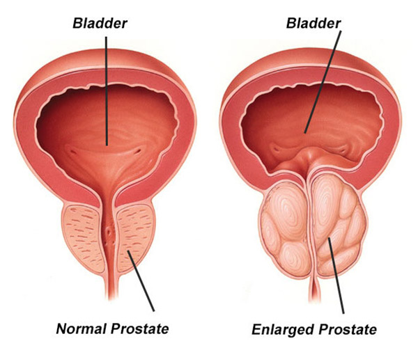
7. Benign Prostatic Hyperplasia (BPH)
Non-cancerous enlargement of the prostate driven by DHT (dihydrotestosterone) in the transition zone.
7.3 Clinical Features
- Obstructive (Voiding): Weak stream, hesitancy, intermittency, incomplete emptying.
- Irritative (Storage): Frequency, Urgency, Nocturia.
Management: Alpha-blockers (Tamsulosin) for rapid relief; 5-alpha-reductase inhibitors (Finasteride) to shrink the prostate. Surgical TURP is the gold standard.
8. Polycystic Kidney Disease (PKD)
Genetic disorder with growth of numerous fluid-filled cysts.
- ADPKD: Autosomal Dominant (Adult onset 30–50 years). Caused by PKD1 (Chr 16) or PKD2 (Chr 4) mutations. Associated with intracranial berry aneurysms.
- ARPKD: Autosomal Recessive (Infancy). Presents with pulmonary hypoplasia (Potter sequence).
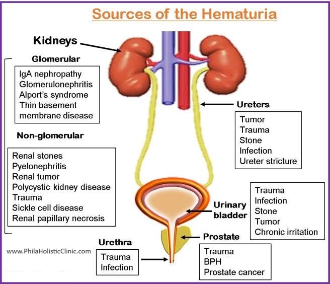
9. Hematuria
Presence of RBCs in urine (>3 RBCs per HPF). Classification by origin:
| Origin | Microscopy Findings | Common Causes |
|---|---|---|
| Glomerular (Medical) | Dysmorphic RBCs, RBC casts, Proteinuria | Glomerulonephritis, IgA Nephropathy |
| Non-Glomerular (Surgical) | Isomorphic (Normal) RBCs, No casts | UTI, Stones, Tumors, Trauma, BPH |
Urine dipstick detects heme but cannot distinguish RBCs from free hemoglobin or myoglobin. Formal microscopy is essential. For adults >40 with unexplained hematuria, cystoscopy is mandatory to exclude malignancy.