Characteristics of Urine
1. INTRODUCTION AND DEFINITION OF URINE
Definition: Urine is a liquid excretory product formed by the kidneys through the processes of glomerular filtration, tubular reabsorption, and tubular secretion. It serves as the primary mechanism for eliminating metabolic waste products, excess water, electrolytes, and foreign substances from the body.
Composition: Urine is approximately 95% water and 5% dissolved solids. The solid components include:
- Nitrogenous wastes: Urea (the largest component), creatinine, uric acid, and ammonia.
- Electrolytes: Sodium, potassium, chloride, bicarbonate, calcium, magnesium, and phosphate.
- Pigments: Urochrome (gives urine its yellow color), urobilinogen, and bilirubin (when present).
- Other substances: Hormones, enzymes, vitamins, drugs, and toxins in trace amounts.
Examination of urine characteristics provides critical information about renal function, metabolic status, hydration, and the presence of infection or systemic disease. Abnormal urine findings often represent the earliest detectable signs of conditions such as diabetes mellitus, urinary tract infection, glomerular disease, and hepatic dysfunction.
2. ANATOMY AND PHYSIOLOGY OF URINE FORMATION
2.1 Overview of the Urinary System
The urinary system consists of two kidneys, two ureters, the urinary bladder, and the urethra. The kidneys are paired, bean-shaped organs located retroperitoneally on either side of the vertebral column, extending from T12 to L3. Each kidney weighs approximately 150 grams in adults and measures about 11 cm in length, 6 cm in width, and 3 cm in thickness.
The functional unit of the kidney is the nephron. Each kidney contains approximately 1 to 1.5 million nephrons. The nephron consists of two main components: the renal corpuscle (comprising the glomerulus and Bowman's capsule) and the renal tubule (including the proximal convoluted tubule, loop of Henle, distal convoluted tubule, and collecting duct).
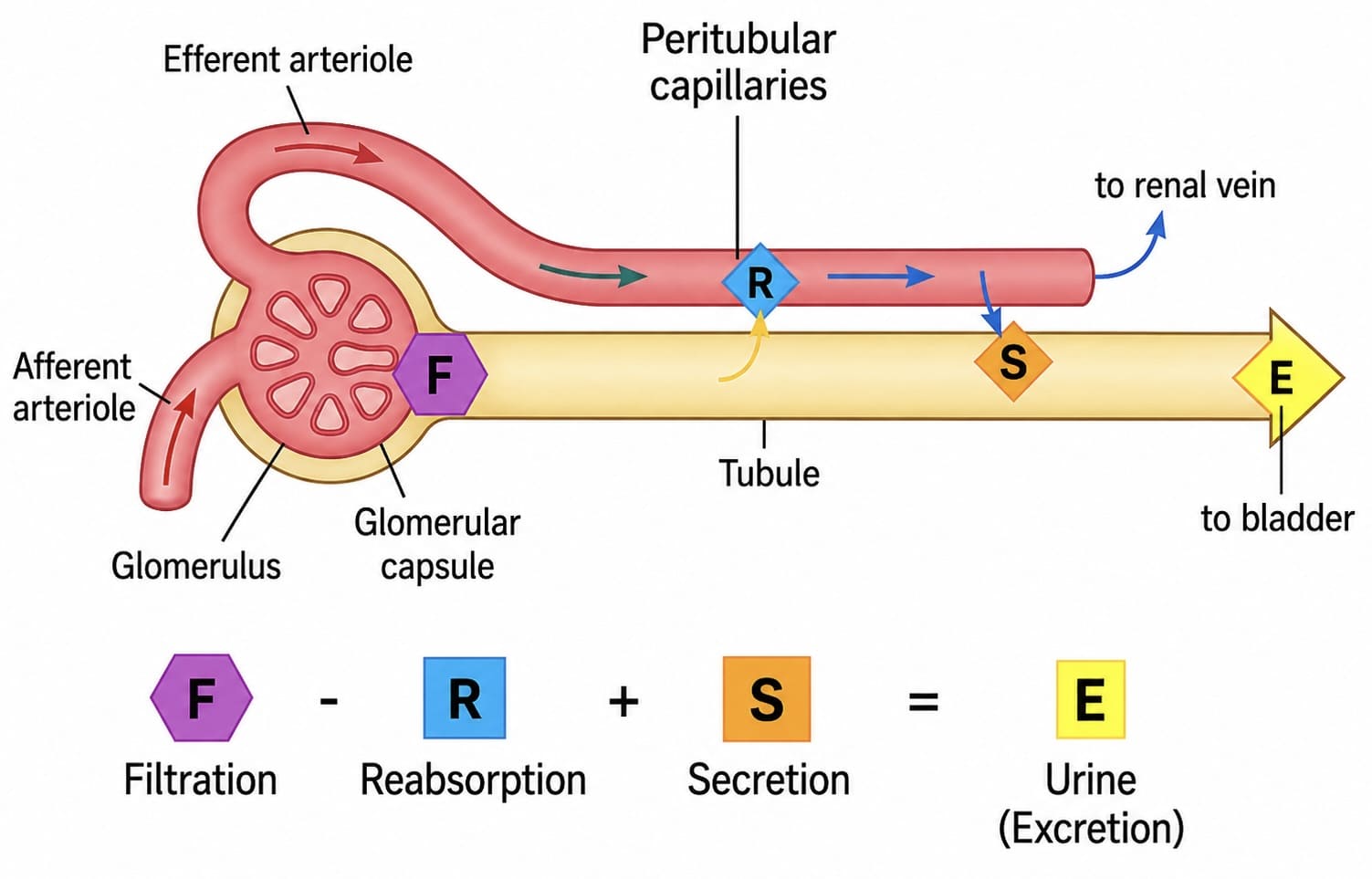
2.2 Three Processes of Urine Formation
- A. Glomerular Filtration: Blood enters the glomerulus through the afferent arteriole. High hydrostatic pressure (~55 mmHg) forces water and small solutes through the filtration membrane into Bowman's capsule. The filtration barrier consists of three layers: fenestrated endothelium, basement membrane, and podocyte slit diaphragm. It prevents the passage of blood cells and large proteins (molecular weight >70,000 Daltons).
- B. Tubular Reabsorption: As filtrate passes through the tubules, essential substances are returned to the bloodstream. The PCT reabsorbs approximately 65% of filtered water, sodium, glucose, and amino acids. The loop of Henle establishes a concentration gradient via the countercurrent multiplier system.
- C. Tubular Secretion: Active transport of substances (H+, K+, creatinine, drugs, and toxins) from peritubular capillaries into the lumen. This is particularly important for acid-base homeostasis.
2.3 Regulation of Urine Formation
- ADH (vasopressin): Released from posterior pituitary; increases water reabsorption in collecting ducts, producing concentrated urine.
- Aldosterone: Secreted by adrenal cortex; promotes sodium reabsorption and potassium secretion.
- ANP: Released from cardiac atria; inhibits sodium reabsorption and promotes diuresis.
- PTH: Regulates calcium and phosphate reabsorption.
3. PHYSICAL CHARACTERISTICS OF URINE
The physical examination is the first step in urinalysis. Characteristics evaluated include color, clarity, odor, volume, specific gravity, pH, and foam.
3.1 Color
Normal urine color ranges from pale yellow to deep amber. The pigment responsible is urochrome (urobilin), a breakdown product of hemoglobin metabolism.
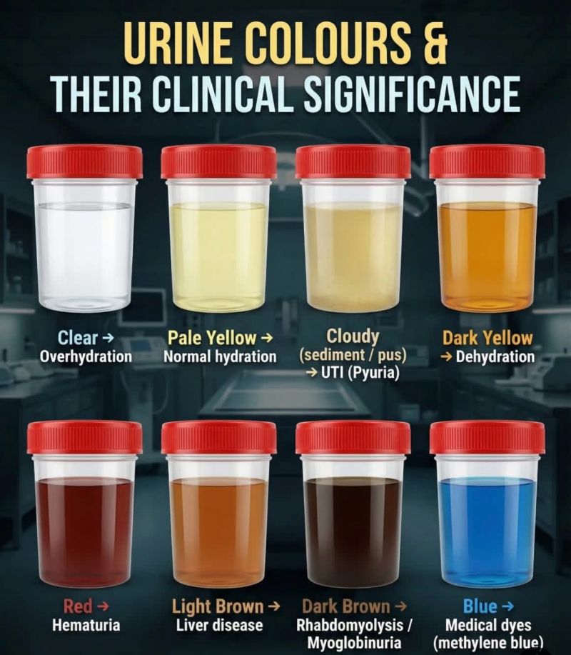
Abnormal Colors and Clinical Significance:
- Red or Pink: May indicate hematuria, hemoglobinuria, or myoglobinuria. Causes include UTI, renal calculi, glomerulonephritis, trauma, and strenuous exercise. Medications like rifampin and phenazopyridine also produce red urine.
- Orange: Associated with dehydration, bilirubinuria (liver disease), or medications like warfarin.
- Green or Blue: Rare. Caused by Pseudomonas aeruginosa UTI, biliverdin (biliary stasis), or propofol.
- Brown or Tea-Colored: Suggests bile pigments (bilirubin), old blood, or myoglobin. Seen in acute viral hepatitis, cirrhosis, and rhabdomyolysis.
- Cloudy or Milky: Results from pyuria (WBCs), bacteriuria, chyluria (lymphatic fluid), or lipiduria. In Uganda, filariasis is an important cause of chyluria.
3.2 Clarity and Odor
- Clarity: Normal urine is clear or translucent. Turbidity results from cellular material, microorganisms, mucus, or crystals. Clinical Note: If cloudiness clears with acetic acid, precipitation of phosphates is likely.
- Odor: Normal urine has a faint aromatic odor.
— Ammoniacal: Bacterial decomposition of urea (UTI).
— Sweet/Fruity: Presence of ketone bodies (DKA).
— Musty/Mousy: Characteristic of phenylketonuria (PKU).
— Maple Syrup: MSUD.
3.4 Volume and Specific Gravity
Normal daily output: 600 to 2,000 mL (average 1,000-1,600 mL). Minimum required: 400-500 mL/day.
- Polyuria (>2,500-3,000 mL/day): Diabetes mellitus, diabetes insipidus, excessive intake.
- Oliguria (<400-500 mL/day): Dehydration, shock, heart failure, ATN, obstruction.
- Anuria (<100 mL/day): Medical emergency; bilateral obstruction or renal cortical necrosis.
- Specific Gravity: Normal 1.002 to 1.035. High (>1.025) in dehydration; low (<1.010) in DI or ATN. A fixed gravity of ~1.010 indicates loss of tubular concentrating ability.
4. CHEMICAL CHARACTERISTICS OF URINE
Commonly performed using reagent-impregnated dipsticks.
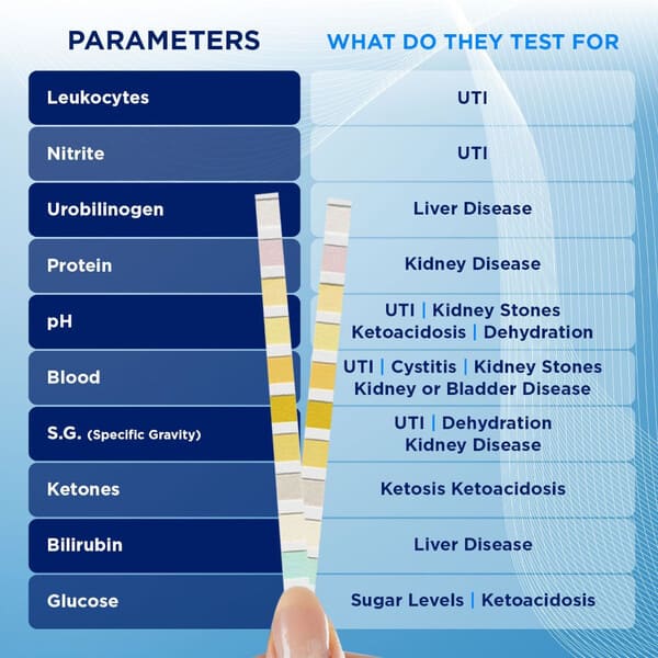
4.1 Proteins
Normal excretion is ≤150 mg/day.
— Glomerular proteinuria: Albumin leaks through damaged barrier (Nephrotic syndrome >3.5 g/day).
— Tubular proteinuria: Failure of reabsorption (e.g., ATN, Fanconi syndrome).
— Overflow proteinuria: Bence Jones proteins in multiple myeloma.
— Functional: Transient after exercise or fever.
4.2 Glucose and Ketones
- Glycosuria: Occurs when glucose exceeds renal threshold (~180 mg/dL). Common in Diabetes Mellitus.
- Ketonuria: Indicates increased lipolysis. Seen in DKA, starvation, and severe diarrhea in children.
4.4 Bilirubin and Urobilinogen
- Bilirubin: Only conjugated (direct) bilirubin appears in urine. Presence indicates hepatocellular damage or biliary obstruction.
- Urobilinogen: Elevated in hemolysis (hemolytic anemia, sickle cell) and hepatic dysfunction. Decreased in complete biliary obstruction.
— Bilirubin (+) / Urobilinogen (-): Obstructive jaundice.
— Bilirubin (-) / Urobilinogen (+): Hemolysis.
5. MICROSCOPIC EXAMINATION OF URINE
Timely examination (within 1-2 hours) is critical for identifying cellular elements and casts.
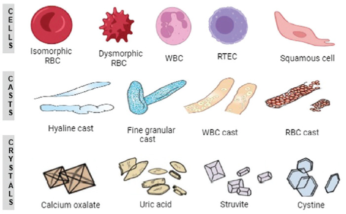
5.1 Cells
- Red Blood Cells: Normal 0–2/HPF. Dysmorphic RBCs suggest glomerular origin; isomorphic suggest lower tract bleeding.
- White Blood Cells: Normal 0–5/HPF. Pyuria (>5/HPF) indicates infection or inflammation.
- Epithelial Cells: Renal tubular epithelial cells are normally absent; their presence indicates tubular damage (ATN).
5.2 Casts
- Hyaline Casts: Composed of Tamm-Horsfall protein; seen in concentrated urine or fever.
- Red Blood Cell Casts: Diagnostic of glomerular bleeding (Acute glomerulonephritis).
- White Blood Cell Casts: Indicate renal parenchymal infection (pyelonephritis).
- Waxy and Broad Casts: Indicate advanced chronic kidney disease and severe tubular atrophy.
- Fatty Casts: "Maltese cross" pattern; associated with Nephrotic Syndrome.
5.3 Crystals
- Calcium Oxalate: Envelope/Dumbbell shaped; increased in ethylene glycol poisoning.
- Triple Phosphate: "Coffin lid" appearance; associated with Proteus UTI.
- Cystine: Hexagonal plates; pathognomonic for cystinuria.
Urinary Schistosomiasis
Caused by Schistosoma haematobium. Identified by terminal-spined eggs (140 x 60 µm). Clinical presentation: terminal hematuria and dysuria. Chronic infection increases risk of squamous cell bladder carcinoma. Best detected in urine collected between 10:00 AM and 2:00 PM.
6. SUMMARY TABLES
Table 2: Abnormal Urine Findings and Differential Diagnosis
| Finding | Differential Diagnosis | Key Clinical Features |
|---|---|---|
| Hematuria | UTI, stones, glomerulonephritis, trauma, tumors, BPH | RBC casts confirm glomerular origin. |
| Pyuria | Bacterial UTI, pyelonephritis, TB, interstitial nephritis | WBC casts indicate renal parenchyma. |
| Proteinuria | Nephrotic syndrome, DM, glomerulonephritis, ATN | >3.5 g/day = nephrotic range. |
| Bilirubinuria | Viral hepatitis, cirrhosis, biliary obstruction | Dark urine with pale stools. |
| Myoglobinuria | Rhabdomyolysis (trauma, crush injury, coma) | Dipstick (+) with NO RBCs on microscopy. |
7. KEY POINTS FOR CLINICAL PRACTICE
- Color and clarity provide immediate clues to hydration and metabolic disorders.
- Specific gravity fixed at 1.010 suggests advanced renal disease.
- Proteinuria is never normal in first-morning specimens.
- RBC casts are pathognomonic for glomerular disease.
- Myoglobinuria and hemoglobinuria produce positive blood dipstick without intact RBCs on microscopy.
- Anuria is a medical emergency requiring urgent investigation and potential dialysis.
