Common Joint Disorders
A Comprehensive Clinical Reference Guide covering Dislocation, Bursitis, Arthritis, Ligament & Meniscal Injuries, Tendon Disorders, and Clinical Red Flags.
5.1 DISLOCATION (LUXATION)
Definition: Complete loss of contact between articulating bone surfaces of a joint. Subluxation refers to a partial dislocation where some articular contact is maintained.
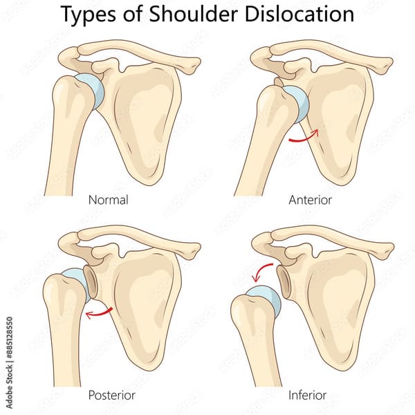
Classification by Direction (Shoulder Focus):
- Anterior: Most common (>90% of shoulder dislocations); the humeral head is displaced anteriorly.
- Posterior: Less common; often associated with seizures or electrocution.
- Inferior (Luxatio Erecta): Rare; the arm is held in a fixed overhead position.
Common Sites & Key Features:
| Joint | Most Common Direction | Key Associations | Special Notes |
|---|---|---|---|
| Shoulder | Anterior (>90%) | Bankart lesion, Hill-Sachs lesion | Recurrence rate 50-90% in young patients. |
| Elbow | Posterior | Coronoid fracture, radial head fracture | Ulnar nerve injury risk. |
| Hip | Posterior | Sciatic nerve injury | Associated with dashboard injury (MVA). |
| Patella | Lateral | MPFL tear | Common in adolescents; often recurrent. |
| Finger (PIP) | Dorsal | Volar plate injury | Common sports injury. |
Causes:
- Trauma: Direct blow, fall on outstretched hand (FOOSH), motor vehicle accidents.
- Congenital Laxity: Developmental dysplasia of the hip (DDH), Marfan syndrome.
- Connective Tissue Disorders: Ehlers-Danlos syndrome, osteogenesis imperfecta.
- Recurrent/Pathological: Seizures (posterior shoulder), ligamentous insufficiency.
Clinical Features:
- Visible deformity with loss of normal joint contour.
- Loss of function: Inability to move the joint actively or passively.
- Severe pain at rest and with any attempted movement.
- Possible neurovascular compromise: MUST assess distal pulses, sensation, and motor function.
Always perform neurovascular examination before and after reduction. Document axillary nerve function (shoulder), ulnar nerve (elbow), sciatic nerve (hip), and popliteal artery (knee).
Management:
- Reduction: Closed reduction (first-line) vs. open reduction (indicated for failed closed reduction, associated fractures, or neurovascular compromise).
- Imaging: Pre-reduction X-rays (AP, lateral, axillary/scapular Y for shoulder); post-reduction films to confirm concentric reduction.
- Immobilization: Sling-and-swath (shoulder), posterior splint (elbow), abduction brace (hip).
- Rehabilitation: Early range of motion (ROM) to prevent stiffness; rotator cuff strengthening for the shoulder.
- Surgical Consideration: Recurrent dislocators, young athletes, Bankart lesions — arthroscopic stabilization.

Associated Injuries to Remember:
- Bankart lesion: Anteroinferior glenoid labrum tear — increases recurrence risk.
- Hill-Sachs lesion: Compression fracture of posterolateral humeral head.
- Coronoid fracture: Part of the "Terrible triad of the elbow" (dislocation + radial head fracture + coronoid fracture).
- Sciatic nerve palsy: Occurs in 10-20% of posterior hip dislocations — check for foot drop.
5.2 BURSITIS
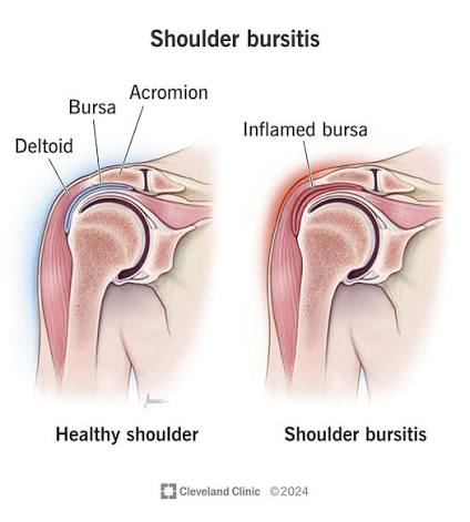
Definition: Inflammation of a bursa — a small, fluid-filled sac that reduces friction between tissues (bone, muscle, tendon, skin). Bursae are strategically located at sites of potential friction throughout the body.
Common Sites & Eponyms:
| Site | Bursa Name | Common Cause | Key Clinical Feature |
|---|---|---|---|
| Shoulder | Subacromial / Subdeltoid | Impingement, overhead activity | Painful arc (60-120° abduction) |
| Elbow | Olecranon | Prolonged pressure, trauma | "Student's elbow" or "Miner's elbow" |
| Knee | Prepatellar | Kneeling (carpet layers) | "Housemaid's knee" |
| Knee | Pes Anserine | Obesity, OA, overuse | Medial knee pain, tender 5cm below joint line |
| Hip | Trochanteric | IT band friction | Lateral hip pain, worse lying on side |
Causes and Clinical Features:
- Repetitive trauma / Overuse: Occupational or sports-related.
- Infection (Septic Bursitis): Staphylococcus aureus is most common; presents with erythema, warmth, fever.
- Crystal Deposition: Gout (monosodium urate) or pseudogout (CPPD).
- Clinical Features: Localized swelling (may be fluctuant), tenderness to palpation, and pain with movement of the adjacent joint (typically worse with specific motions).
Management:
- RICE Protocol: Rest, Ice (15-20 min, 3-4x/day), Compression, Elevation.
- NSAIDs: Ibuprofen or naproxen to reduce inflammation.
- Aspiration: If septic etiology is suspected — send for Gram stain, culture, cell count, and crystal analysis.
- Corticosteroid Injection: Only after excluding infection; inject into the bursa, never the tendon.
- Physical Therapy: Stretching and ergonomic adjustments.
Septic bursitis (especially olecranon and prepatellar) is more common than septic arthritis. Because the bursa is superficial, erythema and warmth are more prominent. Aspiration is both diagnostic and therapeutic.
5.3 ARTHRITIS (GENERAL)
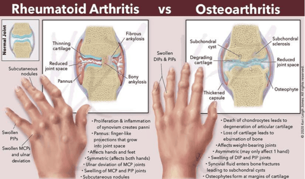
Arthritis refers to joint inflammation. While over 100 types exist, Osteoarthritis and Rheumatoid Arthritis are the most common.
5.3.1 OSTEOARTHRITIS (OA)
Definition: Degenerative joint disease characterized by progressive loss of articular cartilage, subchondral bone sclerosis, and osteophyte formation.
Pathophysiology:
- Cartilage degradation: Loss of proteoglycans leads to decreased resilience.
- Subchondral sclerosis: Bone thickening beneath cartilage.
- Osteophytes: Bony outgrowths at joint margins.
Radiographic Features (Kellgren-Lawrence Grading): Grades 0 (None) to 4 (Large osteophytes, severe JSN, definite deformity).
5.3.2 RHEUMATOID ARTHRITIS (RA)
Definition: Chronic autoimmune inflammatory arthritis characterized by symmetric synovitis, pannus formation, and progressive joint destruction.
Key Features:
- Symmetric involvement: Hands, wrists, and feet (usually spares the DIP joints).
- Morning stiffness >30 minutes: Improves with movement throughout the day.
- Rheumatoid nodules: Subcutaneous nodules over extensor surfaces.
- Treatment: DMARDs (Methotrexate first-line), biologics (TNF inhibitors), and corticosteroids for flares.
5.3.3 SEPTIC ARTHRITIS
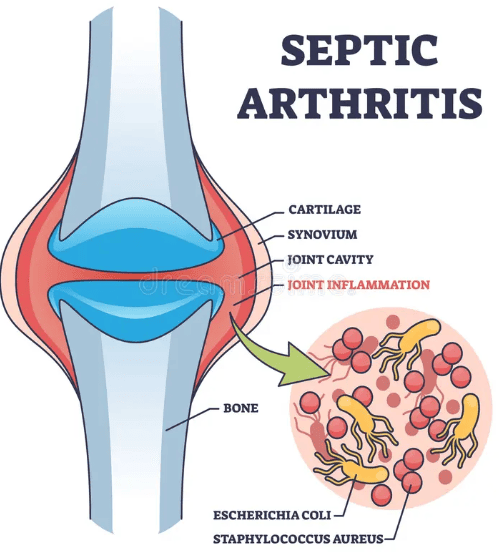
Definition: Bacterial infection of a joint space — a MEDICAL EMERGENCY. Joint destruction can occur within 24-48 hours.
- Presentation: Acute monoarthritis (80-90% of cases), severe pain with minimal movement (patient resists any joint motion), and a hot, swollen joint.
- Management: Urgent joint aspiration for analysis. Start empiric IV antibiotics immediately after aspiration.
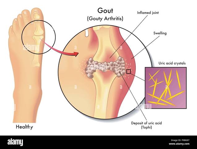
5.3.4 GOUT & PSEUDOGOUT (CRYSTAL DISEASES)
- Gout: Monosodium urate (MSU) crystals; needle-shaped, negatively birefringent. Classically affects the first MTP joint (podagra).
- Pseudogout (CPPD): Calcium pyrophosphate dihydrate crystals; rhomboid, weakly positively birefringent. Often affects the knee or wrist.
5.4 LIGAMENT INJURIES
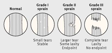
Definition: A sprain is a partial or complete tear of a ligament — the fibrous tissue connecting bone to bone.
Grading System:
| Grade | Pathology | Clinical Features | Stability |
|---|---|---|---|
| I | Stretching / Microtears | Mild pain, minimal swelling | Stable |
| II | Partial tear | Moderate pain, swelling, bruising | Mild laxity |
| III | Complete rupture | Severe pain, significant swelling | Gross instability |
1. ACL Tear (Knee):
- Mechanism: Non-contact pivoting injury, deceleration with valgus stress.
- Signs: "Pop" sound, immediate swelling (hemarthrosis within 2 hours).
- Tests: Lachman test (most sensitive: 85-95%), anterior drawer test, and pivot shift test.
- O'Donoghue's Unhappy Triad: ACL tear + MCL tear + medial meniscus tear.
5.5 MENISCAL INJURIES
Definition: Tear of the meniscus — C-shaped fibrocartilaginous structures that provides shock absorption and stability.
Anatomy: Medial meniscus is less mobile and more commonly torn. The outer third (red-red zone) is vascularized and can heal; the inner two-thirds (white-white zone) is avascular and cannot heal.
Clinical Features:
- Joint line tenderness: The most reliable finding.
- Locking: Inability to fully extend the knee (suggests bucket-handle tear).
- Effusion: Delayed onset (hours to days), unlike ACL tears.
- Positive McMurray's test and Thessaly test.
A locked knee with inability to fully extend is a relative indication for urgent arthroscopy. MRI is the imaging modality of choice for suspected meniscal tears.
5.6 TENDON DISORDERS
Tendinopathy: A spectrum from inflammation (tendinitis) to degenerative failure (tendinosis) to rupture.
Common Sites:
- Supraspinatus: Rotator cuff tendinopathy (Hawkins-Kennedy test).
- Achilles: Achilles tendinopathy; overuse or fluoroquinolone use.
- De Quervain's: Tenosynovitis of the thumb (Finkelstein test).
Tendon Rupture:
Achilles Tendon: Most common major rupture; occurs during push-off. Patient reports 'being kicked in the calf.'
Thompson Test (Simmonds Test): Squeeze the calf while prone. Normal: Foot plantarflexes. Rupture: No movement.
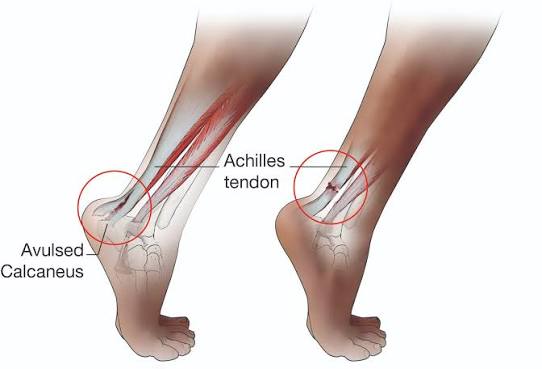
Fluoroquinolone antibiotics (ciprofloxacin) and systemic corticosteroids significantly increase the risk of Achilles tendon rupture.
5.7 QUICK REFERENCE: RED FLAGS
| Red Flag Sign | Possible Diagnosis | Immediate Action |
|---|---|---|
| Hot, swollen, painful single joint | Septic arthritis or Gout | Urgent joint aspiration; empiric antibiotics |
| Joint deformity after trauma | Dislocation or Fracture | X-ray + urgent reduction |
| Numbness/weakness distal to injury | Neurovascular compromise | Emergency surgery consult; limb-threatening |
| Back pain with bowel/bladder dysfunction | Cauda equina syndrome | Emergency MRI + neurosurgical consult |
A hot, swollen, painful joint in any patient is septic arthritis until proven otherwise. Joint aspiration must not be delayed.
5.9 DIFFERENTIAL DIAGNOSIS QUICK REFERENCE
Approach to the Painful Joint:
- Number of Joints: Monoarthritis (1 joint), Oligoarthritis (2-4 joints), or Polyarthritis (>4 joints).
- Acute vs. Chronic: Acute is <2 weeks.
- Inflammatory vs. Non-inflammatory:
— Inflammatory: Morning stiffness >30-60 min, soft swelling, warmth (e.g., RA, Septic).
— Non-inflammatory: Morning stiffness <30 min, hard/bony swelling, no warmth (e.g., OA).
- Always assess neurovascular status before and after any joint manipulation.
- A hot, swollen joint is septic arthritis until proven otherwise.
- Compare to the contralateral side — subtle findings may only be apparent bilaterally.
- Preserve meniscal and ligamentous tissue whenever possible — resection leads to OA.
