Urine Formation
Complete Study Notes covering the coordinated mechanisms of filtration, reabsorption, and secretion.
1. OVERVIEW
Urine formation occurs in the nephron, the functional unit of the kidney. Each human kidney contains approximately 1–1.5 million nephrons. The process involves three coordinated physiological mechanisms:
- Glomerular Filtration: Passive filtration of blood plasma into Bowman's capsule.
- Tubular Reabsorption: The return of useful substances from the tubular fluid back to the blood.
- Tubular Secretion: Active transport of wastes and excess substances from the blood into the tubular fluid.
- Filtrate formed: ~180 L/day (men) / ~150 L/day (women)
- Urine excreted: ~1–2 L/day
- Reabsorbed: ~99% of total filtrate
- GFR (Glomerular Filtration Rate): ~125 mL/min (men) / ~105 mL/min (women)
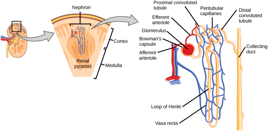
2. GLOMERULAR FILTRATION
2.1 The Process
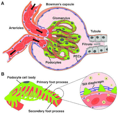
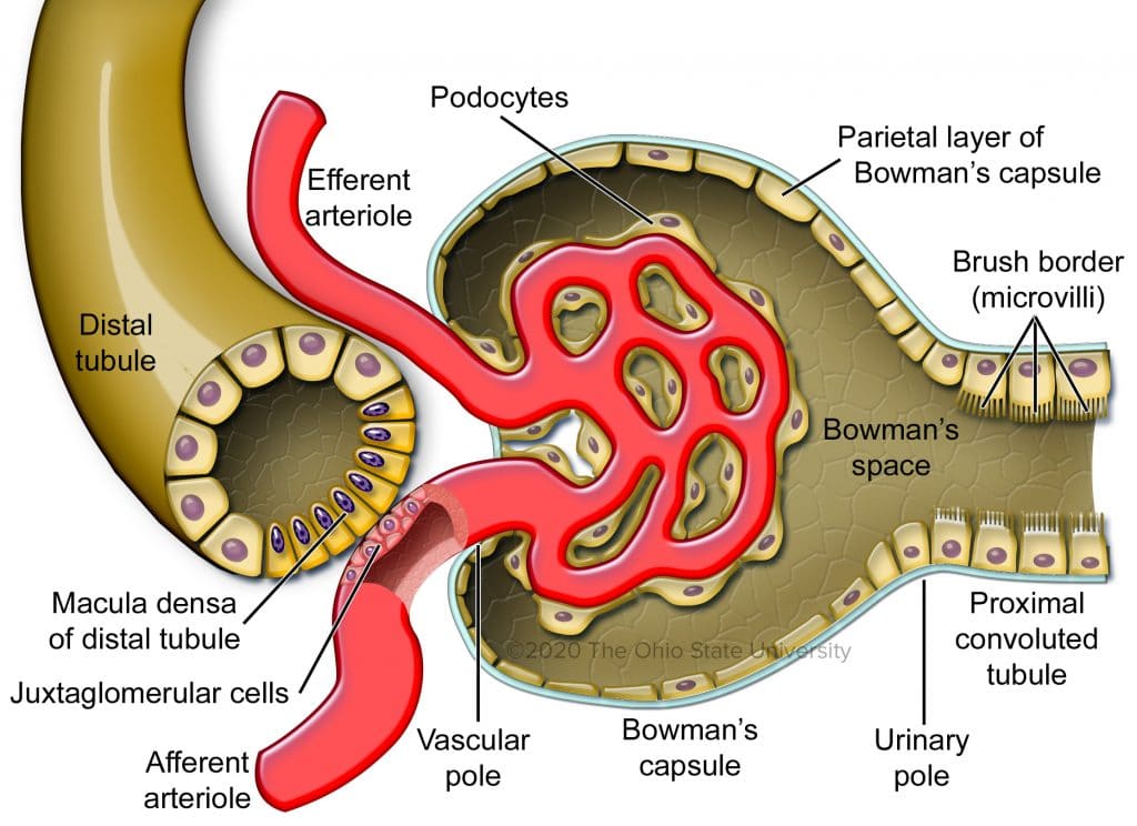
Glomerular filtration is a passive process in which hydrostatic pressure forces water and small solutes from glomerular capillaries across the filtration membrane into Bowman's space. The resulting fluid is called filtrate. Blood enters the glomerulus via the afferent arteriole and exits via the efferent arteriole.
2.2 The Filtration Barrier (Three Layers)
- Layer 1: Fenestrated Endothelium: The capillary endothelium contains large pores (fenestrae, 70–100 nm) that allow plasma to pass but block blood cells.
- Layer 2: Glomerular Basement Membrane: A gel-like layer of type IV collagen and negatively charged proteoglycans. It blocks large proteins and repels negatively charged molecules.
- Layer 3: Podocyte Slit Diaphragms: Specialized epithelial cells with foot-like processes (pedicels) that wrap around capillaries. Gaps between adjacent foot processes (filtration slits, ~25 nm) are bridged by thin slit diaphragms acting as the final molecular sieve.
2.3 What Gets Filtered?
| Passes Freely | Blocked (Retained in Blood) |
|---|---|
| Water, Na+, K+, Cl- | Blood cells (RBCs, WBCs) |
| Glucose, Amino acids | Albumin, Globulins |
| Urea, Creatinine | Platelets |
2.4 Starling Forces (Net Filtration Pressure)
The movement of fluid is determined by Net Filtration Pressure (NFP), calculated as:
NFP = PG − PB − πG
- Glomerular hydrostatic pressure (PG): ~55 mmHg — promotes filtration.
- Bowman's capsule pressure (PB): ~15 mmHg — opposes filtration.
- Plasma colloid osmotic pressure (πG): ~30 mmHg — opposes filtration.
- Net Filtration Pressure: ~10 mmHg.
3. TUBULAR REABSORPTION
Approximately 99% of the filtrate is returned to the blood. Reabsorption occurs along the entire length of the renal tubule.
3.1 Proximal Convoluted Tubule (PCT)
The PCT reabsorbs ~65% of filtered water, sodium, and chloride, and virtually 100% of glucose and amino acids.
- Sodium (Na+): Actively pumped out by Na+/K+-ATPase on the basolateral membrane.
- Glucose & Amino Acids: Reabsorbed by Na+-cotransporters (SGLT) on the apical membrane.
- Water: Follows solutes by osmosis via aquaporin-1 channels.
- Bicarbonate (HCO3-): Reabsorbed indirectly. H+ is secreted into the lumen, combines with HCO3- to form H2CO3, which is converted to CO2 + H2O by carbonic anhydrase. CO2 diffuses into the cell and is reconverted to HCO3-.
Transport Maximum (Tm)
Each carrier has a maximum rate. For glucose, Tm ≈ 375 mg/min. When blood glucose exceeds the renal threshold (~180 mg/dL), glucose appears in the urine (glucosuria).
3.2 Loop of Henle
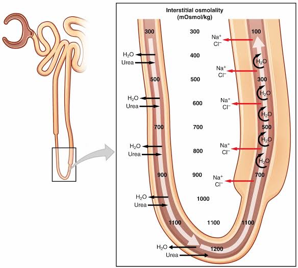
Reabsorbs ~25% of filtered solutes and establishes the medullary osmotic gradient.
| Segment | Permeability | Mechanism |
|---|---|---|
| Thin Descending Limb | Highly permeable to H2O; impermeable to solutes. | Water leaves by osmosis; tubular fluid becomes concentrated. |
| Thin Ascending Limb | Impermeable to H2O; permeable to Na+/Cl-. | Passive salt reabsorption into the interstitium. |
| Thick Ascending Limb (TAL) | Impermeable to H2O; highly permeable to ions. | Active Na+-K+-2Cl- cotransport (NKCC2); dilutes urine. |
3.3 Distal Convoluted Tubule (DCT) & 3.4 Collecting Duct
- DCT: Reabsorbs 5–10% of filtered Na+ and water. Features the Na+-Cl- cotransporter (NCC) and is the site for PTH-stimulated Ca2+ reabsorption. Site of action for thiazide diuretics.
- Collecting Duct: Final site of modification.
— Principal cells: Reabsorb Na+ (aldosterone-dependent), secrete K+, reabsorb water (ADH-dependent via aquaporin-2).
— Intercalated cells: Type A secretes H+; Type B secretes HCO3-.
4. TUBULAR SECRETION
The active transport of substances from blood into tubular fluid, complementing filtration.
| Substance | Site | Mechanism | Importance |
|---|---|---|---|
| H+ | PCT, DCT, Collecting duct | Na+/H+ antiporter; H+-ATPase pump | Acid-base balance; HCO3- reabsorption |
| K+ | Principal cells (CD) | Aldosterone-stimulated secretion | Potassium homeostasis |
| Creatinine | PCT | Passive and active transport | Waste product; used to estimate GFR |
| Uric acid | PCT | Active transport | End product of purine metabolism |
| Drugs/Toxins | PCT | Specific organic acid/base transporters | Elimination of Penicillin, morphine, aspirin |
5. URINE CONCENTRATION AND DILUTION
The kidneys can produce urine ranging from 50 mOsm/L (very dilute) to 1200 mOsm/L (highly concentrated).
5.1 Countercurrent Multiplication
- Single Effect: The TAL pumps Na+, K+, and 2Cl- into the interstitium via NKCC2. Interstitium becomes hyperosmotic.
- Equilibration: Descending limb loses water to the hyperosmotic medulla, concentrating the fluid.
- Multiplication: Concentrated fluid enters the ascending limb, more solutes are pumped out, progressively increasing osmolarity from 300 mOsm/L (cortex) to 1200 mOsm/L (tip).
5.2 Role of Antidiuretic Hormone (ADH)
- Dehydration (High ADH): ADH binds V2 receptors on principal cells. Aquaporin-2 channels are inserted. Water is reabsorbed. Result: concentrated urine (~1200 mOsm/L), small volume (~0.5 L/day).
- Well-hydrated (Low ADH): Little ADH released. No aquaporin-2 inserted. CD remains water-impermeable. Result: dilute urine (~50–100 mOsm/L), large volume.
5.3 Vasa Recta & 5.4 Urea Recycling
The Vasa Recta are hairpin capillaries that act as countercurrent exchangers, preserving the osmotic gradient by preventing "washout."
Urea Recycling: The inner medullary collecting duct is permeable to urea in the presence of ADH. Urea diffuses into the interstitium, contributing 40–50% of medullary osmolarity. Some urea re-enters the loop of Henle, creating a recycling loop.

It’s really nice and understandable, thanks 🙏