Structure and Function of the Lower Urinary Tract
Comprehensive exhaustive notes on the anatomy, histology, and physiology of the lower urinary tract (LUT), focusing on the storage and expulsive functions of the bladder and urethra.
1. Introduction
The lower urinary tract (LUT) comprises the urinary bladder and the urethra. Its two principal functions are to act as a low-pressure reservoir for the storage of urine and to expel urine at high pressure at an appropriate time and under voluntary control.
Urine is produced continuously by the kidneys (~1–2 L/day). The bladder must accommodate this volume without a significant rise in pressure (compliance), then switch rapidly to an expulsive mode during micturition.
2. Gross Anatomy
2.1 The Urinary Bladder
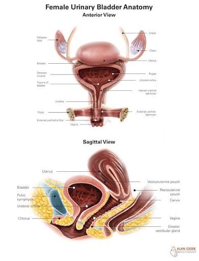
The bladder is a hollow, muscular, distensible organ located in the anterior pelvis, posterior to the pubic symphysis. Adult capacity is typically 400–500 mL, though it can distend to over 600 mL.
Position & Surface Anatomy:
- Empty: Entirely within the pelvis, posterior to the pubic symphysis.
- Full: The dome rises into the abdomen, pushing the peritoneum upward.
A full bladder allows for suprapubic aspiration or catheterization. Because the distended bladder pushes the peritoneum superiorly, a needle can be inserted just above the pubic symphysis into the bladder without entering the peritoneal cavity.
Anatomical Parts & Relations:
- Parts: Apex, Base (Fundus), Body, Neck, Dome, and Trigone.
- Superior: Peritoneum.
- Anterior: Pubic symphysis, retropubic space (Space of Retzius).
- Posterior (Male): Rectum, seminal vesicles, vas deferens.
- Posterior (Female): Uterus (vesicouterine pouch), upper vagina.
- Inferolateral: Pelvic side wall, obturator internus, levator ani.
2.2 The Urethra
The urethra is the only urinary tract organ with significant sexual dimorphism.
Female Urethra:
- Length: ~3–4 cm (short).
- Course: Runs inferiorly from the bladder neck, embedded in the anterior vaginal wall.
- Clinical significance: Its short length and proximity to the anus/vagina predispose females to ascending UTIs.
Male Urethra:
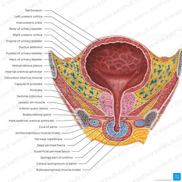
Length: ~18–20 cm. Divided into four parts:
- Pre-prostatic: Above the prostate.
- Prostatic (3–4 cm): Passes through the prostate; contains the urethral crest and seminal colliculus (verumontanum).
- Membranous (1–2 cm): Passes through the deep perineal pouch; surrounded by the external urethral sphincter. This is the narrowest and least distensible part.
- Spongy (penile) (~15 cm): Passes through the corpus spongiosum; receives ducts of bulbourethral (Cowper's) glands.
The membranous urethra is the most susceptible to injury in pelvic fractures. The prostatic urethra is the primary site of obstruction in Benign Prostatic Hyperplasia (BPH).
2.3 The Prostate Gland (Male)
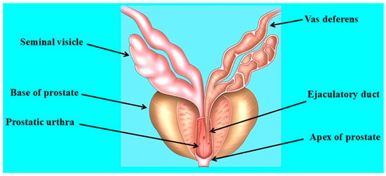
A fibromuscular (30%) and glandular (70%) organ. It is walnut-sized (~20 g) and located inferior to the bladder neck.
McNeal Zonal Anatomy:
- Peripheral Zone (70%): Site where most carcinomas arise.
- Central Zone (25%): Surrounds ejaculatory ducts.
- Transitional Zone (5%): Site where BPH typically arises.
3. Microscopic Structure / Histology
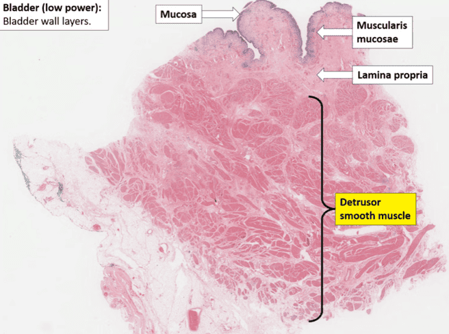
The bladder wall consists of four layers from inside out:
3.1 Mucosa (Urothelium + Lamina Propria)
- Urothelium (Transitional Epithelium): Stratified epithelium (5–7 layers relaxed; 2–3 layers distended). Features umbrella cells that provide a permeability barrier.
- Lamina Propria: Loose connective tissue with myofibroblasts and afferent nerve endings.
3.2 Muscularis (Detrusor Muscle)
- Three indistinct layers: inner longitudinal, middle circular, outer longitudinal.
- The middle circular layer is thickest at the neck, forming the internal urethral sphincter (smooth muscle, involuntary).
The Trigone
The trigone is histologically and functionally distinct. It is derived from the mesonephric duct (not the urogenital sinus). Its mucosa is smooth (no rugae) and contains a dense network of afferent fibers (rich in P2X3 and substance P), acting as a critical sensor for bladder filling and pain.
4. Neurovascular Supply
4.1 Arterial Supply
| Structure | Arterial Supply |
|---|---|
| Bladder (superior) | Superior vesical arteries (from internal iliac/umbilical) |
| Bladder (inferior) | Inferior vesical (male) / vaginal arteries (female) |
| Urethra | Internal pudendal branches |
| Prostate | Inferior vesical, middle rectal arteries |
4.4 Nerve Supply
The LUT receives input from three nervous systems:
| Nerve | Origin | Type | Function |
|---|---|---|---|
| Pelvic nerve | S2–S4 | Parasympathetic | Motor: Detrusor contraction (voiding). Sensory: Fullness/pain. |
| Hypogastric nerve | T12–L2 | Sympathetic | Motor: Detrusor relaxation & internal sphincter contraction (storage). |
| Pudendal nerve | S2–S4 | Somatic | Motor: External sphincter contraction (voluntary continence). |
5. Physiology of the Lower Urinary Tract
The LUT operates in two mutually exclusive phases: storage and voiding.
5.1 Urine Storage Phase
Events:
- Bladder filling (50–400 mL).
- Detrusor relaxation: Mediated by sympathetic stimulation via β3-adrenoceptors.
- Internal sphincter contraction: Sympathetic α-adrenergic stimulation.
- External sphincter contraction: Somatic tonic contraction via pudendal nerve.
- Guarding reflex: Spinal reflexes increase external sphincter tone as volume increases.
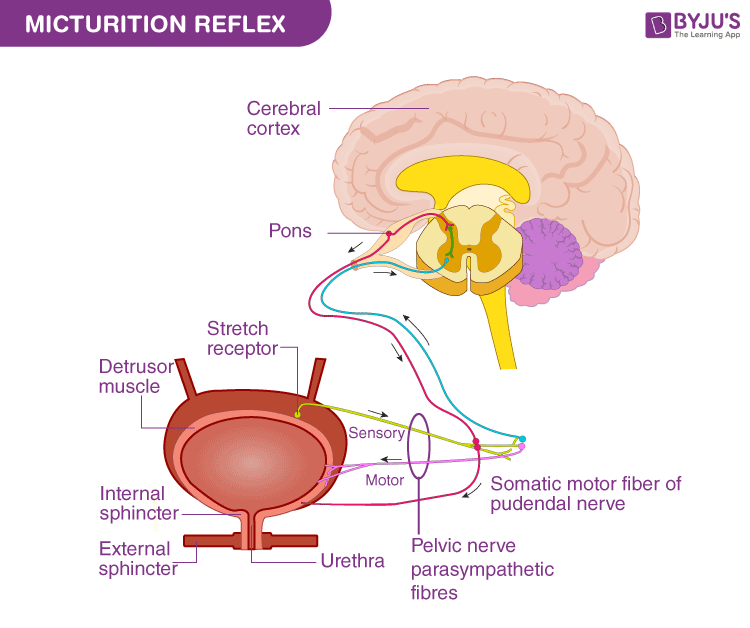
5.2 The Micturition Reflex
Trigger: Bladder volume reaches ~300–400 mL; stretch receptor firing increases dramatically.
The Reflex Arc:
- Afferent: Stretch receptors (Aδ) → Pelvic nerve → Sacral cord (S2-S4) → Ascends to periaqueductal gray (PAG).
- Central Integration: PAG relays to pontine micturition center (PMC) (Barrington's nucleus). The Cerebral Cortex provides voluntary inhibition until socially appropriate.
- Efferent: PMC activates Pelvic nerve (Detrusor contraction) and inhibits Sympathetic/Somatic tone (Sphincter relaxation).
6. Clinical Correlations
6.1 Urinary Tract Infections (UTIs)
Pathophysiology: Ascending infection by coliforms (*E. coli*). High risk in females due to short urethra and proximity to the anus.
6.2 Urinary Incontinence
- Stress Incontinence: Leakage with increased intra-abdominal pressure (e.g., coughing) due to weak pelvic floor.
- Urge Incontinence: Detrusor overactivity; involuntary contraction.
- Overflow Incontinence: Chronic retention with dribbling (e.g., BPH or stricture).
6.4 Neurogenic Bladder
| Lesion Level | Bladder Type | Clinical Features |
|---|---|---|
| Suprapontine (Stroke) | Uninhibited | Urgency, frequency, urge incontinence. |
| Spinal Cord (Above S2) | Reflex / Automatic | Detrusor-sphincter dyssynergia (DSD); risk of upper tract damage. |
| Conus / Cauda Equina | Acontractile | Flaccid bladder, overflow incontinence, painless retention. |
Urethral Stricture
Narrowing of the lumen due to scarring. Common causes in Uganda: Post-gonococcal infection and trauma (pelvic fractures or traumatic catheterization). Leads to obstructive voiding symptoms and chronic retention.
