Doctors Revision

Author name: doctorsrevision@gmail.com

Functions of the skin
Anatomy

Functions of the skin

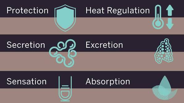
Functions of the Skin A comprehensive study of the integumentary system’s roles in protection, thermoregulation, sensation, excretion, and metabolism, including clinical applications and structural mechanisms. 1. Overview The skin performs a diverse array of functions essential for homeostasis, protection, and interaction with the environment. These functions can be broadly categorized into protective, regulatory, sensory, metabolic, and social functions. Each function is mediated by specific structural components and cellular mechanisms within the skin layers. 2. Protective Functions The skin serves as the body’s first line of defense against external threats. This protective role is multifaceted and involves mechanical, chemical, biological, and ultraviolet barriers. 2.1 Mechanical Barrier Stratum corneum: Provides a tough, keratinized surface that resists abrasion, friction, and minor trauma. Dermal collagen and elastin: Provide tensile strength and elasticity, allowing the skin to withstand stretching and pressure. Hypodermis: Cushions underlying structures against impact and mechanical forces. 2.2 Chemical Barrier The acid mantle: A surface film with a pH of 4.5–6.5 that inhibits the growth of pathogenic bacteria and fungi. Sebum: Produced by sebaceous glands, containing fatty acids with antimicrobial properties. Epidermal lipids: Ceramides, cholesterol, and free fatty acids in the stratum corneum form a waterproof barrier preventing transepidermal water loss (TEWL) and the entry of hydrophilic toxins. 2.3 Biological Barrier Langerhans cells: Dendritic cells in the epidermis that capture antigens and present them to T lymphocytes to initiate adaptive immune responses. Keratinocytes: Produce antimicrobial peptides such as defensins and cathelicidins that directly kill pathogens. Skin microbiome: Commensal bacteria (e.g., Staphylococcus epidermidis) compete with pathogens for resources and produce bacteriocins. 2.4 Ultraviolet (UV) Protection Melanin: Produced by melanocytes; absorbs UV radiation (UVA and UVB) to prevent DNA damage in keratinocytes. Stratum corneum: Scatters and reflects a portion of incident UV radiation. DNA repair: Enzymatic mechanisms in keratinocytes correct UV-induced damage. Clinical Note Barrier Defects Defects in the skin barrier (e.g., atopic dermatitis, ichthyosis) lead to increased transepidermal water loss (TEWL), susceptibility to infections, and allergen penetration. Barrier repair therapy with ceramide-containing emollients is a cornerstone of management. 3. Thermoregulation The skin is the primary organ of thermoregulation, maintaining a core body temperature of approximately 37°C through coordinated vascular, neural, and glandular mechanisms. 3.1 Heat Dissipation (In Hot Conditions) Vasodilation: Dermal blood vessels dilate, increasing blood flow to the skin surface to facilitate heat loss via radiation, conduction, and convection. Eccrine sweating: 2–4 million eccrine glands secrete a hypotonic fluid (99% water, NaCl, urea, lactate). Evaporation of this sweat absorbs latent heat of vaporization (2,430 J/g), effectively cooling the body. 3.2 Heat Conservation (In Cold Conditions) Vasoconstriction: Dermal arterioles constrict, shunting blood away from the skin surface to minimize heat loss. Piloerection: Arrector pili muscles contract, causing hairs to stand erect (“goosebumps”). In humans, this provides minimal insulation but is significant in furry mammals. Subcutaneous fat: Adipose tissue in the hypodermis provides thermal insulation. Clinical Note Anhidrosis (absence of sweating) in burn patients or those with ectodermal dysplasia severely impairs thermoregulation and can lead to life-threatening hyperthermia. Conversely, hyperhidrosis (excessive sweating) causes significant social and functional impairment. 4. Sensation The skin contains an extensive network of sensory nerve endings and specialized receptors that detect touch, pressure, temperature, pain, and itch. 4.1 Mechanoreceptors Meissner corpuscles: Rapidly adapting; located in dermal papillae. Detect light touch and texture (abundant in glabrous skin). Pacinian corpuscles: Rapidly adapting; located in the deep dermis and hypodermis. Detect deep pressure and high-frequency vibration. Merkel discs: Slowly adapting; located in the stratum basale. Detect sustained touch and pressure. Ruffini endings: Slowly adapting; located in the dermis. Detect skin stretch and torque. 4.2 Thermoreceptors and Nociceptors Thermoreceptors: Free nerve endings located at varying depths. Cold receptors are superficial (0.15–0.17 mm); warm receptors are deeper (0.3–0.6 mm). Nociceptors: Free nerve endings that respond to tissue-damaging stimuli (mechanical, thermal, chemical) and mediate protective reflexes. 5. Excretion and Secretion 5.1 Excretion Sweat: Eliminates small amounts of urea, uric acid, ammonia, and lactic acid, contributing to nitrogenous waste elimination. Glandular bypass: In renal failure, the skin can become an important accessory excretory organ for urea, sometimes manifesting as uremic frost. 5.2 Secretion Sebaceous glands: Secrete sebum (lipids, wax esters, squalene), which lubricates skin/hair and provides antimicrobial fatty acids. Apocrine sweat glands: Located in axilla, areola, and anogenital regions. Secrete a protein-rich fluid that is odorless until acted upon by skin bacteria. Ceruminous glands: Found in the external auditory canal; secrete cerumen (earwax) to protect the tympanic membrane. 6. Vitamin D Synthesis The skin is the primary site of Vitamin D synthesis. Exposure to UVB radiation (wavelength 290–315 nm) converts 7-dehydrocholesterol in the epidermis to previtamin D3, which then isomerizes to Vitamin D3 (cholecalciferol). Activation Pathway: Step 1: UV exposure in the skin forms Vitamin D3. Step 2: Transported to the liver for hydroxylation. Step 3: Final hydroxylation in the kidneys to form calcitriol (1,25-dihydroxyvitamin D3). Function: Calcitriol regulates calcium and phosphate metabolism, essential for bone mineralization. Deficiency causes rickets in children and osteomalacia in adults. Clinical Note Dark-skinned individuals require longer sun exposure to synthesize adequate Vitamin D due to higher melanin content. In regions with limited sunlight, dietary supplementation is essential. 7. Social and Psychological Functions Non-verbal communication: Mediated by facial muscles and skin (facial expressions). Self-esteem: Skin appearance, color, and integrity significantly affect body image and mental health. Chronic conditions: Diseases such as acne, psoriasis, and vitiligo can have profound psychosocial impacts beyond their physical manifestations. 8. Summary Table Function Mechanism Clinical Relevance Protection Mechanical (keratin, collagen), Chemical (acid mantle), Biological (Langerhans cells) Barrier defects cause infection, dehydration, photosensitivity Thermoregulation Vasodilation/constriction, eccrine sweating, piloerection, subcutaneous fat Anhidrosis, hyperhidrosis, heat stroke, hypothermia Sensation Meissner, Pacinian, Merkel, Ruffini corpuscles; free nerve endings Neuropathy, chronic pain, anesthesia dolorosa Excretion Sweat (urea, NaCl), sebum, cerumen Uremic frost in renal failure, body odor Vitamin D Synthesis UVB converts 7-dehydrocholesterol to D3 Rickets, osteomalacia, supplementation needs Social/Psychological Facial expression, appearance, body image Psychosocial impact of dermatological disease
Structure of the skin (Epidermis, Dermis, Hypodermis)
Anatomy

Structure of the skin (Epidermis, Dermis, Hypodermis)

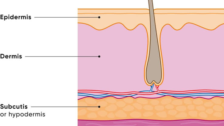
Structure of the Skin Comprehensive medical notes on the integumentary system, detailing the histology, physiology, and clinical anatomy of the epidermis, dermis, and hypodermis. 1. INTRODUCTION The integumentary system is the largest organ system of the human body, comprising the skin and its appendages (hair, nails, and glands). The skin alone covers approximately 1.5–2.0 m² in adults and accounts for about 15% of total body weight. It serves as the primary interface between the internal environment and the external world, performing critical protective, regulatory, and sensory functions. The skin consists of two principal tissues: the epidermis (superficial epithelial layer) and the dermis (deeper connective tissue layer). Beneath the dermis lies the hypodermis (subcutaneous tissue), which is not technically part of the skin but is functionally integrated with it. 2. THE EPIDERMIS The epidermis is the outermost layer of the skin, composed of stratified squamous keratinized epithelium. It is avascular and derives its nutrition by diffusion from the underlying dermis. The epidermis ranges in thickness from 0.05 mm (thin skin) to 1.5 mm (thick skin, e.g., palms and soles). It is continuously renewed through cell division in the basal layer, with complete turnover occurring approximately every 28–40 days. 2.1 Layers of the Epidermis From superficial to deep, the epidermis consists of five distinct layers: Stratum Corneum (Horny Layer) Outermost layer, consisting of 15–30 layers of flattened, anucleate keratinized cells (corneocytes). Cells are filled with keratin and surrounded by a lipid-rich extracellular matrix. Provides the primary barrier against water loss, pathogens, and environmental toxins. Sheds continuously via desquamation; replaced by cells from deeper layers. Stratum Lucidum (Clear Layer) Found only in thick skin (palms, soles, digits). Consists of 3–5 layers of flattened, dead keratinocytes with indistinct cell boundaries. Cells contain eleidin, a transparent substance derived from keratohyalin. Stratum Granulosum (Granular Layer) Composed of 3–5 layers of keratinocytes containing prominent keratohyalin granules. Lamellar granules (Odland bodies) release lipids that form the waterproof barrier. Cells in this layer undergo apoptosis; nuclei and organelles begin to degenerate. Stratum Spinosum (Prickle Cell Layer) Consists of 8–10 layers of polyhedral keratinocytes connected by desmosomes. Cells appear spiny under microscopy due to shrinkage artifacts around desmosomes. Contains Langerhans cells (dendritic antigen-presenting cells of the immune system). Site of initial keratin filament synthesis. Stratum Basale (Basal Layer) Deepest epidermal layer, resting on the basement membrane. Contains a single layer of cuboidal to columnar stem cells (basal cells) that divide mitotically. Contains melanocytes (produce melanin) and Merkel cells (tactile sensation). Anchored to the basement membrane via hemidesmosomes. 2.2 Cell Types in the Epidermis Keratinocytes (90%): Produce keratin; form the structural framework. Melanocytes (5–10%): Synthesize melanin from tyrosine; transfer melanin to keratinocytes via cytocrine secretion. Langerhans cells (3–5%): Dendritic cells involved in immune surveillance and antigen presentation. Merkel cells (<1%): Mechanoreceptors associated with sensory nerve endings; release neurotransmitters. Clinical Note The stratum corneum is the principal barrier to percutaneous drug absorption. Transdermal drug delivery systems exploit this property by using chemical enhancers or microneedles to bypass this layer. 3. THE DERMIS The dermis is the thick, connective tissue layer beneath the epidermis. It provides structural support, nutrition, and sensory innervation to the epidermis. The dermis ranges from 1–4 mm in thickness and contains blood vessels, lymphatics, nerves, glands, and hair follicles. 3.1 Papillary Dermis Superficial layer, directly beneath the epidermis. Composed of loose areolar connective tissue with fine collagen (type III) and elastic fibers. Contains dermal papillae—finger-like projections that interdigitate with epidermal rete ridges. Rich in capillary loops, free nerve endings, and Meissner corpuscles (tactile receptors). 3.2 Reticular Dermis Deeper, thicker layer making up approximately 80% of dermal thickness. Composed of dense irregular connective tissue with thick collagen bundles (type I and III) and elastic fibers. Contains larger blood vessels, lymphatic vessels, nerve bundles, and skin appendages. Contains Pacinian corpuscles (pressure receptors) and Ruffini endings (stretch receptors). 3.3 Components of the Dermis Collagen fibers (70% of dermal dry weight): Provide tensile strength and resistance to tearing. Elastic fibers: Allow the skin to stretch and recoil; deficient in cutis laxa. Ground substance: Gel-like matrix of glycosaminoglycans (hyaluronic acid, dermatan sulfate) that hydrates the tissue. Fibroblasts: Principal cells that synthesize collagen, elastin, and ground substance. Mast cells: Release histamine and other mediators in allergic and inflammatory responses. Macrophages: Phagocytic cells involved in tissue repair and immune defense. Clinical Note The dermo-epidermal junction (basement membrane) is critical for skin integrity. Genetic defects in hemidesmosomal proteins (e.g., collagen VII in dystrophic epidermolysis bullosa) result in blistering diseases. 4. THE HYPODERMIS (SUBCUTANEOUS TISSUE) The hypodermis is the layer of loose connective tissue and adipose tissue beneath the dermis. It is not technically part of the skin but is functionally integrated with it. 4.1 Structure Composed primarily of adipocytes (fat cells) arranged in lobules separated by fibrous septa. Contains loose areolar connective tissue with collagen and elastic fibers. Thickness varies widely (1–10 cm) depending on body site, nutritional status, and sex. 4.2 Functions Energy storage: Adipose tissue is the body’s primary energy reserve. Thermal insulation: Reduces heat loss and maintains core body temperature. Mechanical cushioning: Protects underlying structures from mechanical trauma. Anchorage: Attaches the skin to underlying fascia and muscles. Endocrine function: Adipose tissue secretes leptin, adiponectin, and other hormones. 5. THE DERMO-EPIDERMAL JUNCTION The dermo-epidermal junction is a specialized basement membrane zone that anchors the epidermis to the dermis. It consists of three layers: Lamina lucida (clear layer): Contains laminin-332 and integrins that attach basal keratinocytes. Lamina densa (dense layer): Contains type IV collagen, laminin, and nidogen. Sublamina densa: Contains anchoring fibrils (type VII collagen), microfibrils, and dermal collagen. Hemidesmosomes on basal keratinocytes connect to the lamina lucida via integrins, while anchoring fibrils secure the basement membrane to the dermis. Disruption of this junction results in blistering disorders. 6. CLINICAL RELEVANCE Condition Pathophysiological Basis Psoriasis Accelerated epidermal turnover (3–5 days) results in thickened plaques with silvery scales. Epidermolysis bullosa Genetic defects in basement membrane proteins cause skin fragility and blistering. Scleroderma Excessive collagen deposition in the dermis leads to skin hardening and fibrosis. Lipodystrophy Loss

Disorders of the respiratory system (Asthma, Pneumonia, Emphysema)
Anatomy

Disorders of the respiratory system (Asthma, Pneumonia, Emphysema)

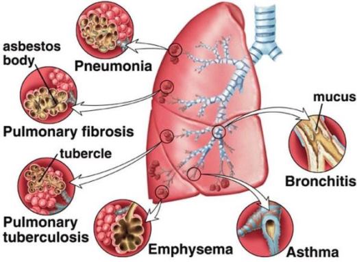
Disorders of the Respiratory System Comprehensive clinical notes covering the etiology, pathophysiology, clinical presentation, and management of major respiratory conditions: Asthma, Pneumonia, and Emphysema. 1. Asthma Asthma is a chronic inflammatory disorder of the airways characterized by reversible airflow obstruction, bronchial hyperresponsiveness, and airway remodeling. It affects approximately 262 million people worldwide and is a leading cause of morbidity in children and young adults. 1.1 Definition and Epidemiology Nature: Chronic condition with variable and recurring symptoms and reversible airflow limitation. Prevalence: 5–10% globally; notably higher in developed countries and urban areas. Onset: Frequently in childhood; may persist or remit and relapse later in life. 1.2 Etiology and Risk Factors Atopic (allergic) asthma: Most common form; associated with IgE-mediated hypersensitivity to allergens such as dust mites, pollen, pet dander, and mold. Non-atopic asthma: Triggered by infections, irritants, exercise, cold air, or stress; involves no allergen sensitization. Occupational asthma: Caused by workplace exposures to isocyanates, flour, wood dust, or chemicals. Risk Factors: Family history of atopy, childhood respiratory infections, obesity, smoking, air pollution, and low birth weight. 1.3 Pathophysiology Acute phase: Allergen exposure triggers IgE-mediated mast cell degranulation, releasing histamine, leukotrienes, prostaglandins, and cytokines. Bronchoconstriction: Smooth muscle contraction narrows airways within minutes. Mucus hypersecretion: Goblet cell hyperplasia and submucosal gland hypertrophy produce thick, tenacious mucus plugs. Airway edema: Increased vascular permeability causes mucosal swelling. Chronic inflammation: Eosinophils, T-helper 2 (Th2) cells, mast cells, and neutrophils infiltrate the airway wall. Airway remodeling: Subepithelial fibrosis, smooth muscle hypertrophy, and basement membrane thickening cause irreversible airflow limitation in long-standing disease. 1.4 Clinical Features Classic triad: Wheeze, cough, and dyspnea. Symptoms: Variable, often worse at night or early morning; triggered by exercise, allergens, or cold air. Physical examination: Prolonged expiratory phase, bilateral wheezing, use of accessory muscles, tachypnea, and tachycardia. Red Flag Severe Acute Asthma Signs of a life-threatening attack include a Silent chest (absence of wheeze due to minimal airflow), cyanosis, altered consciousness, exhaustion, and bradycardia (a pre-arrest sign). 1.5 Diagnosis History: Pattern of symptoms, specific triggers, and family history. Spirometry: Reduced FEV₁ and FEV₁/FVC ratio (<0.70). Diagnosis is supported by significant reversibility (increase in FEV₁ >12% and >200 mL after bronchodilator). 1.7 Management Stepwise Approach (GINA Guidelines): Step 1: SABA as needed. Step 2: Low-dose Inhaled Corticosteroid (ICS) daily + SABA. Step 3: Low-dose ICS/LABA combination or medium-dose ICS. Step 4: Medium-dose ICS/LABA. Step 5: High-dose ICS/LABA + add-on therapy (LAMA, biologics). Clinical Scenario Status Asthmaticus (Acute Severe Asthma) Requires urgent hospitalization. Management includes: — Continuous nebulized salbutamol + ipratropium bromide. — Systemic corticosteroids (Oral prednisolone or IV hydrocortisone). — Oxygen to maintain SpO₂ 94–98%. — IV Magnesium sulfate for bronchodilator effect. — Mechanical ventilation if refractory. 2. Pneumonia Pneumonia is an acute infection of the lung parenchyma, including the alveolar spaces and interstitial tissue. It is a major cause of mortality worldwide, particularly in children under 5 and the elderly. 2.1 Classification Category Definition / Key Feature CAP Community-acquired; acquired outside healthcare facilities. HAP Hospital-acquired; occurs >48 hours after admission. VAP Ventilator-associated; occurs >48 hours after intubation. Lobar Consolidation of an entire lobe (typically S. pneumoniae). Interstitial Inflammation confined to alveolar walls (typical of viruses/Mycoplasma). 2.3 Etiology Pathogen Characteristics Risk Factors S. pneumoniae Most common cause of CAP; Lobar consolidation. Alcoholism, COPD, asplenia. H. influenzae Common cause of bronchopneumonia. COPD, smoking, elderly. Staph. aureus Necrotizing pneumonia; Abscesses. Post-influenza infection, IV drug use. K. pneumoniae Currant jelly sputum; Bulging fissure sign. Alcoholism, Diabetes. M. pneumoniae Atypical; interstitial pattern. Young adults, schools/military. Legionella Atypical; Hyponatremia; GI symptoms. AC systems, hotels, smokers. 2.7 Management General measures: Oxygen to maintain SpO₂ >94% (88–92% in COPD); hydration; analgesia. Antibiotic Therapy (Empirical): — Low risk CAP: Amoxicillin 500 mg TDS for 5 days. — Moderate CAP: Amoxicillin-clavulanate + macrolide. — ICU CAP: Ceftriaxone + azithromycin. Complications Untreated pneumonia can lead to Pleural effusion, Empyema, Lung abscess, Septicemia, and ARDS. 3. Emphysema Emphysema is a form of COPD characterized by the permanent enlargement of airspaces distal to the terminal bronchioles, accompanied by destruction of alveolar walls without significant fibrosis. 3.2 Etiology and Risk Factors Cigarette smoking: Accounts for 80–90% of cases. Alpha-1 antitrypsin (AAT) deficiency: Genetic cause; AAT inhibits neutrophil elastase. Homozygous ZZ genotype causes early-onset panacinar emphysema. Air pollution: Indoor biomass fuel exposure. 3.3 Pathophysiology Protease-antiprotease imbalance: Neutrophil/macrophage proteases (elastase) destroy alveolar walls. Alveolar wall destruction: Loss of elastic fibers and capillary beds; airspaces enlarge and coalesce. Airway collapse: Loss of elastic recoil causes airways to collapse during expiration, leading to dynamic hyperinflation. V/Q mismatch: Reduced surface area causes hypoxemia. 3.4 Types of Emphysema Type Location Associations Centriacinar Central part of acinus; Upper lobes. Cigarette smoking. Panacinar Entire acinus; Lower lobes. AAT deficiency. Paraseptal Distal acinus near pleura. Spontaneous pneumothorax; Subpleural bullae. 3.6 Diagnosis Clinical hallmark: Progressive dyspnea, initially on exertion. Spirometry: Irreversible obstruction (FEV₁/FVC <0.70). Chest X-ray: Hyperinflation, flattened diaphragms, increased retrosternal airspace, and a small heart shadow. CT Chest: Gold standard for detecting low-attenuation areas and bullae. Long-Term Management Oxygen therapy (LTOT): Indicated if PaO₂ ≤55 mmHg. Must be used >15 hours/day to improve survival. Surgical options: Bullectomy (removal of large bullae) or Lung Volume Reduction Surgery (LVRS). 4. Key Points Summary Asthma: Reversible airway obstruction; managed with ICS and SABA. Pneumonia: Infection of parenchyma; CURB-65 guides assessment and antibiotic choice. Emphysema: Alveolar wall destruction and loss of recoil; smoking is the primary cause. Red Flag: The Silent Chest in an asthma attack is a medical emergency indicating near-total absence of airflow.

breathing mechanism
Anatomy

Mechanism of breathing

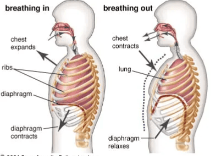
Mechanism of Breathing Complete notes on pulmonary ventilation, including respiratory musculature, pressure-volume dynamics, lung capacities, compliance, and clinical correlations. 1. Introduction Breathing (pulmonary ventilation) is the physical process of moving air into and out of the lungs. It is fundamentally driven by pressure gradients created by changes in thoracic volume. This complex mechanism involves the coordinated action of the respiratory muscles, the elastic properties of the lungs and chest wall, and the patency of the airways. 2. Respiratory Muscles The movement of the thoracic cage and the subsequent change in lung volume are mediated by two sets of muscles categorized by their role in the breathing cycle. 2.1 Primary Muscles of Inspiration Diaphragm: The principal muscle of inspiration. A dome-shaped sheet of skeletal muscle separating the thoracic and abdominal cavities. When it contracts, the dome flattens, increasing the vertical diameter of the thorax and creating negative intrapleural pressure. It accounts for approximately 75% of tidal volume during quiet breathing. External Intercostal Muscles: These muscles elevate the ribs during inspiration, increasing the anteroposterior and lateral diameters of the thorax. This rib movement is often described as the pump-handle and bucket-handle motion. 2.2 Accessory Muscles of Inspiration Used primarily during forced inspiration or when airway resistance is pathologically increased: Sternocleidomastoid: Elevates the sternum. Scalene muscles: Elevate the first two ribs. Serratus anterior and Pectoralis minor: Elevate the ribs when the shoulder girdle is fixed. Erector spinae: Extends the spine, further increasing thoracic volume. 2.3 Muscles of Expiration Quiet expiration: This is a passive process driven by the elastic recoil of the lungs and chest wall. No active muscle contraction is required. Forced expiration (active): — Internal intercostal muscles: Depress the ribs, decreasing thoracic volume. — Abdominal muscles (rectus abdominis, obliques, transversus): Compress the abdomen, pushing the diaphragm upward. — Latissimus dorsi: Depresses the ribs. 3. Mechanics of Inspiration 3.1 Quiet Inspiration Initiated by the contraction of the diaphragm and external intercostals. The sequence of events is as follows: Diaphragm contraction flattens the dome, increasing the vertical thoracic diameter by 1.5 cm (up to 7 cm during deep inspiration). External intercostals elevate the ribs, increasing horizontal diameters. Thoracic volume increases → intrapleural pressure becomes more negative (dropping from -5 cmH₂O to -8 cmH₂O). Alveolar pressure drops below atmospheric pressure (to -1 cmH₂O), creating a pressure gradient that draws air into the lungs. Airflow continues until alveolar pressure equalizes with atmospheric pressure. 3.2 Forced Inspiration Involves the accessory muscles in addition to the primary muscles. This results in a significantly greater increase in thoracic volume and a more negative intrapleural pressure, generating larger pressure gradients and greater airflow. 4. Mechanics of Expiration 4.1 Quiet Expiration A passive process driven by the elastic recoil of the lungs and the relaxation of inspiratory muscles. The sequence includes: The diaphragm relaxes and returns to its dome shape; external intercostals relax, allowing ribs to descend. Thoracic volume decreases → intrapleural pressure becomes less negative. Alveolar pressure rises above atmospheric pressure (to +1 cmH₂O), creating a pressure gradient that drives air out. Airflow continues until alveolar pressure equalizes with atmospheric pressure. 4.2 Forced Expiration An active process requiring the contraction of expiratory muscles. Internal intercostals depress the ribs while abdominal muscles compress the viscera to push the diaphragm upward. Intrapleural pressure becomes positive (reaching +20 to +30 cmH₂O during coughing). This rapid increase in alveolar pressure forces air out at high velocity, essential for coughing, sneezing, and vocalization. 5. Pressure Changes During Breathing Pressure At Rest (End-Expiration) During Inspiration During Expiration Atmospheric pressure 0 cmH₂O 0 cmH₂O 0 cmH₂O Intrapleural pressure -5 cmH₂O -8 cmH₂O -3 cmH₂O Alveolar pressure 0 cmH₂O -1 cmH₂O +1 cmH₂O Transpulmonary pressure +5 cmH₂O +8 cmH₂O +3 cmH₂O Key Definitions Intrapleural pressure: Pressure in the pleural space; always subatmospheric (negative) in healthy individuals, preventing lung collapse. Alveolar (intrapulmonary) pressure: Pressure within the alveoli; fluctuates above and below atmospheric pressure during the cycle. Transpulmonary pressure: The difference between alveolar and intrapleural pressure (Palv – Pip); it maintains alveolar expansion. Transmural pressure: Pressure across the airway wall; keeps airways open during inspiration. 6. Lung Volumes and Capacities 6.1 Static Lung Volumes Tidal volume (TV): Volume of air inhaled or exhaled during quiet breathing; 500 mL in healthy adults. Inspiratory reserve volume (IRV): Additional air that can be forcibly inhaled after a normal inspiration; 2500–3000 mL. Expiratory reserve volume (ERV): Additional air that can be forcibly exhaled after a normal expiration; 1000–1200 mL. Residual volume (RV): Air remaining in the lungs after maximal exhalation; 1000–1200 mL. It prevents alveolar collapse. 6.2 Lung Capacities Inspiratory capacity (IC): TV + IRV (~3000 mL). Functional residual capacity (FRC): ERV + RV (~2300 mL). Important for gas exchange continuity. Vital capacity (VC): IRV + TV + ERV; the maximum air that can be exhaled after maximal inspiration (~4500–5000 mL). Total lung capacity (TLC): VC + RV; total air in lungs at maximal inspiration (~5500–6000 mL). 6.3 Dynamic Volumes Forced vital capacity (FVC): Total volume forcibly exhaled after maximal inspiration. Forced expiratory volume in 1 second (FEV₁): Volume exhaled in the first second of the FVC maneuver. FEV₁/FVC ratio: Normally >0.70 (or 70%). Reduced in obstructive disease (asthma, COPD); normal or increased in restrictive disease. Peak expiratory flow (PEF): Maximum flow rate during forced expiration; measured by peak flow meter. 7. Compliance and Elastic Recoil 7.1 Lung Compliance The distensibility of the lungs; defined as the change in lung volume per unit change in transpulmonary pressure. Normal value: ~200 mL/cmH₂O. Increased compliance: Emphysema (destruction of elastic tissue). Decreased compliance: Pulmonary fibrosis, ARDS, pulmonary edema (stiff lungs). 7.2 Elastic Recoil The tendency of the lungs to collapse inward due to elastic tissue and surface tension. Opposed by the outward recoil of the chest wall. At FRC, these opposing forces are balanced, and no airflow occurs. 7.3 Surface Tension and Surfactant Surface tension at the air-liquid interface in alveoli tends to collapse them (Laplace law: pressure = 2 × tension / radius). Surfactant, produced by Type II pneumocytes, is a phospholipid-protein complex that

Gas exchange and transport
Anatomy

Gas exchange and transport

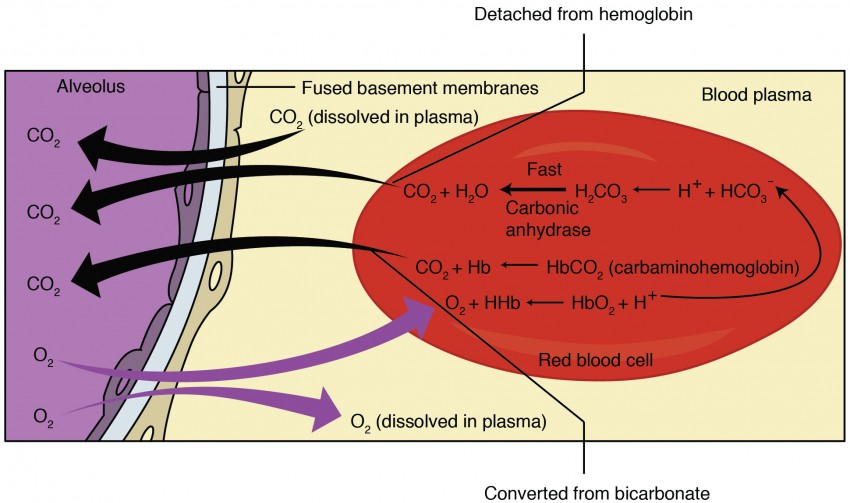
Gas Exchange and Transport Comprehensive notes of respiratory physiology, the dynamics of the blood-air barrier, hemoglobin kinetics, and clinical correlations associated with hypoxemia and acid-base disturbances. 1. Introduction Gas exchange is the physiological process by which oxygen is transferred from alveolar air to pulmonary capillary blood, and carbon dioxide is transferred from blood to alveolar air for elimination. The subsequent transport of these gases in the blood ensures the continuous delivery of oxygen to tissues for aerobic metabolism and the removal of carbon dioxide, a metabolic byproduct. These processes are critically dependent on partial pressure gradients, the structural integrity of the blood-air barrier, and the biochemical properties of hemoglobin. 2. Partial Pressures of Respiratory Gases The partial pressure of a gas is the pressure it would exert if it alone occupied the total volume. Gas exchange occurs strictly down partial pressure gradients via passive diffusion. Location PO₂ (mmHg) PCO₂ (mmHg) Atmospheric air (dry) 159 0.3 Alveolar air 100 40 Arterial blood 95–100 40 Venous blood 40 46 Tissues (rest) ~40 ~46 Expired air 120 27 Key Physiological Rules Humidification: Alveolar PO₂ is lower than atmospheric PO₂ because inspired air is humidified in the upper airways; water vapor dilutes the other gases. O₂ Gradient: The pressure gradient for O₂ is 60 mmHg (Alveolar 100 → Venous blood 40), providing a powerful drive for O₂ uptake. CO₂ Gradient: The pressure gradient for CO₂ is only 6 mmHg (Venous blood 46 → Alveolar 40). Despite this small gradient, CO₂ diffuses rapidly due to its high solubility. 3. External Respiration (Pulmonary Gas Exchange) 3.1 Diffusion Across the Blood-Air Barrier Oxygen diffuses from alveolar air into pulmonary capillary blood, while carbon dioxide diffuses in the opposite direction. Diffusion is extremely efficient due to: Barrier Thickness: The membrane is ultra-thin (0.2–0.5 µm). Surface Area: The total alveolar surface area is massive (70–100 m²). Equilibration Time: Gas equilibration occurs within 0.25 seconds. Since blood spends ~0.75 seconds in the pulmonary capillaries, there is a large safety margin. 3.2 Factors Affecting External Respiration Thickness: Increased in pulmonary fibrosis, edema, or pneumonia, which slows diffusion. Surface Area: Decreased in emphysema (alveolar destruction), pneumonectomy, or atelectasis. Pressure Gradients: Reduced at high altitudes (low inspired PO₂) or in hypoventilation. Diffusion Coefficient: CO₂ is ~20× more soluble than O₂, allowing it to diffuse just as quickly despite its smaller pressure gradient. 3.3 Ventilation-Perfusion (V/Q) Ratio Optimal gas exchange requires matching of alveolar ventilation (V) to pulmonary perfusion (Q). Normal V/Q ratio is ~0.8 (4 L/min ventilation ÷ 5 L/min blood flow). Clinical Correlation V/Q Mismatch High V/Q (Dead Space): Alveoli are ventilated but unperfused. Classic example: Pulmonary Embolism. Low V/Q (Shunt): Alveoli are perfused but unventilated. Seen in atelectasis, airway obstruction, or consolidation. Hypoxic Vasoconstriction: A local compensatory mechanism where alveolar hypoxia causes constriction of local pulmonary arterioles to divert blood to better-ventilated regions. 4. Oxygen Transport in Blood Oxygen is transported in the blood in two forms: dissolved in plasma (1.5%) and bound to hemoglobin (98.5%). 4.1 Hemoglobin Structure and Function Hemoglobin (Hb) is a tetrameric protein (two alpha and two beta chains). Each chain contains a heme group with an iron atom (Fe²⁺) that binds one O₂ molecule. Thus, one Hb molecule can bind four O₂ molecules. 1 gram of Hb can carry 1.34 mL of O₂. 4.2 Oxyhemoglobin Dissociation Curve The relationship between PO₂ and Hb saturation is sigmoid (S-shaped) due to cooperative binding. P₅₀: The PO₂ at which Hb is 50% saturated; normally ~26–27 mmHg. Plateau (PO₂ 60–100 mmHg): Hb remains highly saturated (>90%), providing a buffer against moderate hypoxemia. Steep Portion (PO₂ 10–40 mmHg): Small changes in PO₂ cause large changes in saturation, facilitating O₂ unloading in tissues. 4.3 Factors Shifting the Oxyhemoglobin Curve Factor Right Shift (↓ Affinity) Left Shift (↑ Affinity) Temperature Increased (fever, exercise) Decreased (hypothermia) pH (Bohr effect) Decreased (acidosis) Increased (alkalosis) PCO₂ Increased Decreased 2,3-DPG Increased (chronic hypoxia, anemia) Decreased (stored blood) CO — Increased (CO poisoning) Bohr Effect The Bohr effect states that increased H⁺ (acidosis) and CO₂ decrease Hb affinity for O₂. This is physiologically vital as it promotes O₂ release in metabolically active tissues where CO₂ and acid levels are highest. 5. Carbon Dioxide Transport CO₂ is transported in three forms: dissolved (7%), as bicarbonate (70%), and bound to hemoglobin as carbaminohemoglobin (23%). 5.2 Bicarbonate (HCO₃⁻) Formation This is the most important form of CO₂ transport. The process involves several steps: CO₂ enters the RBC and combines with H₂O to form carbonic acid (H₂CO₃), catalyzed by carbonic anhydrase. H₂CO₃ dissociates into H⁺ and HCO₃⁻. Chloride Shift (Hamburger Phenomenon): HCO₃⁻ diffuses out of the RBC into plasma in exchange for Cl⁻ to maintain electrical neutrality. H⁺ binds to deoxygenated Hb (which acts as a buffer), preventing significant pH changes. Haldane Effect The Haldane effect states that deoxygenated hemoglobin binds CO₂ more readily than oxygenated hemoglobin. This facilitates CO₂ loading in the tissues and CO₂ unloading in the lungs as Hb becomes oxygenated. 6. Internal Respiration (Tissue Gas Exchange) In systemic tissues, O₂ diffuses from capillary blood (PO₂ 95–100 mmHg) into tissues (PO₂ ~40 mmHg). Simultaneously, CO₂ diffuses from tissues (PCO₂ ~46 mmHg) into capillary blood (PCO₂ 40 mmHg). Factors Affecting Delivery: Oxygen delivery (DO₂) depends on cardiac output and arterial O₂ content. Capillary density increases in exercise to shorten diffusion distance, while edema increases it, impairing exchange. 7. Clinical Correlations Hypoxemia Defined as reduced arterial PO₂ (<80 mmHg) or O₂ saturation (<92%). Hypoxic hypoxia: Low inspired O₂ or impaired pulmonary gas exchange. Anemic hypoxia: Reduced Hb (anemia, hemorrhage, CO poisoning). Stagnant hypoxia: Reduced tissue perfusion (shock, heart failure). Histotoxic hypoxia: Impaired cellular O₂ utilization (e.g., cyanide poisoning). Carbon Monoxide Poisoning CO binds to Hb with 240× greater affinity than O₂, forming carboxyhemoglobin (COHb). This shifts the dissociation curve to the left, preventing O₂ unloading in tissues. Features include cherry-red skin, headache, and confusion. Treatment is 100% O₂ or hyperbaric oxygen. Cyanosis Bluish discoloration of skin due to increased deoxygenated Hb (>5 g/dL). Central cyanosis: Due to arterial hypoxemia (cardiac

Organs of the lower respiratory system
Anatomy

Organs of the lower respiratory system

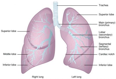
Organs of the Lower Respiratory System Comprehensive anatomical study covering the structural organization, internal architecture, neurovascular supply, and clinical correlations of the trachea, bronchial tree, and lungs. 1. Introduction The lower respiratory tract comprises the trachea, bronchi, bronchioles, and lungs. These structures function primarily to conduct air to the alveoli, where gas exchange occurs. The lower respiratory tract is lined by respiratory epithelium and supported by cartilaginous, muscular, and elastic tissues that maintain airway patency while providing the necessary flexibility for respiration and body movement. 2. The Trachea The trachea (windpipe) is a fibrocartilaginous tube measuring approximately 10–11 cm in length and 2 cm in diameter. it extends from the inferior border of the cricoid cartilage (C6) to the carina at the level of the sternal angle (T4–T5). 2.1 Structure Cartilaginous Support: Composed of 16–20 C-shaped hyaline cartilage rings that prevent airway collapse during inspiration. Trachealis Muscle: The posterior wall is membranous and contains the trachealis muscle (smooth muscle). This arrangement allows the esophagus to expand during swallowing. Histology: Lined by ciliated pseudostratified columnar epithelium with goblet cells. Submucosal glands secrete mucus that traps inhaled particles. Mucociliary Escalator: Ciliary action moves trapped particles upward toward the pharynx for swallowing or expectoration. 2.2 Anatomical Relations Relation Structures Anterior Isthmus of thyroid gland, inferior thyroid veins, sternohyoid and sternothyroid muscles, manubrium sterni. Posterior Esophagus and recurrent laryngeal nerves. Lateral Lobes of the thyroid gland, carotid sheaths. 2.3 Blood Supply and Innervation Arterial Supply: Inferior thyroid arteries. Venous Drainage: Brachiocephalic veins via inferior thyroid veins. Lymphatic Drainage: Pretracheal and paratracheal nodes. Innervation: — Parasympathetic: Vagus nerve (causes bronchoconstriction and increased mucus secretion). — Sympathetic: Sympathetic trunks (causes bronchodilation and reduced secretion). 3. The Bronchial Tree The bronchial tree is the branching system of airways that distributes air from the trachea to the alveoli. 3.1 Main (Primary) Bronchi The trachea bifurcates at the carina into right and left main bronchi. Both are supported by C-shaped cartilage rings and lined by respiratory epithelium. Right main bronchus: Wider, shorter (~2.5 cm), and more vertical than the left. Foreign bodies more commonly enter the right bronchus due to this verticality. Left main bronchus: Longer (~5 cm), narrower, and more horizontal. It passes beneath the aortic arch and anterior to the esophagus. 3.2 Lobar (Secondary) Bronchi Right lung: Three lobar bronchi (superior, middle, lower) corresponding to the three lobes. Left lung: Two lobar bronchi (upper and lower) corresponding to the two lobes. 3.3 Segmental (Tertiary) Bronchi Each lobar bronchus divides into segmental bronchi. There are typically 10 on the right and 8–10 on the left. Each segmental bronchus supplies a bronchopulmonary segment. Key Concept A bronchopulmonary segment is a functionally independent unit with its own artery, vein, and lymphatic drainage. These segments are important surgically because resection of a segment does not compromise adjacent segments. 3.4 Bronchioles Conducting bronchioles: Diameter <1 mm; they lack cartilage and glands. Lined by ciliated cuboidal epithelium. Terminal bronchioles: The most distal part of the conducting zone; each supplies a pulmonary acinus. Respiratory bronchioles: The first part of the respiratory zone; they have scattered alveoli budding directly from their walls. 4. The Lungs The lungs are the paired, cone-shaped organs of respiration located in the thoracic cavity on either side of the mediastinum. 4.1 Gross Anatomy Right lung: Larger and heavier; has three lobes (superior, middle, inferior) separated by horizontal and oblique fissures. Left lung: Smaller due to the cardiac notch; has two lobes (superior and inferior) separated by the oblique fissure. It contains the lingula (homologous to the right middle lobe). Apex: Projects into the root of the neck, 2–4 cm above the clavicle. Base: Concave, resting on the diaphragm. Hilum: Medial surface depression where bronchi, pulmonary vessels, bronchial vessels, lymphatics, and nerves enter and exit. Surfaces: Costal surface (convex, related to ribs) and Mediastinal surface (related to mediastinal structures). 4.2 Bronchopulmonary Segments Lobe Right Lung Segments Left Lung Segments Upper Lobe Apical, Posterior, Anterior Apical-posterior, Anterior, Superior lingular, Inferior lingular Middle Lobe Lateral, Medial — Lower Lobe Superior, Medial basal, Anterior basal, Lateral basal, Posterior basal Superior, Anteromedial basal, Lateral basal, Posterior basal 4.3 Pleura Parietal pleura: Lines the thoracic wall (costal pleura), diaphragm (diaphragmatic pleura), and mediastinum (mediastinal pleura). The cervical pleura (cupula) extends into the neck. Visceral pleura: Covers the lung surface, including the fissures. Pleural cavity: Potential space between parietal and visceral pleurae; contains a thin film of serous fluid (pleural fluid) that reduces friction during breathing. Pleural recesses: Potential spaces not occupied by lungs during quiet breathing, including the costodiaphragmatic recess (largest) and costomediastinal recess. 5. The Respiratory Zone The respiratory zone is where gas exchange occurs. It includes respiratory bronchioles, alveolar ducts, alveolar sacs, and alveoli. 5.1 Alveoli Approximately 300–500 million alveoli exist in adult lungs, providing a total surface area of 70–100 m². Each alveolus is a thin-walled sac (0.2–0.5 µm thick) lined by two types of pneumocytes: Type I pneumocytes: Squamous epithelial cells covering 95% of alveolar surface; the primary site of gas exchange. Type II pneumocytes: Cuboidal cells that produce surfactant (dipalmitoylphosphatidylcholine), which reduces surface tension and prevents alveolar collapse. Alveolar macrophages (dust cells): Phagocytose inhaled particles and pathogens. 5.2 Blood-Air Barrier The barrier between alveolar air and capillary blood consists of: Type I pneumocyte, fused basement membrane, and capillary endothelial cell. The total thickness is 0.2–0.5 µm, allowing for rapid gas diffusion. 5.3 Alveolar-Capillary Network Each alveolus is surrounded by a dense capillary network. Pulmonary arteries carry deoxygenated blood to the alveolar capillaries; pulmonary veins return oxygenated blood to the left atrium. Bronchial arteries (systemic): Supply oxygenated blood to the lung tissue and drain partially into pulmonary veins. 6. Clinical Correlations Clinical Scenarios Tracheal Stenosis: Narrowing of the tracheal lumen due to trauma, prolonged intubation, or granulomatous disease. Presents with stridor, dyspnea, and cough. Atelectasis: Collapse of lung tissue due to airway obstruction, compression, or surfactant deficiency. Types: Obstructive (resorption), Compressive (pleural effusion), Adhesive (ARDS). Pneumothorax: Air in the pleural cavity causing lung collapse. Can be spontaneous (rupture of bleb), traumatic

Organs of the upper respiratory tract
Anatomy

Organs of the upper respiratory tract

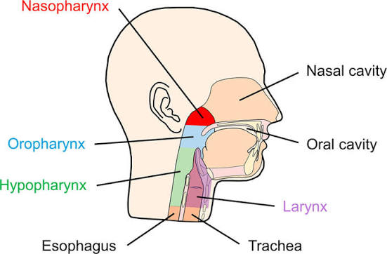
Organs of the Upper Respiratory Tract Complete, exhaustive anatomical study covering the structural organization, mucous membranes, regional compartments, and clinical pathologies of the upper airway. Prepared for Diploma-Level Clinical Medicine Curriculum. code Code 1. Introduction The upper respiratory tract consists of the nose, nasal cavity, paranasal sinuses, pharynx, and larynx. These structures serve as the entry portal for air, conditioning it through warming, humidification, and filtration before it reaches the lower respiratory tract. The upper airway also houses the organs of olfaction (smell) and phonation (voice production). 2. The Nose and Nasal Cavity 2.1 External Nose The external nose is a visible part of the respiratory system that projects from the face. It is composed of: Bone: Nasal bones, frontal processes of the maxillae, and the nasal part of the frontal bone. Cartilage: Septal, lateral, alar, and major/minor cartilages. Nares: The anterior openings (nostrils), bounded laterally by the alae. The bridge is supported by bone, while the tip and alae are cartilaginous and mobile. 2.2 Nasal Cavity The nasal cavity extends from the nostrils anteriorly to the choanae posteriorly, where it communicates with the nasopharynx. It is divided into right and left halves by the nasal septum, which is composed of the perpendicular plate of the ethmoid, the vomer, and the septal cartilage. Boundaries: Floor: Palatine process of the maxilla and horizontal plate of the palatine bone (hard palate). Roof: Nasal bones, cribriform plate of the ethmoid, and the body of the sphenoid. Lateral wall: Composed of several bones and characterized by the presence of conchae. 2.3 Nasal Conchae (Turbinates) The lateral wall contains three bony projections: superior, middle, and inferior conchae. These structures increase the surface area for warming and humidifying inspired air. Beneath each concha is a passage called a meatus: Superior meatus: Receives drainage from the posterior ethmoidal air cells. Middle meatus: Receives drainage from the frontal sinus, maxillary sinus, and anterior/middle ethmoidal air cells. Inferior meatus: Receives drainage from the nasolacrimal duct. 2.4 Nasal Mucosa Respiratory region: Lined with ciliated pseudostratified columnar epithelium with goblet cells. It covers most of the cavity. Mucus traps particles, and cilia move the mucus toward the pharynx (the mucociliary escalator). Olfactory region: Located in the roof and superior concha. It contains olfactory receptor neurons with cilia that bind odorant molecules. Axons pass through the cribriform plate to the olfactory bulb. A rich vascular plexus (cavernous tissue) warms inspired air. 2.5 Paranasal Sinuses These are air-filled cavities within cranial bones that communicate with the nasal cavity: Frontal sinuses: Within the frontal bone; drain into the middle meatus via the frontonasal duct. Maxillary sinuses: The largest; located within the body of the maxilla. Their floor is near the alveolar process of the maxilla, making them vulnerable to dental infection. Ethmoidal sinuses: Multiple small air cells within the ethmoid bone. Sphenoidal sinuses: Within the body of the sphenoid; drain into the sphenoethmoidal recess. Functions: Lighten the skull bones, resonate the voice, and produce mucus. Inflammation is known as sinusitis. 3. The Pharynx The pharynx is a muscular tube (12–14 cm) extending from the base of the skull to the esophagus. It serves as a common passage for air and food. 3.1 Nasopharynx Posterior to the nasal cavity and above the soft palate. Lined by respiratory epithelium. Contains the pharyngeal tonsil (adenoids) on the posterior wall. The auditory (Eustachian) tube opens into the lateral wall, equalizing middle ear pressure. During swallowing, the soft palate elevates to seal the nasopharynx, preventing nasal regurgitation. 3.2 Oropharynx Posterior to the oral cavity, between the soft palate and the epiglottis. Contains the palatine tonsils in the tonsillar fossae. Contains the lingual tonsil at the base of the tongue. Waldeyer ring: A ring of lymphoid tissue (pharyngeal, palatine, and lingual tonsils) surrounding the oropharyngeal inlet for immune defense. 3.3 Laryngopharynx (Hypopharynx) Extends from the tip of the epiglottis to the inferior border of the cricoid cartilage (C6), where it becomes the esophagus. The piriform fossae are recesses on either side of the laryngeal inlet; foreign bodies may lodge here. During swallowing, the larynx elevates and the epiglottis tilts downward to cover the laryngeal inlet, directing food into the esophagus. 4. The Larynx The larynx (voice box) is a cartilaginous structure located at the C3–C6 vertebral levels. It connects the pharynx to the trachea and serves three functions: airway patency, phonation, and protection of the lower airway. 4.1 Laryngeal Cartilages Thyroid cartilage: The largest; forms the laryngeal prominence (Adam’s apple). Composed of two laminae joined anteriorly. Cricoid cartilage: Signet-ring shaped; the only complete cartilaginous ring of the airway. Epiglottis: Leaf-shaped elastic cartilage attached to the thyroid cartilage. During swallowing, it folds down to cover the laryngeal inlet. Arytenoid cartilages: Paired pyramidal cartilages on the cricoid; articulate with the cricoid and move the vocal cords. Corniculate and cuneiform cartilages: Small paired cartilages within the aryepiglottic folds. 4.2 Laryngeal Cavity Vestibule: Space above the vestibular (false) vocal folds. Vestibular folds (false cords): Superior mucosal folds containing vestibular ligaments; no role in phonation. Vocal folds (true cords): Inferior mucosal folds containing vocal ligaments and the vocalis muscle. The space between them is the rima glottidis. Glottis: Comprises the vocal folds and the rima glottidis; the narrowest part of the larynx in adults. Subglottis: Region below the vocal folds extending to the inferior border of the cricoid. 4.3 Intrinsic Muscles Muscle Action on Vocal Cords Cricothyroid Tenses (lengthens) Posterior cricoarytenoid Opens glottis (the only abductor) Lateral cricoarytenoid Closes glottis (adductor) Thyroarytenoid Relaxes (shortens); fine-tunes pitch Arytenoid (transverse/oblique) Closes glottis 4.4 Nerve Supply Superior laryngeal nerve (CN X): Internal branch provides sensory innervation above the cords; external branch supplies the cricothyroid muscle. Recurrent laryngeal nerve (CN X): Motor to all intrinsic muscles except cricothyroid; sensory below the vocal cords. Clinical Note Damage to the recurrent laryngeal nerve causes hoarseness (unilateral) or life-threatening stridor (bilateral) due to vocal cord paralysis. 5. Clinical Correlations Epistaxis (Nosebleed) Most commonly occurs from the Kiesselbach plexus (Little area) on the anterior nasal septum. Causes include trauma, dry air, or

Disorders of Lymphatic system (Lymphadenitis, Hodgkin’s disease)
Anatomy

Disorders of Lymphatic system (Lymphadenitis, Hodgkin’s disease)

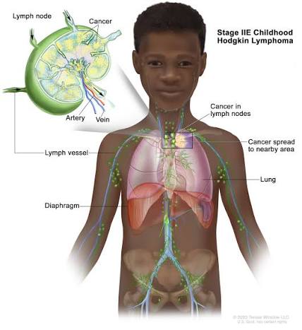
Disorders of the Lymphatic System Complete clinical notes covering the classification, pathophysiology, and management of lymphadenitis and Hodgkin Lymphoma. 1. Lymphadenitis Lymphadenitis is the inflammation of the lymph nodes, usually secondary to infection, immune activation, or malignancy in the drainage territory. It represents a localized immune response and is one of the most common clinical presentations in primary care and general medicine. 1.1 Definition and Classification Acute lymphadenitis: Rapid onset; tender, warm, enlarged nodes; typically bacterial. Chronic lymphadenitis: Persistent enlargement (>4 weeks); often granulomatous or neoplastic. Suppurative lymphadenitis: Progression to abscess formation with central necrosis and pus. Generalized lymphadenitis: Involvement of multiple non-contiguous node groups; suggests systemic disease. Localized lymphadenitis: Confined to a single regional group; indicates local infection or malignancy. 1.2 Causes and Risk Factors Category Common Causes Key Features Bacterial Staphylococcus aureus, Streptococcus pyogenes, Bartonella henselae (cat-scratch), Mycobacterium tuberculosis Acute, tender, erythematous; TB: matted, caseating, cold abscess Viral EBV (infectious mononucleosis), CMV, HIV, cytomegalovirus Bilateral, symmetrical, non-suppurative; often with pharyngitis or rash Fungal Histoplasma, Coccidioides, Sporothrix Endemic regions; chronic or granulomatous presentation Parasitic Toxoplasma gondii, filariasis, leishmaniasis Travel history; eosinophilia may be present Autoimmune SLE, rheumatoid arthritis, sarcoidosis Generalized; associated with other systemic features Malignant Metastatic carcinoma, lymphoma, leukemia Hard, fixed, non-tender; progressive enlargement; systemic B symptoms 1.3 Pathophysiology Infection or antigenic stimulus in the drainage area triggers immune cell recruitment to the regional node. Hyperplasia of germinal centers (B-cell proliferation) and paracortical expansion (T-cell response). Increased blood flow and vascular permeability cause redness, warmth, and swelling. Pressure on the capsule and surrounding tissues produces pain. In bacterial infections, neutrophil infiltration may lead to suppuration and abscess formation. In granulomatous infections (TB, fungi), epithelioid macrophages and giant cells form caseating granulomas. 1.4 Clinical Features Local signs: Enlarged, tender lymph node(s); overlying skin may be erythematous and warm. Systemic signs: Fever, malaise, anorexia; severity correlates with the underlying infection. Condition Distinguishing Features Reactive hyperplasia Recent infection; tender, mobile, bilateral; resolves within weeks Lymphoma Painless, progressive, rubbery; may have B symptoms TB Chronic, matted, cold abscess; exposure history; positive IGRA/TST Metastatic carcinoma Hard, fixed, non-tender; known primary malignancy Kikuchi-Fujimoto disease Young women; cervical nodes; self-limiting; histology diagnostic Castleman disease Localized or multicentric; hypervascular on imaging; biopsy diagnostic 1.7 Management Supportive care: Rest, hydration, analgesia (paracetamol or NSAIDs), warm compresses. Antibiotics: Empirical therapy targeting likely pathogens. — Community-acquired: amoxicillin-clavulanate or cephalexin for 7–10 days. — MRSA risk: clindamycin or trimethoprim-sulfamethoxazole. — Cat-scratch disease: azithromycin if severe; usually self-limiting. — TB lymphadenitis: standard anti-TB regimen (RIPE: rifampicin, isoniazid, pyrazinamide, ethambutol for 2 months, then rifampicin + isoniazid for 4 months). Surgical drainage: Incision and drainage for fluctuant abscesses; needle aspiration may suffice for small collections. Treatment of underlying cause: Antivirals where indicated, antiretroviral therapy for HIV, immunosuppressants for autoimmune disease. Follow-up: Reassess at 2–4 weeks; persistent or enlarging nodes require further investigation. Complications Abscess formation and spontaneous drainage. Bacteremia and sepsis. Chronic sinus tract formation (especially TB). Post-inflammatory fibrosis and persistent node enlargement. Misdiagnosis of underlying malignancy. 2. Hodgkin’s Disease (Hodgkin Lymphoma) Hodgkin lymphoma (HL) is a malignant neoplasm of the lymphatic system characterized by the presence of Reed-Sternberg cells in a background of inflammatory cells. It accounts for approximately 10% of all lymphomas and has a bimodal age distribution with peaks in young adulthood and after age 55. 2.1 Epidemiology and 2.2 Etiology Incidence: 2–3 per 100,000 annually in developed countries. Slight male predominance (M:F ratio 1.3:1). Infectious: Epstein-Barr virus (EBV) implicated in 40–50% of cases; HIV increases risk 10-fold. Genetic: Familial clustering suggests genetic susceptibility; HLA associations identified. Immunodeficiency: Congenital immunodeficiency, post-transplant immunosuppression. Environmental: No strong environmental carcinogen identified; smoking may be a minor risk factor. 2.3 Pathophysiology Originates from germinal center B cells that have undergone failed apoptosis. The neoplastic cell is the Reed-Sternberg (RS) cell or its variant, the Hodgkin cell. RS cells are large, binucleated or multinucleated cells with prominent eosinophilic nucleoli (‘owl-eye’ appearance). RS cells constitute <1% of the tumor mass; the majority is a reactive inflammatory infiltrate. RS cells express CD30 and CD15; they are usually negative for CD20 and CD45 (LCA). Cytokine production (IL-5, IL-10, TGF-beta) by RS cells contributes to the inflammatory background and systemic symptoms. 2.4 Classification (WHO 2016) Subtype Frequency Characteristics Nodular sclerosis (NSHL) 70% of cases Most common in young adults; collagen bands dividing nodules; lacunar RS cell variant; good prognosis. Mixed cellularity (MCHL) 20–25% Older adults and HIV-positive patients; numerous RS cells; EBV association common. Lymphocyte-rich (LRHL) 5% Few RS cells; abundant small lymphocytes; excellent prognosis. Lymphocyte-depleted (LDHL) <5% Rare; elderly or HIV-positive; numerous RS cells, few lymphocytes; aggressive. Nodular lymphocyte-predominant (NLPHL) Distinct entity Popcorn cells (LP cells); CD20+, CD30-; indolent; male predominance. 2.6 Staging (Ann Arbor System) Stage Description I Single lymph node region or single extralymphatic site (IE). II Two or more lymph node regions on the same side of the diaphragm. III Lymph node regions on both sides of the diaphragm; may include spleen (IIIS) or localized extralymphatic site (IIIE). IV Diffuse or disseminated involvement of one or more extralymphatic organs with or without lymph node involvement. Modifiers A: No systemic symptoms. B: Presence of B symptoms (fever, night sweats, weight loss). E: Involvement of a single extranodal site contiguous with a known nodal site. X: Bulky disease (mediastinal mass >1/3 thoracic diameter or nodal mass >10 cm). S: Spleen involvement. 2.7 Investigations Laboratory: Full blood count (anemia, leukocytosis, eosinophilia, or thrombocytosis); ESR and LDH (elevated levels correlate with disease burden); Liver and renal function tests; Serum albumin (low albumin is a poor prognostic factor); HIV and hepatitis B/C serology. Imaging: CT neck/chest/abdomen/pelvis (defines nodal and organ involvement); PET-CT (Gold standard for initial staging and response assessment). Biopsy: Excisional lymph node biopsy is mandatory; fine needle aspiration is insufficient for diagnosis. Immunohistochemistry: CD30+, CD15+, PAX5+ (weak), CD20 variable, CD45 negative. Bone Marrow Biopsy: Required in selected cases (stage IV, cytopenias, or if PET-CT is unavailable). 2.8 Differential Diagnosis Condition Distinguishing Features Non-Hodgkin lymphoma More common; diverse histology; CD30-/CD15-; no RS cells. Infectious mononucleosis Acute onset;

Formation and functions of lymph
Anatomy

Formation and functions of lymph

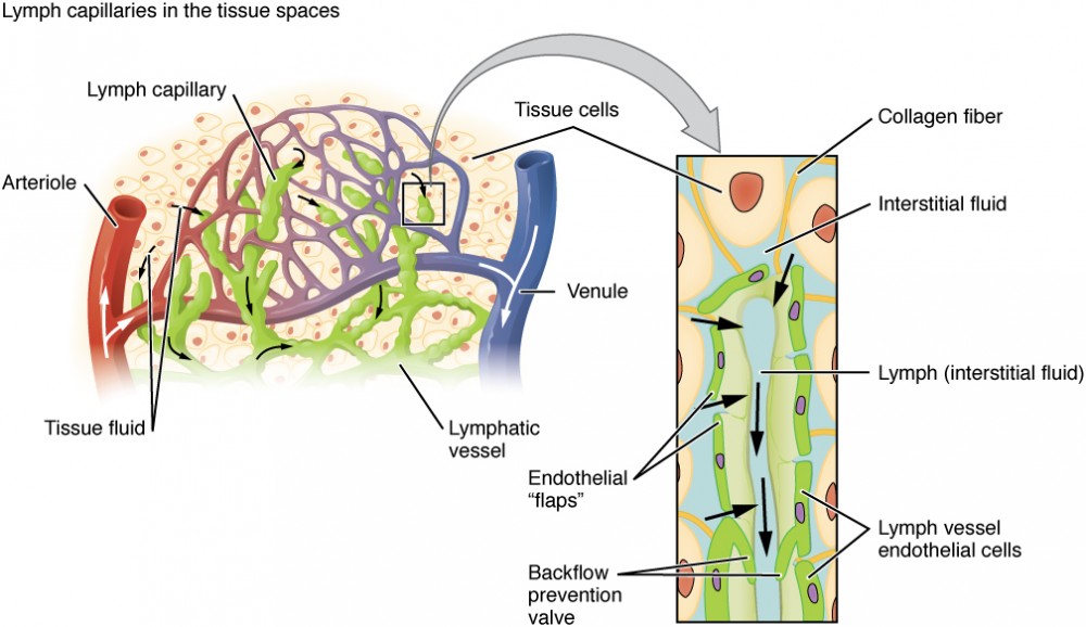
Formation and Functions of Lymph Comprehensive medical notes covering the physiological mechanisms of lymph formation, its chemical composition, circulation dynamics, and clinical relevance. 1. Lymph Formation Lymph is derived from interstitial fluid that has entered lymphatic capillaries. Its formation is a continuous process driven by the dynamics of fluid exchange at blood capillaries and the unique permeability of the lymphatic endothelium. 1.1 Origin: Interstitial Fluid Blood capillaries filter plasma into tissue spaces at the arteriolar end due to hydrostatic pressure exceeding oncotic pressure (Starling forces). Approximately 20 litres of fluid are filtered daily from blood capillaries into tissues. About 17–18 litres are reabsorbed at the venular end where oncotic pressure exceeds hydrostatic pressure. The remaining 2–3 litres constitute the net filtrate that becomes lymph. 1.2 Entry into Lymphatic Capillaries Lymphatic capillaries are blind-ended tubes with highly permeable walls. Their structure is specialized for the uptake of large molecules and fluid: Endothelial cells overlap like shingles; anchoring filaments tether them to surrounding connective tissue. When interstitial pressure rises (due to fluid accumulation or tissue movement), the endothelial junctions open, allowing fluid and particles to enter. Once inside the capillary, the fluid is termed lymph. Particles too large for blood capillaries, such as bacteria, cellular debris, and chylomicrons, can easily enter lymphatic capillaries. 1.3 Factors Promoting Lymph Formation Increased capillary permeability: Caused by inflammation or histamine release. Elevated venous pressure: Seen in heart failure or venous obstruction. Decreased plasma oncotic pressure: Resulting from hypoproteinemia (nephrotic syndrome, liver disease). Increased tissue metabolic activity and vasodilation. 2. Composition of Lymph Lymph closely resembles blood plasma in composition but with lower protein concentration and the addition of cellular elements from tissues. Component Characteristics Clinical Relevance Water and Electrolytes Similar to plasma Reflects plasma status Proteins Lower concentration than plasma (2–3 g/dL); includes albumin, globulins, fibrinogen Protein-losing enteropathies cause hypoproteinemia Lipids Chylomicrons in intestinal lymph (chyle); free fatty acids Milky appearance of chyle after meals; chylothorax if leaked Cells Lymphocytes (predominant), macrophages Reflects immune activity; metastatic cells may be present Cellular Debris Dead cells, bacteria, foreign particles Filtered and phagocytosed in lymph nodes Variations in Lymph Composition: Prenodal lymph: High cellular content, antigens, and debris; low antibody concentration. Postnodal lymph: Lower cellular content; higher antibody concentration due to plasma cell activity in nodes. Intestinal lymph (chyle): Milky appearance due to high lipid content; drains into the cisterna chyli. Liver lymph: High protein content (up to 6 g/dL) due to sinusoidal permeability. 3. Lymph Circulation Lymph flows through a one-way system from peripheral tissues toward the venous angles. Unlike blood, lymph has no central pump and relies on secondary mechanisms for propulsion. 3.1 Pathway of Lymph Flow Tissue spaces → lymphatic capillaries → collecting vessels → lymph nodes → lymphatic trunks → lymphatic ducts → subclavian veins. Lymph passes through multiple lymph nodes, progressively becoming cleaner and more antibody-rich. The entire volume of lymph (2–3 L/day) is returned to the bloodstream within 24 hours. 3.2 Mechanisms of Lymph Propulsion Skeletal muscle pump: Contraction of surrounding muscles compresses lymphatic vessels, propelling lymph forward. Respiratory pump: Changes in intrathoracic pressure during breathing create a suction effect, particularly in the thoracic duct. Intrinsic vessel contractions: Rhythmic contractions of smooth muscle in collecting vessel walls (lymphangion contractions). Valves: Bicuspid valves within collecting vessels prevent backflow, ensuring unidirectional transport. Arterial pulsations: Pulsations of adjacent arteries assist lymph movement in deep vessels. 3.3 Lymphatic Valves Formed by folds of the tunica intima. They are more numerous than venous valves, present every few millimetres. They are critical for preventing retrograde flow, especially in dependent limbs. Valve incompetence contributes to lymphedema and chronic lymphatic insufficiency. 4. Functions of the Lymphatic System The lymphatic system performs four principal functions that are essential for homeostasis and host defense. 4.1 Fluid Homeostasis Returns excess interstitial fluid to the bloodstream, preventing tissue edema. It maintains blood volume by conserving the 2–3 litres of daily capillary filtrate. Failure results in lymphedema, characterized by protein-rich tissue swelling. 4.2 Immune Surveillance and Response Lymph nodes filter lymph and trap foreign antigens, initiating adaptive immune responses. Dendritic cells and macrophages in nodes present antigens to T and B lymphocytes. Memory lymphocytes provide long-term immunity. 4.3 Fat Absorption Specialized lymphatic capillaries in intestinal villi, called lacteals, absorb dietary triglycerides. Lipids are packaged into chylomicrons within enterocytes and extruded into lacteals. Chyle (lipid-rich lymph) drains via intestinal lymphatic trunks to the cisterna chyli and thoracic duct, bypassing the hepatic portal system. Clinical Relevance Damage to intestinal lymphatics causes protein-losing enteropathy and steatorrhea. 4.4 Transport of Cells and Molecules Transports lymphocytes between tissues and lymphoid organs. It carries hormones, enzymes, and metabolic products from tissues to blood and facilitates the dissemination of malignant cells (lymphatic metastasis). 5. Clinical Relevance Lymphedema: Accumulation of protein-rich interstitial fluid due to impaired lymphatic drainage. Primary: Milroy disease (congenital aplasia), lymphedema praecox (puberty onset), lymphedema tarda (adult onset). Secondary: Post-mastectomy axillary node dissection, filariasis (Wuchereria bancrofti), radiation, trauma, recurrent infections. Features: Pitting or non-pitting edema, skin thickening, fibrosis, increased infection risk (cellulitis). Chylothorax and Chylous Ascites: Leakage of chyle into the pleural cavity or peritoneum due to thoracic duct injury (trauma, surgery, malignancy). It presents as milky pleural fluid with elevated triglycerides (>110 mg/dL). Management includes dietary modification (medium-chain triglycerides) or surgical ligation. Protein-Losing Enteropathy: Loss of plasma proteins into the intestinal lumen due to lymphatic obstruction or mucosal disease. Causes include intestinal lymphangiectasia and Crohn disease. Features include hypoalbuminemia, edema, lymphocytopenia, and steatorrhea. 6. Key Points Summary Lymph formation: Formed from excess interstitial fluid (2–3 L/day) entering blind-ended capillaries. Regional Composition: Intestinal lymph (chyle) is lipid-rich; liver lymph is protein-rich. Unidirectional flow: Driven by skeletal muscle/respiratory pumps and intrinsic contractions, assisted by valves. Four principal functions: Fluid homeostasis, immune surveillance, fat absorption, and transport of cells/molecules. Clinical disorders: Include lymphedema, chylothorax, and protein-losing enteropathy.

Structure of Lymphatic system
Anatomy

Structure of Lymphatic system

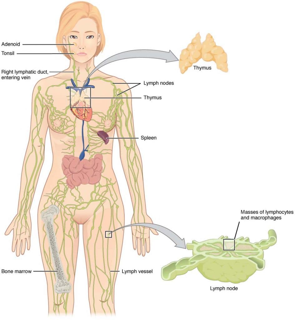
Structure of the Lymphatic System A comprehensive note covering the lymphatic tissues, vessels, and organs. 1. Introduction and Overview The lymphatic system is a network of tissues, vessels, and organs that works in close partnership with the cardiovascular system. It performs essential roles in fluid homeostasis, immune surveillance, and lipid absorption. Unlike the closed circulatory system, the lymphatic system is an open, one-way transport network that returns excess interstitial fluid to the bloodstream. Definition: The lymphatic system comprises lymphatic vessels, lymphoid tissue, lymph nodes, and lymphoid organs that collect and transport lymph—a clear fluid derived from interstitial fluid—back to the venous circulation. Key Components: Lymphatic vessels: Including capillaries, collecting vessels, trunks, and ducts. Lymph nodes: Distributed along vessel pathways for filtration. Lymphoid organs: Classified as primary (bone marrow, thymus) or secondary (spleen, tonsils, MALT). Lymph: The actual fluid transported within the system. Clinical Relevance Understanding lymphatic anatomy is fundamental for interpreting lymphadenopathy, planning cancer staging, assessing lymphedema, and performing safe surgical dissections. 2. Lymphatic Vessels Lymphatic vessels form a hierarchical network that begins as blind-ended capillaries in tissue spaces and progressively converges into larger ducts that drain into the venous system. 2.1 Lymphatic Capillaries Structure: Blind-ended, thin-walled vessels composed of a single layer of overlapping endothelial cells. Mechanism: Endothelial cells are anchored to surrounding connective tissue by fine filaments; increased interstitial pressure opens the junctions to allow fluid entry. Distribution: Present in most tissues except avascular structures (cartilage, epidermis, cornea) and the central nervous system. Specialized Capillaries: Found in the small intestine, these are called lacteals and are responsible for absorbing dietary lipids. 2.2 Collecting Vessels Formed by the union of lymphatic capillaries. Valves: Contain valves (formed by intimal folds) that ensure unidirectional flow toward the heart. Walls: Thin walls with three layers: endothelium, smooth muscle media, and fibrous adventitia. Location: Superficial vessels drain skin and subcutaneous tissue; deep vessels accompany arteries and veins. 2.3 Lymphatic Trunks Formed by the convergence of collecting vessels. There are five principal trunks: Jugular: Drains head and neck. Subclavian: Drains upper limbs. Bronchomediastinal: Drains the thorax. Intestinal: Drains abdominal viscera. Lumbar: Drains lower limbs and pelvis. 2.4 Lymphatic Ducts Thoracic duct: The largest lymphatic vessel (~38–45 cm long). It originates from the cisterna chyli (at L1-L2), ascends through the posterior mediastinum, and drains into the junction of the left subclavian and internal jugular veins. It collects lymph from the entire body except the right upper quadrant. Right lymphatic duct: A short vessel (1–2 cm) that drains into the right venous angle. It collects lymph from the right side of the head and neck, right upper limb, and right thorax. 3. Lymph Nodes Lymph nodes are small, bean-shaped organs (2–10 mm) distributed along lymphatic vessels. They function as filtration stations and sites of immune cell activation. 3.1 Gross Structure Each node is enclosed by a fibrous capsule. Afferent vessels: Penetrate the convex surface. Efferent vessels: Exit at the hilum. An internal framework of trabeculae extends from the capsule, supporting parenchymal tissue. 3.2 Microscopic Architecture Cortex: Outer zone containing lymphoid follicles (B-cell zones). Primary follicles are dense; secondary follicles develop germinal centers after antigen exposure. Paracortex: Deep cortical region rich in T lymphocytes. Contains high endothelial venules (HEVs) that allow lymphocyte entry from blood. Medulla: Innermost zone with medullary cords (plasma cells, B cells, macrophages) and medullary sinuses. Sinuses: Subcapsular, trabecular, and medullary sinuses allow slow lymph flow, facilitating phagocytosis and antigen presentation. 3.3 Regional Lymph Node Groups Region Major Groups Drainage Area Head and Neck Cervical, submandibular, preauricular, parotid Scalp, face, oral cavity, pharynx Upper Limb Axillary, epitrochlear, infraclavicular Breast, upper limb, chest wall Thorax Hilar, mediastinal, paratracheal Lungs, heart, mediastinum Abdomen Mesenteric, para-aortic, celiac GI tract, liver, spleen, kidneys Lower Limb Inguinal, popliteal Lower limb, perineum, external genitalia 4. Lymphoid Organs Lymphoid organs are classified as primary (sites of lymphocyte development) or secondary (sites of immune response activation). 4.1 Primary Lymphoid Organs Bone Marrow: Site of hematopoiesis and B-lymphocyte maturation. Progenitor cells differentiate into mature B cells expressing surface immunoglobulins. Also the source of T-cell progenitors that migrate to the thymus. Thymus: Bilobed organ located in the anterior superior mediastinum. Largest in childhood; undergoes involution after puberty, replaced by adipose tissue. Cortex: Densely packed with immature T lymphocytes (thymocytes). Medulla: Less cellular; contains Hassall corpuscles and mature T cells. Site of T-cell maturation and positive/negative selection (self-tolerance). 4.2 Secondary Lymphoid Organs Spleen: The largest lymphoid organ, located in the left upper quadrant beneath the diaphragm. Functions: Filtration of blood, removal of senescent erythrocytes, immune response to blood-borne pathogens. White pulp: Lymphoid tissue surrounding central arteries; contains periarteriolar lymphoid sheaths (T cells) and lymphoid follicles (B cells). Red pulp: Vascular sinusoids and splenic cords (cords of Billroth); site of blood filtration. Marginal zone: Interface between white and red pulp; rich in macrophages. Tonsils: Aggregates of lymphoid tissue in the oropharynx: palatine, pharyngeal (adenoids), and lingual. Form Waldeyer ring, a first-line defense against inhaled/ingested pathogens. Lack a capsule and have deep crypts lined by epithelium. Mucosa-Associated Lymphoid Tissue (MALT): Diffuse lymphoid tissue in mucosal surfaces (respiratory, GI, GU tracts). Peyer patches: Aggregated lymphoid follicles in the ileum; specialized for sampling intestinal antigens via M cells. Appendix: Contains abundant lymphoid tissue for gut immune surveillance. 5. Clinical Correlations Sentinel Lymph Node Biopsy The sentinel node is the first lymph node to receive drainage from a tumor site. Identification guides cancer staging and surgical planning (e.g., in melanoma or breast cancer). Lymphedema Accumulation of lymph in interstitial spaces due to impaired drainage. Primary: Congenital lymphatic hypoplasia. Secondary: Post-surgical, post-radiation, or caused by filariasis. Splenomegaly Enlargement of the spleen due to infections (malaria, mononucleosis), hematologic disorders, or portal hypertension. May cause hypersplenism with cytopenias. 6. Key Points Summary The lymphatic system is an open, one-way network returning interstitial fluid to venous circulation. Lymphatic capillaries are blind-ended; collecting vessels contain valves. The thoracic duct drains most of the body; the right lymphatic duct drains the right upper quadrant. Lymph nodes filter lymph and activate immune responses via B-cell follicles

Scroll to Top