
On this page
Overview
The integumentary system — skin and its appendages (hair, nails, sweat and sebaceous glands) — is the body's largest organ system. Skin alone covers 1.5–2.0 m² in adults and makes up about 15% of body weight. It is simultaneously a physical barrier, a sensory organ, a thermoregulator, an immune sentinel and a visible window onto systemic disease.
Anchor every layer in three questions: What is it made of? What does it do? Which disease results when it fails?
Learning objectives
- Describe the organisation of the epidermis, dermis and hypodermis.
- Name the layers of the epidermis from deep to superficial and the specialised cell of each layer.
- Compare the papillary and reticular dermis and the receptors within them.
- Explain the structure of the dermo-epidermal junction and its clinical importance.
- Relate each layer to common dermatological and systemic disease.
Introduction
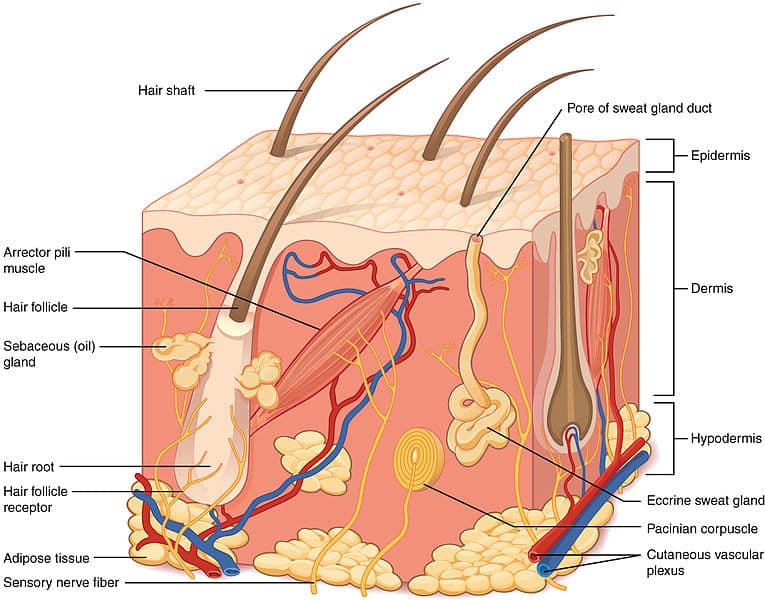
Skin has two principal layers: the epidermis (superficial epithelial layer) and the dermis (deeper connective-tissue layer). Beneath both lies the hypodermis (subcutaneous tissue), which is not strictly part of the skin but is functionally integrated with it. The skin's jobs follow from this architecture: the epidermis keeps the outside out; the dermis nourishes, senses and cushions; the hypodermis insulates and anchors.
The epidermis
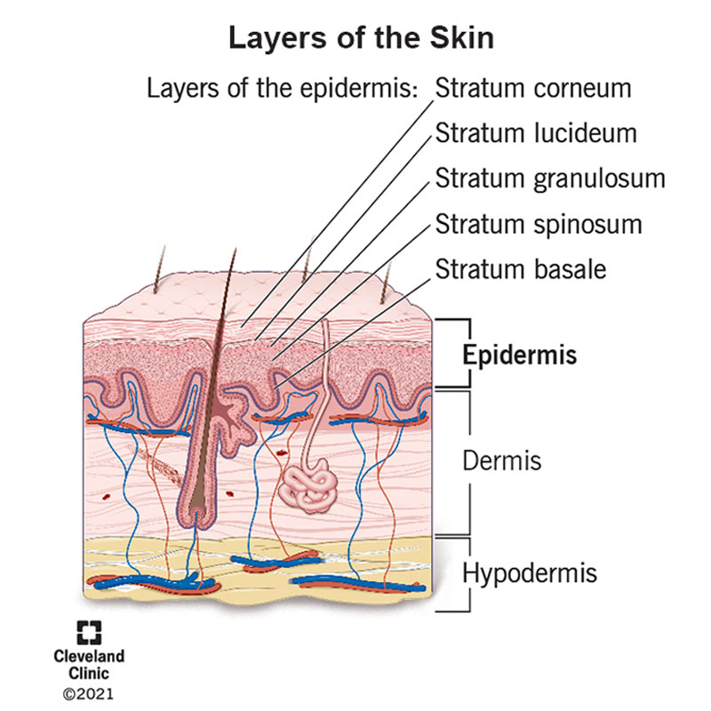
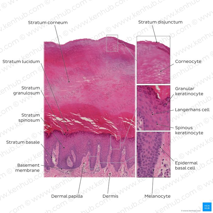
The epidermis is stratified squamous keratinised epithelium. It is avascular — nourished entirely by diffusion from dermal capillaries — and is 0.05 mm in thin skin and up to 1.5 mm in thick skin (palms and soles). Basal cells divide continuously; full epidermal turnover takes about 28–40 days.
From superficial to deep, five layers — "Come, Let's Get Sun Burned":
Stratum corneum (horny layer)
- 15–30 layers of flattened, anucleate, keratin-filled corneocytes in a lipid-rich matrix.
- The main barrier against water loss, pathogens and toxins; sheds by desquamation.
Stratum lucidum (clear layer)
- Only in thick skin (palms, soles); 3–5 layers of dead keratinocytes containing translucent eleidin.
Stratum granulosum (granular layer)
- 3–5 layers of keratinocytes with keratohyalin granules; lamellar (Odland) bodies release lipids that form the waterproof barrier.
- Cells undergo apoptosis here — the last living stage of the keratinocyte.
Stratum spinosum (prickle cell layer)
- 8–10 layers of polyhedral keratinocytes joined by desmosomes (the "spines" of microscopy are shrinkage artefact).
- Houses Langerhans cells — the epidermis's immune sentinels.
Stratum basale (basal layer)
- A single layer of mitotic stem cells on the basement membrane, anchored by hemidesmosomes.
- Contains melanocytes (pigment) and Merkel cells (touch).
Cell types of the epidermis
- Keratinocytes (~90%): make keratin; the structural bricks of the epidermis.
- Melanocytes (5–10%): synthesise melanin from tyrosine and transfer it to keratinocytes by cytocrine secretion; melanin caps nuclei and shields DNA from UV. All skin tones have similar melanocyte numbers — pigmentation reflects melanin production, not cell number.
- Langerhans cells (3–5%): dendritic antigen-presenting cells; take up skin antigens and migrate to lymph nodes — central to contact dermatitis and skin immunity.
- Merkel cells (<1%): slow-adapting mechanoreceptors for fine touch and pressure, clustered in fingertips.
The dermis
The dermis (1–4 mm) is connective tissue that feeds, senses and mechanically supports the epidermis. It has two zones:
Papillary dermis
- Superficial loose areolar tissue with fine type III collagen; forms dermal papillae interdigitating with epidermal rete ridges.
- Houses capillary loops, free nerve endings and Meissner corpuscles (light touch).
Reticular dermis
- Deeper, ~80% of dermal thickness; dense irregular connective tissue with thick type I collagen bundles and elastic fibres.
- Contains the larger vessels, skin appendages, Pacinian corpuscles (deep pressure/vibration) and Ruffini endings (stretch).
Components of the dermis
- Collagen (70% of dermal dry weight): tensile strength; made by fibroblasts.
- Elastic fibres: recoil after stretch; deficient in cutis laxa.
- Ground substance: hyaluronic acid and dermatan sulfate hold water and give the dermis its turgor.
- Mast cells and macrophages: histamine release and phagocytosis in inflammation and allergy.
The hypodermis
The hypodermis (subcutaneous tissue) of adipocytes in fibrous lobules anchors skin to deep fascia. Its functions: energy storage; thermal insulation; mechanical cushioning; anchorage; and endocrine activity (adipose secretes leptin, adiponectin and inflammatory mediators). Its thickness varies from millimetres to centimetres by site, sex and nutrition — and it is the practical site for subcutaneous injections (insulin, heparin, vaccines), where slow absorption from the fat depot smooths drug levels.
The dermo-epidermal junction
The basement membrane zone glues epidermis to dermis in three layers:
- Lamina lucida: laminin-332 and integrins attach basal keratinocyte hemidesmosomes.
- Lamina densa: type IV collagen, laminin and nidogen form the sheet.
- Sublamina densa: type VII collagen anchoring fibrils lock the membrane to dermal collagen.
Clinical relevance
Scroll the table sideways on a smaller screen.
| Condition | Pathophysiological basis | Layer |
|---|---|---|
| Psoriasis | Accelerated turnover (3–5 days vs 28–40) piles immature keratinocytes into scaly plaques | Epidermis |
| Bullous pemphigoid / epidermolysis bullosa | Junctional splitting — autoantibodies or collagen VII/keratin defects | Dermo-epidermal junction |
| Scleroderma | Excess dermal collagen deposition hardens and tightens skin | Dermis |
| Lipodystrophy | Loss of subcutaneous fat with metabolic disturbance | Hypodermis |
| Pressure (decubitus) ulcers | Prolonged pressure ischemia destroys junction and deeper tissue — grade by deepest layer involved | All layers |
| Malignant melanoma | Malignant transformation of melanocytes; prognosis set by Breslow depth of dermal invasion | Epidermis → dermis |
Drug summary table
Scroll the table sideways on a smaller screen.
| Class / drug | Use | Key cautions |
|---|---|---|
| Emollients (petrolatum, aqueous cream, glycerin) | Dry skin, eczema maintenance; apply liberally and often | Fragrance-free for sensitive skin; apply after bathing |
| Topical corticosteroids (hydrocortisone 1% for face/folds; betamethasone or clobetasone for body) | Eczema, psoriasis, contact dermatitis flare | Weakest effective potency; avoid prolonged use (skin atrophy, striae); never on untreated infection; limit on face/groin |
| Topical antifungals (clotrimazole cream 1–2% BD; miconazole) | Tinea, candidiasis, pityriasis versicolor | Continue 1–2 weeks after clearing; oral agents (fluconazole/itraconazole) for extensive disease |
| Mupirocin 2% | Localised bacterial infection (impetigo); also nasal decolonisation of MRSA | Short courses; resistance emerges with prolonged use |
| Benzoyl peroxide | Mild–moderate acne; antibacterial and keratolytic | Bleaches fabric; start low strength to limit irritation |
| Calamine lotion | Itch in chickenpox, insect bites, mild dermatitis | Symptomatic only; dries weeping lesions |
Clinical scenario
Scenario 1 — Itchy plaques in the flexures
A 34-year-old farmer has itchy, well-demarcated silvery-scaled plaques on the elbows and knees, some now appearing at a scar site (Koebner phenomenon). The history of rapid turnover and the silvery scale point to psoriasis. Management: emollients, a potent topical steroid (e.g., betamethasone) on the body plaques short-term, vitamin D analogues (calcipotriol) as maintenance, and tar or salicylic acid for thick scale; refer extensive disease for phototherapy or systemic agents. You explain the disease as "epidermal cells dividing four times too fast" — the layer-based logic makes the treatment make sense.
Scenario 2 — Blisters in an elderly patient
An 80-year-old has tense bullae on flexor surfaces and groin, with intense itching but little mucosal involvement. Biopsy with immunofluorescence shows subepidermal blistering with basement-membrane antibodies — bullous pemphigoid, an autoantibody attack on the hemidesmosomes you revised in the junction section. Treat with superpotent topical corticosteroids or oral prednisolone under specialist guidance, check for triggers (new drugs, neurological disease association), and nurse the skin gently to prevent secondary infection. Contrast with pemphigus vulgaris: flaccid blisters, mucosal disease, and antibodies against desmoglein between keratinocytes.
Key points
References
- Junqueira's Basic Histology — integument (Library reference.)
- Rook's Textbook of Dermatology — structure and function of skin (Library reference.)
- Goodman & Gilman's The Pharmacological Basis of Therapeutics — topical and percutaneous drug delivery (Library reference.)
- British National Formulary — topical corticosteroid and antifungal guidance (Library reference.)
Further reading
For deeper study, compare this lesson with current guidance and standard references from recognised medical and public-health organisations.
For education only. Apply current Uganda Clinical Guidelines, local protocols and specialist advice in clinical practice.