
On this page
1. Overview
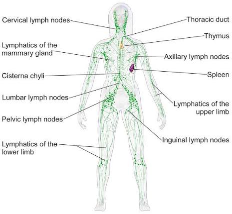
The lymphatic system is an open, one-way drainage network that partners with the cardiovascular system to return interstitial fluid to the blood. Its three jobs are fluid homeostasis (~10% of the fluid leaving capillaries returns as lymph, 4 L/day), immune surveillance, and lipid absorption (dietary fat via intestinal lacteals).
- Lymph: clear fluid derived from interstitial fluid — water, electrolytes, proteins, lipids (chylomicrons from the gut), lymphocytes and antigen-presenting cells.
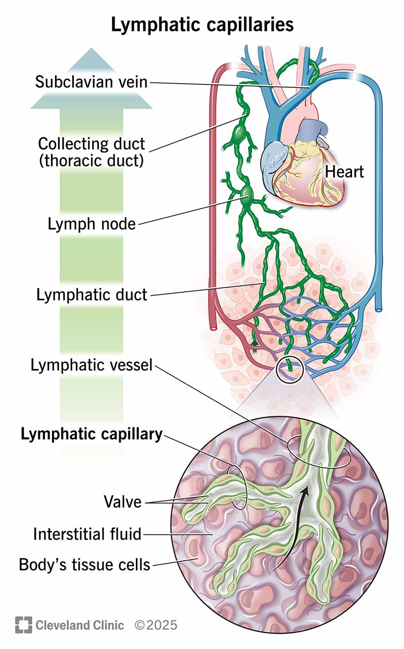
- Components: lymphatic capillaries → collecting vessels (with valves) → trunks → two ducts; lymph nodes along the vessels; lymphoid organs.
2. Lymphatic vessels
2.1 Lymphatic capillaries
- Structure: blind-ended, thin-walled tubes of a single endothelial layer with overlapping endothelial cells and anchoring filaments tethered to surrounding connective tissue.
- One-way inlet valve: rising interstitial pressure opens the cell junctions (fluid and particles enter); falling pressure seals them shut — lymph cannot leak back out.
- Absent from avascular tissues (epidermis, cartilage, cornea) and the CNS, which drains via the glymphatic/CSF route.
- Lacteals are specialised intestinal capillaries that absorb chylomicrons — giving post-prandial (milky) chyle.
2.2 Collecting vessels and trunks
- Capillaries unite into valved collecting vessels whose smooth-muscle walls and valves (intimal folds) guarantee one-way flow; lymph is propelled by skeletal-muscle pumping, respiration and intrinsic vessel contractions.
- Convergence forms five trunks: jugular (head/neck), subclavian (upper limb), bronchomediastinal (thorax), intestinal (abdominal viscera) and lumbar (lower limbs, pelvis).
2.3 The two ducts
Scroll the table sideways on a smaller screen.
| Duct | Form / origin | Drains | Terminates |
|---|---|---|---|
| Thoracic duct | Largest lymphatic vessel (~38–45 cm); begins at the cisterna chyli (L1–L2), right of the aorta, ascends through the aortic hiatus and posterior mediastinum, crosses to the left at T4–T5 | Entire body except the right arm, right side of the thorax, right side of head and neck (and the right lung/heart partially) | Junction of the left subclavian and left internal jugular veins (left venous angle) |
| Right lymphatic duct | Short (1–2 cm), formed by the right jugular, subclavian and bronchomediastinal trunks | Right upper quadrant: right head/neck, right upper limb, right thorax | Right venous angle |
3. Lymph nodes
Bean-shaped filters (2–10 mm, up to 2 cm) scattered along the collecting vessels — hundreds in the body, concentrated in the neck, axilla, groin, mediastinum and mesentery.
3.1 Structure (inside → out)
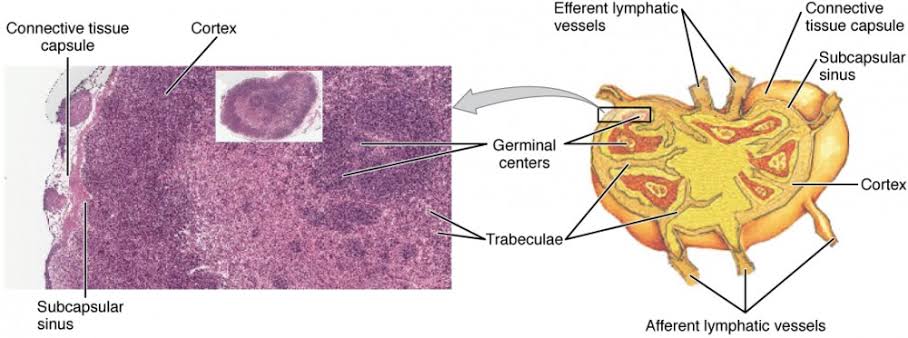
- Capsule of dense connective tissue with trabeculae extending inward; subcapsular sinus just beneath.
- Afferent lymphatics enter the convex surface; efferents leave at the hilum (where arteries, veins and nerves also pass).
- Cortex: lymphoid follicles — B-cell zones. Primary follicles are resting; antigen exposure creates germinal centres (secondary follicles) where B cells proliferate, undergo somatic hypermutation and class-switch — the dark zone of proliferating blasts, light zone of selection.
- Paracortex: T-cell-rich deep cortex with high endothelial venules (HEVs) — cuboidal endothelial "gates" through which lymphocytes continuously enter from blood.
- Medulla: medullary cords (plasma cells, B cells, macrophages) and medullary sinuses draining toward the efferent vessel.
- Lymph percolates slowly through the sinuses, maximising contact with macrophages and dendritic cells — pathogens and tumour cells are trapped here.
3.2 Regional groups and drainage
Scroll the table sideways on a smaller screen.
| Region | Major groups | Drainage territory |
|---|---|---|
| Head and neck | Occipital, pre/post-auricular, parotid, submandibular, submental, superficial and deep cervical chains | Scalp, face, oral cavity, pharynx, larynx, thyroid |
| Upper limb / chest wall | Epitrochlear, axillary (pectoral, subscapular, apical), infraclavicular | Hand, forearm, arm, breast (lateral), chest wall |
| Thorax | Paratracheal, tracheobronchial, hilar, mediastinal | Lungs, trachea, oesophagus, heart (partial) |
| Abdomen | Celiac, superior/inferior mesenteric, para-aortic | Stomach, liver, spleen, pancreas, gut, kidneys, gonads |
| Lower limb | Popliteal, superficial and deep inguinal | Leg, thigh, perineum, external genitalia |
4. Lymphoid organs
Primary organs generate lymphocytes; secondary organs house the immune response to antigen.
4.1 Primary — bone marrow and thymus
- Bone marrow: all blood cells are born here (haematopoiesis); B cells mature in the marrow itself; T-cell progenitors emigrate to the thymus.
- Thymus: bilobed organ in the anterior superior mediastinum, largest in childhood, involutes after puberty (replaced by fat). Cortex packed with immature thymocytes; medulla with Hassall corpuscles (concentrically keratinised epithelial cells unique to the thymus). Site of positive selection (MHC-restricted survival) and negative selection (deletion of self-reactive clones) → central tolerance.
4.2 Secondary — spleen, tonsils, MALT
- Spleen (largest lymphoid organ, left hypochondrium): filters blood, not lymph. White pulp — periarteriolar lymphoid sheaths (T cells) and follicles (B cells); marginal zone — macrophage-rich interface sampling blood-borne antigen; red pulp — splenic cords (cords of Billroth) and sinusoids where old/damaged red cells and intra-erythrocytic parasites (e.g., Plasmodium) are cleared.
- Tonsils (palatine, pharyngeal/adenoids, lingual): form Waldeyer's ring guarding the oro/nasopharynx; uncapsulated with deep crypts lined by reticulated epithelium that traps antigen.
- MALT (mucosa-associated lymphoid tissue): diffuse lymphoid aggregates in the respiratory, gut and genitourinary mucosa. Peyer patches in the ileum sample intestinal antigen through specialised M cells; the appendix is a major MALT site.
5. Clinical correlations
5.1 Lymphadenopathy — a structured approach
- Localised vs generalised — generalised (>2 non-contiguous regions) suggests systemic disease: HIV, TB, lymphoma, leukaemia, SLE, sarcoidosis, mononucleosis.
- Character: size, tenderness (inflammation), consistency (rubbery = lymphoma; hard/fixed = malignancy; matted = TB), warmth, overlying skin.
- Territory: match the node group to its drainage area (e.g., left supraclavicular = Virchow's node — abdominal/ thoracic malignancy; axillary with arm infection).
- Red flags: painless progressive enlargement, hard fixed nodes, night sweats, weight loss, fever — biopsy.
5.2 Infections of the lymphatic system
- Lymphangitis: red streaking along a limb toward regional nodes with fever — treat the source (usually Streptococcus; penicillin/flucloxacillin).
- Lymphadenitis: tender regional nodes draining an infected area.
- Lymphatic filariasis (Wuchereria bancrofti): mosquito-borne; adult worms in lymphatics cause lymphedema and elephantiasis — a major cause in Uganda. Diagnosis by night blood smear (microfilariae) or antigen testing; treatment diethylcarbamazine (DEC) 6 mg/kg/day × 12 days (or single-dose ivermectin + albendazole per MDA protocols).
- TB lymphadenitis (scrofula): painless matted cervical nodes that may suppurate and scar; biopsy for AFB/culture.
5.3 Cancer staging
The sentinel node — the first node draining a tumour — is identified (blue dye/radioisotope) and biopsied in melanoma and breast cancer to guide axillary clearance. Nodal status (e.g., TNM "N") determines stage and treatment in most solid tumours; lymphatic invasion also seeds distant nodes — e.g., gastric cancer to left supraclavicular nodes (Virchow/Troisier sign).
5.4 Lymphedema
- Primary: congenital hypoplasia of lymphatics (Milroy disease).
- Secondary: lymphatic destruction — filariasis, surgery/radiation (post-mastectomy arm swelling), tumour infiltration, recurrent cellulitis.
- Management: skin care, compression, elevation, physiotherapy, treating the cause; antibiotics promptly for cellulitis — repeated infection progressively destroys residual lymphatics.
5.5 Splenic conditions
- Splenomegaly in Uganda commonly reflects malaria, schistosomal portal hypertension, leukaemia/lymphoma, TB, EBV; massive spleen think chronic myeloid leukaemia, myelofibrosis, visceral leishmaniasis.
- Hypersplenism: enlarged spleen plus cytopenias (anaemia, leukopenia, thrombocytopenia) with compensatory marrow hyperplasia.
- Hyposplenism/post-splenectomy: loss of filtering → risk of overwhelming infection by encapsulated organisms (S. pneumoniae, H. influenzae, N. meningitidis) — vaccinate and educate patients (septicemia can be fulminant within hours; carry alert information).
6. Key points
References
- Standring, Gray's Anatomy — lymphatic system chapter. (Library reference.)
- Abbas, Cellular and Molecular Immunology — lymphoid organs and tissues. (Library reference.)
- WHO — lymphatic filariasis fact sheet and management guidelines. (Library reference.)
- Uganda Clinical Guidelines, Ministry of Health. (Library reference.)
For education only. Apply current Uganda Clinical Guidelines, local protocols and specialist advice in clinical practice.
Best ever