Formation and Functions of Lymph
Comprehensive medical notes covering the physiological mechanisms of lymph formation, its chemical composition, circulation dynamics, and clinical relevance.
1. Lymph Formation
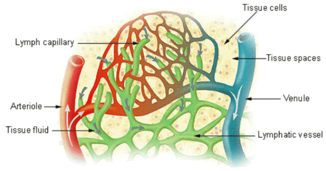
Lymph is derived from interstitial fluid that has entered lymphatic capillaries. Its formation is a continuous process driven by the dynamics of fluid exchange at blood capillaries and the unique permeability of the lymphatic endothelium.
1.1 Origin: Interstitial Fluid
Blood capillaries filter plasma into tissue spaces at the arteriolar end due to hydrostatic pressure exceeding oncotic pressure (Starling forces).
- Approximately 20 litres of fluid are filtered daily from blood capillaries into tissues.
- About 17–18 litres are reabsorbed at the venular end where oncotic pressure exceeds hydrostatic pressure.
- The remaining 2–3 litres constitute the net filtrate that becomes lymph.
1.2 Entry into Lymphatic Capillaries
Lymphatic capillaries are blind-ended tubes with highly permeable walls. Their structure is specialized for the uptake of large molecules and fluid:
- Endothelial cells overlap like shingles; anchoring filaments tether them to surrounding connective tissue.
- When interstitial pressure rises (due to fluid accumulation or tissue movement), the endothelial junctions open, allowing fluid and particles to enter.
- Once inside the capillary, the fluid is termed lymph.
- Particles too large for blood capillaries, such as bacteria, cellular debris, and chylomicrons, can easily enter lymphatic capillaries.
1.3 Factors Promoting Lymph Formation
- Increased capillary permeability: Caused by inflammation or histamine release.
- Elevated venous pressure: Seen in heart failure or venous obstruction.
- Decreased plasma oncotic pressure: Resulting from hypoproteinemia (nephrotic syndrome, liver disease).
- Increased tissue metabolic activity and vasodilation.
2. Composition of Lymph
Lymph closely resembles blood plasma in composition but with lower protein concentration and the addition of cellular elements from tissues.
| Component | Characteristics | Clinical Relevance |
|---|---|---|
| Water and Electrolytes | Similar to plasma | Reflects plasma status |
| Proteins | Lower concentration than plasma (2–3 g/dL); includes albumin, globulins, fibrinogen | Protein-losing enteropathies cause hypoproteinemia |
| Lipids | Chylomicrons in intestinal lymph (chyle); free fatty acids | Milky appearance of chyle after meals; chylothorax if leaked |
| Cells | Lymphocytes (predominant), macrophages | Reflects immune activity; metastatic cells may be present |
| Cellular Debris | Dead cells, bacteria, foreign particles | Filtered and phagocytosed in lymph nodes |
Variations in Lymph Composition:
- Prenodal lymph: High cellular content, antigens, and debris; low antibody concentration.
- Postnodal lymph: Lower cellular content; higher antibody concentration due to plasma cell activity in nodes.
- Intestinal lymph (chyle): Milky appearance due to high lipid content; drains into the cisterna chyli.
- Liver lymph: High protein content (up to 6 g/dL) due to sinusoidal permeability.
3. Lymph Circulation
Lymph flows through a one-way system from peripheral tissues toward the venous angles. Unlike blood, lymph has no central pump and relies on secondary mechanisms for propulsion.
3.1 Pathway of Lymph Flow
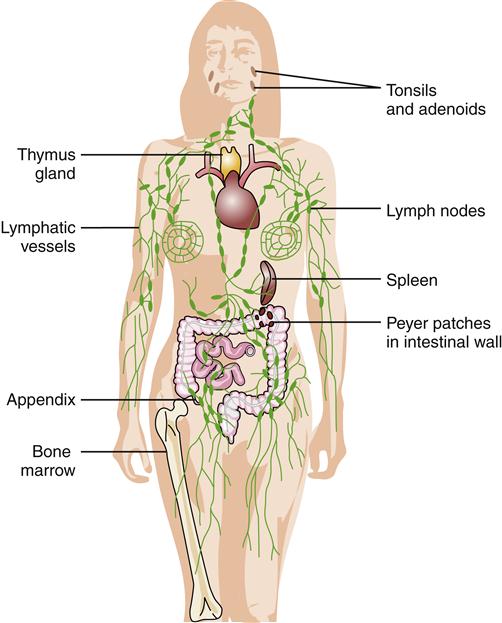
Tissue spaces → lymphatic capillaries → collecting vessels → lymph nodes → lymphatic trunks → lymphatic ducts → subclavian veins.
Lymph passes through multiple lymph nodes, progressively becoming cleaner and more antibody-rich. The entire volume of lymph (2–3 L/day) is returned to the bloodstream within 24 hours.
3.2 Mechanisms of Lymph Propulsion
- Skeletal muscle pump: Contraction of surrounding muscles compresses lymphatic vessels, propelling lymph forward.
- Respiratory pump: Changes in intrathoracic pressure during breathing create a suction effect, particularly in the thoracic duct.
- Intrinsic vessel contractions: Rhythmic contractions of smooth muscle in collecting vessel walls (lymphangion contractions).
- Valves: Bicuspid valves within collecting vessels prevent backflow, ensuring unidirectional transport.
- Arterial pulsations: Pulsations of adjacent arteries assist lymph movement in deep vessels.
3.3 Lymphatic Valves
Formed by folds of the tunica intima. They are more numerous than venous valves, present every few millimetres. They are critical for preventing retrograde flow, especially in dependent limbs. Valve incompetence contributes to lymphedema and chronic lymphatic insufficiency.
4. Functions of the Lymphatic System
The lymphatic system performs four principal functions that are essential for homeostasis and host defense.
4.1 Fluid Homeostasis
Returns excess interstitial fluid to the bloodstream, preventing tissue edema. It maintains blood volume by conserving the 2–3 litres of daily capillary filtrate. Failure results in lymphedema, characterized by protein-rich tissue swelling.
4.2 Immune Surveillance and Response
Lymph nodes filter lymph and trap foreign antigens, initiating adaptive immune responses. Dendritic cells and macrophages in nodes present antigens to T and B lymphocytes. Memory lymphocytes provide long-term immunity.
4.3 Fat Absorption
Specialized lymphatic capillaries in intestinal villi, called lacteals, absorb dietary triglycerides. Lipids are packaged into chylomicrons within enterocytes and extruded into lacteals. Chyle (lipid-rich lymph) drains via intestinal lymphatic trunks to the cisterna chyli and thoracic duct, bypassing the hepatic portal system.
Damage to intestinal lymphatics causes protein-losing enteropathy and steatorrhea.
4.4 Transport of Cells and Molecules
Transports lymphocytes between tissues and lymphoid organs. It carries hormones, enzymes, and metabolic products from tissues to blood and facilitates the dissemination of malignant cells (lymphatic metastasis).
5. Clinical Relevance
Lymphedema:
Accumulation of protein-rich interstitial fluid due to impaired lymphatic drainage.
- Primary: Milroy disease (congenital aplasia), lymphedema praecox (puberty onset), lymphedema tarda (adult onset).
- Secondary: Post-mastectomy axillary node dissection, filariasis (Wuchereria bancrofti), radiation, trauma, recurrent infections.
- Features: Pitting or non-pitting edema, skin thickening, fibrosis, increased infection risk (cellulitis).
Chylothorax and Chylous Ascites:
Leakage of chyle into the pleural cavity or peritoneum due to thoracic duct injury (trauma, surgery, malignancy). It presents as milky pleural fluid with elevated triglycerides (>110 mg/dL). Management includes dietary modification (medium-chain triglycerides) or surgical ligation.
Protein-Losing Enteropathy:
Loss of plasma proteins into the intestinal lumen due to lymphatic obstruction or mucosal disease. Causes include intestinal lymphangiectasia and Crohn disease. Features include hypoalbuminemia, edema, lymphocytopenia, and steatorrhea.
6. Key Points Summary
- Lymph formation: Formed from excess interstitial fluid (2–3 L/day) entering blind-ended capillaries.
- Regional Composition: Intestinal lymph (chyle) is lipid-rich; liver lymph is protein-rich.
- Unidirectional flow: Driven by skeletal muscle/respiratory pumps and intrinsic contractions, assisted by valves.
- Four principal functions: Fluid homeostasis, immune surveillance, fat absorption, and transport of cells/molecules.
- Clinical disorders: Include lymphedema, chylothorax, and protein-losing enteropathy.
