Mechanism of Breathing
Complete notes on pulmonary ventilation, including respiratory musculature, pressure-volume dynamics, lung capacities, compliance, and clinical correlations.
1. Introduction
Breathing (pulmonary ventilation) is the physical process of moving air into and out of the lungs. It is fundamentally driven by pressure gradients created by changes in thoracic volume. This complex mechanism involves the coordinated action of the respiratory muscles, the elastic properties of the lungs and chest wall, and the patency of the airways.
2. Respiratory Muscles
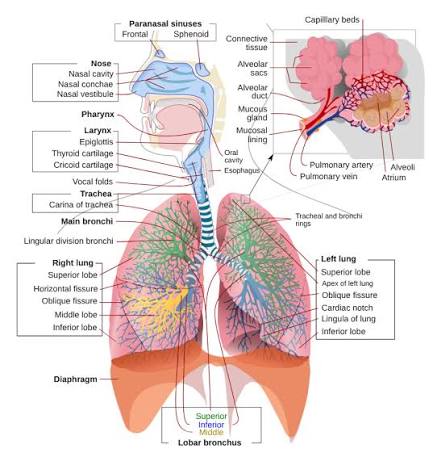
The movement of the thoracic cage and the subsequent change in lung volume are mediated by two sets of muscles categorized by their role in the breathing cycle.
2.1 Primary Muscles of Inspiration
- Diaphragm: The principal muscle of inspiration. A dome-shaped sheet of skeletal muscle separating the thoracic and abdominal cavities. When it contracts, the dome flattens, increasing the vertical diameter of the thorax and creating negative intrapleural pressure. It accounts for approximately 75% of tidal volume during quiet breathing.
- External Intercostal Muscles: These muscles elevate the ribs during inspiration, increasing the anteroposterior and lateral diameters of the thorax. This rib movement is often described as the pump-handle and bucket-handle motion.
2.2 Accessory Muscles of Inspiration
Used primarily during forced inspiration or when airway resistance is pathologically increased:
- Sternocleidomastoid: Elevates the sternum.
- Scalene muscles: Elevate the first two ribs.
- Serratus anterior and Pectoralis minor: Elevate the ribs when the shoulder girdle is fixed.
- Erector spinae: Extends the spine, further increasing thoracic volume.
2.3 Muscles of Expiration
- Quiet expiration: This is a passive process driven by the elastic recoil of the lungs and chest wall. No active muscle contraction is required.
- Forced expiration (active):
— Internal intercostal muscles: Depress the ribs, decreasing thoracic volume.
— Abdominal muscles (rectus abdominis, obliques, transversus): Compress the abdomen, pushing the diaphragm upward.
— Latissimus dorsi: Depresses the ribs.
3. Mechanics of Inspiration
3.1 Quiet Inspiration
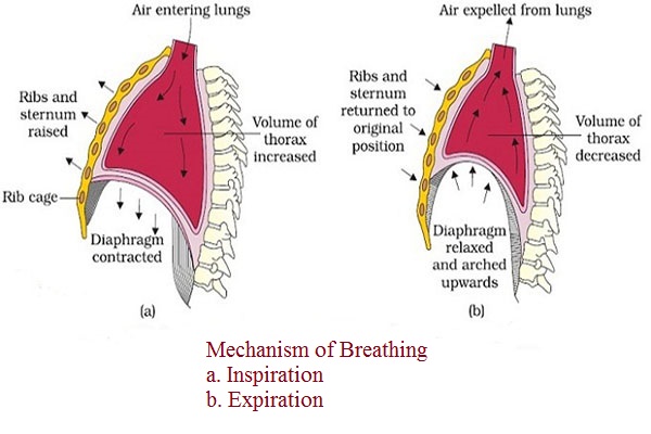
Initiated by the contraction of the diaphragm and external intercostals. The sequence of events is as follows:
- Diaphragm contraction flattens the dome, increasing the vertical thoracic diameter by 1.5 cm (up to 7 cm during deep inspiration).
- External intercostals elevate the ribs, increasing horizontal diameters.
- Thoracic volume increases → intrapleural pressure becomes more negative (dropping from -5 cmH₂O to -8 cmH₂O).
- Alveolar pressure drops below atmospheric pressure (to -1 cmH₂O), creating a pressure gradient that draws air into the lungs.
- Airflow continues until alveolar pressure equalizes with atmospheric pressure.
3.2 Forced Inspiration
Involves the accessory muscles in addition to the primary muscles. This results in a significantly greater increase in thoracic volume and a more negative intrapleural pressure, generating larger pressure gradients and greater airflow.
4. Mechanics of Expiration
4.1 Quiet Expiration
A passive process driven by the elastic recoil of the lungs and the relaxation of inspiratory muscles. The sequence includes:
- The diaphragm relaxes and returns to its dome shape; external intercostals relax, allowing ribs to descend.
- Thoracic volume decreases → intrapleural pressure becomes less negative.
- Alveolar pressure rises above atmospheric pressure (to +1 cmH₂O), creating a pressure gradient that drives air out.
- Airflow continues until alveolar pressure equalizes with atmospheric pressure.
4.2 Forced Expiration
An active process requiring the contraction of expiratory muscles. Internal intercostals depress the ribs while abdominal muscles compress the viscera to push the diaphragm upward. Intrapleural pressure becomes positive (reaching +20 to +30 cmH₂O during coughing). This rapid increase in alveolar pressure forces air out at high velocity, essential for coughing, sneezing, and vocalization.
5. Pressure Changes During Breathing
| Pressure | At Rest (End-Expiration) | During Inspiration | During Expiration |
|---|---|---|---|
| Atmospheric pressure | 0 cmH₂O | 0 cmH₂O | 0 cmH₂O |
| Intrapleural pressure | -5 cmH₂O | -8 cmH₂O | -3 cmH₂O |
| Alveolar pressure | 0 cmH₂O | -1 cmH₂O | +1 cmH₂O |
| Transpulmonary pressure | +5 cmH₂O | +8 cmH₂O | +3 cmH₂O |
- Intrapleural pressure: Pressure in the pleural space; always subatmospheric (negative) in healthy individuals, preventing lung collapse.
- Alveolar (intrapulmonary) pressure: Pressure within the alveoli; fluctuates above and below atmospheric pressure during the cycle.
- Transpulmonary pressure: The difference between alveolar and intrapleural pressure (Palv - Pip); it maintains alveolar expansion.
- Transmural pressure: Pressure across the airway wall; keeps airways open during inspiration.
6. Lung Volumes and Capacities
6.1 Static Lung Volumes
- Tidal volume (TV): Volume of air inhaled or exhaled during quiet breathing; 500 mL in healthy adults.
- Inspiratory reserve volume (IRV): Additional air that can be forcibly inhaled after a normal inspiration; 2500–3000 mL.
- Expiratory reserve volume (ERV): Additional air that can be forcibly exhaled after a normal expiration; 1000–1200 mL.
- Residual volume (RV): Air remaining in the lungs after maximal exhalation; 1000–1200 mL. It prevents alveolar collapse.
6.2 Lung Capacities
- Inspiratory capacity (IC): TV + IRV (~3000 mL).
- Functional residual capacity (FRC): ERV + RV (~2300 mL). Important for gas exchange continuity.
- Vital capacity (VC): IRV + TV + ERV; the maximum air that can be exhaled after maximal inspiration (~4500–5000 mL).
- Total lung capacity (TLC): VC + RV; total air in lungs at maximal inspiration (~5500–6000 mL).
6.3 Dynamic Volumes
- Forced vital capacity (FVC): Total volume forcibly exhaled after maximal inspiration.
- Forced expiratory volume in 1 second (FEV₁): Volume exhaled in the first second of the FVC maneuver.
- FEV₁/FVC ratio: Normally >0.70 (or 70%). Reduced in obstructive disease (asthma, COPD); normal or increased in restrictive disease.
- Peak expiratory flow (PEF): Maximum flow rate during forced expiration; measured by peak flow meter.
7. Compliance and Elastic Recoil
7.1 Lung Compliance
The distensibility of the lungs; defined as the change in lung volume per unit change in transpulmonary pressure. Normal value: ~200 mL/cmH₂O.
- Increased compliance: Emphysema (destruction of elastic tissue).
- Decreased compliance: Pulmonary fibrosis, ARDS, pulmonary edema (stiff lungs).
7.2 Elastic Recoil
The tendency of the lungs to collapse inward due to elastic tissue and surface tension. Opposed by the outward recoil of the chest wall. At FRC, these opposing forces are balanced, and no airflow occurs.
7.3 Surface Tension and Surfactant
Surface tension at the air-liquid interface in alveoli tends to collapse them (Laplace law: pressure = 2 × tension / radius).
Surfactant, produced by Type II pneumocytes, is a phospholipid-protein complex that reduces surface tension. Its functions include:
- Prevents alveolar collapse.
- Reduces the work of inspiration.
- Stabilizes alveoli of different sizes.
- Prevents pulmonary edema by reducing fluid transudation.
Neonatal Respiratory Distress Syndrome
Surfactant deficiency in premature infants causes atelectasis and respiratory failure.
8. Airway Resistance
Resistance to airflow is determined primarily by airway radius (Poiseuille law: resistance ∝ 1/radius⁴). Bronchial smooth muscle tone regulates airway caliber:
- Bronchodilation: Sympathetic stimulation (β₂-adrenergic receptors), epinephrine.
- Bronchoconstriction: Parasympathetic stimulation (muscarinic receptors), histamine, leukotrienes, cold air, irritants.
The highest resistance occurs in medium-sized bronchi (generations 2–8), not in the smallest airways.
9. Clinical Correlations
Air enters the pleural space, equalizing intrapleural pressure with atmospheric pressure. The lung collapses due to loss of negative intrapleural pressure.
Tension pneumothorax: One-way valve effect causes progressive accumulation of air, mediastinal shift, and cardiovascular collapse.
Multiple rib fractures causing a segment of chest wall to move paradoxically (inward during inspiration, outward during expiration). Impairs ventilation and causes hypoxemia.
— Unilateral: Often asymptomatic; elevated hemidiaphragm on X-ray.
— Bilateral: Severe dyspnea, orthopnea, respiratory failure; may require non-invasive ventilation.
10. Key Points Summary
- Breathing is driven by pressure gradients created by changes in thoracic volume.
- Inspiration is active; diaphragm contraction is primary.
- Quiet expiration is passive (elastic recoil); forced expiration is active.
- Intrapleural pressure is always negative in health.
- Lung volumes include TV, IRV, ERV, and RV; capacities include IC, FRC, VC, and TLC.
- FEV₁/FVC ratio distinguishes obstructive (<0.70) from restrictive disease.
- Surfactant reduces surface tension, preventing alveolar collapse.
- Airway resistance is inversely proportional to the fourth power of airway radius.
