Doctors Revision

Author name: doctorsrevision@gmail.com

Disorders of blood circulation (Heart failure, Arteriosclerosis)
Anatomy

Disorders of blood circulation (Heart failure, Arteriosclerosis)

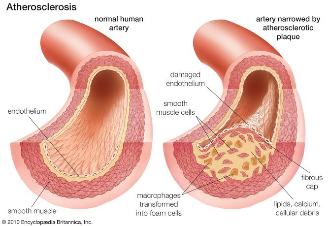
Disorders of Blood Circulation Complete study notes on Heart Failure and Arteriosclerosis, covering definitions, classifications, pathophysiology, clinical manifestations, investigations, and the “Four Pillars” of modern management. 1. HEART FAILURE (HF) 1.1 Definition Heart Failure (HF) is a clinical syndrome characterized by structural or functional abnormalities of the heart that result in impaired ventricular filling or ejection of blood. It is not a single disease but a constellation of signs and symptoms arising from inadequate cardiac output to meet the metabolic demands of the body, or from elevated cardiac filling pressures at rest or with exertion. 1.2 Classification A. By Ejection Fraction (2022 AHA/ACC/HFSA Guidelines) Type LVEF Pathophysiological Description HFrEF (Reduced) ≤ 40% Systolic dysfunction; impaired contractility. HFmrEF (Mildly Reduced) 41–49% Heart Failure with mildly reduced Ejection Fraction; intermediate group. HFpEF (Preserved) ≥ 50% Diastolic dysfunction; impaired relaxation and filling. HFimpEF (Improved) > 40% Previously HFrEF with subsequent LVEF > 40% on repeated measurement. B. By AHA/ACC/HFSA Stages (Progressive Framework) Stage A: At risk for HF but without symptoms, structural heart disease, or cardiac biomarkers. Stage B: Structural heart disease but no prior or current signs/symptoms of HF (e.g., prior MI, LV hypertrophy, valvular disease). Stage C: Structural heart disease with prior or current symptoms of HF. Stage D: Marked HF symptoms that interfere with daily life; recurrent hospitalizations despite optimized medical therapy (GDMT). C. By NYHA Functional Classification (Symptom Severity) Class I: No limitation of physical activity. Ordinary activity does not cause symptoms. Class II: Slight limitation. Comfortable at rest. Ordinary activity causes fatigue, palpitation, dyspnea, or chest pain. Class III: Marked limitation. Comfortable at rest. Less than ordinary activity causes symptoms. Class IV: Unable to perform any physical activity without symptoms. Symptoms present at rest. 1.3 Etiology and Risk Factors Category Specific Causes Ischemic heart disease Myocardial infarction, chronic angina, hibernating myocardium. Hypertension Chronic pressure overload leading to LV hypertrophy and remodeling. Valvular disease Aortic stenosis/regurgitation, mitral stenosis/regurgitation. Cardiomyopathies Dilated (alcohol, viral, genetic), hypertrophic, restrictive. Arrhythmias Atrial fibrillation (tachycardia-induced cardiomyopathy). Toxins/Drugs Alcohol, cocaine, anthracyclines (doxorubicin), trastuzumab. Metabolic Diabetes, thyroid disease, obesity, Thiamine deficiency (Beriberi). High-output states Anemia, hyperthyroidism, arteriovenous fistula. 4.1.4 Pathophysiology A. Left-Sided Heart Failure Forward failure (low output): Reduced cardiac output → tissue hypoperfusion → fatigue, weakness, oliguria, altered mental status. Backward failure (pulmonary congestion): LV fails to pump effectively → increased left ventricular end-diastolic pressure (LVEDP) → increased left atrial pressure → pulmonary venous congestion → Pulmonary Edema. B. Right-Sided Heart Failure Commonly caused by Left-sided HF, pulmonary hypertension, or chronic lung disease (Cor Pulmonale). Right ventricle fails to pump effectively → increased right ventricular and right atrial pressure → Systemic Venous Congestion. Clinical consequences: Jugular venous distension (JVD), hepatomegaly, peripheral edema (ankles, sacrum), and ascites. C. Neurohormonal Activation Sympathetic Nervous System: Increased norepinephrine → increased heart rate and contractility. Chronic activation causes myocardial toxicity and arrhythmias. RAAS System: Decreased renal perfusion → Renin release → Angiotensin II (vasoconstriction) + Aldosterone (sodium/water retention) → Volume Overload. Natriuretic Peptides (BNP, NT-proBNP): Released from stretched myocardium to promote vasodilation and diuresis (compensatory). Key Concept Cardiac Remodeling Concentric Hypertrophy: Increased wall thickness due to pressure overload (HTN, Aortic Stenosis). Eccentric Hypertrophy: Chamber dilation due to volume overload (MI, Mitral Regurgitation). 4.1.5 Clinical Features Symptom/Sign Left-Sided HF Right-Sided HF Dyspnea Exertional, Orthopnea, PND Less prominent Cough Dry or productive (pink frothy) Not typical Edema Pulmonary edema (crackles) Peripheral pitting edema JVP Normal or elevated Elevated (key sign) Heart Sounds S3 gallop Loud P2 (pulmonic) Clinical Alert Acute Pulmonary Edema (Severe Left HF) Presents with severe dyspnea at rest, anxiety, diaphoresis, and pink frothy sputum. This is a medical emergency requiring immediate intervention (Oxygen, Diuretics, Vasodilators). 4.1.6 Investigations Chest X-ray: Cardiomegaly (ratio > 50%), pulmonary congestion, Kerley B lines, and pleural effusions. ECG: Evidence of arrhythmias (Atrial Fibrillation), LV hypertrophy, Q waves (prior MI), or LBBB. Echocardiography: Gold standard for assessing LVEF, chamber size, and valvular function. BNP / NT-proBNP: Elevated levels (> 400 pg/mL BNP) support HF; levels < 100 pg/mL make HF unlikely. Troponins: Elevated in acute decompensation or myocardial stretch/necrosis. 4.1.7 Management of Heart Failure A. Guideline-Directed Medical Therapy (GDMT) — “The Four Pillars” For HFrEF, the standard of care includes the simultaneous or rapid sequence initiation of: ARNI (Sacubitril/Valsartan): Or ACEI/ARB; inhibits RAAS and neprilysin. Beta-blockers: (Carvedilol, Bisoprolol, Metoprolol succinate) to block sympathetic overactivation. MRA: (Spironolactone, Eplerenone) to block aldosterone. SGLT2 Inhibitors: (Dapagliflozin, Empagliflozin) to improve cardiovascular outcomes. B. Acute Management Oxygen therapy if hypoxemic. IV loop diuretics (Furosemide) to reduce fluid volume. Vasodilators (Nitrates) if hypertensive. Inotropes (Dobutamine) if in cardiogenic shock. 4.2 ARTERIOSCLEROSIS 4.2.1 Definition Arteriosclerosis is an umbrella term for the thickening, hardening, and loss of elasticity of arterial walls. Atherosclerosis is a specific type involving atheromatous plaque formation in the tunica intima. 4.2.2 Types of Arteriosclerosis Type Affected Vessels Key Feature Atherosclerosis Large and medium arteries Lipid-rich plaques in tunica intima; inflammatory. Monckeberg medial sclerosis Medium-sized muscular arteries Calcium deposits in tunica media; no luminal narrowing. Arteriolosclerosis Small arteries and arterioles Hyaline or hyperplastic thickening; associated with HTN/Diabetes. 4.2.3 Atherosclerosis Pathophysiology Response-to-Injury Hypothesis: Endothelial injury: Caused by smoking, hypertension, or diabetes. Lipid accumulation: LDL enters the intima and becomes oxidized (Ox-LDL). Inflammatory response: Macrophages engulf Ox-LDL to become Foam Cells. Smooth muscle proliferation: Migration from media to intima; production of collagen. Plaque progression: Fibrous cap forms over a lipid core. Plaque Morphology Feature Stable Plaque Unstable (Vulnerable) Plaque Fibrous cap Thick, well-developed Thin or absent Lipid core Small Large (> 40% of volume) Risk Gradual narrowing (Angina) Rupture → Thrombosis (MI/Stroke) Summary & Key Clinical Pearls The Four Pillars of HFrEF therapy (ARNI, beta-blocker, MRA, SGLT2i) are all mortality-reducing. BNP/NT-proBNP are essential biomarkers for excluding heart failure. Atherosclerosis is a systemic disease; management of one territory (e.g., coronary) requires modification of all vascular beds. Statins are indicated for all patients with established atherosclerotic disease regardless of baseline LDL. Ankle-brachial index (ABI) < 0.9 indicates Peripheral Artery Disease (PAD).
Types of circulation (Pulmonary, Systemic, Fetal)
Anatomy

Types of circulation (Pulmonary, Systemic, Fetal)

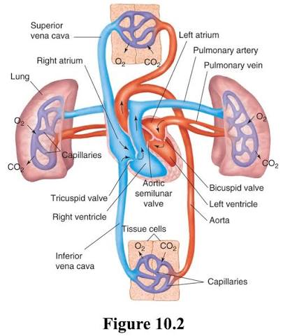
Types of Circulation Complete study notes covering Pulmonary, Systemic, and Fetal circulation, including physiological mechanisms, anatomical pathways, special circulatory systems, and clinical correlations. 3.1 Introduction The circulatory system is responsible for transporting blood throughout the body. In adults, there are two primary circuits: pulmonary circulation (blood flow between the heart and lungs) and systemic circulation (blood flow between the heart and all body tissues). During fetal development, a specialized fetal circulation exists to bypass the non-functioning lungs and liver. 3.2 Pulmonary Circulation 3.2.1 Definition and Purpose Pulmonary circulation is the portion of the cardiovascular system that transports deoxygenated blood from the right side of the heart to the lungs and returns oxygenated blood to the left side of the heart. Purpose: Gas exchange: Loading oxygen and unloading carbon dioxide in pulmonary capillaries. Blood volume reservoir: Pulmonary vessels contain approximately 9% of total blood volume. Filtration: Removal of small blood clots and air bubbles by pulmonary capillaries. 3.2.2 Pathway of Pulmonary Circulation The precise anatomical route follows this sequence: Right ventricle → Pulmonary valve → Pulmonary trunk → Right and left pulmonary arteries → Lobar and segmental arteries → Pulmonary capillaries (alveoli) → Pulmonary venules → Pulmonary veins (4 total: 2 from each lung) → Left atrium Key Features Pulmonary arteries carry deoxygenated blood (exception to the general rule). Pulmonary veins carry oxygenated blood (exception to the general rule). Pulmonary circulation is a low-pressure, low-resistance system. Pulmonary artery pressure: 15-30 mmHg systolic, 4-12 mmHg diastolic. Pulmonary vascular resistance is approximately 1/10 of systemic resistance. 3.2.3 Physiology of Pulmonary Circulation Hypoxic vasoconstriction: Unique response of pulmonary vessels: alveolar hypoxia causes local vasoconstriction. Directs blood away from poorly ventilated alveoli toward well-ventilated regions. Optimizes ventilation-perfusion (V/Q) matching. Contrast with systemic circulation: hypoxia causes vasodilation. Gravity effects: In upright position, blood flow is greater at lung bases than apices. Zone 1 (apex): Alveolar pressure > arterial pressure; minimal flow (potential for alveolar dead space). Zone 2 (mid-lung): Arterial pressure > alveolar pressure > venous pressure; intermittent flow. Zone 3 (base): Arterial and venous pressures > alveolar pressure; continuous flow. 3.2.4 Clinical Correlation Clinical Pearl Pulmonary Embolism (PE) Obstruction of pulmonary arteries by thrombus (usually from deep vein thrombosis). Symptoms include sudden dyspnea, pleuritic chest pain, tachypnea, tachycardia, and hypoxemia. Diagnosis via CT pulmonary angiography (CTPA), D-dimer, and Wells score. Management involves anticoagulation or thrombolysis in massive PE. Pulmonary Hypertension Mean pulmonary artery pressure > 20 mmHg (2022 ESC/ERS guidelines). Causes include left heart disease, lung disease, or idiopathic factors. Leads to right ventricular hypertrophy and eventually right heart failure (cor pulmonale). 3.3 Systemic Circulation 3.3.1 Definition and Purpose Systemic circulation is the portion of the cardiovascular system that transports oxygenated blood from the left side of the heart to all body tissues and returns deoxygenated blood to the right side of the heart. Purpose: Delivery of oxygen and nutrients to all body tissues. Removal of carbon dioxide and metabolic waste products. Transport of hormones from endocrine glands to target organs. Regulation of body temperature (thermoregulation via blood flow to skin). Maintenance of fluid and electrolyte balance. 3.3.2 Pathway of Systemic Circulation Left ventricle → Aortic valve → Ascending aorta → Aortic arch → Descending thoracic aorta → Abdominal aorta → Common iliac arteries → Arteries of lower limbs → Arterioles → Capillary beds (all tissues) → Venules → Veins → Inferior vena cava (from below heart) + Superior vena cava (from above heart) → Right atrium 3.3.3 Major Arterial Branches From aortic arch: Brachiocephalic trunk → right subclavian + right common carotid. Left common carotid → head and neck. Left subclavian → left upper limb. From descending aorta: Intercostal arteries → chest wall. Celiac trunk → liver, stomach, spleen. Superior mesenteric artery → small intestine, proximal colon. Renal arteries → kidneys. Inferior mesenteric artery → distal colon, rectum. Common iliac arteries → pelvis and lower limbs. 3.3.4 Major Venous Drainage Superior vena cava: Formed by union of brachiocephalic veins. Drains head, neck, upper limbs, chest wall, and upper thoracic organs. Inferior vena cava: Formed by union of common iliac veins. Drains lower limbs, pelvis, abdomen, kidneys, and liver (hepatic veins). Clinical Note Hepatic Portal System A unique venous system: capillaries (GI tract) → portal vein → capillaries (liver) → hepatic veins → IVC. Carries nutrient-rich, deoxygenated blood from digestive organs to the liver for processing. This is a critical site for first-pass metabolism and detoxification. 3.3.5 Special Circulations Coronary circulation: Supplies heart muscle itself. Arises from ascending aorta; drains into right atrium via coronary sinus. Cerebral circulation: Internal carotid arteries and vertebral arteries form the Circle of Willis. Protected by the blood-brain barrier (tight junctions in continuous capillaries). Renal circulation: High blood flow (20-25% of cardiac output) for filtration. Features two capillary beds in series: glomerular capillaries (filtration) and peritubular capillaries (reabsorption). Splanchnic circulation: Blood flow to GI tract, liver, spleen, and pancreas. Highly variable; increases after meals (active hyperemia). 3.3.6 Regulation of Systemic Circulation Short-term regulation (seconds to minutes): Baroreceptor reflex: carotid sinus and aortic arch receptors detect pressure changes; mediate heart rate and vasomotor tone via autonomic nervous system. Chemoreceptor reflex: carotid and aortic bodies detect changes in O2, CO2, and pH. Local autoregulation: myogenic response (Bayliss effect) and metabolic factors. Long-term regulation (hours to days): Renin-angiotensin-aldosterone system (RAAS): regulates blood volume and pressure. Antidiuretic hormone (ADH/vasopressin): regulates water reabsorption in kidneys. Atrial natriuretic peptide (ANP): promotes sodium and water excretion. 3.4 Fetal Circulation 3.4.1 Overview Fetal circulation is distinctly different from adult circulation because the fetus does not use its lungs for gas exchange. Instead, the placenta serves as the site of oxygenation, nutrient delivery, and waste removal. The fetal heart and blood vessels contain special shunts that bypass the lungs and liver. 3.4.2 Key Structures in Fetal Circulation Placenta: Organ of exchange between maternal and fetal circulations. Maternal oxygenated blood and nutrients diffuse across placental barrier into fetal blood. Fetal waste products (CO2, urea) diffuse into maternal blood. Connected to fetus via umbilical cord. Umbilical Cord: Contains two umbilical

Structure and functions of blood vessels
Anatomy

Structure and functions of blood vessels

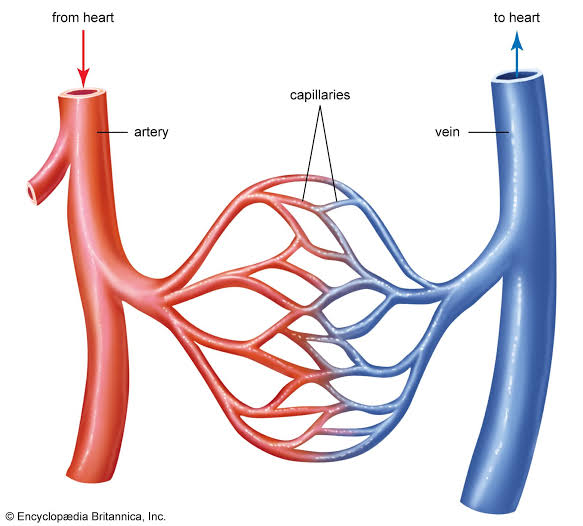
Structure and Functions of Blood Vessels Complete study notes covering the histology of vessel walls, classification of the vascular tree, hemodynamics, capillary exchange mechanisms, and major systemic vessels. 2.1 Introduction Blood vessels form a closed system of tubes that transport blood between the heart and body tissues. There are five main types: arteries, arterioles, capillaries, venules, and veins. Together, they regulate blood flow, blood pressure, and the exchange of substances between blood and tissues. 2.2 General Structure of Blood Vessels Most blood vessels share a common three-layered (tunica) wall structure: Layer Name Composition Function Tunica externa Outer layer Connective tissue (collagen) with elastic fibers Protection, anchoring, nerve/vessel passage Tunica media Middle layer Smooth muscle, elastic fibers (lamellae) Vasoconstriction, vasodilation, pressure regulation Tunica intima Inner layer Endothelium (simple squamous epithelium) on basement membrane Smooth surface for blood flow; prevents clotting Key Histological Difference Capillaries consist only of a single layer of endothelium (tunica intima) and a basement membrane, allowing for efficient nutrient and gas exchange. 2.3 Arteries Arteries carry blood away from the heart. They are classified by size and structure into three categories: 2.3.1 Elastic (Conducting) Arteries Largest arteries: Aorta, pulmonary trunk, brachiocephalic, common carotid, subclavian, common iliac arteries. Structure: Tunica media contains abundant elastic fibers (lamellae) with less smooth muscle. Function: Conduct blood from heart to medium-sized arteries; elastic recoil maintains continuous blood flow during diastole (Windkessel effect). 2.3.2 Muscular (Distributing) Arteries Medium-sized arteries: Radial, ulnar, femoral, tibial, coronary arteries. Structure: Tunica media contains more smooth muscle (40-60% of wall thickness) and less elastic tissue. Function: Distribute blood to specific organs and tissues; vasoconstriction and vasodilation regulate blood flow to organs. 2.3.3 Arterioles Smallest arteries: 10-100 um diameter. Structure: Tunica media has 1-3 layers of smooth muscle cells. Function: Major resistance vessels; regulate blood flow into capillary beds; primary site of peripheral vascular resistance. Precapillary sphincters: Rings of smooth muscle that control capillary blood flow. Arterial Blood Pressure Systolic pressure: 90-120 mmHg (peak pressure during ventricular systole). Diastolic pressure: 60-80 mmHg (minimum pressure during ventricular diastole). Mean arterial pressure (MAP): ~93 mmHg = diastolic + 1/3 pulse pressure. Pulse pressure: systolic – diastolic (normally 30-40 mmHg). 2.4 Capillaries Capillaries are the smallest blood vessels (5-10 um diameter) and form extensive networks (capillary beds) between arterioles and venules. They are the primary site of exchange between blood and tissues. 2.4.1 Structure Wall composed of a single layer of endothelial cells on a basement membrane. No tunica media or tunica externa. Total surface area: Approximately 600 m² (enormous exchange capacity). Total length: Approximately 96,000 km if all capillaries were placed end-to-end. 2.4.2 Types of Capillaries Type Structure Location Function Continuous Endothelial cells joined by tight junctions; complete basement membrane Muscle, skin, lungs, brain (blood-brain barrier) Most common; controlled exchange of water, ions, small molecules Fenestrated Endothelial cells have pores (fenestrae); thin/continuous basement membrane Kidneys, intestines, endocrine glands Rapid exchange of fluids, solutes; filtration, absorption Sinusoidal Large, irregular lumen; incomplete basement membrane; gaps between endothelial cells Liver, spleen, bone marrow, lymph nodes Allow passage of large molecules, cells (RBCs, WBCs) 2.4.3 Capillary Exchange Mechanisms Diffusion: Primary mechanism for exchange of gases, nutrients, and waste products. — Lipid-soluble: (O2, CO2, steroid hormones) pass directly through endothelial membranes. — Water-soluble: (glucose, amino acids, ions) pass through intercellular clefts or fenestrae. Transcytosis: Transport of large molecules (proteins, hormones) via vesicles (endocytosis + exocytosis). Bulk Flow (Filtration and Reabsorption): Driven by hydrostatic and osmotic pressure differences (Starling forces). — At arterial end: net filtration (fluid moves out into interstitium). — At venous end: net reabsorption (fluid moves back into capillary). — Approximately 85% of filtered fluid is reabsorbed; 15% returns via lymphatic vessels. Starling Equation Net filtration = Kf x [(Pc – Pi) – (πc – πi)] Pc = capillary hydrostatic pressure Pi = interstitial hydrostatic pressure πc = plasma colloid osmotic pressure πi = interstitial colloid osmotic pressure 2.5 Veins and Venules Veins carry blood toward the heart. They have larger lumens and thinner walls than corresponding arteries. 2.5.1 Venules Smallest veins: 10-100 um; formed by union of capillaries. Postcapillary venules: Major site of leukocyte extravasation during inflammation. Tunica media is very thin (1-2 layers of smooth muscle). 2.5.2 Veins Medium and large veins have all three tunics, but tunica media is thinner than in arteries. Tunica externa is the thickest layer in large veins. Luminal diameter is larger than corresponding arteries. Blood pressure is low (approximately 10-15 mmHg in vena cava). Blood volume capacity is high (approximately 60% of total blood volume at rest). Venous Return Mechanisms: Skeletal muscle pump: Contraction of surrounding muscles compresses veins, pushing blood toward the heart (one-way valves prevent backflow). Respiratory pump: During inspiration, thoracic pressure decreases and abdominal pressure increases, drawing blood toward the heart. One-way valves: Prevent backflow of blood, especially in lower limbs. Gravity: Aided by upright posture and venous tone. Clinical Correlation Varicose Veins Dilated, tortuous, elongated superficial veins (commonly saphenous veins). Caused by incompetent venous valves, increased venous pressure, or weakened vein walls. Risk factors: prolonged standing, pregnancy, obesity, family history. Complications: venous stasis ulcers, thrombophlebitis, bleeding. 2.6 Comparison of Blood Vessels Feature Arteries Capillaries Veins Wall thickness Thick Very thin (1 cell) Thin Tunica media Thick (smooth muscle + elastic) Absent Thin Lumen diameter Smaller than vein Smallest (5-10 um) Larger than artery Blood pressure High (80-120 mmHg) Low (20-40 mmHg) Very low (5-15 mmHg) Blood velocity Fast Very slow Slow Blood volume Low (~15%) Low (~5%) High (~60%) Valves None (except near heart) None Present (especially in limbs) Function Transport blood away from heart Exchange of gases, nutrients, wastes Return blood to heart; blood reservoir 2.7 Major Blood Vessels of the Body 2.7.1 Aorta and Major Arteries Ascending aorta: Gives rise to right and left coronary arteries. Aortic arch: — Brachiocephalic trunk -> right subclavian + right common carotid — Left common carotid artery — Left subclavian artery Thoracic aorta: Bronchial, esophageal, mediastinal, posterior intercostal arteries. Abdominal aorta: — Celiac trunk (liver, stomach, spleen) — Superior mesenteric artery (small intestine, proximal

Structure and functions of the heart
Anatomy

Structure and functions of the heart

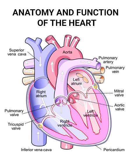
Structure and Functions of the Heart Complete study notes covering macroscopic and microscopic anatomy, the cardiac cycle, neurovascular supply, and physical examination correlations. 1. Introduction The heart is a hollow, muscular, cone-shaped organ located in the mediastinum, between the lungs. Approximately two-thirds of its mass lies to the left of the midline. It functions as a double pump maintaining continuous blood circulation through the pulmonary and systemic circuits. Key Parameters: Size: Approximately 12 cm long, 9 cm wide, 6 cm thick. Weight: Approximately 250-350 g in adult males; slightly less in females. Location: Posterior to the sternum, anterior to the vertebral column at levels T5-T8. Apex: Directed anteriorly, inferiorly, and to the left (located at the 5th intercostal space, midclavicular line). Base: Directed posteriorly, superiorly, and to the right. 2. Structure of the Heart 2.1 Pericardium The heart is enclosed within the pericardium, a double-walled sac consisting of two main layers: Fibrous pericardium: A tough, outer layer of dense connective tissue that prevents overfilling and anchors the heart to surrounding structures. Serous pericardium: Parietal layer: Lines the inner surface of the fibrous pericardium. Visceral layer (epicardium): Covers the external surface of the heart. Pericardial cavity: The potential space between the parietal and visceral layers containing 10-20 mL of serous fluid to reduce friction. 2.2 Heart Wall The heart wall consists of three distinct layers: Layer Name Characteristics Outer Epicardium Visceral layer of serous pericardium; contains coronary vessels, nerves, and adipose tissue. Middle Myocardium Cardiac muscle tissue; the thickest layer; responsible for the pumping action/contraction. Inner Endocardium Endothelium-lined; smooth surface that reduces friction and prevents blood clot formation; covers valves. 2.3 Chambers of the Heart The heart has four chambers: two atria (upper) and two ventricles (lower). Right Atrium: Receives deoxygenated blood from systemic circulation via the superior vena cava (head, neck, limbs), inferior vena cava (abdomen, pelvis, lower limbs), and coronary sinus (heart wall). Contains the fossa ovalis (remnant of fetal foramen ovale). Houses the sinoatrial (SA) node — the natural pacemaker. Right Ventricle: Receives blood from the right atrium via the tricuspid valve. Pumps blood through the pulmonary valve into the pulmonary trunk. Wall is thinner than the left ventricle as it pumps against low pulmonary resistance (15-30 mmHg). Contains the moderator band (septomarginal trabecula) carrying the right bundle branch. Left Atrium: Receives oxygenated blood from the lungs via four pulmonary veins (two from each lung). Smaller but thicker-walled than the right atrium. Left Ventricle: Receives oxygenated blood from the left atrium via the mitral (bicuspid) valve. Pumps blood through the aortic valve into the ascending aorta for systemic circulation. Wall is 2-3 times thicker than the right ventricle to pump against high systemic pressure (80-120 mmHg). Forms the apex of the heart. 3. Valves and Blood Flow 3.1 Atrioventricular (AV) Valves Tricuspid Valve (Right AV): Three cusps (anterior, posterior, septal). Prevents backflow to RA during ventricular systole. Mitral (Bicuspid) Valve (Left AV): Two cusps (anterior, posterior). Most commonly affected by rheumatic heart disease and mitral valve prolapse. Both are anchored by chordae tendineae to papillary muscles of the ventricles. 3.2 Semilunar Valves Pulmonary Valve: Three semilunar cusps. Located between RV and pulmonary trunk. Aortic Valve: Three semilunar cusps (right, left, non-coronary). Right and left coronary cusps give rise to the coronary arteries. Mechanism of Valve Closure AV valves close when ventricular pressure exceeds atrial pressure during systole. Chordae tendineae prevent the cusps from everting into the atria. Semilunar valves close when arterial pressure exceeds ventricular pressure during diastole. 3.3 Fibrous Skeleton of the Heart A dense connective tissue framework that: Provides attachment for valves and cardiac muscle fibers. Electrically insulates atria from ventricles. The AV bundle (Bundle of His) is the only normal electrical connection between atria and ventricles. 4. Coronary Circulation 4.1 Arterial Supply Right Coronary Artery (RCA): Arises from the right aortic sinus. Supplies the RA, most of the RV, SA node (in 60%), AV node (in 80%), and posterior 1/3 of the septum. Left Coronary Artery (LCA): Arises from the left aortic sinus. Divides into: Left Anterior Descending (LAD): Supplies anterior 2/3 of septum, apex; known as the “widow maker” when occluded. Circumflex artery: Supplies LA and lateral/posterior walls of the LV. Coronary Dominance Dominance is determined by which artery gives rise to the posterior interventricular artery (PDA). — Right-dominant (85%): PDA from RCA. — Left-dominant (10%): PDA from Circumflex. — Co-dominant (5%): PDA from both. 4.2 Venous Drainage Great cardiac vein: Accompanies LAD; drains into coronary sinus. Middle cardiac vein: Accompanies posterior interventricular artery. Coronary sinus: Drains into the right atrium (main venous return). 5. Functions and Physiology 5.1 The Cardiac Cycle Sequence of events in one heartbeat (~0.8 seconds at 75 bpm): Atrial Systole (0.1 sec): SA node fires; atria contract, forcing 20-30% of blood into ventricles (“atrial kick”). Isovolumetric Ventricular Contraction (0.05 sec): Ventricular pressure rises; AV valves close (S1 sound). All valves closed. Ventricular Ejection (0.25 sec): Pressure exceeds arterial pressure; semilunar valves open. SV is ~70 mL. Isovolumetric Ventricular Relaxation (0.08 sec): Semilunar valves close (S2 sound). All valves closed. Ventricular Filling (0.4 sec): AV valves open; blood flows passively from atria. 5.2 Heart Sounds Sound Timing Cause Clinical Significance S1 (Lubb) Beginning of systole Closure of AV valves Loud in mitral stenosis; soft in mitral regurgitation. S2 (Dubb) End of systole Closure of Semilunar valves Splitting: normal (A2 then P2 on inspiration). S3 Early diastole Rapid ventricular filling Normal in children; pathological in heart failure. S4 Late diastole Atrial contraction against stiff ventricle Indicates decreased compliance (HTN, AS). 5.3 Cardiac Output and Stroke Volume Cardiac Output (CO): Volume of blood pumped per minute (SV x HR). Normal: 4.0-8.0 L/min. Ejection Fraction (EF): Percentage of end-diastolic volume ejected. Normal: 55-70%. Factors Affecting Stroke Volume: Preload: Frank-Starling mechanism (increased venous return stretches muscle, increasing force). Contractility: Force of contraction (increased by Sympathetics, Digoxin). Afterload: Systemic vascular resistance (pressure the heart must pump against). 6. Conduction System Sinoatrial (SA) Node: Pace-maker; generates impulses at 60-100 bpm. Atrioventricular (AV) Node: Located in

Common blood disorders (Anaemia, Leukemia, Hemophilia)
Anatomy

Common blood disorders (Anaemia, Leukemia, Hemophilia)

Common Blood Disorders Complete study notes covering the classification, pathophysiology, investigation, and management of Anaemia, Leukemia, and Hemophilia. 4.1 ANAEMIA 4.1.1 Definition Anaemia is defined as a reduction in the oxygen-carrying capacity of blood due to decreased hemoglobin (Hb) concentration, reduced red blood cell (RBC) count, or low hematocrit levels relative to the physiological needs of the individual. 4.1.2 WHO 2024 Diagnostic Criteria The World Health Organization updated hemoglobin cutoffs in 2024 using systematic review and GRADE methodology. These thresholds are critical for clinical diagnosis: Population Hb Threshold (g/dL) Children 6-59 months <11.0 (revised to <10.5 for 6-23 months) Children 5-12 years <11.5 Non-pregnant women <12.0 Pregnant women (all trimesters) <11.0 Pregnant women (2nd trimester) <10.5 Men <13.0 Severity Classification (WHO): Mild: 10.0 — threshold Moderate: 7.0 — 9.9 Severe: <7.0 4.1.3 Classification by Morphology (MCV) Type MCV (fL) MCH (pg) Common Causes Microcytic hypochromic <80 fL <27 pg Iron deficiency, thalassemia, sideroblastic anemia, chronic disease Normocytic normochromic 80-100 fL 27-33 pg Acute blood loss, hemolytic anemia, chronic disease, aplastic anemia, renal failure Macrocytic >100 fL >33 pg B12 deficiency, folate deficiency, liver disease, alcoholism, hypothyroidism 4.1.4 Classification by Pathophysiology Decreased production: Iron deficiency, B12/folate deficiency, aplastic anemia, chronic disease, renal failure. Increased destruction (hemolysis): Sickle cell disease, thalassemia, autoimmune hemolytic anemia, G6PD deficiency, malaria. Blood loss: Trauma, menorrhagia, GI bleeding (peptic ulcer, hookworm, colorectal cancer). 4.1.5 Iron Deficiency Anaemia (IDA) Etiology: Chronic blood loss (menorrhagia, GI bleeding, hookworm infestation), inadequate dietary intake (vegetarian/vegan diets), malabsorption (celiac disease, gastrectomy), and increased demand (pregnancy, infancy, adolescence). Clinical Features: General: Pallor (conjunctival, palmar, tongue), fatigue, weakness, dizziness. Cardiovascular: Tachycardia, palpitations, systolic flow murmur, heart failure (severe). Epithelial: Angular cheilitis, glossitis, koilonychia (spoon-shaped nails), pica. Neurological: Headache, irritability, reduced work capacity. Investigations: CBC: Low Hb, low MCV, low MCH, low MCHC, high RDW. Peripheral smear: Microcytic, hypochromic RBCs; pencil cells; target cells. Iron studies: Low serum iron, low ferritin, high TIBC, low transferrin sat. Reticulocytes: Low or inappropriately normal. Management: Oral iron: Ferrous sulfate 200 mg TDS (or 65 mg elemental iron). Dietary advice: Iron-rich foods (red meat, leafy greens); Vitamin C enhances absorption. Treat underlying cause: Control bleeding, treat hookworm, manage menorrhagia. Parenteral iron: For malabsorption, intolerance, or non-compliance. Transfusion: Reserved for severe anemia (Hb <7 g/dL) with hemodynamic compromise. 4.1.6 Megaloblastic Anaemia Causes: — B12 deficiency: Pernicious anemia (autoimmune), gastrectomy, ileal disease, vegan diet. — Folate deficiency: Poor nutrition, alcoholism, malabsorption, pregnancy, hemolysis. Clinical Features: General anemia symptoms plus: B12 deficiency: Neurological — subacute combined degeneration of spinal cord: loss of vibration/position sense, ataxia, spasticity, dementia; glossitis. Folate deficiency: No neurological symptoms; neural tube defects in pregnancy. Investigations: CBC: Low Hb, high MCV (>100 fL), pancytopenia possible. Peripheral smear: Macrocytic RBCs, hypersegmented neutrophils (>5 lobes). B12/Folate level: Low (B12 <200 pg/mL; Folate <3 ng/mL). Anti-IF antibodies: Positive in pernicious anemia. IMPORTANT Never give folate alone if B12 deficiency is possible — it corrects the anemia but allows neurological damage to progress. 4.1.7 Sickle Cell Disease Autosomal recessive hemoglobinopathy (HbS). Point mutation in beta-globin gene (Glu -> Val at position 6). RBCs sickle under low oxygen, causing vaso-occlusion and hemolysis. Clinical: Painful crises, dactylitis, splenic sequestration, acute chest syndrome, stroke, priapism. Diagnosis: Hb electrophoresis. Management: Hydration, analgesia, oxygen, hydroxyurea, blood transfusion, penicillin prophylaxis in children. Key Clinical Pearls — Anaemia Always investigate the cause of iron deficiency in adult males and postmenopausal females — GI malignancy must be excluded. Never give folate alone if B12 deficiency is possible. Peripheral blood smear is essential for morphological classification. 4.2 LEUKEMIA 4.2.1 Definition Leukemia is a malignant neoplasm of hematopoietic stem cells characterized by uncontrolled proliferation of abnormal white blood cells (blasts) in the bone marrow and peripheral blood, with subsequent suppression of normal hematopoiesis. 4.2.2 Classification Classification Acute Chronic Myeloid Acute Myeloid Leukemia (AML) Chronic Myeloid Leukemia (CML) Lymphoid Acute Lymphoblastic Leukemia (ALL) Chronic Lymphocytic Leukemia (CLL) — Acute leukemias: Rapid onset, immature blasts (>20% in bone marrow), fatal without treatment. — Chronic leukemias: Insidious onset, more mature cells, slower progression. 4.2.3 Etiology and Risk Factors Genetic: Down syndrome (ALL risk 10-20x), Fanconi anemia, Bloom syndrome. Radiation: Atomic bomb survivors, therapeutic radiation. Chemicals: Benzene, alkylating agents, topoisomerase II inhibitors. Viruses: HTLV-1 — associated with adult T-cell leukemia/lymphoma. Prior chemo: Myelodysplastic syndrome progressing to AML. 4.2.4 Clinical Features Result from bone marrow failure and organ infiltration: System Manifestations General Fatigue, malaise, weight loss, fever, night sweats Anemia Pallor, dyspnea, tachycardia Infections Recurrent bacterial, viral, fungal infections (neutropenia) Bleeding Petechiae, ecchymoses, epistaxis, gingival bleeding (thrombocytopenia) Organomegaly Hepatomegaly, splenomegaly (especially CML, ALL) Lymphadenopathy Generalized lymph node enlargement (ALL, CLL) Bone pain Metaphyseal pain in children with ALL CNS Headache, cranial nerve palsies, meningismus (CNS involvement in ALL) Other Gum hypertrophy (AML M4/M5), skin infiltration (chloromas), testicular enlargement (ALL relapse) 4.2.5 Investigations CBC: Anemia, thrombocytopenia, high/low WBC; blasts may be seen. Peripheral blood smear: Blasts (large, high N:C ratio, fine chromatin, prominent nucleoli, Auer rods in AML). Bone marrow aspirate/biopsy: Hypercellular marrow with >20% blasts (diagnostic gold standard). Flow cytometry (immunophenotyping): Identifies cell lineage (CD3, CD7 = T-cell; CD19, CD20 = B-cell; CD13, CD33 = myeloid). Cytogenetics/Karyotyping: Philadelphia chromosome t(9;22) in CML; t(8;21), inv(16) in AML. Lumbar puncture: CSF cytology for CNS involvement (ALL). Chest X-ray: Mediastinal mass (T-cell ALL). 4.2.6 Management Chemotherapy: Induction, consolidation, maintenance; combination regimens (e.g., 7+3 for AML: cytarabine + anthracycline). Targeted therapy: Tyrosine kinase inhibitors (imatinib, dasatinib) for BCR-ABL1+. Immunotherapy: CAR-T cell therapy, blinatumomab (CD19-directed) for B-ALL. Stem cell transplant: Allogeneic transplant for high-risk or relapsed disease. Supportive care: Blood transfusions, infection prophylaxis, growth factors (G-CSF), tumor lysis syndrome prevention. Clinical Pearl Acute leukemia is a medical emergency. Tumor lysis syndrome can be fatal without prophylaxis (hydration, allopurinol, rasburicase). 4.3 HEMOPHILIA 4.3.1 Definition Hemophilia is a group of inherited bleeding disorders caused by deficiency of specific coagulation factors, resulting in impaired secondary hemostasis and prolonged bleeding. 4.3.2 Classification Type Deficient Factor Inheritance Incidence Hemophilia A (Classic) Factor VIII X-linked recessive 1 in 5,000-10,000 male births (80%) Hemophilia B (Christmas Disease) Factor IX

Composition and functions of blood
Anatomy

Composition and functions of blood

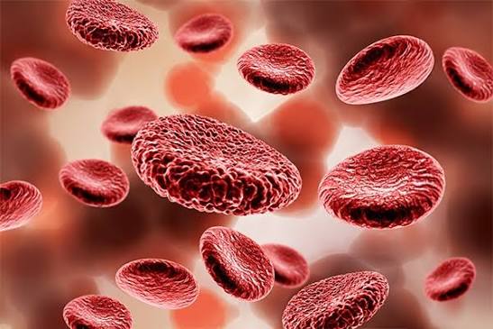
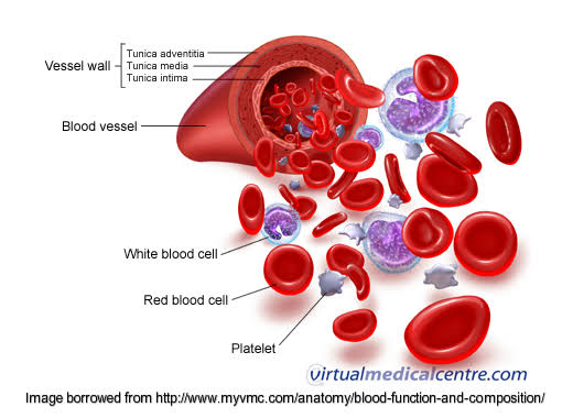
Composition and Functions of Blood Complete study notes covering the definition, general characteristics, detailed composition of plasma and formed elements, hematopoiesis, and clinical correlations. 1. Composition and Functions of Blood 1.1 Definition and General Characteristics Blood is a specialized connective tissue consisting of cells suspended in a liquid extracellular matrix called plasma. It circulates through the heart, arteries, capillaries, and veins, maintaining homeostasis throughout the body. Key Parameters: Total blood volume: ~7-8% of body weight (5-6 L in a 70 kg adult male). pH: 7.35-7.45 (slightly alkaline). Temperature: ~38°C (slightly higher than core body temperature). Viscosity: 3-5 times greater than water. Color: Bright red (oxygenated) to dark red (deoxygenated). Specific gravity: 1.055-1.065. 1.2 Composition of Blood Blood consists of two principal components: Component Percentage Constituents Plasma 55% Water (92%), proteins (7%), solutes (1%) Formed elements 45% Erythrocytes (99%), leukocytes, platelets Buffy coat <1% Leukocytes and platelets (after centrifugation) 1.2.1 Plasma Plasma is the straw-colored liquid portion of blood. Water (92%): Serves as solvent for transport. Plasma Proteins (7%): Albumin: Liver origin; maintains colloid osmotic pressure; transports fatty acids, bilirubin, and drugs. Globulins: Alpha/beta transport lipids, iron, copper; gamma = immunoglobulins. Fibrinogen: Liver origin; essential for blood clotting (converted to fibrin). Other Solutes (1%): Electrolytes (Na+, K+, Ca2+, Mg2+, Cl-, HCO3-, HPO4^2-) Nutrients (glucose, amino acids, fatty acids, vitamins) Waste products (urea, uric acid, creatinine, bilirubin) Respiratory gases (O2, CO2) and hormones 1.2.2 Erythrocytes (Red Blood Cells) Structure: Biconcave disc shape; 7-8 um in diameter. Anucleate (no nucleus) and lack organelles in mature form. Cell membrane contains spectrin and actin for flexibility through capillaries. Cytoplasm filled with hemoglobin (~280 million molecules per cell). Hemoglobin: Molecular weight: 64,458 Da. Four polypeptide chains (2 alpha, 2 beta in adults = HbA) + four heme groups. Each heme contains Fe2+ binding one O2 molecule. Normal types: HbA (95-98%), HbA2 (2-3%), HbF (<1% in adults). Function: Transport of O2 from lungs to tissues and CO2 from tissues to lungs. Normal Values: Parameter Adult Male Adult Female RBC count 4.5-5.5 x 10^12/L 4.0-5.0 x 10^12/L Hemoglobin 13.5-17.5 g/dL 12.0-16.0 g/dL Hematocrit 40-54% 37-47% MCV 80-100 fL 80-100 fL MCH 27-33 pg 27-33 pg MCHC 32-36 g/dL 32-36 g/dL Lifespan: 120 days. Senescent RBCs removed by macrophages in spleen, liver, and bone marrow. 1.2.3 Leukocytes (White Blood Cells) Leukocytes are nucleated cells critical for immune defense. Type % Key Features Functions Neutrophils 50-70% Multilobed nucleus (2-5); Fine lilac granules Phagocytosis of bacteria; first responders to acute inflammation Eosinophils 1-4% Bilobed nucleus; red-orange granules Defense against parasites; modulate allergic responses Basophils 0.5-1% S-lobed nucleus; deep blue granules Release histamine and heparin; mediate allergic reactions Lymphocytes 20-40% Large round nucleus; scant cytoplasm B cells = antibodies; T cells = cellular immunity; NK cells = tumors Monocytes 2-8% Kidney-shaped nucleus; gray cytoplasm Differentiate into macrophages; phagocytosis; antigen presentation Normal total WBC count: 4,000-11,000 cells/uL (4.0-11.0 x 10^9/L). 1.2.4 Platelets (Thrombocytes) Small cell fragments (2-4 um) derived from megakaryocytes in bone marrow. Anucleate but contain mitochondria, lysosomes, and granules. Normal count: 150,000-400,000/uL (150-400 x 10^9/L). Lifespan: 8-10 days. Functions: Formation of platelet plugs during hemostasis. Clot retraction through actin and myosin filaments. Release of growth factors (PDGF) promoting tissue repair. 1.3 Hematopoiesis Hematopoiesis is the process of blood cell formation. In adults, it occurs primarily in red bone marrow (sternum, ribs, vertebrae, pelvis, proximal femurs, and humeri). Stages: Pluripotent hematopoietic stem cells — self-renewal and differentiation. Multipotent progenitor cells — myeloid and lymphoid lineages. Committed progenitor cells — specific cell lines. Precursor cells (blasts) — immature forms with specific morphology. Mature cells — released into circulation. Key Regulatory Factors: Erythropoietin (EPO): Kidney production; stimulates erythropoiesis. Colony-stimulating factors (CSFs): Stimulate granulocyte/monocyte production. Thrombopoietin (TPO): Stimulates megakaryocyte and platelet production. Interleukins: Regulate lymphocyte development and immune responses. 1.4 Functions of Blood Function Mechanism Clinical Relevance Transport Carries O2, CO2, nutrients, hormones, waste products, heat Hypoxia in anemia; uremia in renal failure Regulation Buffers pH (bicarbonate); regulates temperature; maintains osmotic pressure Acid-base disorders; edema in hypoalbuminemia Protection Immune function (WBCs, antibodies); hemostasis (platelets, clotting factors) Immunodeficiency; bleeding disorders 1.5 Clinical Correlation: Complete Blood Count (CBC) The CBC is the most commonly ordered hematological investigation. It provides: RBC count, hemoglobin, hematocrit. RBC indices (MCV, MCH, MCHC, RDW). WBC count with differential. Platelet count. Peripheral blood smear morphology. Clinical Pearl A peripheral blood smear examination remains essential for confirming automated CBC findings and identifying morphological abnormalities such as sickle cells, schistocytes, or blast cells. Summary — Composition and Functions of Blood Key Points to Remember Blood volume: 5-6 L in adults. Plasma vs Formed elements: 55% vs 45%. RBC Lifespan: 120 days. Platelet Lifespan: 8-10 days. Hematopoiesis: Occurs in red bone marrow, regulated by EPO, TPO, and cytokines. Primary functions: Transport, Regulation, and Protection.

Types of blood groups
Anatomy

Types of blood groups

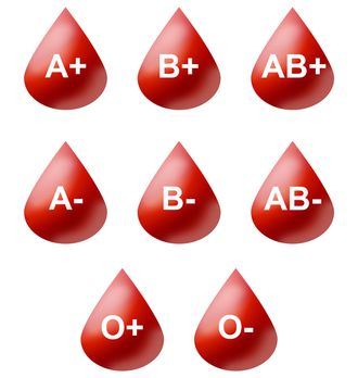
Types of Blood Groups Complete study notes covering the genetics, biochemistry, compatibility, and clinical significance of the ABO and Rh systems, as well as minor blood group systems and cross-matching procedures. 2.1 Introduction Blood groups are determined by antigenic substances (glycoproteins and glycolipids) present on the surface of erythrocytes. The two most clinically significant systems are the ABO and Rh systems. Historical Note ABO system: Discovered by Karl Landsteiner in 1900 (Nobel Prize, 1930). Rh system: Identified by Landsteiner and Wiener in 1939. 2.2 ABO Blood Group System The ABO system is governed by three alleles (I^A, I^B, and i) located on chromosome 9. 2.2.1 Genetics and Biochemistry Genotype Phenotype RBC Antigens Plasma Antibodies I^A I^A or I^A i A A antigen Anti-B I^B I^B or I^B i B B antigen Anti-A I^A I^B AB A and B antigens None ii O None (H only) Anti-A and Anti-B Biochemical Basis: The H antigen (precursor) is present on all RBCs. I^A allele codes for an enzyme adding N-acetylgalactosamine = A antigen. I^B allele codes for an enzyme adding galactose = B antigen. i allele is non-functional; no modification of H antigen occurs. Antibody Characteristics: Anti-A and Anti-B are naturally occurring (develop by 3-6 months of age). They are IgM antibodies, capable of fixing complement. Cause rapid intravascular hemolysis. Primary cause of acute hemolytic transfusion reactions. Bombay Phenotype (hh) Rare individuals lack H antigen due to FUT1 gene mutations. They type as O but possess anti-H, anti-A, and anti-B antibodies, making blood transfusion extremely difficult as they can only receive blood from other Bombay phenotype donors. 2.2.2 ABO Compatibility for Transfusion Recipient Can Receive From Can Donate To A A, O A, AB B B, O B, AB AB A, B, AB, O (univ. rec) AB only O O only A, B, AB, O (univ. donor) **Universal donor:** O negative (no A, B, or D antigens). **Universal recipient:** AB positive (no anti-A, anti-B, or anti-D antibodies). 2.3 Rh Blood Group System The Rh system is the second most important blood group system clinically, particularly in obstetrics. 2.3.1 Genetics and Antigens Determined by RHD and RHCE genes on chromosome 1. The D antigen is the most immunogenic Rh antigen. Rh-positive: D antigen present on RBCs. Rh-negative: D antigen absent. Prevalence: Rh-positive: ~85% Caucasians, 92% African Americans, 99% East Asians. Rh-negative: ~15% Caucasians, 8% African Americans,

Mechanism of blood clotting
Anatomy

Mechanism of blood clotting

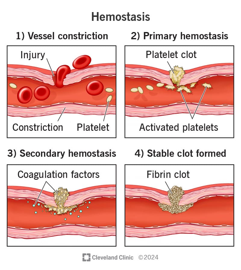
Mechanism of Blood Clotting Complete study notes covering the physiological phases of hemostasis, the biochemical coagulation cascade, modern cell-based models, fibrinolysis, and clinical laboratory diagnostics. 3.1 Definition and Overview Hemostasis is the physiological process that stops bleeding from a damaged blood vessel. It is a rapid, localized, and carefully regulated process that involves three overlapping phases: Vascular spasm (vasoconstriction) Platelet plug formation (primary hemostasis) Coagulation cascade (secondary hemostasis) Clot stabilization and fibrinolysis 3.2 Vascular Spasm This is the immediate reflex constriction of a damaged vessel to reduce blood loss. It is most effective in smaller vessels and is mediated by: Local myogenic spasm: Direct damage to vascular smooth muscle triggers contraction. Endothelin release: Released from damaged endothelial cells. Neural reflexes: Triggered by pain receptors. 3.3 Platelet Plug Formation (Primary Hemostasis) This process converts a temporary leak into a mechanical plug through three distinct steps: Step 1: Adhesion Vessel injury exposes subendothelial collagen. von Willebrand factor (vWF) binds to the exposed collagen. Platelet glycoprotein Ib (GPIb) receptors bind to vWF, causing platelets to adhere to the damaged wall. Step 2: Activation Adhesion triggers a platelet shape change (pseudopod formation). Release of granule contents: — Dense granules: Release ADP, serotonin, and Ca2+. — Alpha granules: Release fibrinogen, factor V, vWF, and PDGF. Synthesis and release of thromboxane A2 (TXA2), a potent vasoconstrictor and platelet activator. Platelet membrane expresses glycoprotein IIb/IIIa (GPIIb/IIIa) receptors. Step 3: Aggregation GPIIb/IIIa receptors bind fibrinogen, linking platelets together. This forms a loose platelet plug (white thrombus), which is later reinforced by fibrin. Clinical Correlation Antiplatelet Therapy Aspirin irreversibly inhibits COX-1, blocking the synthesis of Thromboxane A2 (TXA2). This effectively reduces platelet aggregation and forms the basis for secondary prevention of myocardial infarction and stroke. 3.4 Coagulation Cascade (Secondary Hemostasis) Secondary hemostasis involves a series of enzymatic reactions involving plasma proteins called clotting factors. Most factors are synthesized in the liver. Factors II, VII, IX, and X are Vitamin K-dependent. 3.4.1 Clotting Factors Factor Name Source Function I Fibrinogen Liver Converted to fibrin (clot structure) II Prothrombin Liver Converted to Thrombin (central enzyme) III Tissue Factor Tissue Initiates the Extrinsic Pathway IV Calcium (Ca2+) Diet Cofactor for multiple reactions V Proaccelerin Liver Cofactor for Factor X activation VII Proconvertin Liver Initiates extrinsic pathway VIII Antihemophilic A Liver Cofactor for Factor IX (Deficient in Hemophilia A) IX Antihemophilic B Liver Activates Factor X (Deficient in Hemophilia B) X Stuart-Prower Liver Forms Prothrombinase complex XI PTA Liver Activates Factor IX XII Hageman Factor Liver Initiates Intrinsic Pathway XIII Fibrin-stabilizing Liver Cross-links fibrin monomers *Note: Factor VI was originally assigned to activated factor V but is no longer used in modern nomenclature. 3.4.2 The Three Pathways A. Extrinsic Pathway (Tissue Factor Pathway) Trigger: Tissue injury exposing Tissue Factor (Factor III) to blood. Sequence: Factor III + Factor VII activates Factor X. Speed: Fast-acting; generates thrombin within seconds. Lab Test: Prothrombin Time (PT). B. Intrinsic Pathway (Contact Activation Pathway) Trigger: Blood exposure to negatively charged subendothelial surfaces. Sequence: Factor XII → XI → IX (with VIII as cofactor) → activates Factor X. Speed: Slower; requires several minutes. Lab Test: Activated Partial Thromboplastin Time (aPTT). C. Common Pathway Both pathways converge at Factor X activation. Factor Xa + Va (Prothrombinase complex) + Ca2+ + phospholipids converts Prothrombin (II) to Thrombin (IIa). Thrombin converts Fibrinogen (I) to Fibrin monomers. Fibrin monomers polymerize into a mesh. Factor XIIIa cross-links the fibrin to create a stable, permanent clot. 3.4.3 Cell-Based Model (Modern Understanding) Modern hematology emphasizes that coagulation occurs in three phases on cell surfaces: Initiation: Tissue factor-bearing cells activate small amounts of X and thrombin. Amplification: Thrombin activates platelets and cofactors (V, VIII, XI). Propagation: Large-scale thrombin generation (thrombin burst) on platelet surfaces produces a massive fibrin clot. 3.5 Clot Retraction and Repair Platelets contain actin and myosin filaments (contractile proteins). Clot contraction pulls wound edges together. This squeezes serum out of the clot (Serum = Plasma without clotting factors). Platelet-Derived Growth Factor (PDGF) stimulates fibroblast migration and tissue repair. 3.6 Fibrinolysis (Clot Removal) Fibrinolysis prevents excessive clot propagation and removes the clot once healing has occurred. Mechanism: Plasminogen (inactive) is incorporated into the clot during formation. Tissue plasminogen activator (t-PA) from endothelial cells converts plasminogen to Plasmin. Plasmin digests fibrin into fibrin degradation products (FDPs). D-dimer is a specific FDP from cross-linked fibrin. Diagnostic Value D-dimer Elevated D-dimer levels indicate active fibrinolysis and are used to screen for thrombotic conditions like DVT or Pulmonary Embolism (PE). However, it is non-specific and can also be elevated in infection, pregnancy, or malignancy. 3.7 Natural Anticoagulant Mechanisms Mechanism Action Deficiency State Antithrombin III Inhibits thrombin and factors Xa, IXa Thrombophilia Protein C Inactivates factors Va and VIIIa Thrombophilia; Warfarin skin necrosis Protein S Cofactor for Protein C Thrombophilia TFPI Inhibits Tissue Factor-VIIa complex — Fibrin Clot Adsorbs 85-90% of thrombin to prevent spread — 3.8 Clinical Laboratory Tests of Coagulation Test Measures Normal Range Prolonged In… PT Extrinsic + Common (VII, X, V, II, I) 11–13 sec Warfarin use, Vit K deficiency, Liver disease aPTT Intrinsic + Common (XII, XI, IX, VIII, X, V, II, I) 25–35 sec Heparin use, Hemophilia, vWD INR Standardized PT ratio 0.9–1.1 Warfarin monitoring TT Fibrinogen → Fibrin conversion 15–19 sec Hypofibrinogenemia, DIC D-dimer Fibrin degradation products <0.5 ug/mL DVT, PE, DIC, Post-surgery Key Comparison Hemophilia A vs. B Hemophilia A: Deficiency of Factor VIII. Hemophilia B: Deficiency of Factor IX. Both present with bleeding into joints (hemarthrosis), muscle hematomas, and a prolonged aPTT with a normal PT.

Muscle disorders (Myositis, Strain, Paralysis)
Anatomy

Muscle disorders (Myositis, Strain, Paralysis)

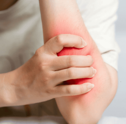
Muscle Disorders: Myositis, Strain, and Paralysis A comprehensive clinical resource for the Muscular System (Sub-topic 2.3), detailing the pathophysiology, classification, diagnosis, and management of inflammatory, mechanical, and neurological muscle conditions. 1. INTRODUCTION Muscle disorders represent a significant category of clinical conditions encountered in general medical practice. These disorders may result from inflammation, mechanical injury, or neurological dysfunction. This chapter examines three specific entities: myositis (inflammation of muscle), muscle strain (mechanical injury to fibers), and paralysis (loss of function due to neurological impairment). 2. MYOSITIS Myositis is defined as the inflammation of skeletal muscle. It may be acute or chronic, localized or generalized, occurring as an isolated condition or as part of a systemic disease. Involvement may include muscle fibers, connective tissue, or intramuscular blood vessels. 2.1 Classification and Types Infectious Myositis: Caused by bacterial, viral, fungal, or parasitic organisms. Bacterial myositis (pyomyositis): Common in tropical regions and immunocompromised states; Staphylococcus aureus is the most common pathogen. Viral myositis: Caused by influenza, coxsackievirus, EBV, or HIV. Parasitic myositis: Includes trichinellosis (Trichinella spiralis) and toxoplasmosis. Idiopathic Inflammatory Myopathies (IIM): Autoimmune disorders including: Dermatomyositis: Features proximal muscle weakness and characteristic skin rashes (heliotrope rash on eyelids, Gottron papules over knuckles). Associated with malignancy and interstitial lung disease. Polymyositis: Progressive, symmetric proximal weakness without skin involvement. Inclusion body myositis: Most common in older adults; causes asymmetric weakness of finger flexors and quadriceps. Often refractory to treatment. Drug-induced Myositis: Triggered by statins (cholesterol-lowering), colchicine, chloroquine, and zidovudine. Ranges from myalgia to severe rhabdomyolysis. 2.2 Pathophysiology The mechanism varies by etiology. In infectious forms, organisms invade tissue directly, triggering an acute response with neutrophils and macrophages releasing proteolytic enzymes. In autoimmune forms, autoreactive T cells target muscle antigens leading to fiber necrosis. In dermatomyositis, a complement-mediated microangiopathy causes ischemic damage to the fibers. 2.3 Clinical Features The hallmark is proximal muscle weakness (shoulders and hips). Patients report difficulty climbing stairs, rising from chairs, or lifting objects overhead. Muscle pain (myalgia): Severe in infectious; mild/absent in idiopathic. Muscle tenderness: Palpation reveals tenderness and potential swelling. Systemic: Fatigue, fever (infectious), and dysphagia (pharyngeal involvement). Dermatological: Heliotrope rash and shawl sign (dermatomyositis). 2.4 Diagnosis and Management Blood tests: Creatine Kinase (CK) is the most sensitive marker for damage. Autoantibodies (anti-Jo-1, anti-Mi-2) support IIM diagnosis. Imaging: MRI (T2-weighted/STIR) is the modality of choice for identifying inflammation. Muscle Biopsy: The gold standard; shows inflammatory infiltration and fiber necrosis. Management Principle Autoimmune Myositis: First-line treatment is high-dose corticosteroids (prednisone 1 mg/kg/day). Steroid-sparing agents (methotrexate, azathioprine) are added for resistant cases. Drug-induced: Immediate discontinuation of the offending agent. Severe cases require aggressive hydration to prevent acute kidney injury. 3. MUSCLE STRAIN A muscle strain (pulled muscle) is an injury to a muscle or its tendons caused by overstretching or excessive force. Strains involve muscles/tendons, whereas sprains involve ligaments. 3.1 Etiology and Vulnerability Occurs during sudden acceleration or deceleration. Common mechanisms include direct trauma, muscle fatigue, and inadequate warm-up. The hamstrings (biceps femoris) are most vulnerable as they cross two joints (hip and knee). 3.2 Classification by Severity Grade Severity Pathology Healing Time Grade I Mild Few fibers torn; minimal swelling; no strength loss. 1–2 weeks Grade II Moderate Moderate fibers torn; intact muscle; noticeable weakness/bruising. 3–6 weeks Grade III Severe Complete rupture of muscle or tendon; palpable defect; complete loss of function. 3–6 months 3.4 Management: The PRICE Protocol In the acute phase (first 48 hours): P — Protection: Use splints or crutches. R — Rest: Avoid activities that cause pain (relative rest). I — Ice: Apply cold packs for 15–20 mins every 2–3 hours. C — Compression: Elastic bandages to minimize swelling. E — Elevation: Raise limb above heart level to reduce edema. Surgical Note Surgical management is reserved for Grade III strains with complete rupture, particularly involving the Achilles tendon or pectoralis major in athletes. 4. PARALYSIS Paralysis is the complete or partial loss of muscle function resulting from damage anywhere along the motor pathway, from the motor cortex to the neuromuscular junction (NMJ). 4.1 Classification By Distribution: — Monoplegia: One limb. — Hemiplegia: One side of the body. — Paraplegia: Both lower limbs. — Quadriplegia: All four limbs. By Muscle Tone: — Flaccid: Loss of tone (hypotonia), absent reflexes, muscle atrophy. Caused by LMN lesions. — Spastic: Increased tone (hypertonia), hyperreflexia, clonus, Babinski sign. Caused by UMN lesions. 4.2 Causes and Clinical Features Upper Motor Neuron (UMN) Lesions: Originate in the cortex. Causes: Stroke, MS, Cerebral Palsy. Features: Spasticity, weakness in extensors (upper) and flexors (lower). Lower Motor Neuron (LMN) Lesions: Originate in the anterior horn. Causes: Polio, GBS, Nerve trauma. Features: Fasciculations and severe neurogenic atrophy. Clinical Differentiation Facial Paralysis In Bell’s palsy (LMN), the entire half of the face is affected (cannot wrinkle forehead). In a Stroke (UMN), the forehead is spared because the frontalis muscle receives bilateral cortical innervation. 4.6 Summary Comparison Table: UMN vs. LMN Feature Upper Motor Neuron (UMN) Lower Motor Neuron (LMN) Muscle Tone Increased (Spasticity) Decreased (Flaccidity) Reflexes Hyperreflexia Hyporeflexia or absent Babinski Sign Present (Upgoing toe) Absent Atrophy Mild (Disuse) Severe (Neurogenic) Fasciculations Absent Present Surgical Emergency Cauda Equina Syndrome Compression of nerve roots below L1–L2. Red flags: saddle anesthesia, bladder/bowel dysfunction. Decompression within 48 hours is required to improve outcomes. 5. KEY POINTS SUMMARY Myositis: CK is the most sensitive lab marker; MRI is the choice for imaging. Muscle Strain: Grade III is a complete rupture requiring potential surgery; hamstrings are most frequently injured. Paralysis: UMN lesions result in spasticity; LMN lesions result in flaccidity and fasciculations. Bell’s Palsy: An LMN facial nerve lesion affecting the entire ipsilateral half of the face.

Physiology of muscle contraction
Anatomy

Physiology of muscle contraction

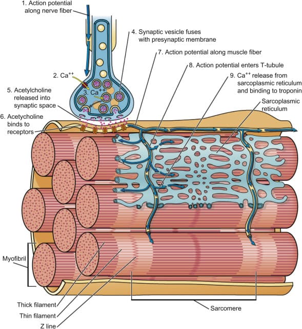
Physiology of Muscle Contraction Complete detailed notes on the mechanisms and regulation of skeletal muscle contraction, covering the neuromuscular junction, sarcomere architecture, sliding filament theory, and clinical correlations. 1. Introduction Muscle contraction is a complex physiological process that converts chemical energy into mechanical force. Understanding these mechanisms is essential for diagnosing neuromuscular disorders and managing patients with muscle-related conditions. This study covers the structural and functional integration of the nervous and muscular systems. 2. The Neuromuscular Junction (NMJ) The Neuromuscular Junction (NMJ) is a specialized synapse between a motor neuron and a skeletal muscle fiber. It is the critical site where neural signals initiate muscular action. 2.1 Structure of the NMJ Presynaptic Terminal: The axon terminal of the motor neuron containing numerous synaptic vesicles filled with acetylcholine (ACh). It features voltage-gated calcium channels in its membrane. Synaptic Cleft: A narrow extracellular space (~50 nm wide). It contains the enzyme acetylcholinesterase (AChE), which terminates the signal by hydrolyzing ACh. Postsynaptic Membrane (Motor End Plate): The specialized region of the sarcolemma containing nicotinic acetylcholine receptors (nAChR). These are concentrated in junctional folds that maximize surface area. Each nAChR consists of five subunits (two alpha, one beta, one gamma/delta, one epsilon). 2.2 Events at the NMJ (Step-by-Step) Step 1 — Arrival of action potential: The impulse reaches the presynaptic terminal, opening voltage-gated calcium (Ca2+) channels. Step 2 — Calcium influx and vesicle fusion: Ca2+ triggers the exocytosis of ACh into the synaptic cleft. Step 3 — ACh binding: ACh diffuses across the cleft and binds to the alpha subunits of nAChR on the motor end plate. Step 4 — Depolarization: Receptors open, allowing sodium (Na+) influx and potassium (K+) efflux. This produces an End-Plate Potential (EPP). Step 5 — Muscle fiber action potential: The EPP is typically suprathreshold, triggering a general action potential that spreads across the sarcolemma. Step 6 — ACh hydrolysis: AChE breaks down ACh into acetate and choline, preventing continuous stimulation. Key Points ACh is the primary neurotransmitter of the NMJ. The EPP is always suprathreshold under normal physiological conditions. AChE is vital for the precise control of muscle relaxation. 3. Sarcomere Structure The sarcomere is the basic functional unit of skeletal muscle contraction, defined as the region between two adjacent Z-discs. 3.1 Thin Filaments (Actin) Composed primarily of Actin. G-actin (globular) polymerizes into F-actin (filamentous). In the resting state, myosin-binding sites are blocked by tropomyosin. The Troponin complex regulates this: Troponin T: Binds to tropomyosin. Troponin I: Inhibits the actin-myosin interaction. Troponin C: Binds calcium ions. 3.2 Thick Filaments (Myosin) Composed of Myosin II. Each molecule consists of two heavy chains (forming the tail and heads) and four light chains. Each head contains an ATP-binding site and an actin-binding site. 3.3 Bands and Zones A band: The dark band representing the full length of thick filaments. I band: The light band containing only thin filaments; bisected by the Z-disc. H zone: The center of the A band containing only thick filaments (no overlap). M line: Protein line in the center of the H zone that anchors thick filaments. Z-disc: Anchors thin filaments; defines the sarcomere boundary. 4. The Sliding Filament Theory Proposed by Huxley and Hanson (1954), this theory states that muscle shortening occurs because thin filaments slide past thick filaments without the individual filaments changing length. 4.1 The Cross-Bridge Cycle Step 1 — Cross-bridge formation: Calcium binds to Troponin C, moving tropomyosin and exposing binding sites. The myosin head (bound to ADP + Pi) attaches to actin. Step 2 — Power stroke: Release of Pi triggers the head to pivot, pulling the thin filament ~10 nm toward the M line. ADP is released. Step 3 — Cross-bridge detachment: A new ATP binds to the myosin head, causing it to release from actin. Step 4 — Reactivation: Myosin hydrolyzes ATP into ADP + Pi, “re-cocking” the head into its high-energy state. Rigor Mortis ATP is required for detachment. In the absence of ATP (after death), cross-bridges remains permanently bound, resulting in the muscle stiffness known as rigor mortis. 5. Excitation-Contraction Coupling This is the process by which an electrical action potential triggers the mechanical release of calcium for contraction. 5.1 Steps of Coupling Action potential propagation: The signal travels down the T-tubules to reach the interior of the fiber. DHP Receptor activation: Voltage-gated Dihydropyridine (DHP) receptors in the T-tubule sense the change in voltage. Ryanodine Receptor (RyR) opening: DHP receptors are mechanically coupled to RyR channels on the Sarcoplasmic Reticulum (SR). Opening RyR allows Ca2+ to flood the cytosol. Calcium-Troponin binding: Ca2+ binds to Troponin C, initiating the cross-bridge cycle. SERCA Reuptake: Relaxation occurs when Ca2+ is actively pumped back into the SR by the Sarcoplasmic/Endoplasmic Reticulum Calcium ATPase (SERCA). 6. Energy Sources for Muscle Contraction Muscles require a continuous supply of ATP, but only store a few seconds’ worth. Rapid regeneration occurs via three systems: Energy System Substrate ATP Yield Duration Oxygen? Phosphocreatine Phosphocreatine 1 ATP per PCr 10–15 seconds No Anaerobic Glycolysis Glucose/Glycogen 2 ATP per glucose 1–2 minutes No Aerobic Respiration Glucose, Fatty Acids 30–32 ATP per glucose Hours Yes 7. Types of Muscle Contraction Isotonic Contraction: Muscle changes length while tension remains constant. Concentric: Muscle shortens (e.g., upward phase of a bicep curl). Eccentric: Muscle lengthens while generating tension (e.g., controlled lowering of a weight). Isometric Contraction: Muscle generates tension without changing length (e.g., holding a heavy object stationary). Isokinetic Contraction: Muscle contracts at a constant velocity against variable resistance (requires specialized equipment). 8. Clinical Relevance Pathology Myasthenia Gravis An autoimmune disorder where antibodies block or destroy nicotinic ACh receptors at the NMJ. Results in progressive muscle weakness that worsens with activity. Diagnosed via the edrophonium test. Pathology Malignant Hyperthermia A genetic disorder of Ryanodine Receptors (RyR) triggered by volatile anesthetics. Causes massive, uncontrolled calcium release from the SR, leading to sustained contraction, hyperthermia, and fatal hyperkalemia. Treated with dantrolene (RyR antagonist). Pathology Botulism Produced by Clostridium botulinum. The toxin cleaves SNARE proteins, preventing the release of ACh from presynaptic terminals. Results in flaccid

Scroll to Top