Tricarboxylic Acid (TCA) Cycle (Krebs Cycle / Citric Acid Cycle)

Tricarboxylic Acid (TCA) Cycle (Krebs Cycle / Citric Acid Cycle)

Tricarboxylic Acid (TCA) Cycle: (Krebs Cycle / Citric Acid Cycle)

The Tricarboxylic Acid (TCA) Cycle (Krebs Cycle / Citric Acid Cycle)

The Tricarboxylic Acid (TCA) cycle, also famously known as the Krebs cycle or the Citric Acid Cycle, is a central hub of metabolism. It's a metabolic superhighway where the breakdown products of carbohydrates, fats, and proteins converge for final oxidation.

Central Role in Aerobic Respiration:

The TCA cycle is the second major stage of aerobic respiration. Unlike glycolysis, the TCA cycle requires oxygen indirectly to function, as its products (NADH and FADH₂) ultimately feed into the electron transport chain, which absolutely depends on oxygen. Without the ETC running, the NAD⁺ and FAD needed for the TCA cycle would not be regenerated, and the cycle would grind to a halt.

Main Function: Complete Oxidation of Acetyl-CoA:

The primary catabolic function of the TCA cycle is the complete oxidation of acetyl-CoA to carbon dioxide (CO₂). This acetyl-CoA is primarily derived from:

  • Carbohydrates: Pyruvate (from glycolysis) is converted to acetyl-CoA.
  • Fats: Fatty acids are broken down into acetyl-CoA via beta-oxidation.
  • Proteins: Certain amino acids are degraded into acetyl-CoA or other TCA cycle intermediates.

As acetyl-CoA is oxidized, the cycle captures the released energy in the form of high-energy electron carriers (NADH and FADH₂) and a small amount of GTP (which is interconvertible with ATP). These carriers are then channeled into the Electron Transport Chain (ETC) to drive the synthesis of the vast majority of cellular ATP.

Amphibolic Nature (Both Catabolic and Anabolic Roles):

One of the most fascinating aspects of the TCA cycle is its amphibolic nature, meaning it serves both catabolic (breakdown) and anabolic (synthesis) roles.

  • Catabolism: It catabolizes acetyl-CoA to CO₂, generating ATP, NADH, and FADH₂.
  • Anabolism: Many of the intermediates are precursors for various biosynthetic pathways. For example:
    • Citrate can be used for fatty acid and cholesterol synthesis.
    • α-Ketoglutarate is a precursor for several amino acids (e.g., glutamate).
    • Succinyl-CoA is used in the synthesis of porphyrins (like heme).
    • Oxaloacetate is a precursor for amino acids and glucose (via gluconeogenesis).

Because these intermediates are often "siphoned off" for synthesis, the cell has mechanisms (called anaplerotic reactions) to replenish them.

Location:

The location of the TCA cycle is critical to its function and regulation.

  • Mitochondrial Matrix: In eukaryotic cells, the entire TCA cycle takes place within the mitochondrial matrix, the innermost compartment of the mitochondrion.
  • Why is this significant?
    • Proximity to ETC: NADH and FADH₂ are produced directly where they are needed, ensuring efficient energy transfer to the ETC on the inner membrane.
    • Isolation and Concentration: Confining the cycle within the matrix allows for the concentration of substrates and enzymes.
    • Coupling with Oxidative Phosphorylation: This spatial arrangement is essential for the effective coupling of the TCA cycle with ATP production.

In prokaryotic cells, which lack mitochondria, the TCA cycle enzymes are found in the cytosol.

Entry Point: Pyruvate Dehydrogenase Complex (PDC)

The Pyruvate Dehydrogenase Complex (PDC) is a critical bridge between glycolysis and the TCA cycle. It's not part of the TCA cycle itself, but it's an absolutely essential prerequisite for aerobic glucose metabolism to proceed.

Irreversible Conversion of Pyruvate to Acetyl-CoA:

The PDC catalyzes an irreversible oxidative decarboxylation of pyruvate to form acetyl-CoA. This reaction is a metabolic crossroads: once pyruvate is converted to acetyl-CoA, it cannot be converted back to glucose. The fate of glucose is committed to complete oxidation or fatty acid synthesis.

  • Location: This reaction also takes place in the mitochondrial matrix. Pyruvate from glycolysis is transported into the matrix by a specific translocase protein.

Overall Reaction and Coenzymes Involved:

The PDC is a complex of three distinct enzymes and five different coenzymes. The overall reaction is:

Pyruvate + CoA + NAD⁺ → Acetyl-CoA + CO₂ + NADH + H⁺

The Five Coenzymes (or Prosthetic Groups):

  • Thiamine Pyrophosphate (TPP): From vitamin B1 (thiamine). Decarboxylates pyruvate.
  • Lipoate (Lipoamide): Transfers the acetyl group to CoA.
  • Flavin Adenine Dinucleotide (FAD): From vitamin B2 (riboflavin). Re-oxidizes the reduced lipoamide.
  • Nicotinamide Adenine Dinucleotide (NAD⁺): From vitamin B3 (niacin). Re-oxidizes FADH₂.
  • Coenzyme A (CoA): From vitamin B5 (pantothenic acid). Accepts the acetyl group.

Regulation of the PDC:

Because this step is irreversible, the PDC is a crucial point of regulation.

  • Allosteric Regulation:
    • Inhibitors (high energy signals): Acetyl-CoA, NADH, ATP.
    • Activators (low energy signals): CoA, NAD⁺, AMP.
  • Covalent Modification (Phosphorylation/Dephosphorylation): This is the primary long-term regulatory mechanism.
    • A PDC Kinase adds a phosphate group to INACTIVATE the PDC. The kinase is activated by high energy signals (ATP, NADH, Acetyl-CoA).
    • A PDC Phosphatase removes the phosphate group to ACTIVATE the PDC. The phosphatase is activated by Ca²⁺ and insulin.

In summary, when the cell has plenty of energy, the PDC is turned off. When energy is needed, the PDC is activated.


The Cycle Itself (Key Steps, Enzymes, and Products)

The cycle consists of eight enzymatic steps, leading to the complete oxidation of the two carbons from acetyl-CoA and the regeneration of oxaloacetate.

Overall Summary of One Turn of the Cycle:

  • A four-carbon oxaloacetate condenses with a two-carbon acetyl unit (from acetyl-CoA) to yield a six-carbon citrate.
  • Citrate is isomerized and then oxidatively decarboxylated to form five-carbon α-ketoglutarate, releasing one molecule of CO₂ and producing NADH.
  • α-ketoglutarate is oxidatively decarboxylated to yield four-carbon succinyl-CoA, releasing another CO₂ and producing NADH.
  • Succinyl-CoA is converted to succinate, generating one GTP (or ATP).
  • Oxaloacetate is regenerated from succinate through a series of steps involving fumarate and malate, producing one FADH₂ and one NADH.

In total, two carbon atoms (from acetyl-CoA) enter the cycle, and two carbon atoms leave as CO₂. Energy is captured as 3 NADH, 1 FADH₂, and 1 GTP (ATP).

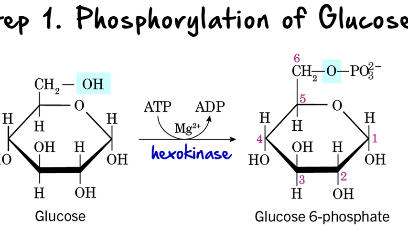
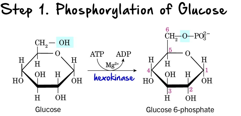
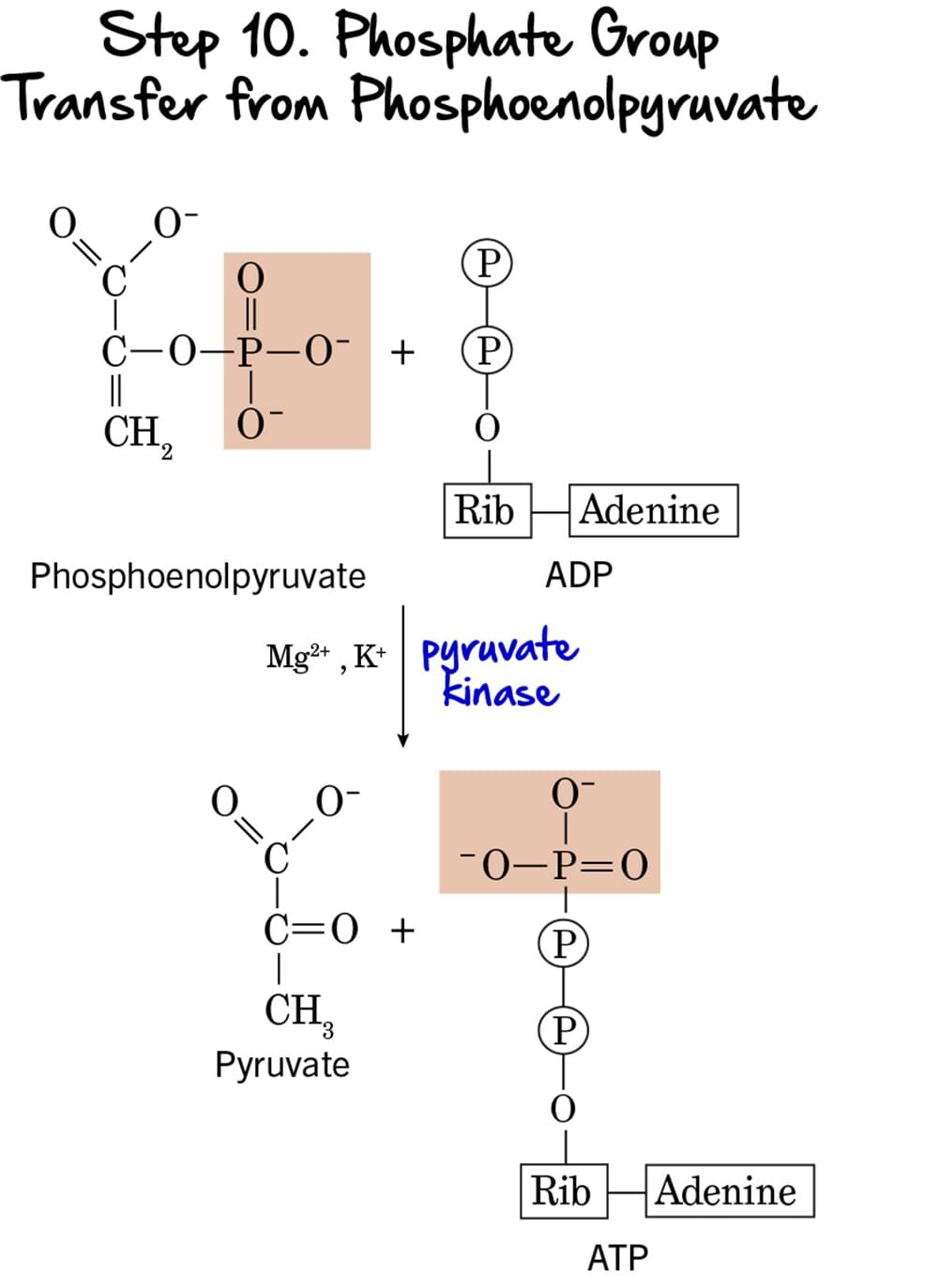
The Reactions of the Citric Acid Cycle

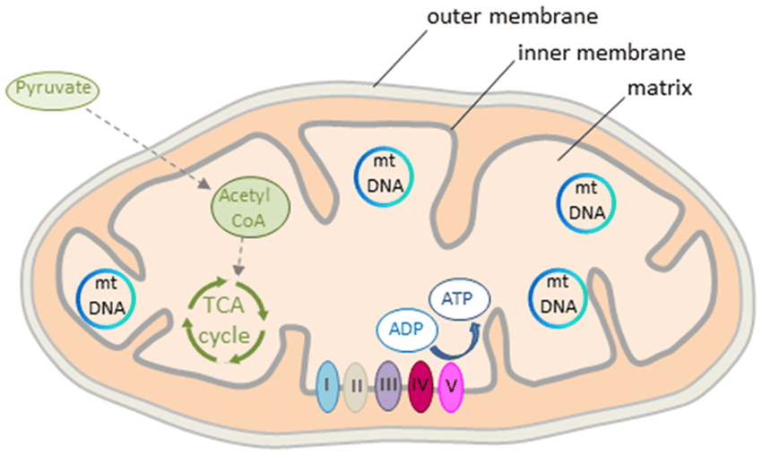
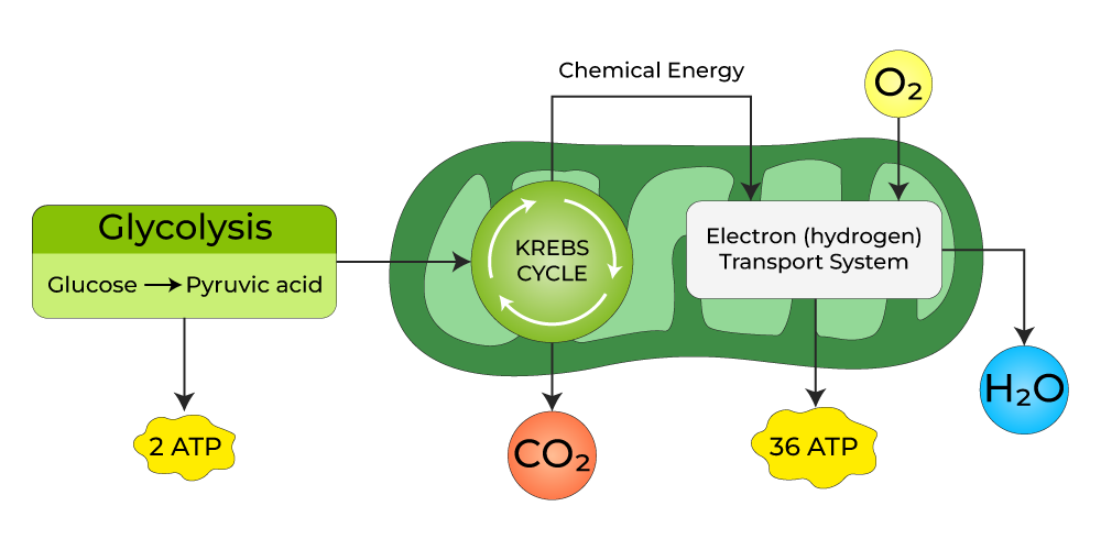
Now that Acetyl-CoA has been generated in the mitochondrial matrix, it enters the eight-step cyclical pathway. Each turn of the cycle processes one molecule of Acetyl-CoA, systematically oxidizing its two carbons to CO₂ while capturing high-energy electrons in the form of NADH and FADH₂. Let's examine each step.


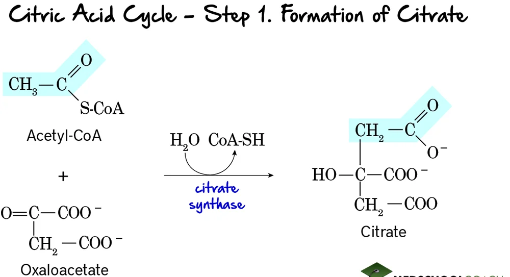
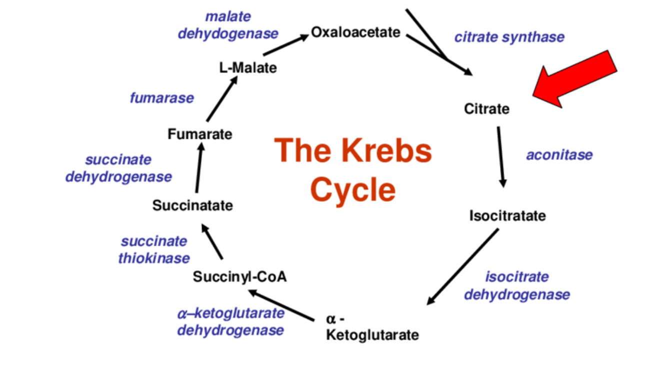
Step 1: Formation of Citrate

This is the entry point for the 2-carbon acetyl group into the citric acid cycle. It involves the condensation of a 2-carbon unit with a 4-carbon molecule to form a 6-carbon molecule.

Reaction:

Acetyl-CoA (a 2-carbon molecule) condenses with Oxaloacetate (a 4-carbon molecule). This reaction, accompanied by the hydrolysis of the thioester bond in Acetyl-CoA, forms Citrate, a 6-carbon tricarboxylic acid.

Key Features of Step 1:

  • Enzyme: The reaction is catalyzed by Citrate Synthase. This is a crucial regulatory enzyme of the TCA cycle.
  • Reactants: Acetyl-CoA and Oxaloacetate.
  • Product: Citrate and Coenzyme A (CoA-SH).
  • Type of Reaction: This is a condensation reaction.
  • Irreversibility: The reaction is highly exergonic and essentially irreversible under cellular conditions, making it a key control point.
  • Regulation: Citrate synthase is allosterically inhibited by high levels of ATP, NADH, succinyl-CoA, and its own product, citrate. These are all signals that the cell has a high energy charge and an abundance of metabolic intermediates.

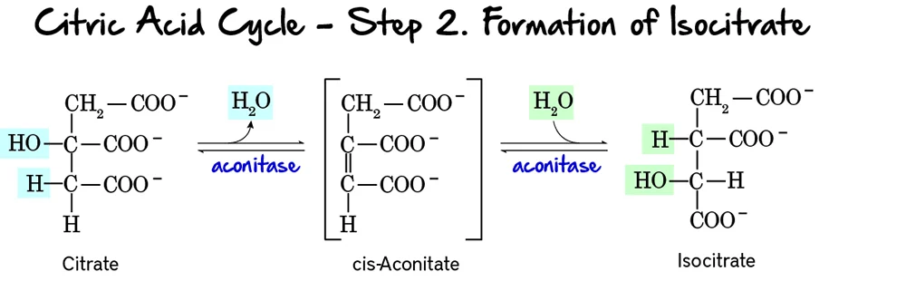
Step 2: Formation of Isocitrate

This step involves the isomerization of citrate to isocitrate. This rearrangement is crucial because the hydroxyl group of isocitrate is positioned to be oxidized in the next step.

Reaction:

Citrate, a tertiary alcohol, is isomerized to Isocitrate, a secondary alcohol. The reaction occurs in two substeps: first, a molecule of water is removed to form cis-Aconitate, and then water is re-added in a different position.

Key Features of Step 2:

  • Enzyme: The enzyme catalyzing this reversible reaction is Aconitase. It contains an iron-sulfur cluster essential for its activity.
  • Reactant: Citrate.
  • Product: Isocitrate.
  • Type of Reaction: An isomerization, specifically an intramolecular rearrangement involving dehydration and rehydration.
  • Reversibility: The reaction is reversible, but the subsequent steps quickly consume isocitrate, pulling the reaction forward.

Purpose of this Step:

  • Preparation for Oxidation: Citrate, being a tertiary alcohol, is not readily oxidizable. The isomerization to isocitrate, a secondary alcohol, positions the hydroxyl group at a carbon that can be easily oxidized in the next step.
  • Stereospecificity: Aconitase catalyzes a stereospecific conversion, meaning it produces a specific isomer of isocitrate.

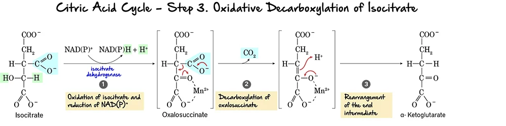
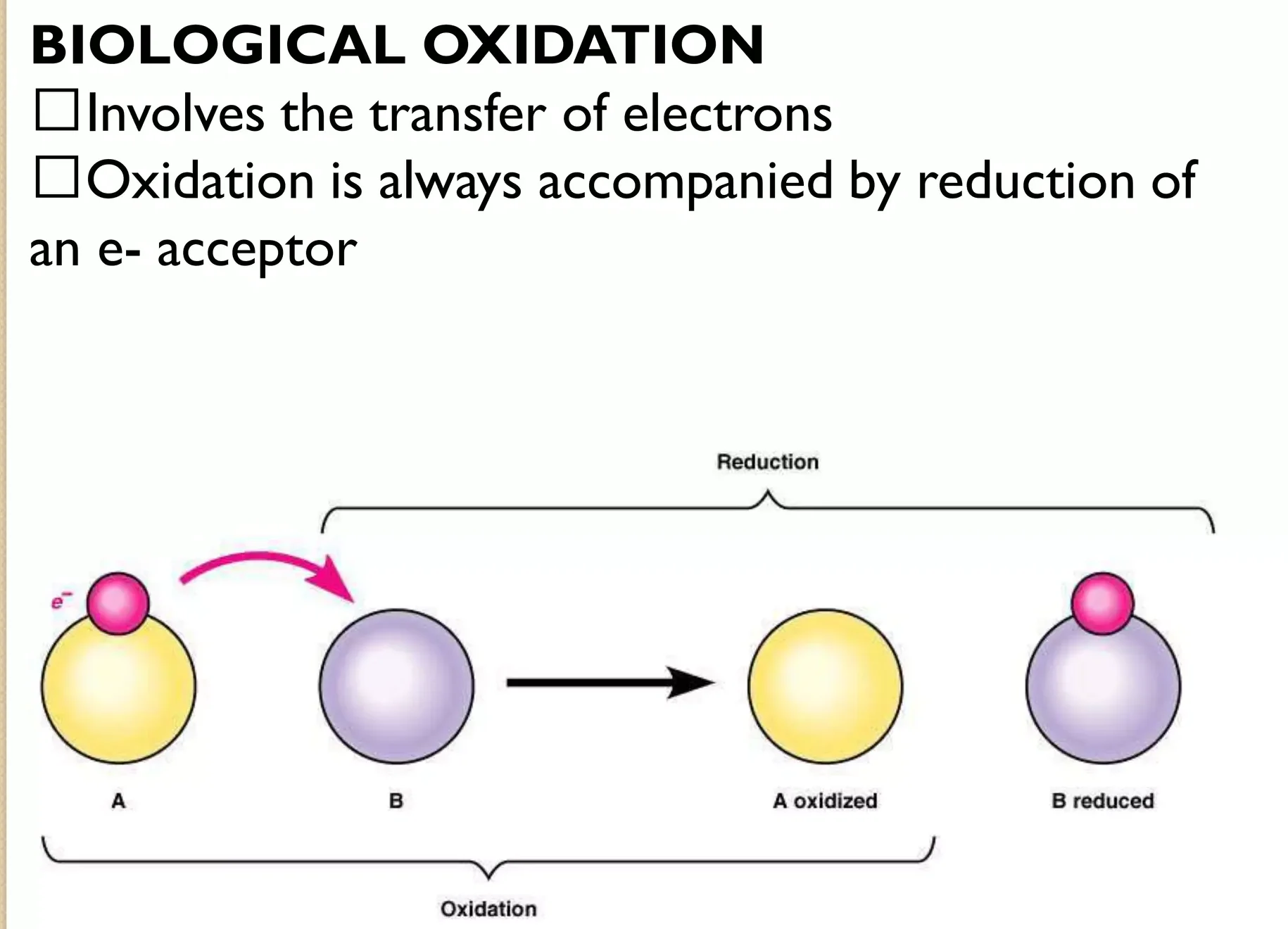
Step 3: Oxidative Decarboxylation of Isocitrate

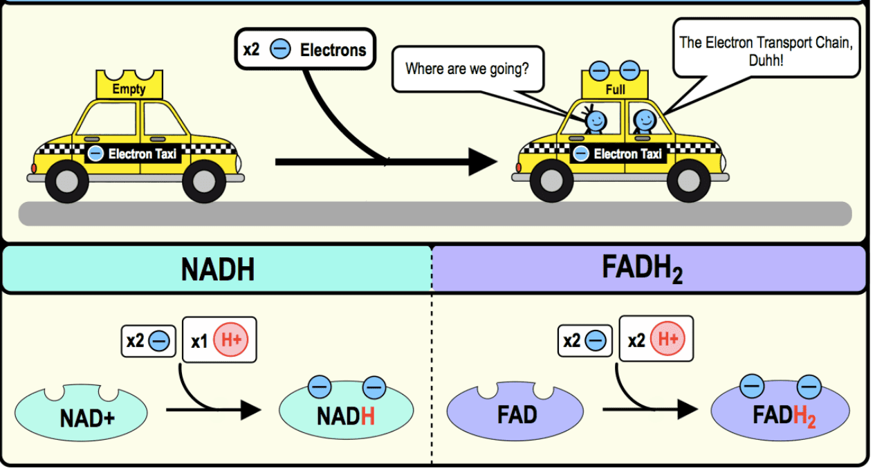
This is the first oxidative step in the citric acid cycle, where the first molecule of carbon dioxide is released and the first NADH is produced.

Reaction:

Isocitrate undergoes an oxidative decarboxylation reaction. This involves two main parts:

  1. Oxidation: The hydroxyl group on isocitrate is oxidized to a keto group, reducing NAD⁺ to NADH.
  2. Decarboxylation: The beta-keto acid intermediate, Oxalosuccinate, immediately loses a molecule of carbon dioxide (CO₂), forming alpha-Ketoglutarate.

Key Features of Step 3:

  • Enzyme: The reaction is catalyzed by Isocitrate Dehydrogenase. This enzyme requires Mn²⁺ as a cofactor.
  • Reactant: Isocitrate.
  • Product: alpha-Ketoglutarate and CO₂.
  • Electron Carriers Reduced: NAD⁺ is reduced to NADH.
  • ATP Change: 0 ATP directly produced.

Purpose of this Step:

  • CO₂ Release: This is the first of two carbon atoms released as CO₂ from the original acetyl unit.
  • NADH Production: The generation of NADH is vital for ATP production via oxidative phosphorylation.
  • Regulation: Isocitrate Dehydrogenase is a crucial regulatory enzyme. It is allosterically activated by ADP (indicating low energy) and inhibited by ATP and NADH (indicating high energy).

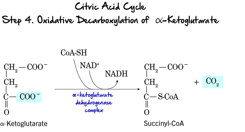
Step 4: Oxidative Decarboxylation of alpha-Ketoglutarate

This is the second and final oxidative decarboxylation step in the citric acid cycle. It is remarkably similar in mechanism to the pyruvate dehydrogenase complex reaction.

Reaction:

alpha-Ketoglutarate undergoes oxidative decarboxylation to form Succinyl-CoA. This complex reaction involves the release of another molecule of CO₂, the reduction of NAD⁺ to NADH, and the incorporation of Coenzyme A.

Key Features of Step 4:

  • Enzyme: This reaction is catalyzed by the alpha-Ketoglutarate Dehydrogenase Complex. This is a multi-enzyme complex requiring several coenzymes (Thiamine pyrophosphate (TPP), Lipoamide, FAD, NAD⁺, and CoA-SH).
  • Reactant: alpha-Ketoglutarate.
  • Product: Succinyl-CoA, CO₂, and NADH.
  • Electron Carriers Reduced: NAD⁺ is reduced to NADH.
  • ATP Change: 0 ATP directly produced.

Purpose of this Step:

  • CO₂ Release: This is the second and last carbon atom released as CO₂. At this point, both carbons from the initial acetyl-CoA have been fully oxidized.
  • NADH Production: This NADH contributes significantly to ATP production.
  • Formation of a High-Energy Thioester: The formation of succinyl-CoA, with its high-energy thioester bond, primes the molecule for the substrate-level phosphorylation step that follows.
  • Regulation: The alpha-Ketoglutarate Dehydrogenase Complex is another regulatory point. It is inhibited by its products, Succinyl-CoA and NADH, and also by high ATP levels.

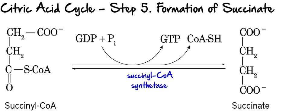
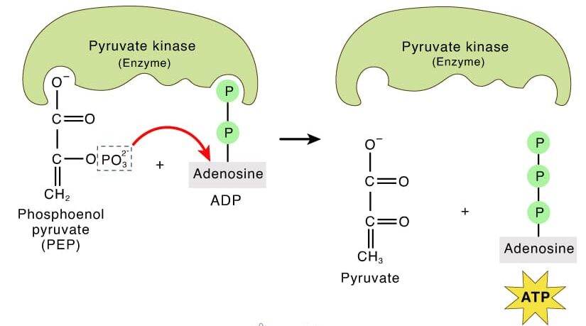
Step 5: Formation of Succinate (Substrate-Level Phosphorylation)

This is the only step in the Citric Acid Cycle that directly generates a high-energy phosphate compound (GTP or ATP) through substrate-level phosphorylation.

Reaction:

The high-energy thioester bond of Succinyl-CoA is hydrolyzed. The energy released drives the phosphorylation of GDP to GTP. Coenzyme A is released, and Succinate is formed.

Key Features of Step 5:

  • Enzyme: Succinyl-CoA Synthetase (also known as Succinate Thiokinase).
  • Reactant: Succinyl-CoA.
  • Products: Succinate, CoA-SH, and GTP (or ATP).
  • Energy Production: 1 GTP is produced per turn. GTP can be readily converted to ATP (GTP + ADP ↔ GDP + ATP).

Purpose of this Step:

  • ATP/GTP Generation: This provides a direct energy yield for the cell.
  • Regeneration of Succinate: Succinate is now available for further processing.
  • Removal of CoA: The release of free CoA is important for other enzyme complexes to function.

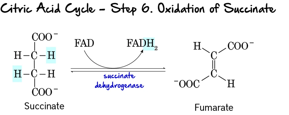
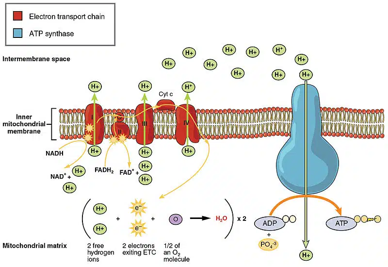
Step 6: Oxidation of Succinate

This is the second oxidative step in the cycle, where electrons are transferred to FAD, producing FADH₂.

Reaction:

Succinate is oxidized to Fumarate through the removal of two hydrogen atoms. These hydrogens are accepted by FAD, which is reduced to FADH₂. This reaction forms a double bond in fumarate.

Key Features of Step 6:

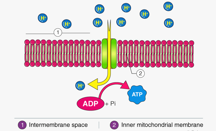
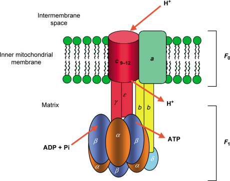
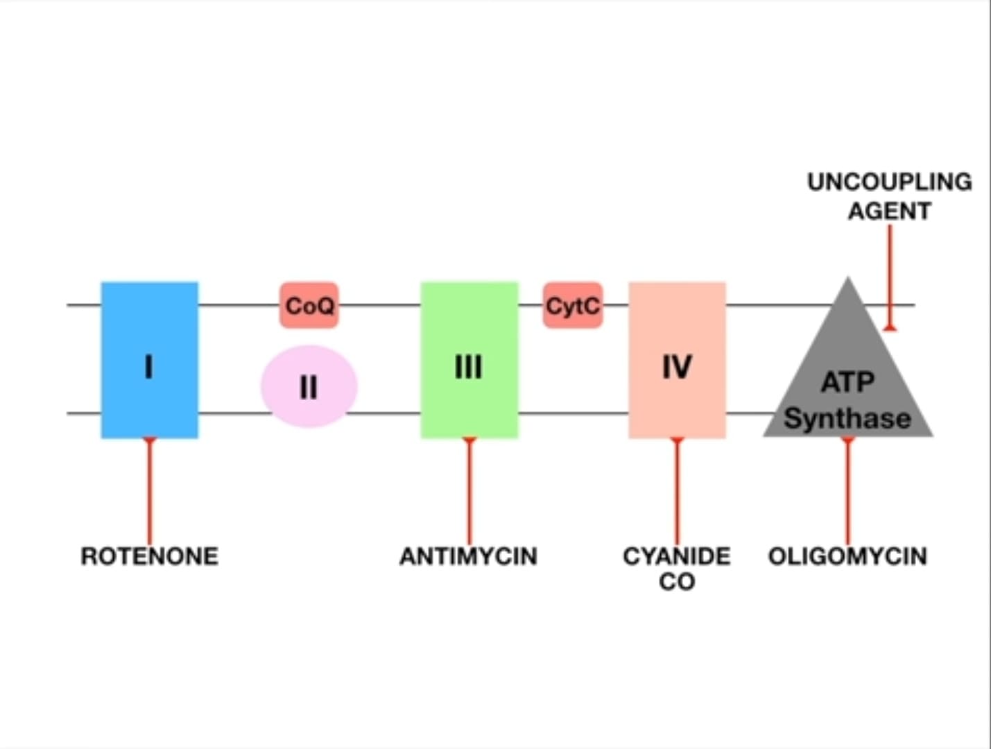
  • Enzyme: Succinate Dehydrogenase. This enzyme is unique as it is an integral protein of the inner mitochondrial membrane and is directly part of the electron transport chain (Complex II).
  • Reactant: Succinate.
  • Products: Fumarate and FADH₂.
  • Electron Carriers Reduced: FAD is reduced to FADH₂.
  • ATP Change: 0 ATP directly produced.

Purpose of this Step:

  • FADH₂ Production: FADH₂ is another high-energy electron carrier that will donate its electrons to the ETC. It yields fewer ATP than NADH because it enters the ETC at a lower energy level.
  • Connection to Electron Transport Chain: Being part of Complex II directly links the Citric Acid Cycle to the ETC, facilitating efficient electron transfer.

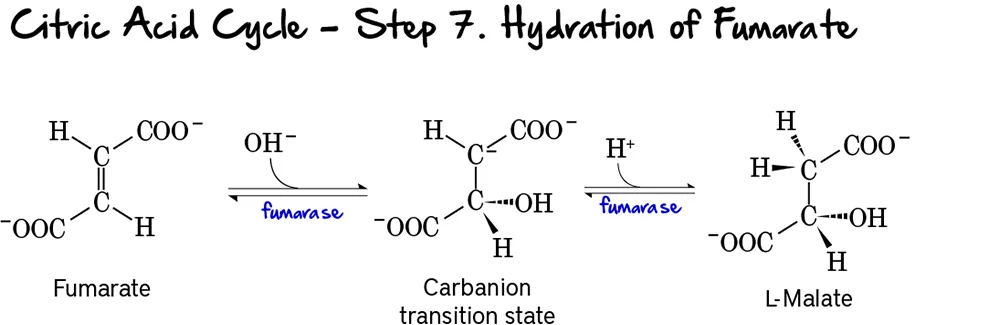
Step 7: Hydration of Fumarate

This step involves the stereospecific addition of water across the double bond of fumarate, forming L-malate.

Reaction:

Fumarate undergoes a hydration reaction, where a molecule of water is added across its double bond. This reaction forms L-Malate.

Key Features of Step 7:

  • Enzyme: Fumarase (also known as Fumarate Hydratase).
  • Reactant: Fumarate.
  • Product: L-Malate.
  • Type of Reaction: This is a hydration reaction.
  • Stereospecificity: Fumarase is highly stereospecific, forming specifically L-malate (not D-malate).
  • Reversibility: This reaction is reversible.

Purpose of this Step:

  • Preparation for Oxidation: The addition of water creates a hydroxyl group on L-malate, which is necessary for the subsequent oxidation step.
  • Regeneration of Oxaloacetate: This step is crucial for setting up the regeneration of oxaloacetate.

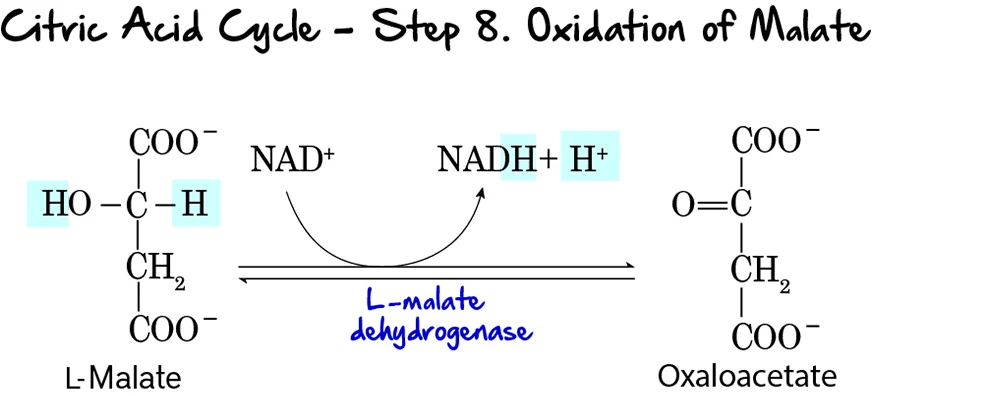
Step 8: Oxidation of Malate

This is the final step of the Citric Acid Cycle, regenerating oxaloacetate and producing the last NADH of the cycle.

Reaction:

L-Malate is oxidized to Oxaloacetate. During this oxidation, NAD⁺ is reduced to NADH and H⁺. This completes the regeneration of oxaloacetate, which is now ready to condense with another molecule of acetyl-CoA.

Key Features of Step 8:

  • Enzyme: L-Malate Dehydrogenase.
  • Reactant: L-Malate.
  • Products: Oxaloacetate and NADH + H⁺.
  • Electron Carriers Reduced: NAD⁺ is reduced to NADH.
  • Reversibility: This reaction has a highly positive standard free energy change (ΔG°'), making it thermodynamically unfavorable. However, in the cell, the rapid consumption of oxaloacetate by citrate synthase (Step 1) pulls this reaction forward.

Purpose of this Step:

  • Regeneration of Oxaloacetate: This is the most critical function, ensuring the cycle can continue to operate.
  • NADH Production: This produces the third and final molecule of NADH generated directly within the cycle (per acetyl-CoA).
  • Completion of the Cycle: With the regeneration of oxaloacetate, the cycle is complete.

Energy Yield

The primary purpose of breaking down glucose is to generate ATP. While glycolysis and the TCA cycle directly produce a small amount, the vast majority of ATP is generated indirectly through oxidative phosphorylation, utilizing the NADH and FADH₂ produced.

Let's summarize the yield from one molecule of glucose through the complete process.

1. Glycolysis (Cytosol):

  • Net ATP: 2 ATP (via substrate-level phosphorylation)
  • NADH: 2 NADH

2. Pyruvate Oxidation (PDC - Mitochondrial Matrix):

Since one glucose yields two pyruvate molecules, this reaction occurs twice.

  • CO₂: 2 CO₂ (1 per pyruvate)
  • NADH: 2 NADH (1 per pyruvate)

3. TCA Cycle (Mitochondrial Matrix):

Since two acetyl-CoA molecules enter the cycle (from one glucose), the cycle runs twice.

Per turn of the cycle (i.e., per Acetyl-CoA):

  • GTP/ATP: 1 GTP (equivalent to 1 ATP)
  • NADH: 3 NADH
  • FADH₂: 1 FADH₂
  • CO₂: 2 CO₂

For two turns of the cycle (i.e., per Glucose):

  • GTP/ATP: 2 GTP (equivalent to 2 ATP)
  • NADH: 6 NADH
  • FADH₂: 2 FADH₂
  • CO₂: 4 CO₂

4. Total Yield (Direct and Indirect) per Glucose Molecule:

Let's consolidate the reduced coenzymes and directly produced ATP:

Stage ATP/GTP (Direct) NADH FADH₂ CO₂
Glycolysis 2 2 0 0
Pyruvate Oxidation (x2) 0 2 0 2
TCA Cycle (x2) 2 6 2 4
TOTAL (before ETC) 4 10 2 6

5. Contribution to ATP Generation via Oxidative Phosphorylation:

Now, we account for the ATP generated from NADH and FADH₂ through the ETC. Standard estimations are:

  • 1 NADH ≈ 2.5 ATP
  • 1 FADH₂ ≈ 1.5 ATP

Using these conversion factors:

  • ATP from 10 NADH: 10 NADH * 2.5 ATP/NADH = 25 ATP
  • ATP from 2 FADH₂: 2 FADH₂ * 1.5 ATP/FADH₂ = 3 ATP

6. Overall Theoretical Maximum ATP Yield from One Glucose Molecule:

Source ATP Yield
Direct ATP/GTP 4
From 10 NADH (via ETC) 25
From 2 FADH₂ (via ETC) 3
TOTAL ATP ~32 ATP

Key Takeaways:

  • Most ATP is made indirectly: The vast majority is produced through oxidative phosphorylation, driven by NADH and FADH₂.
  • Complete Oxidation: The 6 carbons from glucose are completely oxidized to 6 molecules of CO₂.
  • Efficiency: The TCA cycle is incredibly efficient at extracting energy from acetyl-CoA and channeling it into electron carriers for maximal ATP production.

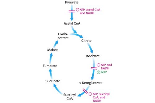
Regulation of the TCA Cycle

The TCA cycle is meticulously regulated to ensure that energy production aligns with the cell's demand. Regulation primarily occurs at the irreversible steps through allosteric control. The cycle slows down when the cell has ample ATP and speeds up when energy is needed.

Key Control Points within the Cycle:

Three main enzymes catalyze irreversible reactions and are thus primary targets for regulation:

  1. Citrate Synthase (Step 1)
  2. Isocitrate Dehydrogenase (Step 3) - Often considered a major rate-limiting step.
  3. α-Ketoglutarate Dehydrogenase Complex (Step 4)

Mechanisms of Regulation:

The TCA cycle's activity is finely tuned by several mechanisms, with allosteric modulation being the primary mode of control.

Allosteric Modulators:

This is the primary mode of regulation. Various molecules signal the cell's energy status, directly binding to and altering the activity of the regulatory enzymes.

Citrate Synthase (Step 1)

Activated by:

  • ADP (low energy)

Inhibited by:

  • ATP, NADH, Succinyl-CoA, Citrate (all are high energy signals or products).

Isocitrate Dehydrogenase (Step 3)

Activated by:

  • ADP (low energy).
  • Ca²⁺ (signals muscle contraction / energy demand).

Inhibited by:

  • ATP, NADH (high energy signals).

α-Ketoglutarate Dehydrogenase (Step 4)

Activated by:

  • Ca²⁺ (signals energy demand).

Inhibited by:

  • Succinyl-CoA, NADH (product inhibition).
  • ATP (high energy signal).

Other Regulatory Mechanisms:

  • Covalent Modification: This is not a common regulatory mechanism for the core TCA cycle enzymes in eukaryotes. However, it is the primary control method for the Pyruvate Dehydrogenase Complex (PDC), which controls the entry of acetyl-CoA into the cycle.
  • Supply of Acetyl-CoA: The activity of the PDC is a critical determinant of the flux into the TCA cycle, effectively controlling the primary fuel input.

Relating Regulation to Cellular Energy State:

The overall regulation ensures the TCA cycle's activity is finely tuned to the cell's energy demands:

  • High Energy State (High ATP/ADP, High NADH/NAD⁺): When the cell has abundant energy, the products (ATP, NADH, citrate, succinyl-CoA) accumulate and act as allosteric inhibitors, slowing down the cycle to conserve fuel.
  • Low Energy State (Low ATP/ADP, Low NADH/NAD⁺): When the cell needs energy, ATP and NADH levels drop, while ADP/AMP levels rise. These conditions act as allosteric activators. Ca²⁺ also acts as a key activator, signaling increased metabolic activity, especially in contracting muscles.
  • Substrate Availability: The availability of acetyl-CoA and oxaloacetate also influences the flux through the cycle. The maintenance of oxaloacetate levels is crucial and involves anaplerotic reactions.

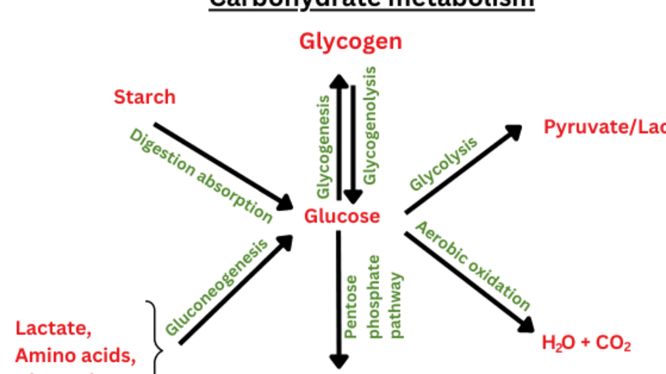
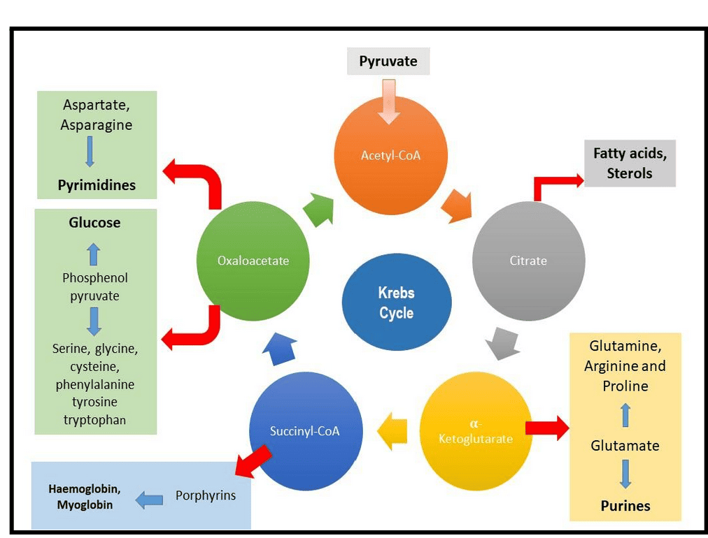
Amphibolic Nature and Anaplerotic Reactions

The TCA cycle is often presented as a purely catabolic pathway, but this is only half the story. The TCA cycle is, in fact, amphibolic, meaning it functions in both catabolic (breakdown) and anabolic (biosynthetic) processes.

1. Amphibolic Nature

The intermediates of the TCA cycle are not just steps on the way to CO₂; they are also crucial precursors for the biosynthesis of a wide variety of essential biomolecules.

  • Catabolic Role: The primary catabolic role involves the complete oxidation of acetyl-CoA to CO₂, generating NADH, FADH₂, and GTP (ATP).
  • Anabolic Role (Examples of Intermediates as Precursors):
    • Citrate: Can be transported out of the mitochondria to serve as a precursor for fatty acid and steroid biosynthesis.
    • α-Ketoglutarate: A direct precursor for the synthesis of several non-essential amino acids (e.g., glutamate, glutamine) and purines.
    • Succinyl-CoA: An intermediate used in the synthesis of porphyrins, which are components of heme (in hemoglobin).
    • Fumarate & Oxaloacetate: Precursors for several non-essential amino acids. Oxaloacetate is also a starting point for gluconeogenesis (synthesis of glucose).

The diagram below illustrates some of these connections:

Diagram illustrating the amphibolic connections of the TCA cycle.

2. Anaplerotic Reactions

Because TCA cycle intermediates are frequently drawn off for biosynthesis, the cycle would quickly stop if these intermediates were not replenished. Reactions that replenish the intermediates of a metabolic pathway are called anaplerotic reactions (from Greek: "to fill up").

The most important anaplerotic reaction in mammals involves the replenishment of oxaloacetate:

  • Pyruvate Carboxylase: This enzyme catalyzes the carboxylation of pyruvate to oxaloacetate.
    Pyruvate + HCO₃⁻ + ATP → Oxaloacetate + ADP + Pi
  • Location: Primarily in the mitochondrial matrix of liver and kidney cells.
  • Activator: This enzyme is allosterically activated by acetyl-CoA. This is a crucial regulatory link: when there's an abundance of acetyl-CoA but the cycle is low on oxaloacetate, pyruvate carboxylase steps in to produce more, ensuring the acetyl-CoA can be processed.
  • Significance: This reaction is vital because if oxaloacetate is drained for anabolic processes (like gluconeogenesis), its replenishment ensures the TCA cycle can continue to operate.

Other anaplerotic reactions exist, such as replenishing intermediates from the breakdown of certain amino acids (e.g., glutamate to α-ketoglutarate). This highlights the incredible interconnectedness and flexibility of metabolism.

Biochemistry: TCA/Krebs Cycle Exam
Logo

Biochemistry: TCA/Krebs Cycle Exam

Test your knowledge with these 40 questions.

Glycolysis step 1

Krebs Cycle for Slow learners

TCA Cycle Adventure - Complete Game (Stages 1-10)
1
2
3
4
5
6
7
8
9
10
🎯 Score
0
⭐ High Score
0
🔥 Streak
0
0
NADH Produced
0
FADH₂ Produced
0
GTP Produced
0
CO₂ Released

Stage 1: The Great Entry 🚪

Acetyl-CoA + Oxaloacetate → Citrate: Committing to the Cycle!

Acetyl-CoA (C₂)
CH₃
C=O
S-CoA
O
2-Carbon fuel
High-energy thioester
🔗
Citrate Synthase
Oxaloacetate (C₄)
C=O
CH₂
CH₂
C=O
O
O
O
4-Carbon acceptor
Regenerated later
⚡ Energy Investment: Thioester bond hydrolysis
The high-energy thioester bond in Acetyl-CoA powers this condensation
🎮 INSERTING YOUR TOKEN!
Acetyl-CoA is your "game token" - you insert it into the machine (citrate synthase) where it merges with the existing 4C platform. This forms a 6C citrate molecule and commits you to playing through the entire cycle!

🎯 Key Points:

  • C-C bond formation - only step that builds a bond
  • ✅ Irreversible - commitment step to the cycle
  • ✅ Oxaloacetate acts as a catalyst (regenerated)
  • ✅ Energy from thioester bond makes reaction favorable

📝 Challenge Questions - Stage 1

Q1: Why is this step irreversible?
Q2: What happens to the CoA group?
Q3: Why is oxaloacetate called a catalyst?

Stage 2: The Isomerization Shuffle 🔄

Citrate → Isocitrate: Getting Ready for Oxidation!

Citrate (C₆)
COOH
CH₂
COH
CH₂
COOH
CH₂
O
O
O
O
6-Carbon
Tertiary alcohol (unstable)
🎲
Aconitase
Isocitrate (C₆)
COOH
CH₂
CH
CH₂
COOH
CH₂
O
O
O
O
OH
6-Carbon
Secondary alcohol (oxidizable!)
⚡ Energy Status: Near equilibrium
Two-step process via cis-aconitate intermediate
🔧 ADJUSTING THE SPARK PLUG!
Citrate's hydroxyl group is in the wrong position (tertiary). Aconitase "moves" it via dehydration then rehydration, converting it to isocitrate's secondary alcohol - now perfectly positioned for oxidation!

🎯 Key Points:

  • Isomerization: Same formula, different structure
  • ✅ Moves OH group from tertiary → secondary carbon
  • ✅ Involves dehydration then rehydration
  • ✅ Activates molecule for the first oxidation step

📝 Challenge Questions - Stage 2

Q1: Why is this rearrangement necessary?
Q2: What is the intermediate?
Q3: This reaction involves...

Stage 3: First Oxidation Strike ⚡

Isocitrate → α-Ketoglutarate: First NADH & CO₂!

Isocitrate (C₆)
COOH
CH₂
CH(OH)
CH₂
COOH
CH₂
O
O
O
O
OH
6-Carbon
Ready to oxidize
Isocitrate Dehydrogenase
α-Ketoglutarate (C₅)
COOH
C=O
CH₂
COOH
CH₂
O
O
O
O
5-Carbon
α-keto acid
+1
NADH + H⁺
CO₂ Released
🎉 FIRST ENERGY HARVEST!
  • Oxidation: Isocitrate loses 2 electrons
  • Decarboxylation: One carbon leaves as CO₂
  • NAD⁺ reduced: To NADH + H⁺ (energy carrier)
  • Product: α-Ketoglutarate (5 carbons)
🎯 FIRST PRIZE CLAW!
You're playing a claw machine (isocitrate dehydrogenase). You grab the electron prize (NADH) and drop the waste carbon (CO₂) into the chute. You walk away with one less carbon but an energy-rich NADH battery!

📝 Challenge Questions - Stage 3

Q1: What is the carbon count change?
Q2: What is the energy product?
Q3: What type of reaction is this?

Stage 4: Second Oxidation Blast 💥

α-Ketoglutarate → Succinyl-CoA: Second NADH & CO₂!

α-Ketoglutarate (C₅)
COOH
C=O
CH₂
COOH
CH₂
O
O
O
O
5-Carbon α-keto acid
Next to be oxidized
💥
α-Ketoglutarate Dehydrogenase Complex
Succinyl-CoA (C₄)
COOH
CH₂
CH₂
COOH
S-CoA
O
O
O
O
4-Carbon
High-energy thioester
+1
NADH + H⁺
CO₂ Released
+
CoA-SH Added
🎉 SECOND ENERGY CAPTURE!
  • Oxidative decarboxylation (like Stage 3)
  • ✅ Uses same enzyme complex as pyruvate dehydrogenase
  • ✅ Produces high-energy thioester (succinyl-CoA)
  • ✅ 2nd NADH and 2nd CO₂ per acetyl-CoA
🗑️ SECOND TRASH COMPACTOR!
Another carbon gets kicked out as CO₂ waste, and you grab another NADH energy prize! The enzyme complex is identical to the one that converts pyruvate → acetyl-CoA.

📝 Challenge Questions - Stage 4

Q1: Total CO₂ released so far?
Q2: What makes succinyl-CoA special?
Q3: This enzyme is similar to...

Stage 5: Energy Cash-Out 💰

Succinyl-CoA → Succinate: The TCA Cycle's Only Direct ATP!

Succinyl-CoA (C₄)
COOH
CH₂
CH₂
COOH
S-CoA
O
O
O
O
High-energy thioester
Ready to release energy
💸
Succinyl-CoA Synthetase
Succinate (C₄)
COOH
CH₂
CH₂
COOH
O
O
O
O
Standard acid
Lower energy
+1
GTP → GDP + Pi
CoA-SH Released
🎉 SUBSTRATE-LEVEL PHOSPHORYLATION!
  • ✅ Thioester bond energy transferred to GTP
  • ✅ GTP can phosphorylate ADP → ATP (nucleoside diphosphate kinase)
  • Only direct energy currency produced in TCA cycle
  • ✅ Equivalent to 1 ATP per acetyl-CoA
🏦 CASHING A CHECK!
Your succinyl-CoA is a cashier's check (high-energy thioester). The bank (succinyl-CoA synthetase) cashes it into GTP cash. You can immediately convert GTP to ATP at the currency exchange!

📝 Challenge Questions - Stage 5

Q1: What is unique about this step?
Q2: GTP is equivalent to...
Q3: The CoA is...

Stage 6: FADH₂ Battery Charge 🔋

Succinate → Fumarate: Membrane-Bound Oxidation!

Succinate (C₄)
COOH
CH₂
CH₂
COOH
O
O
O
O
Saturated dicarboxylic acid
No double bonds
Succinate Dehydrogenase
(Complex II)
Fumarate (C₄)
COOH
=CH
=CH
COOH
O
O
O
O
Trans-unsaturated
Double bond formed
+1
FADH₂
FAD is reduced
🎉 THIRD ENERGY CAPTURE!
  • Dehydrogenation: Removes 2 H atoms (2e⁻ + 2H⁺)
  • ✅ Creates trans double bond (stereospecific)
  • FAD reduced → FADH₂ (electron carrier)
  • ✅ Enzyme is Complex II of electron transport chain
🔌 RECHARGING THE BATTERY!
Succinate dehydrogenase is like a wireless charging pad. As succinate passes over it, 2 hydrogen atoms (electrons) jump onto FAD, recharging it to FADH₂. This "battery" will later power the electron transport chain!

📝 Challenge Questions - Stage 6

Q1: What is the key change in succinate?
Q2: Where is this enzyme located?
Q3: FADH₂ holds...

Stage 7: Water Addition Splash 💧

Fumarate → L-Malate: Stereospecific Hydration!

Fumarate (C₄)
COOH
=CH
=CH
COOH
O
O
O
O
Trans double bond
No OH groups
💧
Fumarase
L-Malate (C₄)
COOH
CH(OH)
CH₂
COOH
O
O
O
O
OH
Hydroxy-dicarboxylic acid
OH added to C2
⚡ Energy Status: Near equilibrium
Adds H₂O across double bond in trans configuration
💦 FILLING THE TANK!
Fumarase is like a water station at a car wash. As fumarate drives through, it gets a precise water spray added to its double bond, becoming malate - perfectly positioned for the final oxidation step!

🎯 Key Points:

  • Hydration: Adds H₂O across double bond
  • Stereospecific: Only L-malate formed (not D-)
  • ✅ No energy carriers produced
  • ✅ Prepares molecule for final oxidation

📝 Challenge Questions - Stage 7

Q1: What is added to fumarate?
Q2: This step is...
Q3: Energy carriers produced?

Stage 8: Final Oxidation & Regeneration 🏁

Malate → Oxaloacetate: Third NADH & Cycle Complete!

L-Malate (C₄)
COOH
CH(OH)
CH₂
COOH
O
O
O
O
OH
Hydroxy-dicarboxylic acid
Ready for final oxidation
🏁
Malate Dehydrogenase
Oxaloacetate (C₄)
C=O
CH₂
CH₂
C=O
O
O
O
O
4-Carbon keto-dicarboxylic acid
Cycle can restart!
+1
NADH + H⁺
Oxaloacetate regenerated!
🎉 CYCLE COMPLETE!
  • 3rd NADH produced per acetyl-CoA
  • Oxaloacetate regenerated (catalyst restored)
  • ✅ Cycle ready for next acetyl-CoA
  • ✅ Net reaction complete!
⭕ COMING FULL CIRCLE!
You've come full circle! Malate dehydrogenase is the exit ramp that drops you back at the starting line (oxaloacetate), but not before grabbing one last NADH prize. The platform is ready for the next acetyl-CoA player!

📝 Challenge Questions - Stage 8

Q1: Why is this step highly favorable?
Q2: Total NADH per acetyl-CoA?
Q3: The cycle is now...

Stage 9: Energy Accounting 📊

Counting the Total Energy Harvested per Acetyl-CoA!

Per Acetyl-CoA Input
CH₃
C=O
S-CoA
O
2-Carbon fuel
Starting material
📊
Energy Audit
Total Energy Yield
3 NADH × 2.5 ATP = 7.5 ATP
1 FADH₂ × 1.5 ATP = 1.5 ATP
1 GTP = 1 ATP
Total ≈ 10 ATP!
Plus 2 CO₂ released
3
NADH (7.5 ATP)
1
FADH₂ (1.5 ATP)
1
GTP (1 ATP)
🎯 NET REACTION PER ACETYL-CoA:
Acetyl-CoA + 3 NAD⁺ + FAD + GDP + Pi + 2 H₂O →
2 CO₂ + 3 NADH + FADH₂ + GTP + CoA-SH + 3 H⁺
🏦 COUNTING THE LOOT!
After completing the cycle, you count your winnings: 3 high-value NADH bonds (7.5 ATP), 1 FADH₂ bond (1.5 ATP), and 1 cash GTP (1 ATP). Total ≈ 10 ATP per acetyl-CoA! Not bad for one spin through the cycle!

📝 Challenge Questions - Stage 9

Q1: Why does NADH produce more ATP than FADH₂?
Q2: What happened to the 2 carbons from acetyl-CoA?
Q3: The cycle is efficient because...

Stage 10: Master Control Panel 🎛️

Regulation & Integration: The Power Plant Command Center!

Regulatory Enzymes
Citrate Synthase
Isocitrate DH
α-Ketoglutarate DH
Three control points
Rate-limiting steps
🎛️
Regulatory Network
Key Regulators
Inhibitors: ATP, NADH, succinyl-CoA
Activators: ADP, Ca²⁺
Feedback inhibition
Allosteric control
Responds to cell energy status
⚡ Regulation Principle: Energy Status
High [ATP]/[NADH] = "Energy rich" → Slow down cycle
High [ADP]/[Ca²⁺] = "Energy needed" → Speed up cycle

🎯 Key Regulatory Features:

  • Citrate Synthase: Inhibited by ATP, NADH, succinyl-CoA (feedback)
  • Isocitrate DH: Activated by ADP/Ca²⁺, inhibited by ATP/NADH
  • α-Ketoglutarate DH: Inhibited by ATP, NADH, succinyl-CoA
  • Amphibolic: Both catabolic (energy) and anabolic (biosynthesis)
  • Intermediates feed: Amino acid synthesis, fatty acid synthesis
🤖 SMART FACTORY CONTROL!
The TCA cycle is like a smart factory that adjusts production based on inventory levels. When ATP "inventory" is full, production slows down. When ADP "orders" pile up, production accelerates. The factory also produces "spare parts" (intermediates) for other factories (biosynthetic pathways)!

📝 Challenge Questions - Stage 10

Q1: Why does high ATP inhibit the cycle?
Q2: What activates the cycle?
Q3: "Amphibolic" means...
Carbohydrate Metabolism and Glycolysis

Carbohydrate Metabolism and Glycolysis

Carbohydrate Metabolism: Glycolysis

Glycolysis: The Embden-Meyerhof Pathway

Building upon our understanding of bioenergetics, we now delve into Glycolysis, also known as the Embden-Meyerhof Pathway (EMP). This fundamental metabolic pathway is the initial step in breaking down glucose to generate energy in nearly all living organisms.

Glycolysis (from Greek "glykys" = sweet, "lysis" = splitting) is the process where one molecule of glucose (a 6-carbon sugar) is broken down into two molecules of pyruvate (a 3-carbon compound). This process releases a small amount of energy, which is captured as ATP and NADH.

  • Location: All the reactions of glycolysis occur in the cytoplasm of the cell. This means it doesn't require mitochondria.
  • Key Roles:
    • Energy for Mitochondria-Lacking Tissues: It's the primary way tissues without mitochondria (like red blood cells, cornea, and lens) produce ATP.
    • Brain's Energy Source: The brain relies heavily on glucose, and glycolysis is its initial step in energy extraction.
  • Anaerobic vs. Aerobic Fates:
    • Without Oxygen (Anaerobic): Pyruvate is converted to lactate, providing a quick, albeit limited, energy supply (2 net ATP per glucose).
    • With Oxygen (Aerobic): Pyruvate enters the mitochondria for further breakdown in the Citric Acid Cycle and Oxidative Phosphorylation, which yields a much larger amount of ATP.

Energy Yield and Thermodynamics of Glycolysis

Glycolysis is an energy-releasing (exergonic) pathway.

Overall Chemical Transformation:

Glucose + 2 NAD⁺ → 2 Pyruvate + 2 NADH + 2H⁺

This reaction generates energy that is used to produce ATP:

2 ADP + 2 Pi → 2 ATP + 2 H₂O

Free Energy Changes (Standard Biological Conditions, ΔG°'):

  • Energy released from glucose conversion to pyruvate: ΔG°' = -146 kJ/mol
  • Energy required to form 2 ATP: 2 × (30.5 kJ/mol) = 61 kJ/mol
  • Overall Net Free Energy Change: ΔG°' (overall) = -146 kJ/mol + 61 kJ/mol = -85 kJ/mol

Thermodynamic Summary:

The significantly negative overall ΔG°' indicates that glycolysis is an exergonic reaction that proceeds spontaneously and is largely irreversible under standard conditions.

Glycolysis releases only a small fraction of the total potential energy stored in glucose. Specifically:

"5.2% of the total free energy that can be released by glucose is released in glycolysis."

The complete oxidation of glucose yields much more energy (ΔG°' = -2840 kJ/mol), meaning the majority of glucose's energy remains in pyruvate and NADH, awaiting further aerobic processing.

Fates of Glucose in Living Systems

Once glucose enters a cell, it has 4 primary metabolic fates, depending on the organism's immediate needs:

  • Storage: Glucose can be linked to form large storage polymers like Glycogen in animals or Starch in plants.
  • Structural Synthesis: Glucose derivatives can be used to synthesize polysaccharides that form structural components, such as the extracellular matrix.
  • Oxidation via Pentose Phosphate Pathway (PPP): Glucose is converted to Ribose 5-phosphate. This pathway is vital for producing NADPH (for biosynthesis and protecting from oxidative damage) and Ribose 5-phosphate (for synthesizing nucleotides like DNA and RNA).
  • Oxidation via Glycolysis (Energy Production): Glucose is broken down to Pyruvate, serving as the initial step for ATP production.

Historical Discovery of Glycolysis

The elucidation of glycolysis was a monumental achievement, marking it as one of the first and "oldest" metabolic pathways to be fully understood.

  • Louis Pasteur (1854-1864): Observed that fermentation was caused by microorganisms. His "Pasteur effect" noted that organisms use less glucose in the presence of oxygen because aerobic respiration is far more efficient.
  • Eduard Buchner (1897): Revolutionized biochemistry by demonstrating that yeast extracts, even without living cells, could carry out fermentation, proving that enzymes were responsible.
  • Harden and Young (1905): Made two key discoveries: inorganic phosphate is essential for fermentation, and yeast extracts could be separated into heat-stable small molecules ("Co-zymase," later identified as NAD+, ATP, ADP) and heat-labile protein enzymes ("Zymase").
  • By 1940: Through the combined efforts of many scientists, including Gustav Embden, Otto Meyerhof, and Jacob Parnas, the complete step-by-step pathway of glycolysis was definitively established.

Click here for the Glycolysis Game

Digestion and Absorption of Dietary Carbohydrates

Before carbohydrates can be used by the body, complex forms (polysaccharides and disaccharides) must be broken down into monosaccharides for absorption. This process begins in the mouth and continues in the small intestine.

1. Digestion

Digestion involves the enzymatic hydrolysis of glycosidic bonds.

  • In the Mouth:
    • Mechanical digestion (chewing).
    • Salivary alpha-amylase (Ptyalin): Begins the breakdown of starch into smaller polysaccharides (dextrins) and some maltose. It's inactivated by stomach acid.
  • In the Stomach: No significant carbohydrate digestion occurs here due to the acidic environment.
  • In the Small Intestine: This is where the bulk of carbohydrate digestion takes place.
    • Pancreatic alpha-amylaseme: Continues breaking down starch and dextrins into maltose and other small polymers.
    • Brush Border Enzymes: Located on the microvilli of intestinal cells, these are responsible for the final breakdown into monosaccharides.
      • Maltase: Hydrolyzes maltose → two glucose molecules.
      • Sucrase: Hydrolyzes sucrose → one glucose and one fructose.
      • Lactase: Hydrolyzes lactose → one glucose and one galactose.
      • Alpha-dextrinase (Isomaltase): Hydrolyzes alpha-1,6 bonds in limit dextrins, releasing glucose.

The end products are almost exclusively monosaccharides: glucose, fructose, and galactose.

2. Absorption

Monosaccharides are absorbed by intestinal epithelial cells (enterocytes) and then transported into the bloodstream.

  • Glucose and Galactose Absorption:
    • Primarily absorbed by secondary active transport via the SGLT1 (Sodium-Glucose Cotransporter 1) protein. This requires energy and co-transports Na⁺ ions.
    • From the enterocyte, they exit into the bloodstream via facilitated diffusion through the GLUT2 transporter.
  • Fructose Absorption:
    • Absorbed solely by facilitated diffusion via the GLUT5 transporter. This does not require energy.
    • From the enterocyte, it also exits into the bloodstream via the GLUT2 transporter.

3. Transport to the Liver

Once absorbed, these monosaccharides enter the hepatic portal vein, which carries them directly to the liver. The liver is the primary site for fructose and galactose metabolism, converting them into glucose or its intermediates.

Clinical Significance:

  • Lactose Intolerance: A deficiency of the enzyme lactase, leading to maldigestion of lactose.
  • Pancreatic Insufficiency: Conditions like cystic fibrosis can lead to maldigestion of starch.
  • SGLT1 Deficiency: A rare genetic disorder where glucose and galactose cannot be absorbed.

Fates of Absorbed Monosaccharides (Especially Glucose)

After absorption, our monosaccharides (primarily glucose) enter the bloodstream. The body then has several crucial "fates" or pathways for this glucose, depending on energy needs and hormonal signals.

Visualizing the "Fates":

Imagine glucose as a central hub. From this hub, it can be directed down different "roads":

  • Road 1: "Burn it for immediate power!" (Glycolysis → TCA → ETC)
  • Road 2: "Store it for a quick pick-me-up!" (Glycogenesis)
  • Road 3: "Pack it away for a rainy day!" (Conversion to Fat)
  • Road 4: "Build other essential parts!" (Pentose Phosphate Pathway)

Energy Production (Oxidation):

  • Goal: To generate ATP.
  • Pathways: Glycolysis → Pyruvate Oxidation → TCA Cycle → Oxidative Phosphorylation.
  • When: Continuously in most cells, especially during high energy demand.

Storage as Glycogen (Glycogenesis):

  • Goal: To store excess glucose for later use.
  • Where: Primarily in the liver and skeletal muscles.
  • When: When blood glucose is high (e.g., after a meal), stimulated by insulin.

Conversion to Fat (Lipogenesis):

  • Goal: To store excess energy in a long-term form when glycogen stores are full.
  • Pathways: Glucose is converted to Acetyl-CoA, which is then used for fatty acid synthesis and stored as triglycerides in adipose tissue.
  • When: When carbohydrate intake consistently exceeds energy needs.

Formation of Other Biomolecules (e.g., via Pentose Phosphate Pathway):

  • Goal: To provide precursors for other essential molecules.
  • Pathway: Pentose Phosphate Pathway (PPP) / Hexose Monophosphate Shunt (HMP Shunt):
    • Produces NADPH: Crucial for biosynthesis (e.g., fatty acids) and protecting cells from oxidative stress.
    • Produces Ribose-5-phosphate: A key component of nucleotides (DNA, RNA) and coenzymes (ATP, NADH).
  • When: Continuously in cells with high demand for NADPH (e.g., liver, adipose tissue) or nucleotide synthesis.

Stages of Glycolysis: An Overview

Glycolysis proceeds through a sequence of ten enzyme-catalyzed reactions, typically divided into two main stages:

Stage 1: Energy Investment (Reactions 1-5)

This initial stage is a preparatory phase where the glucose molecule is modified and split. It requires an input of energy.

Key events:

  • Phosphorylation of Glucose: Glucose is phosphorylated (a phosphate group is added) to trap it within the cell and increase its reactivity.
  • Isomerization and Second Phosphorylation: The phosphorylated glucose is rearranged and then phosphorylated again, forming a doubly phosphorylated fructose molecule.
  • Cleavage: This 6-carbon molecule is then cleaved into two molecules of glyceraldehyde-3-phosphate (a 3-carbon compound).

Energy Cost: This stage involves an investment of two molecules of ATP. These ATP molecules are consumed to add the phosphate groups, effectively "priming" the molecule for later energy extraction.

Stage 2: Energy Payoff (Reactions 6-10)

In this stage, the two glyceraldehyde-3-phosphate molecules are converted into pyruvate, generating ATP and NADH.

Key events:

  • Oxidation and Phosphorylation: The two molecules of glyceraldehyde-3-phosphate undergo oxidation and further phosphorylation.
  • ATP Production: Energy released is used to generate ATP directly through substrate-level phosphorylation.
  • Formation of Pyruvate: The final product is two molecules of pyruvate.

Energy Gain: This stage produces a total of four molecules of ATP and two molecules of NADH (nicotinamide adenine dinucleotide, an electron carrier) per glucose molecule.

Net Energy Yield of Glycolysis

Considering both stages, the overall net gain from glycolysis per molecule of glucose is:

  • Net ATP: 4 ATP produced - 2 ATP invested = 2 Net ATP
  • Net NADH: 2 NADH (These will be used to generate more ATP later in aerobic respiration).

VI. Importance of Phosphorylated Intermediates

The fact that many intermediates in glycolysis are phosphorylated serves several critical purposes:

  • Trapping within the Cell: The addition of a negatively charged phosphate group makes these intermediates hydrophilic and unable to easily cross the nonpolar cell membrane. This inhibits their diffusion out of the cell, ensuring they remain available for metabolic processing.
  • Conservation of Free Energy: The phosphate group forms a "high-energy" bond in certain intermediates. The energy stored in these bonds can be directly transferred to ADP to form ATP during substrate-level phosphorylation, as seen in Stage 2 of glycolysis.
  • Facilitation of Catalysis: The phosphate groups act as binding sites for enzymes. They help position the substrate correctly in the active site and contribute to the overall binding energy, thus facilitating the enzyme-catalyzed reactions. The negative charges also alter the electronic configuration of the molecule, making it more reactive.

Click Here To play the Game of Glycolysis

Glycolysis: Step 1 - Phosphorylation of Glucose

The first step in glycolysis is a crucial preparatory reaction, where glucose is activated and trapped within the cell.

Reaction:

Glucose is phosphorylated on its carbon 6 (C6) hydroxyl group to form Glucose 6-phosphate (G6P). This reaction consumes one molecule of ATP.

Key Features of Step 1:

  • Enzyme: The phosphorylation is catalyzed by kinases, which are enzymes that transfer a phosphate group from ATP.
    • Hexokinase: Found in most tissues. It has a high affinity for glucose, meaning it can efficiently phosphorylate glucose even at low concentrations. It is inhibited by its product, glucose-6-phosphate.
    • Glucokinase: Primarily found in the liver and pancreatic beta cells. It has a lower affinity for glucose, acting only when blood glucose levels are high. It is not inhibited by glucose-6-phosphate, allowing the liver to continue taking up glucose. Both enzymes require Mg²⁺ as a cofactor.
  • Intermediate Formed: Glucose 6-phosphate
  • ATP Change: -1 ATP (One ATP molecule is consumed). This is the first "investment" in the energy-investment phase.

Purpose of Phosphorylation:

  • Traps Glucose in the Cell: The addition of a negatively charged phosphate group prevents glucose 6-phosphate from easily crossing the cell membrane. Once phosphorylated, glucose is effectively "locked" inside the cell.
  • Activates Glucose: The phosphate group makes glucose more reactive and unstable, priming it for subsequent enzymatic reactions.

Step 2 - Isomerization of Glucose 6-Phosphate

Following the phosphorylation of glucose, the next step involves a rearrangement of the molecule from an aldose to a ketose.

Reaction:

Glucose 6-phosphate (an aldose) is isomerized (rearranged) into Fructose 6-phosphate (a ketose). This reaction is reversible.

Key Features of Step 2:

  • Enzyme: The reaction is catalyzed by Phosphohexose Isomerase (also known as Phosphoglucose Isomerase or PGI). It requires Mg²⁺ as a cofactor.
  • Intermediate Formed: Fructose 6-phosphate
  • ATP Change: 0 ATP (No ATP is consumed or produced in this step).

Purpose of Isomerization:

This isomerization is crucial because it sets up the molecule for the next two steps in glycolysis:

  • It creates a primary alcohol group at carbon 1 (C1) of fructose 6-phosphate, which can then be phosphorylated in the next step.
  • It prepares the molecule for symmetric cleavage in a later step (Step 4), allowing it to be split into two 3-carbon units. If glucose 6-phosphate were cleaved directly, it would result in unequal 2-carbon and 4-carbon fragments.

Step 3 - Phosphorylation of Fructose 6-Phosphate

This is a critical and highly regulated step in glycolysis, often considered the "committed step" of the pathway.

Reaction:

Fructose 6-phosphate undergoes a second phosphorylation, this time at its carbon 1 (C1) hydroxyl group, to form Fructose 1,6-bisphosphate. This reaction consumes another molecule of ATP.

Key Features of Step 3:

  • Enzyme: The enzyme catalyzing this reaction is Phosphofructokinase-1 (PFK-1). This is a crucial enzyme and a major regulatory point. It requires ATP as the phosphate donor and Mg²⁺ as a cofactor.
  • Intermediate Formed: Fructose 1,6-bisphosphate
  • ATP Change: -1 ATP (Another ATP molecule is invested, bringing the total to 2 ATP).

Purpose of this Step:

  • Commitment to Glycolysis: The formation of Fructose 1,6-bisphosphate is the committed step. Once formed, this molecule is generally destined to proceed through the rest of the glycolytic pathway.
  • Preparation for Cleavage: Having phosphate groups on both ends (C1 and C6) is essential for the symmetrical cleavage that occurs in the next step.

Regulation of PFK-1:

PFK-1 is a key control point because its activity dictates the overall rate of glycolysis. It is allosterically regulated:

  • Activation:
    • High AMP (Adenosine Monophosphate): Indicates low cellular energy, so PFK-1 is activated to increase ATP production.
    • Fructose 2,6-bisphosphate: A potent allosteric activator, signaling high levels of available glucose.
  • Inhibition:
    • High ATP: Indicates ample cellular energy. ATP binds to an allosteric site on PFK-1, reducing its activity.
    • High Citrate: An intermediate of the Krebs Cycle, indicating that energy precursors are abundant, thus signaling to slow glycolysis.

Step 4 - Cleavage of Fructose 1,6-bisphosphate

After two phosphorylation steps and an isomerization, the 6-carbon sugar is now ready to be split into two 3-carbon molecules, marking the true "lysis" of glycolysis.

Reaction:

Fructose 1,6-bisphosphate (a 6-carbon sugar) is cleaved into two distinct 3-carbon phosphorylated sugars:

  • Glyceraldehyde 3-phosphate (GAP), an aldose sugar.
  • Dihydroxyacetone phosphate (DHAP), a ketose sugar.

Key Features of Step 4:

  • Enzyme: The enzyme catalyzing this reversible cleavage is Aldolase. The name refers to its ability to catalyze an aldol cleavage reaction.
  • Intermediates Formed: Glyceraldehyde 3-phosphate (GAP) and Dihydroxyacetone phosphate (DHAP).
  • ATP Change: 0 ATP (No ATP is consumed or produced).

Purpose of the Cleavage:

This step is crucial because it takes the single 6-carbon sugar and converts it into two 3-carbon molecules. These two molecules will then proceed through the second, "energy payoff" stage. The previous isomerization to fructose 6-phosphate (Step 2) was essential to enable this symmetrical cleavage into two triose phosphates, making the rest of the pathway more efficient.

Step 5 - Interconversion of Triose Phosphates

Following the cleavage of Fructose 1,6-bisphosphate (Step 4), two different 3-carbon sugars are produced: Dihydroxyacetone phosphate (DHAP) and Glyceraldehyde 3-phosphate (GAP). However, only GAP can directly proceed into the next steps of glycolysis. This step ensures that both molecules can be utilized.

Reaction:

Dihydroxyacetone phosphate (DHAP), a ketose, is reversibly isomerized into Glyceraldehyde 3-phosphate (GAP), an aldose.

Key Features of Step 5:

  • Enzyme: The enzyme catalyzing this reversible isomerization is Triose Phosphate Isomerase (TPI). This enzyme is remarkably efficient, catalyzing the reaction at a rate close to the diffusion limit.
  • Intermediate Formed: Through this reaction, all the carbon atoms from the initial glucose molecule are now in the form of Glyceraldehyde 3-phosphate (GAP). From one glucose molecule, we now have two molecules of GAP ready to enter the energy payoff phase.
  • ATP Change: 0 ATP (No ATP is consumed or produced).

Purpose of the Interconversion:

This isomerization is crucial because:

  • Ensures Efficient Pathway Progression: Only Glyceraldehyde 3-phosphate can move forward. By converting DHAP to GAP, the cell ensures that all carbon atoms from the original glucose are processed efficiently.
  • Maintains Balance: The reaction is reversible, maintaining an equilibrium between DHAP and GAP, although the subsequent rapid consumption of GAP drives the equilibrium towards GAP formation.

End of Energy Investment Phase

This concludes the Energy Investment Phase (Reactions 1-5). We have now invested 2 ATP and converted one 6-carbon glucose into two 3-carbon Glyceraldehyde 3-phosphate molecules. The pathway is now ready to enter the Energy Payoff Phase.

The Energy Payoff Phase:

Step 6 (Oxidation and Phosphorylation of Glyceraldehyde 3-Phosphate)

We're now entering the Energy Payoff Phase of glycolysis! This is where the cell starts to recover its ATP investment and generate reducing power. Step 6 is the first reaction in this phase, and it's a crucial one as it involves both an oxidation event and the formation of a high-energy phosphate compound.

Reaction:

Each molecule of Glyceraldehyde 3-phosphate (GAP) undergoes a two-part transformation:

  • Oxidation: The aldehyde group of GAP is oxidized to a carboxyl group.
  • Phosphorylation: An inorganic phosphate (Pᵢ) group (not from ATP) is added to this newly formed carboxyl group, creating an acyl phosphate bond.

Key Features of Step 6:

  • Enzyme: The enzyme catalyzing this reaction is Glyceraldehyde 3-phosphate Dehydrogenase.
  • Intermediate Formed: 1,3-Bisphosphoglycerate (1,3-BPG).
  • ATP Change: 0 ATP directly.
  • NADH Production: +1 NADH is produced per molecule of GAP. Since each glucose yields two GAP molecules, this step generates a total of 2 NADH per glucose.

Purpose of this Step:

  • Generation of Reducing Power (NADH): This is the only redox reaction in glycolysis. The electrons released during the oxidation of GAP are captured by NAD⁺, forming NADH. NADH is a crucial electron carrier that will later produce ATP in the electron transport chain (under aerobic conditions).
  • Formation of a High-Energy Phosphate Bond: The newly formed bond at carbon 1 of 1,3-BPG is an acyl phosphate bond. This is a high-energy bond, meaning its hydrolysis releases significant free energy, which will be harnessed in the next step to synthesize ATP.
  • Primer for ATP Synthesis: By creating 1,3-BPG with its high-energy phosphate, this step sets the stage for the first ATP generation in glycolysis via substrate-level phosphorylation.

Step 7 - Substrate-Level Phosphorylation

This step marks the first direct production of ATP in glycolysis, utilizing the high-energy phosphate bond generated in the previous step.

Reaction:

The high-energy phosphate group from the C1 position of 1,3-Bisphosphoglycerate (1,3-BPG) is transferred to ADP, forming ATP. The remaining molecule is 3-Phosphoglycerate.

Key Features of Step 7:

  • Enzyme: The reaction is catalyzed by Phosphoglycerate Kinase. It requires Mg²⁺ as a cofactor.
  • Intermediate Formed: 3-Phosphoglycerate.
  • ATP Change: +1 ATP is generated per molecule of 1,3-BPG. Since two molecules of 1,3-BPG are produced from each glucose, this step generates a total of 2 ATP per glucose.
  • Mechanism: This is a classic example of substrate-level phosphorylation. ATP is formed directly from the transfer of a high-energy phosphate group from a substrate (1,3-BPG) to ADP.

Purpose of this Step:

  • ATP Generation: This is the first actual ATP gain in glycolysis, partially recovering the energy invested in the preparatory phase.
  • Energy Recovery: The energy released from the hydrolysis of the high-energy acyl phosphate bond in 1,3-BPG is efficiently captured to synthesize ATP.

Step 8 - Migration of the Phosphate Group

After the first ATP-generating step, the molecule undergoes a structural rearrangement of its phosphate group to prepare for the next high-energy bond formation.

Reaction:

The phosphate group on 3-Phosphoglycerate moves from the carbon at position 3 to the carbon at position 2, forming 2-Phosphoglycerate. This is an intramolecular rearrangement.

Key Features of Step 8:

  • Enzyme: The reaction is catalyzed by Phosphoglycerate Mutase. Mutases are a class of isomerases that catalyze the transfer of a functional group within the same molecule. This enzyme requires Mg²⁺ as a cofactor.
  • Intermediate Formed: 2-Phosphoglycerate.
  • ATP Change: 0 ATP (No ATP is consumed or produced).

Purpose of the Phosphate Migration:

This rearrangement is crucial for the subsequent steps:

  • Positions for Dehydration: Moving the phosphate group to the C2 position places it in a strategic location to allow for the formation of a high-energy phosphate bond in the next step. It creates the necessary conditions for the dehydration reaction that follows.
  • Increased Energy Potential: While 2-phosphoglycerate itself doesn't contain a high-energy bond, its structure is primed to become one through the elimination of water.

Step 9 - Dehydration of 2-Phosphoglycerate

Following the migration of the phosphate group, the molecule undergoes a dehydration reaction, which significantly raises the phosphoryl transfer potential of the phosphate group.

Reaction:

A molecule of water (H₂O) is removed from 2-Phosphoglycerate. This dehydration reaction creates a double bond within the molecule and forms the high-energy compound Phosphoenolpyruvate (PEP), which contains an "enol phosphate" bond.

Key Features of Step 9:

  • Enzyme: The reaction is catalyzed by Enolase.
  • Intermediate Formed: Phosphoenolpyruvate (PEP).
  • ATP Change: 0 ATP (No ATP is consumed or produced).

Purpose of the Dehydration:

  • Creation of a High-Energy Phosphate Bond: This is the most important outcome. The removal of water redistributes energy within the molecule, transforming a low-energy phosphate bond into a high-energy enol phosphate bond. The ΔG°' for the hydrolysis of PEP's phosphate bond is one of the highest in biochemistry.
  • Preparation for ATP Synthesis: By forming PEP, the molecule is now poised to donate its phosphate group to ADP to generate ATP in the final step of glycolysis.

Clinical Relevance:

Enolase is inhibited by fluoride ions. This property is exploited in clinical settings: when blood samples are collected for glucose measurement, fluoride is often added to the collection tube to prevent glycolysis by red blood cells, ensuring the measured glucose concentration is accurate.

Step 10 - Phosphate Group Transfer from Phosphoenolpyruvate

This is the second and final ATP-generating step in glycolysis, again utilizing substrate-level phosphorylation to produce ATP and the ultimate end-product, pyruvate.

Reaction:

The high-energy phosphate group from Phosphoenolpyruvate (PEP) is transferred to ADP, yielding another molecule of ATP. The product remaining is Pyruvate. The initial enol form of pyruvate immediately tautomerizes to the more stable keto form.

Key Features of Step 10:

  • Enzyme: The reaction is catalyzed by Pyruvate Kinase, another key regulatory enzyme. It requires Mg²⁺ and K⁺ as cofactors.
  • Intermediate Formed: Pyruvate.
  • ATP Change: +1 ATP is generated per molecule of PEP. Since two molecules of PEP are produced from each glucose, this step generates a total of 2 ATP per glucose.
  • Mechanism: This is the second instance of substrate-level phosphorylation in glycolysis.

Purpose of this Step:

  • ATP Generation: This step provides the second net gain of ATP, completing the energy payoff.
  • Formation of Pyruvate: Pyruvate is the end-product of glycolysis. Its fate depends on oxygen availability. Under aerobic conditions, it enters the mitochondria; under anaerobic conditions, it can be fermented.
  • Irreversible Step & Regulation: This reaction is essentially irreversible, making Pyruvate Kinase a crucial regulatory enzyme.

Regulation of Pyruvate Kinase:

Pyruvate kinase is tightly regulated to control the flow of carbon through glycolysis:

  • Activation:
    • Fructose 1,6-bisphosphate: This is a classic example of feed-forward activation. The product of PFK-1 (an earlier step) activates pyruvate kinase, ensuring intermediates are quickly processed.
  • Inhibition:
    • High ATP: Signals abundant energy.
    • Acetyl-CoA: An indicator of high energy status.
    • Long-chain fatty acids: Another alternative fuel source.

Summary of Glycolysis (Net Reaction per glucose):

Glucose + 2 NAD⁺ + 2 ADP + 2 Pᵢ

2 Pyruvate + 2 NADH + 2 H⁺ + 2 ATP

Glycolysis has now broken down one 6-carbon glucose molecule into two 3-carbon pyruvate molecules, produced a net of 2 ATP molecules, and generated 2 NADH molecules for further energy production.

Net Energy Yield of Glycolysis (per molecule of Glucose)

  • ATP Consumed: 2 (Step 1 and Step 3)
  • ATP Produced: 4 (Step 7 x 2, Step 10 x 2)
  • Net ATP: 2 ATP
  • NAD+ Reduced: 2 (Step 6 x 2)
  • Net NADH: 2 NADH

This 2 net ATP and 2 NADH are the immediate energy harvest from glycolysis. The fate of pyruvate and NADH depends on the presence of oxygen.

Key Enzymes of Glycolysis: A Summary Table

Step # Enzyme Name Reaction Catalyzed Key Characteristics / Regulation
1 Hexokinase (I, II, III) Glucose → G6P Found in most tissues; High affinity for glucose; Inhibited by its product, G6P (feedback inhibition). Traps glucose in the cell.
1 Glucokinase (IV) Glucose → G6P Liver & pancreas; Low affinity (glucose sensor); Not inhibited by G6P; Induced by insulin. Important for glycogen/fat synthesis.
3 Phosphofructokinase-1 (PFK-1) F6P → F1,6BP RATE-LIMITING STEP. Activated by: High AMP, Fructose-2,6-bisphosphate. Inhibited by: High ATP, Citrate.
10 Pyruvate Kinase PEP → Pyruvate Activated by: Fructose-1,6-bisphosphate (feed-forward). Inhibited by: High ATP, Acetyl-CoA, fatty acids. Deficiency can cause hemolytic anemia.

Additional Notes on Isoenzymes:

Isoenzymes (or isozymes) are different forms of an enzyme that catalyze the same reaction but are encoded by different genes, allowing for fine-tuning of metabolic control in different organs.

  • Hexokinase vs. Glucokinase: Glucokinase's low affinity in the liver means it only works significantly when glucose is abundant, allowing other tissues (like the brain) to get glucose first when supplies are limited.
  • Pyruvate Kinase L-type (Liver) vs. M-type (Muscle): The liver (L-type) form can be inhibited by glucagon (a hormone signaling low blood sugar) to conserve glucose. The muscle (M-type) form is not, as muscles prioritize their own energy supply.

Differentiating Between Aerobic and Anaerobic Glycolysis

The primary challenge after glycolysis is to regenerate NAD⁺ from NADH. If NAD⁺ is not regenerated, glycolysis will stop.

1. Anaerobic Glycolysis (No Oxygen Present)

When oxygen is scarce (e.g., in vigorously contracting muscles or red blood cells), cells convert pyruvate to lactate to regenerate NAD⁺.

  • Reaction: Pyruvate is reduced to lactate.
  • Enzyme: Lactate Dehydrogenase (LDH)
  • Key Process: In this reaction, NADH is oxidized back to NAD⁺.
    Pyruvate + NADH + H⁺ → Lactate + NAD⁺
  • Net Products (per glucose): 2 ATP, 2 Lactate
  • Physiological Significance:
    • Allows rapid, short-term ATP production, vital for tissues like red blood cells (no mitochondria) and muscles during intense exercise.
    • Cori Cycle: Lactate from muscle goes to the liver, is converted back to glucose (gluconeogenesis), and returned to the muscle.
    • Limited efficiency; lactate accumulation can lead to fatigue.

2. Aerobic Glycolysis (Oxygen Present)

When oxygen is abundant, pyruvate and NADH are further oxidized in the mitochondria to generate much more ATP.

  • Fate of Pyruvate:
    • Pyruvate is transported into the mitochondrial matrix.
    • It is converted to Acetyl-CoA by the Pyruvate Dehydrogenase Complex (PDC).
    • Acetyl-CoA then enters the Tricarboxylic Acid (TCA) Cycle.
  • Fate of NADH:
    • Cytoplasmic NADH cannot directly enter the mitochondria. Its electrons are transferred via shuttle systems:
      • Malate-Aspartate Shuttle (heart, liver): More efficient, yields mitochondrial NADH.
      • Glycerol-3-Phosphate Shuttle (muscle, brain): Less efficient, yields mitochondrial FADH₂.
    • The mitochondrial NADH and FADH₂ then donate their electrons to the ETC for Oxidative Phosphorylation.

Overall ATP Yield (Aerobic vs. Anaerobic):

  • Anaerobic Glycolysis: Net 2 ATP per glucose.
  • Aerobic Glycolysis (and subsequent oxidation): Approximately 30-32 ATP per glucose.

Summary of Pyruvate Fates:

  • Anaerobic Conditions: Pyruvate → Lactate (to regenerate NAD⁺).
  • Aerobic Conditions: Pyruvate→ Acetyl-CoA → TCA Cycle (for complete oxidation and much more ATP).

Regulation of Glycolysis

The body doesn't just run metabolic pathways at full throttle. A sophisticated system of regulation ensures that glucose is utilized efficiently and ATP is produced only when needed. Glycolysis is primarily regulated at three irreversible steps, each catalyzed by a key enzyme:

  • Hexokinase / Glucokinase (Step 1)
  • Phosphofructokinase-1 (PFK-1) (Step 3)
  • Pyruvate Kinase (Step 10)

These enzymes act as "gatekeepers" that can be turned up or down through two main mechanisms:

1. Allosteric Control (Immediate, Short-Term Regulation)

Allosteric regulation involves molecules binding to an enzyme at a site other than the active site, causing a conformational change that either increases (activator) or decreases (inhibitor) its activity. This provides rapid feedback based on the cell's immediate energy status.

Hexokinase (Step 1)

Inhibited by:

  • Glucose-6-phosphate (its own product, providing feedback inhibition).

PFK-1 (Step 3)

(Rate-Limiting Step)

Activated by:

  • AMP & ADP (signals low energy).
  • Fructose-2,6-bisphosphate (signals high glucose).

Inhibited by:

  • ATP (signals high energy).
  • Citrate (signals TCA cycle is full).

Pyruvate Kinase (Step 10)

Activated by:

  • Fructose-1,6-bisphosphate (feed-forward activation).

Inhibited by:

  • ATP, Acetyl-CoA, Fatty Acids (all signal high energy).
  • Alanine.

2. Hormonal Control (Longer-Term, Systemic Regulation)

Hormones, primarily insulin and glucagon, regulate glycolysis (especially in the liver) to maintain whole-body blood glucose homeostasis. They achieve this mainly by changing the amount or activity of key enzymes.

Insulin (High Blood Glucose)

Promotes Glycolysis:

  • Increases the synthesis (gene expression) of glucokinase, PFK-1, and pyruvate kinase in the liver.
  • Activates PFK-2, which produces Fructose-2,6-bisphosphate, a powerful activator of PFK-1.

Glucagon (Low Blood Glucose)

Inhibits Glycolysis (in the Liver):

  • Decreases the synthesis of key glycolytic enzymes.
  • Inactivates Pyruvate Kinase via phosphorylation, redirecting intermediates towards making new glucose (gluconeogenesis).
  • Inactivates PFK-2, reducing levels of the PFK-1 activator Fructose-2,6-bisphosphate.

3. Gene Expression (Long-Term Adaptation)

The rates of synthesis of glycolytic enzymes can also be regulated at the level of gene transcription. For instance, in conditions of chronic high glucose or in some cancers, the expression of glycolytic enzymes can be upregulated.

Clinical Relevance of Glycolysis

Understanding glycolysis is not just an academic exercise; it's essential for comprehending the pathophysiology of numerous diseases and for developing therapeutic strategies.

1. Cancer (The Warburg Effect)

Many cancer cells exhibit significantly increased rates of glycolysis, even with sufficient oxygen ("Warburg Effect").

Why? While less efficient, rapid glycolysis provides ATP and metabolic intermediates needed for fast proliferation. In low-oxygen tumors, HIF-1 boosts glycolytic enzymes.

Clinical Application: PET Scans use a radioactive glucose analog (FDG) that is avidly taken up by cancer cells, making them "light up" on the scan to locate tumors.

2. Red Blood Cell Metabolism

Mature red blood cells (RBCs) lack mitochondria and are entirely dependent on anaerobic glycolysis for ATP.

ATP in RBCs is used to:

  • Maintain ion gradients (Na⁺/K⁺ pump).
  • Preserve the biconcave shape.

A side-product, 2,3-BPG, is crucial as it binds to hemoglobin and facilitates oxygen release to tissues.

3. Pyruvate Kinase Deficiency

A genetic defect in the pyruvate kinase enzyme, primarily affecting RBCs.

Consequences: Insufficient ATP production in RBCs leads to failure of ion pumps, cell swelling, and premature destruction (hemolysis).

Clinical Presentation: Chronic hemolytic anemia, characterized by fatigue, jaundice, and an enlarged spleen.

4. Lactic Acidosis

A metabolic condition with an accumulation of lactate in the blood, leading to a decrease in blood pH.

Causes:

  • Tissue Hypoxia (Type A): Most common; caused by shock, severe anemia, or intense exercise. Tissues switch to anaerobic glycolysis, overproducing lactate.
  • Mitochondrial Dysfunction (Type B): Defects in the ETC cause pyruvate to be shunted to lactate.

It is a serious condition that can lead to organ dysfunction.

5. Diabetes Mellitus (Indirect Relevance)

While not a direct defect in glycolysis, its regulation is profoundly affected in diabetes.

  • Insulin Resistance (Type 2): Cells become less responsive to insulin, which impairs glucose uptake and reduces the stimulation of glycolytic enzymes, leading to higher blood glucose.
  • Insulin Deficiency (Type 1): Lack of insulin means glucose cannot be efficiently taken up by many tissues, and liver glycolysis is not stimulated, contributing to hyperglycemia.
Biochemistry: Glycolysis Exam
Logo

Biochemistry: Glycolysis Exam

Test your knowledge with these 40 questions.

Glycolysis step 1

Glycolysis for Slow learners

Glycolysis Adventure - Complete Game (Stages 1-10)
1
2
3
4
5
6
7
8
9
10
🎯 Score
0
⭐ High Score
0
🔥 Streak
0

Stage 1: Glucose Activation 🚀

The First Investment - Adding a Phosphate Group

Glucose (C₆H₁₂O₆)
C
C
C
C
C
O
O
O
O
O
O
Starting molecule
Free glucose in cell
🔄
Hexokinase
Glucose-6-Phosphate
C
C
C
C
C
O
O
O
O
O
O
P
Activated molecule
Trapped in cell ⚡
💰 Energy Investment: Uses 1 ATP → ADP + Pi
🎮 GAME ANALOGY
Theme park ticket activation! You pay $1 (ATP) to stamp your ticket (phosphate) so you can't lose it outside the park. Now you're trapped inside and ready for rides!

🎯 Key Points:

  • ✅ Traps glucose inside cell (phosphate can't cross membrane)
  • ✅ Investment: 1 ATP
  • ✅ Enzyme: Hexokinase
  • ✅ Rate-limiting step

📝 Challenge Questions - Stage 1

Q1: Why does the cell "spend" ATP at the start?
Q2: What if hexokinase was missing?
Q3: The phosphate group is like...

Stage 2: The Isomerization Shuffle 🔄

Rearranging Atoms - Preparing for the Big Split!

Glucose-6-Phosphate (Aldose)
C1
C2
C3
C4
C5
O
O
O
P
6-membered ring
Carbonyl at C1
🎲
Phosphoglucose Isomerase
Fructose-6-Phosphate (Ketose)
C2
C3
C4
C5
C=O
O
O
O
P
5-membered ring + tail
Carbonyl at C2
🎮 PARTY SETUP!
Same furniture, but you rearrange to make space for dancing. The carbonyl group moves from C1 to C2 - crucial for the upcoming split!

📝 Challenge Questions - Stage 2

Q1: Why is isomerization necessary?
Q2: What does "isomerization" mean?
Q3: Difference between G6P and F6P?

Stage 3: The Power Boost ⚡

Second ATP Investment - The Commitment Step!

Fructose-6-Phosphate
C2
C3
C4
C5
C=O
O
O
O
P
ONE phosphate
At carbon #6
Phosphofructokinase-1 (PFK-1)
COMMITMENT ENZYME
Fructose-1,6-Bisphosphate
C2
C3
C4
C5
C=O
O
O
O
P
P
TWO phosphates!
At #1 AND #6
💰 Energy Investment - Second Payment
ATP ⚡→ ADP + Pi
Total invested: 2 ATP

🎯 What is a "Commitment Step"?

  • ⚠️ Irreversible - Cannot go backward
  • ⚠️ Rate-limiting - Most controlled step
  • ⚠️ Point of no return - Glycolysis MUST finish
🎮 FINAL BOSS UPGRADE!
You're about to face the boss! You spend gold (2nd ATP) to activate "Mega Weapon". PFK-1 is the shopkeeper who controls this upgrade. No refund = you're committed to the fight!

📝 Challenge Questions - Stage 3

Q1: Why is this the "Commitment Step"?
Q2: What makes PFK-1 special?
Q3: If PFK-1 was blocked?

Stage 4: The Great Split 💥

From 6-Carbon to Two 3-Carbon Molecules!

Fructose-1,6-Bisphosphate
C2
C3
P
C4
C=O
O
O
C5
P
C6
6 CARBONS
Ready to cleave!
⚔️
Aldolase
"The Splitter"
Glyceraldehyde-3-P (G3P)
P
C1
C2
C3
O
O
O
3-Carbon
Will become G3P
Dihydroxyacetone-P (DHAP)
P
C4
C5
C6
O
O
O
3-Carbon
Will convert to G3P
⚡ Energy Status: No gain/loss
Just breaking in half - like splitting a candy bar!
🍫 SPLITTING A CANDY BAR!
You have a 6-piece candy bar (F1,6BP). You break it exactly in half → two 3-piece halves (G3P & DHAP). Same total pieces, just separated!

🎯 Key Points:

  • C-C bond broken between C3 and C4
  • ✅ Creates TWO 3-carbon molecules
  • ✅ DHAP must convert to G3P (next step)
  • No energy change - just cleavage

📝 Challenge Questions - Stage 4

Q1: What happens in the aldolase reaction?
Q2: Where is the bond broken?
Q3: Energy outcome of this step?

Stage 5: Twin Conversion 🔄→⚡

DHAP → G3P: Getting Two for the Price of One!

Dihydroxyacetone-P (DHAP)
P
C=O
CHOH
CH₂OH
O
O
O
DHAP
Ketone structure
🔀
Triose Phosphate Isomerase
"The Converter"
Glyceraldehyde-3-P (G3P)
P
CHO
HC-OH
CH₂OH
O
O
O
G3P
Aldehyde structure
⚡ Energy Status: Near equilibrium
Very fast reaction - both DHAP and G3P are present

🎯 CRITICAL INSIGHT

Now we have TWO G3P molecules! Even though glycolysis splits F1,6BP into one G3P and one DHAP, DHAP quickly converts to G3P. So effectively, we have TWO identical 3-carbon molecules moving forward. This means everything from now on happens TWICE per glucose!

🎯 TWO FOR ONE DEAL!
You split a candy bar and get one milk chocolate half (G3P) and one dark chocolate half (DHAP). But you only like milk chocolate! So you trade the dark piece with a friend who converts it to milk chocolate. Now you have two identical pieces!

🎯 Key Points:

  • ✅ DHAP and G3P are isomers
  • ✅ DHAP converts to G3P (not much G3P → DHAP)
  • ✅ Reaction is fast and reversible
  • Result: Two G3P molecules per glucose!

📝 Challenge Questions - Stage 5

Q1: What is the outcome of this step?
Q2: Why is this conversion important?
Q3: How many G3P molecules per glucose?

🏆 You've Completed Stages 1-5!

0
Points Earned
150
Max Points
0%
Accuracy

🎯 What You've Achieved:

  • Stage 1: Activated glucose with hexokinase (1 ATP spent)
  • Stage 2: Rearranged to fructose-6-phosphate via isomerization
  • Stage 3: Committed with PFK-1 (2nd ATP invested = 2 total)
  • Stage 4: Split the 6-carbon molecule into two 3-carbon pieces
  • Stage 5: Converted DHAP → G3P (now 2 identical G3P per glucose)
💡 Ready for the Energy Payoff!
The investment phase is complete. Now you'll harvest energy and produce ATP!

Stage 6: Energy Harvest Begins ⚡

G3P → 1,3-BPG: Oxidation & First Energy Capture!

Glyceraldehyde-3-P (G3P)
P
CHO
HC-OH
CH₂OH
O
O
O
3-Carbon Aldehyde
Ready to oxidize
⚗️
Glyceraldehyde-3-P Dehydrogenase
1,3-Bisphosphoglycerate
P
C=O
HC-OH
CH₂OH
P
O
O
NEW PHOSPHATE!
High-energy bond
+1
NADH + H⁺
NAD⁺ Used
🎉 FIRST ENERGY PAYOFF!
  • Oxidation: G3P loses electrons (aldehyde → carboxylic acid)
  • Phosphate added: Creates high-energy acyl phosphate bond
  • NAD⁺ reduced: To NADH + H⁺ (electron carrier for later!)
🔋 CHARGING A BATTERY!
You're cracking open a glow stick (G3P). The chemical reaction releases light energy (NADH) and creates a high-energy intermediate (1,3-BPG) that you'll use later. The NADH is like storing that energy in a rechargeable battery for the cell!

🎯 Key Points:

  • Inorganic phosphate (Pi) added - NOT from ATP!
  • ✅ Creates high-energy acyl phosphate bond
  • ✅ Produces NADH + H⁺ (energy carrier)
  • ✅ This happens TWICE per glucose (2 G3P molecules)

📝 Challenge Questions - Stage 6

Q1: What is the energy product of this step?
Q2: Where does the new phosphate come from?
Q3: How many NADH per glucose?

Stage 7: First ATP Payday 💰

1,3-BPG → 3-PG: Substrate-Level Phosphorylation #1!

1,3-Bisphosphoglycerate
P
C=O
HC-OH
CH₂OH
P
O
O
HIGH ENERGY!
Acyl phosphate bond
💸
Phosphoglycerate Kinase
"The Payday"
3-Phosphoglycerate
P
C=O
HC-OH
CH₂OH
O
O
O
O
Lower energy
Phosphate transferred to ADP
+1
ATP Generated
ADP + Pi
🎉 SUBSTRATE-LEVEL PHOSPHORYLATION!
  • High-energy phosphate bond transfers directly to ADP
  • ✅ Makes ATP without oxygen (anaerobic!)
  • ✅ Happens TWICE per glucose = 2 ATP total
  • Recoups the 2 ATP invested earlier!
🏦 CASHING A CHECK!
You have a high-energy cashier's check (1,3-BPG). You go to the bank (phosphoglycerate kinase) and transfer that energy into cash (ATP). The check becomes regular paper (3-PG) after you cash it. Since you had two checks, you get 2 cash payments!

🎯 Key Points:

  • Substrate-level phosphorylation: Direct phosphate transfer
  • No oxygen needed - can happen anaerobically
  • ✅ Each G3P → 1 ATP, so 2 ATP per glucose
  • ✅ This breaks even with the 2 ATP invested

📝 Challenge Questions - Stage 7

Q1: What is substrate-level phosphorylation?
Q2: How many ATP from this step per glucose?
Q3: Why is this called "payday"?

Stage 8: Phosphate Shuffle 🎲

3-PG → 2-PG: Preparing for the Final Payoff!

3-Phosphoglycerate
P
C=O
HC-OH
CH₂OH
O
O
O
O
Phosphate on C3
(end of molecule)
🔄
Phosphoglycerate Mutase
"The Shuffler"
2-Phosphoglycerate
C=O
HC-OH
CH₂OH
P
O
O
O
O
Phosphate moved to C2
(middle position)
⚡ Energy Status: No net gain/loss
Very fast reaction - both DHAP and G3P are present
🔧 ADJUSTING THE ENGINE!
You're fine-tuning an engine before the final race. You move a spark plug from the back (C3) to the center (C2) to get better ignition. Same parts, better position for maximum power output later!

🎯 Key Points:

  • Rearrangement only: Phosphate moves from C3 to C2
  • Mutase enzyme: Moves functional groups within molecule
  • ✅ Prepares molecule for dehydration (next step)
  • Near equilibrium - very fast reaction

📝 Challenge Questions - Stage 8

Q1: What happens in this step?
Q2: Why is this shuffle necessary?
Q3: Energy change?

Stage 9: Power Concentration 💧→⚡

2-PG → Phosphoenolpyruvate (PEP): Removing Water to Store Energy!

2-Phosphoglycerate
C=O
HC-OH
CH₂OH
P
O
O
O
O
Low energy
Phosphate-ester bond
💧
Enolase
"The Dehydrator"
Phosphoenolpyruvate (PEP)
C=O
C=C
CH₂
P
O
O
O
VERY HIGH ENERGY!
Enol phosphate bond
⚡ Energy Status: MASSIVE ENERGY GAIN
Removing water concentrates energy into the phosphate bond, making it the highest energy bond in glycolysis!
💧 WATER REMOVED (DEHYDRATION)
  • ✅ 2-PG loses an -OH group and an -H atom = H₂O removed
  • ✅ Creates enol phosphate bond - extremely unstable/high-energy
  • ✅ This is the most energetic phosphate bond in the pathway
  • ✅ Perfect for making ATP in the final step!
🔥 CONCENTRATING FUEL!
You have dilute juice (2-PG) and you remove water to make super-concentrated syrup (PEP). The same amount of "stuff" is there, but now it's packed with much more potential energy! This concentrated fuel will power the final big ATP generation.

🎯 Key Points:

  • Dehydration: Removes H₂O from molecule
  • ✅ Creates enol phosphate - highest energy bond
  • ✅ Catalyzed by enolase (needs Mg²⁺)
  • Irreversible under cellular conditions

📝 Challenge Questions - Stage 9

Q1: What makes PEP special?
Q2: What is removed in this step?
Q3: Why is dehydration important here?

Stage 10: Grand Finale 🏆

PEP → Pyruvate: The Big ATP Payoff!

Phosphoenolpyruvate (PEP)
C=O
C=C
CH₂
P
O
O
O
HIGHEST ENERGY!
Ready to transfer
💥
Pyruvate Kinase
"The Jackpot"
Pyruvate
C=O
C=O
CH₃
O
O
O
O
FINAL PRODUCT!
3-Carbon pyruvate
+2
ATP Generated
ADP + Pi
🎉 SUBSTRATE-LEVEL PHOSPHORYLATION #2!
The highest energy phosphate bond in glycolysis transfers to ADP. This is the most exergonic reaction in the pathway - so much energy released that it's irreversible! Since we have 2 PEP molecules, we get 2 ATP hereTotal: 4 ATP per glucose!
🎰 JACKPOT WIN!
You have two lottery tickets with the highest possible prize (PEP). You go to the lottery office (pyruvate kinase) and cash them in. Each ticket gives you $2 (ATP). Since you have two tickets, you win $4 total! The office keeps the used tickets (pyruvate).

🎯 Key Points:

  • Irreversible reaction - huge energy release
  • ✅ Uses the highest-energy phosphate bond in glycolysis
  • ✅ Produces 2 ATP per glucose (1 per PEP)
  • Total ATP: 4 produced - 2 invested = NET GAIN of 2 ATP!

📝 Challenge Questions - Stage 10

Q1: Why is this reaction irreversible?
Q2: Net ATP gain per glucose?
Q3: What happens to the phosphate from PEP?

🏆 Glycolysis Complete - All 10 Stages!

4
ATP Produced
-2
ATP Invested
+2
Net ATP
2
NADH Produced
0
Total Score

🎯 What You've Achieved:

  • Stages 1-3 (Investment): Spent 2 ATP to activate and trap glucose
  • Stage 4: Split 6C molecule → two 3C molecules
  • Stage 5: Converted DHAP → G3P (now 2 identical molecules)
  • Stage 6: Oxidized G3P → captured 2 NADH + H⁺
  • Stage 7: First ATP payoff! 2 ATP produced (break even)
  • Stage 8: Rearranged phosphate for final payoff
  • Stage 9: Removed water → concentrated energy into PEP
  • Stage 10: Jackpot! 2 more ATP = +2 net ATP per glucose!
🚀 Next Steps: The 2 NADH can produce 5-6 more ATP in mitochondria (cellular respiration) - that's where the real energy harvest begins!
Bioenergetics and Metabolism Intro

Bioenergetics and Metabolism Intro

Bioenergetics &: Metabolism

Bioenergetics

Bioenergetics is the specialized field that studies how living organisms acquire, transform, and utilize energy. It's essentially the application of the principles of thermodynamics – the study of energy and its effects on matter – to biological processes.

Concepts of Energy

Energy: At its core, energy is the capacity to do work. In biological systems, "work" can encompass a vast array of activities: muscle contraction, nerve impulse transmission, synthesizing complex molecules, maintaining body temperature, and transporting substances across cell membranes.

Forms of Energy:

  • Kinetic Energy: The energy of motion. Examples include heat energy, light energy, and mechanical energy (e.g., a moving muscle).
  • Potential Energy: Stored energy, or the energy of position. This is the energy that could do work. In biology, the most crucial form is chemical energy, stored within the bonds of molecules like ATP, glucose, and fats.

Thermodynamics and Chemical Reactions

Thermodynamics provides the framework for understanding energy changes during chemical reactions.

Exergonic vs. Endergonic Reactions:

  • Exergonic Reactions (Energy-Releasing): These reactions release energy into the surroundings. They are spontaneous.
  • Endergonic Reactions (Energy-Consuming/Requiring): These reactions require an input of energy to proceed. They are non-spontaneous.

Free Energy (ΔG): The "Usable" Energy

  • Free Energy (G): Represents the portion of a system's energy that is available to do work.
  • Change in Free Energy (ΔG): The difference in free energy between the products and reactants.
    • A negative ΔG: The reaction is exergonic and spontaneous.
    • A positive ΔG: The reaction is endergonic and non-spontaneous.
    • ΔG = 0: The reaction is at equilibrium.

Enthalpy and Entropy:


The change in free energy (ΔG) is determined by two other thermodynamic quantities: Enthalpy and Entropy. The relationship is expressed by the Gibbs Free Energy Equation:

ΔG = ΔH − TΔS

Where:

  • ΔG = Change in Free Energy
  • ΔH = Change in Enthalpy
  • T = Absolute Temperature (in Kelvin)
  • ΔS = Change in Entropy
  • Enthalpy (ΔH): Represents the change in heat content.
    • Negative ΔH (Exothermic): Heat is released.
    • Positive ΔH (Endothermic): Heat is absorbed.
  • Entropy (ΔS): Represents the change in randomness or disorder. The universe tends towards maximum entropy (Second Law of Thermodynamics).
    • Positive ΔS: The system becomes more disordered.
    • Negative ΔS: The system becomes more ordered.

In biological systems, reactions often lead to a temporary decrease in entropy locally (e.g., building a complex protein). However, this is always accompanied by a greater increase in disorder in the surroundings, maintaining the Second Law of Thermodynamics overall.

Standard Free Energy (ΔG°) and Biological Standard Free Energy (ΔG°')

  • Standard Free Energy (ΔG°): The change in free energy under standard conditions (1 M concentration, 25° C, 1 atm pressure).
  • Biological Standard Free Energy (ΔG°'): In biology, a modified standard condition is used to better reflect physiological conditions:
    • pH 7.0 (neutral).
    • All other conditions remain the same as ΔG°.

The actual free energy change (ΔG) in a living cell will depend on the actual concentrations of reactants and products. However, ΔG°' is a useful reference point.

Biological Energy Transformations: Coupled Reactions

Living organisms power endergonic reactions through coupled reactions, where the liberation of energy from an exergonic reaction is used to drive an endergonic one.


ATP Hydrolysis: A Classical Example of an Exergonic Reaction:

The hydrolysis of Adenosine Triphosphate (ATP) to Adenosine Diphosphate (ADP) and inorganic phosphate (Pi) is a highly exergonic reaction, releasing a significant amount of free energy (approx. -7.3 kcal/mol or -30.5 kJ/mol under standard biological conditions, ΔG°'). This released energy is then used to fuel various endergonic processes in the cell.

Additive Nature of Free Energy Changes in Pathways

Biochemical pathways consist of a series of sequential reactions. The overall free energy change (ΔG) for an entire pathway is the sum of the ΔG values of all individual reactions.

Crucially, even if some individual reactions are endergonic (positive ΔG), the pathway can still proceed as long as the sum of all ΔG values for the entire pathway is negative. This is achieved by coupling endergonic steps with highly exergonic steps, effectively "pulling" the pathway forward.

Cellular Metabolic Reactions

Metabolism refers to all the chemical reactions that occur in living organisms to maintain life. These processes allow organisms to grow, reproduce, maintain their structures, and respond to their environments. Cellular metabolism is a highly organized and interconnected network of reactions that takes place within the cell.

Types of Metabolism

There are two main types of metabolic reactions:

  • Catabolism (Breakdown): The process of breaking down complex molecules into simpler ones, usually releasing energy in the process. This energy is then captured and stored in molecules like ATP (adenosine triphosphate).
    • Analogy: Demolition – taking apart a complex building to get raw materials and energy.
    • Example: The breakdown of glucose into carbon dioxide and water to produce ATP.
  • Anabolism (Build-up/Synthesis): The process of building complex molecules from simpler ones, which typically requires an input of energy (often supplied by ATP).
    • Analogy: Construction – using raw materials and energy to build a complex structure.
    • Example: The synthesis of proteins from amino acids, or DNA from nucleotides.

These two processes are linked: the energy released during catabolism fuels anabolism, creating a continuous cycle of energy transformation and matter recycling within the cell.

Characteristics of Cellular Metabolic Reactions

  • Enzyme-Catalyzed: Almost all metabolic reactions are catalyzed by specific enzymes. Enzymes allow reactions to occur quickly and efficiently at physiological temperatures and pH.
  • Highly Regulated: Metabolic pathways are tightly controlled to ensure that cells only produce what they need, when they need it. Regulation occurs at various levels:
    • Enzyme activity: Allosteric regulation, feedback inhibition, covalent modification (e.g., phosphorylation).
    • Enzyme synthesis: Gene expression can be turned on or off.
    • Substrate availability: The presence or absence of reactants can dictate reaction rates.
  • Occur in Pathways: Metabolic reactions are rarely isolated events. Instead, they occur in a series of sequential, interconnected steps called metabolic pathways. The product of one reaction often serves as the substrate for the next.
    • Linear pathways: A → B → C → D
    • Branched pathways: A → B → C and A → B → D
    • Cyclic pathways: A → B → C → A (e.g., Krebs cycle)
  • Energy Transformations: A central theme of metabolism is energy transformation. Cells capture energy from their environment (from sunlight or food) and convert it into a usable form, primarily ATP.
  • Location-Specific: Many metabolic pathways are compartmentalized within specific organelles of the cell. This allows for efficient regulation and prevents conflicting reactions from occurring simultaneously.
    • Cytosol: Glycolysis, pentose phosphate pathway.
    • Mitochondria: Krebs cycle, oxidative phosphorylation, fatty acid oxidation.
    • Endoplasmic Reticulum: Lipid synthesis, protein folding.
    • Lysosomes: Degradation of macromolecules.

The Central Role of ATP (Adenosine Triphosphate)

ATP is often called the "energy currency" of the cell. It's a nucleotide consisting of adenine, a ribose sugar, and three phosphate groups. The energy stored in ATP is primarily in the high-energy phosphate bonds.

  • ATP Hydrolysis: When the terminal phosphate bond of ATP is broken (hydrolyzed) to form ADP (adenosine diphosphate) and inorganic phosphate (Pi), a significant amount of free energy is released (exergonic reaction).
    ATP + H₂O → ADP + Pi + Energy
  • ATP Synthesis: The reverse reaction, the synthesis of ATP from ADP and Pi, requires energy input (endergonic reaction) and is typically coupled to energy-releasing catabolic processes.
    ADP + Pi + Energy → ATP

ATP provides the energy for a wide range of cellular activities, including:

  • Muscle contraction
  • Active transport across membranes
  • Synthesis of macromolecules (anabolism)
  • Nerve impulse transmission
  • Maintaining body temperature

How ATP is Made: The Two Main Strategies

Cells primarily use two distinct strategies to "recharge" ADP into ATP: Substrate-Level Phosphorylation and Oxidative Phosphorylation.

A. Substrate-Level Phosphorylation: The "Direct Hand-Off" Method:

  • The Concept: Imagine a relay race where one runner directly hands the baton (a phosphate group) to the next runner (ADP) who then crosses the finish line (becomes ATP).
  • Mechanism: It's a metabolic reaction where an enzyme directly transfers a phosphate group from a high-energy donor compound (the "substrate") to an ADP molecule, forming ATP. No complex electron transfers or oxygen are directly involved.
  • Key Features:
    • Direct Transfer: The phosphate comes straight from another molecule.
    • Quicker Source: It's a relatively fast way to produce ATP.
    • Lower Yield: It only generates a small amount of ATP.
    • Exergonic Reaction: The breaking of the high-energy bond in the substrate molecule releases enough energy to power the formation of ATP.
  • Where it Happens:
    • Cytoplasm (during Glycolysis): Examples include the conversion of 1,3-bisphosphoglycerate to 3-phosphoglycerate and phosphoenolpyruvate to pyruvate.
    • Mitochondria (during the Krebs Cycle): The conversion of succinyl-CoA to succinate yields GTP, which is quickly converted to ATP.
  • A "substrate" molecule with a phosphate group attached passes that phosphate directly to an ADP molecule, creating ATP. The enzyme simply facilitates this direct transfer.

B. Oxidative Phosphorylation: The "Conveyor Belt" Energy Factory:

  • The Concept: This is the cell's main power plant, producing the vast majority of ATP. It uses a sophisticated system of electron transport and a "proton pump" to generate energy.
  • Mechanism: A complex series of reactions that uses the energy from electrons (stripped from food molecules) to create a proton gradient, which then drives ATP synthesis.
  • Key Features:
    • Major Source of ATP: Generates most of the ATP in aerobic organisms.
    • Aerobic Respiration Hallmark: It absolutely requires oxygen.
    • Highly Conserved: Found in nearly all complex life forms.
  • Where it Happens: Exclusively within the mitochondria, specifically on the inner mitochondrial membrane.

How it Works (The "Conveyor Belt" Analogy):

  • Electron Delivery (NADH & FADH₂): Imagine tiny delivery trucks, NADH and FADH₂, loaded with high-energy electrons from the breakdown of food. They arrive at the inner mitochondrial membrane.
  • The Electron Transport Chain (ETC): These trucks unload their electrons onto a series of protein complexes called the ETC. Think of it as a conveyor belt. As electrons "fall" down this chain, they release small bursts of energy.
  • Proton Pumping & Electrochemical Gradient: The energy released is used by some complexes to act as proton pumps, forcing protons (H⁺ ions) from the mitochondrial matrix into the intermembrane space. This creates a highly concentrated "pool" of protons, a difference in charge and concentration known as an electrochemical gradient.
    Analogy: This is like pumping water uphill to create a reservoir behind a dam. The stored water (protons) has potential energy.
  • ATP Synthase (The "Turbine"): The accumulated protons flow back across the membrane through a specific channel: the enzyme ATP synthase. This enzyme acts like a tiny turbine. As protons flow through it, the enzyme spins, driving the reaction of combining ADP and Pi to synthesize a large amount of ATP.
    Analogy: The water in the reservoir flows down through a turbine (ATP synthase) to generate electricity (ATP).
  • Oxygen's Role: At the very end of the ETC, the electrons combine with protons and oxygen to form water. Oxygen is the final electron acceptor. Without it, the ETC would back up, and ATP production would halt.

What Oxidative Phosphorylation Produces:

  • Lots of ATP: This is its primary and most vital product.
  • Reactive Oxygen Species (ROS): As a side effect, the ETC can sometimes prematurely pass electrons to oxygen, leading to the formation of highly reactive molecules like superoxide and hydrogen peroxide.
    Analogy: Think of a factory with some exhaust fumes. While efficient, there are inevitable byproducts.
  • Impact of ROS: While cells have defenses against ROS, an excess can be damaging. ROS can harm DNA, proteins, and lipids, contributing to aging and various diseases.

Biological Oxidation, the Electron Transport Chain, and Oxidative Phosphorylation

Imagine your body as a high-performance engine, and food as its fuel. Just as an engine burns fuel to produce mechanical energy, your cells "burn" food molecules to produce the chemical energy currency: ATP. This isn't a quick, explosive burn, however. It's a highly controlled process called Biological Oxidation, culminating in the Electron Transport Chain (ETC) and Oxidative Phosphorylation – the cellular equivalent of a meticulously managed power plant.

I. Biological Oxidation:

Before we get to the power plant, we need to understand the concept of "burning" food in biology. This isn't fire; it's a series of chemical reactions where molecules lose electrons.

Oxidation vs. Reduction – The Dance of Electrons:

  • Oxidation: In chemistry, oxidation means a molecule loses electrons.
  • Reduction: Conversely, reduction means a molecule gains electrons.
  • They're Inseparable: Oxidation and reduction always happen together. If one molecule is oxidized, another must be reduced. These are called redox reactions.
  • In Living Systems: Often, biological oxidation involves the removal of hydrogen atoms (a proton and an electron).

Analogy: Imagine a person (a molecule) carrying a heavy backpack (hydrogen atoms/electrons). When they "oxidize," they pass the backpack to someone else. They become lighter (oxidized), and the person who receives it becomes heavier (reduced).

The Energy Principle: Your food molecules (like glucose) are rich in electrons. By gradually removing these electrons (oxidizing the food), cells can harvest the energy stored within them.

Example: The transformation of ferrous iron (Fe²⁺) to ferric iron (Fe³⁺):

Fe²⁺ → Fe³⁺ + e⁻

Here, Fe²⁺ is oxidized. This electron then moves on to reduce another molecule.

The Electron Shuttles: Cells use special "electron taxis" or "shuttles" like NADH (from NAD⁺) and FMNH₂ (from FMN). These molecules pick up electrons (become reduced) from the breakdown of food and deliver them to the Electron Transport Chain.

II. Overview of the Electron Transport Chain:

The ETC is where energy-rich electrons converge to generate ATP.

  • Fueling the System: Carbohydrates, fats, and amino acids are first broken down.
  • Making the "Electron Taxis": During this breakdown, electrons are extracted and loaded onto NADH and FADH₂. Think of these as fully charged battery packs.
  • The Power Plant (ETC): These charged taxis deliver their electrons to the ETC, a series of protein complexes that act like a miniature hydroelectric dam.
  • The Flow of Energy: As electrons "fall" through this chain, their energy is gradually released.
  • Oxygen: The Final Acceptor: At the end of the line, the spent electrons are accepted by oxygen (O₂), combining with protons (H⁺) to form water (H₂O). This highlights why we breathe oxygen.
  • ATP Generation: The energy released from this electron flow is then used to synthesize large amounts of ATP.
  • Waste Products: The final "exhaust" from burning our food is carbon dioxide (CO₂) and water (H₂O).

III. Metabolic Breakdown of Energy-Yielding Molecules: Collecting the Good Stuff

How different food sources contribute to the ETC.

  • Food as Raw Material: Sugar, fat, or protein are broken down through various metabolic pathways (glycolysis, Krebs cycle, beta-oxidation).
  • Harvesting Electrons: The key output is not directly ATP, but rather our "energy-rich reduced coenzymes" – predominantly NADH and FADH₂. These molecules are the direct carriers of the energy that will feed the ETC.
  • Heat Generation: Not all energy is captured perfectly. Some is always dissipated as heat. This "excess energy generates heat," which helps maintain our body temperature.

IV. The Mitochondria:

The entire process of the ETC and oxidative phosphorylation is confined to a specialized organelle: the mitochondrion.

  • The "Power House" Design: Mitochondria are the "powerhouses of the cell" because they generate most of the cell's ATP.
  • Mitochondrial Organization (Like a Factory Layout):
    • Outer Membrane: The factory's outer wall. It's relatively porous.
    • Intermembrane Space: The corridor between the outer and inner walls. This space is for building up a high concentration of protons.
    • Inner Mitochondrial Membrane: The most important part for ATP production. It's highly selective and impermeable to most ions (like H⁺, K⁺, Na⁺). This impermeability is vital for maintaining the proton gradient.
  • Cristae: Increasing Efficiency: The inner membrane is intricately folded into shelf-like structures called cristae.
    • Analogy: Imagine building shelves in a small factory to maximize space. The cristae increase the surface area of the inner membrane, allowing more room for the ETC complexes and ATP synthase enzymes.
  • Mitochondrial Matrix: The factory floor, the innermost compartment. This is a busy place, rich in enzymes that carry out:
    • The citric acid cycle (Krebs cycle)
    • Beta-oxidation of fatty acids
    • Oxidation of amino acids

Essentially, the matrix is where much of the initial "fuel processing" happens to prepare electrons for the ETC.

V. The Electron Transport Chain (ETC):

Located entirely within the inner mitochondrial membrane.

The ETC is a precisely arranged series of protein complexes and mobile electron carriers that work together like a bucket brigade or an assembly line.

  • The Goal: To take electrons from NADH and FADH₂, pass them down a chain, and use the energy released to make ATP.
  • The Components (The "Workers" on the Assembly Line):
    • Four Major Protein Complexes (I, II, III, IV): Large, multi-subunit proteins embedded in the inner membrane.
    • Complex V (ATP Synthase): The final machine that synthesizes ATP.
    • Two Mobile Carriers (Coenzyme Q and Cytochrome c): Small delivery carts ferrying electrons between the larger complexes.
  • The Flow:
    • Electron Acceptance: Each complex in the chain accepts electrons from the preceding component.
    • Electron Donation: It then donates those electrons to the next component.
    • The "Electron Affinity Gradient": The complexes are arranged in a specific order of increasing electron affinity, ensuring a downhill flow of electrons.
    • Oxygen: The Ultimate Magnet: Electrons keep moving down this energy gradient until they reach the end, where they combine with oxygen (our final electron acceptor) to form water.
    • Energy Harvest: The gradual release of energy as electrons move down this chain is harnessed to pump protons and ultimately make ATP.

VI. The Electron Carriers: NADH, FADH₂, and More

These are the molecules that bring and pass electrons through the ETC.

  • NADH and FADH₂: The Primary Electron Taxis:
    • Nicotinamide Adenine Dinucleotide (NAD⁺/NADH):
      • Derived from niacin (Vitamin B3).
      • NADH is the reduced form (carrying 2 electrons) and is primarily involved in the ETC.
    • Flavin Adenine Dinucleotide (FAD/FADH₂):
      • Derived from riboflavin (Vitamin B2).
      • FADH₂ is the reduced form and also carries 2 electrons to the ETC.
    • NADPH: Related to NADH but used for building molecules (anabolic reactions), not typically for ETC.
  • Complex I: NADH Dehydrogenase – The First Port of Entry
    • This is where NADH drops off its electrons.
    • It contains the enzyme NADH dehydrogenase, a flavoprotein (contains FMN) with iron-sulfur clusters.
    • The Reaction: NADH + H⁺ + FMN → NAD⁺ + FMNH₂. Electrons are passed to Coenzyme Q (CoQ).
    • Crucial Action: Complex I uses the energy from these electrons to pump protons (H⁺) from the matrix into the intermembrane space.
  • Complex II: Succinate Dehydrogenase – A Unique Entry Point
    • This is where FADH₂ delivers its electrons.
    • It is called Succinate dehydrogenase, unique because it's both an enzyme in the Krebs cycle and a component of the ETC.
    • The Reaction: Succinate + FAD → Fumarate + FADH₂. FADH₂ is formed directly within Complex II.
    • Electrons are passed to Coenzyme Q (CoQ).
    • Important Difference: Unlike Complex I, Complex II does NOT pump protons. This is why FADH₂ contributes less to ATP production than NADH.
  • Coenzyme Q (CoQ) / Ubiquinone – The Mobile Shuttle
    • CoQ is a small, lipid-soluble molecule that acts like a ferry boat, moving within the inner mitochondrial membrane.
    • It collects electrons from both Complex I and Complex II.
    • It then delivers these electrons to Complex III.
  • Cytochromes – The Iron Carriers
    • Cytochromes are proteins that contain an iron-containing heme group.
    • The iron atom can switch between Ferric (Fe³⁺, oxidized) and Ferrous (Fe²⁺, reduced) states.
    • This reversible change (Fe³⁺ ↔ Fe²⁺) allows them to efficiently pick up and release electrons one at a time.
  • Complex III and IV: The Final Steps
    • Complex III (Cytochrome bc₁ complex):
      • Receives electrons from CoQ.
      • Passes electrons to Cytochrome c (another mobile carrier).
      • Crucially, Complex III also pumps protons.
    • Complex IV (Cytochrome c oxidase):
      • Receives electrons from Cytochrome c.
      • This is the terminal complex. Here, electrons are finally passed to oxygen (O₂), which combines with protons to form water (H₂O).
      • Crucially, Complex IV also pumps protons.

VII. Oxidative Phosphorylation: Turning the Proton Flow into ATP

The "oxidative" part (electron transport) is now coupled to the "phosphorylation" part (making ATP).

  • The Link: The transport of electrons through Complexes I, III, and IV releases free energy, which is used to pump protons.
  • The Proton Gradient – A Stored Battery:
    • As electrons move, Complexes I, III, and IV act as proton pumps, moving H⁺ ions from the matrix to the intermembrane space.
    • Analogy: This is like using energy to pump water uphill into a reservoir. You're creating a high concentration of water (protons) on one side with potential energy. This difference in proton concentration is the electrochemical proton gradient.
  • The Chemiosmotic Hypothesis: Proposed by Peter Mitchell, this explains how the proton gradient drives ATP synthesis. "Chemi" refers to the chemical gradient, and "osmotic" to movement across a membrane.
  • ATP Synthase (Complex V) – The Turbine:
    • The inner membrane is impermeable to protons. The only way for protons to flow back down their gradient is through ATP synthase.
    • Analogy: ATP synthase is like the turbine at the base of our dam. As the protons (water) flow back down, the turbine spins.
    • ATP Synthesis: The spinning of ATP synthase uses this mechanical energy to catalyze the formation of ATP from ADP and Pi.
  • Sites of Oxidative Phosphorylation (Proton Pumping Sites):
    • There are three main "power strokes" or sites where enough energy is released to pump protons:
    • At Complex I, when NADH donates electrons.
    • At Complex III, as electrons move from CoQ to cytochrome c.
    • At Complex IV, as electrons finally reach oxygen.

The Mechanics of ATP Production:
Proton Gradients, ATP Synthase, Inhibitors, Uncouplers, and Shuttles

We've established that the Electron Transport Chain (ETC) is a sophisticated system for moving electrons and generating energy. Now, let's connect the dots to how that energy is actually converted into ATP, how the system can be sabotaged, and how electrons from outside the mitochondria get into this vital pathway.

I. The Proton Gradient: The Engine's Potential Energy

Imagine a hydroelectric dam. The water held behind the dam represents potential energy. In our cellular power plant, this "potential energy" is stored in a proton gradient.

  • The Inner Mitochondrial Membrane: A Selective Barrier: The inner mitochondrial membrane is a highly specialized barrier. It's impermeable to protons (H⁺) and hydroxyl ions (OH⁻). This impermeability is absolutely critical.
    Analogy: Think of it as a perfectly sealed wall in our dam. If the wall were leaky, you couldn't build up pressure.
  • Proton Pumping: Building the Pressure: As electrons move through the ETC (at Complexes I, III, and IV), energy is released. This energy is used to actively pump protons (H⁺) from the mitochondrial matrix into the intermembrane space.
    Analogy: This is like giant pumps moving water from the riverbed up into the reservoir behind the dam.
  • The Result: An Electrochemical Gradient: This pumping action creates two things:
    1. A chemical gradient: There are now many more protons in the intermembrane space than in the matrix.
    2. An electrical gradient: Protons are positively charged, so the intermembrane space becomes positively charged relative to the matrix.
    Together, these form the electrochemical proton gradient. This gradient represents a huge store of potential energy.
  • The Purpose: This gradient is developed due to the electron flow in the ETC. Its sole purpose is to drive the synthesis of ATP from ADP and Pi.

II. Coupling of Electron Transport and ATP Synthesis

The core principle is how the electron flow is coupled to ATP synthesis.

  • The "Coupling" Concept: Imagine two gears meshing together. One gear (electron transport) turns the other gear (ATP synthesis). They are linked. Protons (H⁺) are pumped from the matrix into the intermembrane space, creating the proton gradient. Then, ATP synthase allows protons to flow back into the matrix, powering the enzyme to produce ATP. This is the essence of chemiosmotic coupling.

III. Outline of Chemiosmotic Hypothesis

This is the accepted model for how oxidative phosphorylation works.

  • The Core Idea: Energy from the ETC (electron flow) creates a proton gradient across the inner mitochondrial membrane. The potential energy stored in this gradient is then used by ATP synthase to make ATP.
  • Analogy: It's the entire hydroelectric dam in one picture. The electron "river" (ETC) powers the pumps to create the proton "reservoir" (intermembrane space), and the water flowing back through the "turbines" (ATP synthase) generates "electricity" (ATP).

IV. Enzyme System for ATP Synthesis: ATP Synthase

The star of the show is a remarkable molecular machine called ATP synthase.

  • The Master Enzyme: ATP synthase (also called Complex V) is an enzyme in the inner mitochondrial membrane that utilizes the proton gradient for the synthesis of ATP. It's also known as ATPase because it can hydrolyze ATP to ADP and Pi.
  • Structure: ATP synthase has two functional parts:
    • F₀ (F-zero) component: Embedded in the membrane; it's the proton channel.
    • F₁ (F-one) component: Protrudes into the matrix; this is the catalytic head where ATP is made.
  • Structure of Mitochondrial ATP Synthase:
    • The entire enzyme is a single F₀F₁ complex.
    • The F₀ domain contains a rotating ring of 'c' subunits. A central stalk (γ subunit) is attached to this ring and extends into the F₁ head.
    • The F₁ domain is the catalytic part, a sphere-like structure made of three α subunits and three β subunits. The β subunits are where ATP synthesis occurs.
    Analogy: Think of F₀ as the spinning rotor of a water wheel and F₁ as the gears attached to it. The central stalk (γ subunit) is the axle connecting them.
  • The Binding Change Model: This is the ingenious mechanism by which ATP synthase works. It's a rotating molecular motor.
    1. Proton Flux Drives Rotation: The flow of protons through the F₀ channel causes the 'c' subunit ring and the central γ stalk to rotate.
    2. Conformational Changes: This rotation causes the three β subunits in the F₁ head to change their shape sequentially. There are three conformations:
      • Loose (L) conformation: Binds ADP and Pi loosely.
      • Tight (T) conformation: Binds ADP and Pi tightly and catalyzes the formation of ATP.
      • Open (O) conformation: Releases the newly synthesized ATP.
    3. The Cycle: Each β subunit cycles through L → T → O conformations. For every full rotation of the γ subunit, three ATP molecules are produced.
    Analogy: Imagine a car engine with three cylinders firing in sequence. The proton flow is the fuel that makes the engine turn.

V. Summary of Energy Production

This integrates all the major metabolic pathways:

  • Glycolysis (Cytoplasm): Breaks down glucose into pyruvate, producing a small amount of ATP and NADH.
  • Krebs Cycle / Citric Acid Cycle (Mitochondrial Matrix): Further oxidizes pyruvate, producing more NADH, FADH₂, and some GTP (converted to ATP).
  • Electron Transport Chain & Oxidative Phosphorylation (Inner Mitochondrial Membrane): The grand finale, where the vast majority of ATP is produced using the NADH and FADH₂ from the earlier steps.

VI. Site-Specific Inhibitors of the ETC

The ETC is so critical that interfering with it can be deadly. Various compounds act as inhibitors, blocking specific points in the chain.

  • Mechanism of Inhibition: Inhibitors bind to a component of the ETC, blocking electron flow.
  • Upstream Accumulation: Components before the blockage accumulate electrons (become reduced).
  • Downstream Oxidation: Components after the blockage become oxidized (lack electrons).
  • No ATP: Blocking the ETC immediately halts ATP production.

Examples of Inhibitors:

  • Inhibitors of NADH Dehydrogenase (Complex I):
    • Rotenone (pesticide), Amytal (barbiturate), Piericidin A (antibiotic).
    • Effect: Blocks electron transfer from Complex I to CoQ.
  • Inhibitors of Cytochrome b-c₁ complex (Complex III):
    • Antimycin A (antibiotic), BAL (British Anti-Lewisite).
    • Effect: Blocks electron transfer from Complex III to cytochrome c.
  • Inhibitors of Cytochrome Oxidase (Complex IV): These are particularly dangerous.
    • Carbon Monoxide (CO): Binds tightly to the Fe²⁺ in cytochrome a₃, preventing oxygen from binding.
    • Cyanide (CN⁻): Binds tightly to the Fe³⁺ in cytochrome a₃.
    • Azide: Also inhibits Complex IV.
    • Effect: Blocks electron transfer to oxygen, effectively shutting down the entire ETC. This is why cyanide poisoning causes rapid death due to histotoxic hypoxia.

VII. Inhibitors of Oxidative Phosphorylation (Uncouplers)

Unlike inhibitors that stop electron transport, uncouplers allow electron transport to continue, but they disconnect it from ATP synthesis.

  • The Uncoupling Effect: Normally, electron transport is tightly "coupled" with ATP synthesis. Uncouplers disconnect these two processes.
    Analogy: Imagine a dam where the pumps (ETC) are still moving water (protons), but the turbine (ATP synthase) is no longer connected to the generator. The water flows, but no electricity is made.
  • Mechanism: Uncouplers are typically lipophilic weak acids that carry protons across the inner mitochondrial membrane, bypassing ATP synthase. They make the membrane permeable to protons.
    Analogy: They poke holes in our dam wall. Protons leak back without passing through ATP synthase.
  • The Result:
    • Electron transport continues (often at an accelerated rate).
    • No ATP is produced.
    • Energy is released as heat.

Examples of Uncouplers:

  • 2,4-Dinitrophenol (DNP): A notorious uncoupler once used as a weight-loss drug, leading to dangerous hyperthermia (overheating).
  • Valinomycin, gramicidin, nigericin (antibiotics).
  • Pentachlorophenol (PCP), FCCP, dicumarol, aspirin (at high doses).

Physiological Uncouplers:

  • Some uncoupling can occur naturally in the body.
  • Thermogenin (UCP1): A protein found in brown adipose tissue (BAT).
  • Bilirubin: Also identified as an uncoupler.

Significance of Uncoupling:

  • Heat Generation: Uncoupling is crucial for maintaining body temperature, especially in hibernating animals and newborns.
  • Brown Adipose Tissue (BAT): This specialized tissue is rich in mitochondria and thermogenin. When activated, BAT uncouples oxidative phosphorylation, leading to rapid heat production without shivering.

VIII. Transport of Reducing Equivalents – Shuttle Pathways

Glycolysis happens in the cytoplasm, producing NADH. The ETC is in the mitochondria. The inner mitochondrial membrane is impermeable to NADH. So, how do these cytosolic electrons get into the ETC? Through shuttle systems!

  • The Problem: NADH produced in the cytosol cannot directly cross the inner mitochondrial membrane.
  • The Solution: Shuttle systems transport the "reducing equivalents" (electrons) from cytosolic NADH into the mitochondria.
    Analogy: It's like having a cargo ship in a port (cytosol), but the factory (mitochondria) is inland. You need smaller trucks (shuttle systems) to deliver the goods.

1. Glycerol 3-Phosphate Shuttle:

  • Mechanism:
    1. In the cytosol, an enzyme oxidizes NADH to NAD⁺, using the electrons to reduce dihydroxyacetone phosphate (DHAP) to glycerol 3-phosphate.
    2. Glycerol 3-phosphate moves to the inner mitochondrial membrane.
    3. Here, a different enzyme oxidizes glycerol 3-phosphate back to DHAP, transferring the electrons to FAD, forming FADH₂.
    4. This FADH₂ then delivers its electrons to Coenzyme Q in the ETC.
  • ATP Yield: Because these electrons enter the ETC at the level of FADH₂ (bypassing Complex I), they result in the synthesis of ~2 ATPs per cytosolic NADH.

2. Malate-Aspartate Shuttle:

This shuttle is more complex but more efficient, primarily active in the heart and liver.

  • Mechanism:
    1. In the cytosol, oxaloacetate (OAA) accepts electrons from NADH to become malate.
    2. Malate enters the mitochondrial matrix.
    3. Inside the matrix, an enzyme oxidizes malate back to OAA, regenerating NADH inside the matrix.
    4. This mitochondrial NADH can now directly enter Complex I of the ETC.
    5. (A complex cycle involving aspartate is used to return the OAA back to the cytosol).
  • ATP Yield: Since the electrons are delivered to NADH within the matrix, they enter at Complex I, yielding ~3 ATPs per cytosolic NADH.
Biochemistry: Biological Oxidation & ETC Exam
Logo

Biochemistry: Biological Oxidation & ETC

Test your knowledge with these 40 questions.

Biochemical Techniques

Biochemical Techniques in Biochemistry

Biochemical Techniques: Lab

Advanced Techniques in Clinical Chemistry

Clinical chemistry laboratories are at the forefront of medical diagnostics, utilizing sophisticated instrumentation and methodologies to analyze biological samples. The goal is to provide accurate, precise, and timely results that aid in disease diagnosis, prognosis, treatment monitoring, and prevention. The advent of computerization and automation has revolutionized these labs, dramatically increasing productivity and improving the quality of services. A deep understanding of the underlying principles and instrumental theories is paramount for laboratory professionals to effectively operate and troubleshoot these systems, ensuring the highest standard of patient care.

A diverse range of analytical techniques are employed in clinical chemistry, each tailored to specific analytes and diagnostic needs. The most fundamental and widely used methods include:

  • Electrophoresis
  • Chromatography
  • Spectrophotometry
  • Mass Spectrometry
  • Fluorometry
  • Nephelometry
  • Turbidimetry
  • Biochip (Protein and DNA Chip/Array)
  • Biosensor

Let's embark on a detailed exploration of each of these techniques, starting with Electrophoresis.

Electrophoresis: Principles and Applications in Clinical Chemistry

Electrophoresis refers to the migration of charged solutes or particles in a liquid or a porous supporting medium, such as cellulose acetate sheets or agarose gel film, under the influence of an electrical field. This fundamental biophysical technique is widely used for separating and analyzing macromolecules, primarily proteins and nucleic acids, based on their charge, size, and shape.

Theory of Electrophoresis: The Driving Forces

The movement of charged particles in an electric field is governed by fundamental electrochemical principles.

Key Definitions:

  • Anode: The positively charged electrode. Negatively charged molecules (anions) migrate towards the anode.
  • Cathode: The negatively charged electrode. Positively charged molecules (cations) migrate towards the cathode.
  • Isoelectric Point (pI) of a Molecule: This is the specific pH at which a molecule carries no net electrical charge. At its pI, a molecule will not move in an electrical field.
  • Ampholyte or Zwitterion: A molecule that possesses both acidic and basic functional groups (e.g., proteins with NH₂ and COOH groups). These molecules can carry a net positive, net negative, or zero charge depending on the pH.

Mechanism of Migration:

  • In a solution more acidic than its pI, a protein will take on a net positive charge and migrate toward the cathode (negative electrode).
  • In a solution more alkaline (basic) than its pI, a protein will take on a net negative charge and migrate toward the anode (positive electrode).

Factors Influencing the Rate of Migration:

The velocity (v) of a charged molecule is influenced by several factors:

  • The Net Electrical Charge of the Molecule: The primary determinant. Molecules with a greater net charge will migrate faster.
  • The Size and Shape of the Molecule: Larger and more irregularly shaped molecules experience greater frictional resistance and migrate slower.
  • The Electric Field Strength: A stronger electric field (higher voltage) leads to faster migration but also generates more heat.
  • The Characteristics of the Supporting Medium: The type, concentration, and pore size of the medium (e.g., agarose gel) create a sieve-like effect that impacts migration.
  • The Operation Temperature: Higher temperatures decrease buffer viscosity, which increases migration rates, but excessive heat can cause sample denaturation and band distortion.

Description of an Electrophoresis System

Schematic Diagram Components:

  1. Two Buffer Boxes with Baffle Plates: These reservoirs hold the buffer, which maintains a constant pH and conducts the current.
  2. Electrodes: Made of inert materials like platinum, these are connected to the power supply to create the electric field.
  3. Electrophoresis Support: The medium where separation occurs (e.g., agarose gel, cellulose acetate).
  4. Wicks (Strips): Porous materials that connect the support to the buffer, ensuring continuous electrical contact.
  5. Cover: Minimizes evaporation, maintains stable temperature, and protects the system.

Direct Current (DC) Power Supply: This component provides the electrical energy and can be set to constant voltage, constant current, or constant power (often preferred as it controls heat generation).

Automated Electrophoresis Systems

Highly automated systems have revolutionized clinical labs by improving throughput and reproducibility.

  • Evolution: From labor-intensive manual techniques, electrophoresis has evolved with prepackaged gels and integrated platforms.
  • Example: Analyzers like the Rapid Electrophoresis (REP) Analyzer feature automated sample application, programmed running conditions, automated staining, and integrated densitometry for quantitative analysis, streamlining the entire workflow.

Different Types of Electrophoresis

1. Starch Gel Electrophoresis

  • Principle: Separates macromolecules based on both surface charge and molecular size, using a gel matrix made from potato starch.
  • Limitations in Clinical Labs: Preparation is technically difficult and gels are opaque, hindering visualization. Reproducibility is poor.
  • Current Status: Rarely used in modern clinical labs, largely superseded by agarose and polyacrylamide methods.

2. Agarose Gel Electrophoresis

  • Principle: A convenient method using agarose, a purified polysaccharide, as the supporting medium. The gel forms a porous matrix. For proteins, separation is based on charge; for nucleic acids, it's primarily size.
  • Advantages: Lower affinity for proteins (clearer separations), optically clear after drying (excellent for densitometry), easy preparation, and a wide range of pore sizes.
  • Successful Applications in Clinical Chemistry:
    • Serum Proteins Electrophoresis (SPEP): The most common application, separating serum proteins (albumin, α₁, α₂, β, and γ-globulins).
    • Hemoglobin Variants: Separation of normal and abnormal hemoglobins (e.g., HbA, HbS, HbC).
    • Isoenzymes: Separation of different forms of enzymes like LDH and CK.
    • Lipoprotein Fractions: Separation of VLDL, LDL, and HDL.
    • Nucleic Acids: Fundamental for DNA and RNA analysis.

3. Cellulose Acetate Electrophoresis (CAE)

  • Principle: Uses a highly porous membrane made from cellulose acetate. Separation is based on net charge and size.
  • Advantages of CAE:
    • Speed of Separation: Relatively rapid (20 minutes to 1 hour).
    • Transparency and Storage: Membranes become transparent after treatment, allowing for easy densitometric scanning, and can be stored as a permanent record.
    • Small Sample Volumes: Requires relatively small amounts of sample.
  • Applications: Similar to agarose, used for rapid screening of serum proteins and hemoglobin variants.
  • Comparison to Agarose: While CAE is faster, agarose often provides better resolution. However, for quick, routine separations, CAE remains a viable option.

Chromatography: Principles of High-Resolution Separation

Chromatography is a family of laboratory techniques for the separation of mixtures. The mixture is dissolved in a fluid called the mobile phase, which carries it through a structure holding another material called the stationary phase. The various constituents of the mixture travel at different speeds, causing them to separate. The separation is based on the differential partitioning of components between the stationary and mobile phases.

The fundamental principle of chromatography:

The process relies on the differing affinities of various sample components for the stationary phase versus the mobile phase. Components that interact more strongly with the stationary phase will move more slowly, while those that preferentially stay in the mobile phase will move faster. This differential migration leads to separation.

Basic Principle

  • Mobile Phase: A solvent or gas that carries the sample through the system.
  • Stationary Phase: A solid or a liquid coated on a solid support, which is typically packed into a column or spread on a flat surface.
  • Separation Mechanism: Components of the sample continuously partition between the two phases. Each component has a characteristic equilibrium constant for this distribution, leading to different retention times (how long a component stays in the system), thus achieving separation.

Key Types of Chromatography Used in Clinical Chemistry

1. Paper Chromatography (PC)

  • Principle: One of the oldest forms. The stationary phase is a sheet of filter paper (cellulose), and the mobile phase is a solvent that moves up the paper by capillary action. Separation occurs due to differences in polarity and partitioning.
  • Mechanism: Sample is spotted on the paper. As the mobile phase moves up, components with higher affinity for the mobile phase travel further, while those with higher affinity for the stationary phase travel shorter distances.
  • Clinical Applications (Historical/Teaching): Used in the past to screen for aminoacidopathies (e.g., PKU) and to separate sugars in urine.
  • Limitations: Low resolution, slow, difficult to quantify, and limited sample capacity.
  • Current Status: Largely replaced by more advanced techniques in modern clinical labs, though still valuable as a teaching tool.

2. Thin Layer Chromatography (TLC)

  • Principle: Similar to paper chromatography, but the stationary phase is a thin layer of adsorbent material (e.g., silica gel, alumina) coated onto a rigid support (e.g., glass plate).
  • Mechanism: Sample is spotted on the plate, which is then placed in a chamber with the mobile phase. The solvent ascends, separating components based on differential adsorption and solubility.
  • Advantages over Paper Chromatography: Faster separation, better resolution, wider choice of stationary phases, and higher sensitivity.
  • Clinical Applications: Rapid screening for drugs of abuse in urine, lipid analysis, and screening for certain inborn errors of metabolism.
  • Current Status: Still used for rapid, qualitative, or semi-quantitative screening tests where high throughput and precise quantification are not critical.

3. Column Chromatography

This is a broad category where the stationary phase is packed into a column. This technique offers much higher resolution.

a. Ion-Exchange Chromatography (IEC)

  • Principle: Separation is based on the reversible electrostatic interaction between charged molecules in the sample and oppositely charged groups on an insoluble stationary phase (resin).
  • Mechanism:
    • Cation Exchange: Positively charged sample molecules bind to a negatively charged stationary phase.
    • Anion Exchange: Negatively charged sample molecules bind to a positively charged stationary phase.
    Molecules are eluted (released) by changing the ionic strength (e.g., increasing salt concentration) or pH of the mobile phase.
  • Clinical Applications: A cornerstone test for measuring Hemoglobin A1c (HbA1c) in diabetes management. Also used for separating isoenzymes (CK, LD) and amino acids.

b. Gel Filtration Chromatography (Size Exclusion Chromatography, SEC)

  • Principle: Separation is based purely on the size and shape of molecules. The stationary phase consists of porous beads with a controlled range of pore sizes.
  • Mechanism:
    • Larger molecules: Cannot enter the pores and pass around the beads, eluting first.
    • Smaller molecules: Can enter the pores, taking a more tortuous path, and elute later.
    There are no chemical interactions between the sample and the stationary phase.
  • Clinical Applications: Separating protein fractions of different molecular weights, removing high molecular weight substances, or for desalting samples.

c. High-Performance Liquid Chromatography (HPLC)

  • Principle: An advanced form of column chromatography using a high-pressure pump to force a liquid mobile phase through a column packed with very fine particles. The small particle size provides a huge surface area, leading to highly efficient separations.
  • Mechanism: Sample is injected into the mobile phase stream. Components partition between the stationary and mobile phases under high pressure and are detected as they exit the column.
  • Key Features: High-pressure pumps, specialized columns packed with fine particles (2-5 μm diameter), and highly sensitive detectors (e.g., UV-Vis, fluorescence).
  • Modes of HPLC:
    • Reversed-Phase HPLC (RP-HPLC): The most common mode.
      • Stationary Phase: Nonpolar (e.g., C18 hydrocarbon chains).
      • Mobile Phase: Polar (e.g., water/methanol).
      • Separation: Based on hydrophobicity. More nonpolar components are retained longer.
      • Clinical Applications: Separation and quantification of drugs, hormones, and vitamins; therapeutic drug monitoring.
    • Normal-Phase HPLC (NP-HPLC): Less common.
      • Stationary Phase: Polar (e.g., silica).
      • Mobile Phase: Nonpolar (e.g., hexane).
      • Separation: Based on polarity. More polar components are retained longer.
  • Advantages: High resolution and sensitivity, accuracy, precision, versatility, and can be fully automated.
  • Limitations: Expensive instrumentation, can require time-consuming method development, and requires skilled personnel.

Chromatography: Principles, Techniques, and Clinical Applications

Chromatography is a collective term for a set of laboratory techniques for the separation of mixtures. The mixture is dissolved in a fluid called the mobile phase, which carries it through a structure holding another material called the stationary phase. The various constituents of the mixture travel at different speeds, causing them to separate. The separation is based on the differential partitioning of components between these two phases.

Theory of Chromatography: Differential Partitioning

The fundamental principle underlying all chromatographic separations is the differential partitioning (or distribution) of individual components of a sample mixture between a stationary phase and a mobile phase.

  • Stationary Phase: This is a fixed, immobile phase, which can be a solid, a gel, or a liquid coated on a solid support.
  • Mobile Phase: This is a fluid (a liquid or a gas) that carries the sample components through or over the stationary phase.
  • Separation Mechanism: Components with a higher affinity for the stationary phase will interact more strongly and move more slowly. Conversely, components with a higher affinity for the mobile phase will move more quickly. This difference in migration rates leads to separation.

Key Chromatographic Terminology:

  • Analyte: The substance whose presence or quantity is being determined.
  • Chromatogram: A visual output, typically a graph of detector response versus retention time.
  • Retention Time (Rₜ): The time taken for a specific analyte to pass through the system.
  • Resolution: A measure of the ability to separate two adjacent peaks.
  • Eluent: The mobile phase solvent entering the column.
  • Eluate: The mobile phase leaving the column.

Factors Affecting Chromatographic Separation:

  • Nature of Stationary Phase: Its chemical composition, particle size, and surface area determine interactions.
  • Nature of Mobile Phase: Its solvent strength, polarity, and pH dictate how strongly analytes are carried.
  • Column Dimensions: Length and diameter affect separation efficiency and sample capacity.
  • Flow Rate: The speed of the mobile phase.
  • Temperature: Affects viscosity, solubility, and kinetics.

Types of Chromatography Used in Clinical Chemistry

A. Gas Chromatography (GC)

  • Principle: The mobile phase is an inert gas (e.g., helium, nitrogen), and the stationary phase is a liquid coated inside a capillary column. The sample must be volatile and thermally stable. Separation occurs as components partition between the gas and liquid phases at elevated temperatures.
  • Instrumentation: Includes a carrier gas supply, a heated injector port, a temperature-controlled oven with the column, and a detector.
    • Common detectors include the Flame Ionization Detector (FID), Electron Capture Detector (ECD), and a Mass Spectrometer (for GC-MS).
  • Clinical Applications of GC:
    • Drug Monitoring and Toxicology: Detection of drugs of abuse and their metabolites.
    • Volatile Organic Compounds (VOCs): Analysis of compounds in breath or blood.
    • Steroid Analysis: Quantification of steroid hormones (often requiring derivatization).
    • Fatty Acid and Amino Acid Profiling: For nutritional studies or diagnosis of metabolic disorders.

B. High-Performance Liquid Chromatography (HPLC) / Ultra-High Performance Liquid Chromatography (UHPLC)

  • Principle: The mobile phase is a liquid, and the stationary phase is a solid packing material with very small, uniformly sized particles. High pressure is used to force the mobile phase through the column, which significantly increases efficiency and speed. UHPLC uses even smaller particles and higher pressures.
  • Instrumentation: Includes a solvent reservoir, high-pressure pump, injector, column, and a detector.
    • Common detectors include UV-Vis, Diode Array Detector (DAD), Fluorescence, and a Mass Spectrometer (for LC-MS).
  • Clinical Applications of HPLC/UHPLC:
    • Therapeutic Drug Monitoring (TDM): Quantification of drug levels (e.g., anticonvulsants, immunosuppressants).
    • Vitamins: Analysis of both water-soluble and fat-soluble vitamins.
    • Hormones: Measurement of steroid hormones, catecholamines, and thyroid hormones.
    • Amino Acids and Organic Acids: Diagnosis of inborn errors of metabolism.
    • Hemoglobinopathies: Separation and quantification of hemoglobin variants.

Sub-types of HPLC based on Separation Mechanism:

  • Reversed-Phase HPLC (RP-HPLC):
    • Principle: The most common mode. The stationary phase is nonpolar (e.g., C18), and the mobile phase is polar (e.g., water/methanol). Separation is based on hydrophobic interactions. Nonpolar analytes are retained longer.
    • Applications: Widely used for analyzing drugs, vitamins, and hormones.
  • Normal-Phase HPLC (NP-HPLC):
    • Principle: The stationary phase is polar (e.g., silica), and the mobile phase is nonpolar (e.g., hexane). Polar analytes are retained longer.
    • Applications: Useful for separating very polar compounds that are poorly retained in RP-HPLC.
  • Ion-Exchange Chromatography (IEC):
    • Principle: The stationary phase contains charged functional groups. Separation is based on the reversible electrostatic attraction between charged analytes and the oppositely charged stationary phase.
    • Applications: Separation of charged molecules like proteins, amino acids, and hemoglobin variants (e.g., measuring HbA₁c).
  • Size-Exclusion Chromatography (SEC) / Gel Filtration Chromatography:
    • Principle: The stationary phase consists of porous particles. Separation is based on the size of the molecules. Larger molecules are excluded from the pores and elute first. Smaller molecules enter the pores and elute later.
    • Applications: Separation of macromolecules like proteins based on their size; useful for determining molecular weight.
  • Affinity Chromatography:
    • Principle: Highly specific. The stationary phase has a ligand (e.g., an antibody) that has a specific, reversible binding affinity for a target analyte.
    • Applications: Purification of specific proteins. A common clinical example is using boronate affinity chromatography to measure glycated hemoglobin (HbA₁c).

Advantages of Chromatography in Clinical Chemistry:

  • High Resolution: Ability to separate complex mixtures.
  • High Sensitivity: Can detect and quantify analytes at very low concentrations.
  • Specificity: Highly selective, especially when coupled with mass spectrometry.
  • Versatility: Can analyze a wide range of compounds.
  • Quantitative Accuracy: Provides precise and accurate results.
  • Automation: Modern systems are highly automated for high throughput.

Limitations of Chromatography:

  • Cost: Instrumentation can be expensive.
  • Sample Preparation: Often requires extensive sample preparation.
  • Method Development: Can be time-consuming and requires expertise.
  • Troubleshooting: Complex systems can be challenging to troubleshoot.

Spectrophotometry: Measuring Light Absorption for Quantitative Analysis

Spectrophotometry is an analytical technique used to measure the absorption or transmission of electromagnetic radiation (light) by a substance, typically in the ultraviolet (UV), visible, or infrared (IR) regions. It quantifies how much light of a specific wavelength is absorbed by an analyte in a solution, allowing for the determination of the analyte's concentration.

Basic Principles of Light and Absorption

At its core, spectrophotometry relies on the interaction of light with matter.

  • Electromagnetic Radiation (Light): Light is a form of energy that travels in waves. Its key characteristics include:
    • Wavelength (λ): The distance between successive crests of a wave, measured in nanometers (nm).
    • Energy (E): Inversely proportional to wavelength (E = hc/λ). Shorter wavelengths (e.g., UV) carry higher energy.
    • Spectrum: The range of all electromagnetic radiation, including UV (100-400 nm) and Visible (400-700 nm) light.
  • Interaction of Light with Matter: When light passes through a solution, it can be absorbed, transmitted, reflected, or scattered. Spectrophotometry is based on absorption, where analyte molecules absorb photons of specific wavelengths.
  • Chromophores: Molecules that absorb light in the UV or visible region are called chromophores. Many clinically relevant analytes are chromophores (e.g., bilirubin, hemoglobin), or they can be chemically modified to form them.

Quantitative Relationship: Beer-Lambert Law

The fundamental law governing spectrophotometric analysis is the Beer-Lambert Law (or Beer's Law), which states that the absorbance of a solution is directly proportional to the concentration of the analyte and the path length of the light through the solution.

Mathematical Expression:

A = ϵ ⋅ b ⋅ c

Where:

  • A = Absorbance (dimensionless)
  • ϵ = Molar absorptivity (or molar extinction coefficient) (L⋅mol⁻¹⋅cm⁻¹). This is a constant for a given substance at a specific wavelength.
  • b = Path length of the light through the solution (typically 1 cm).
  • c = Concentration of the absorbing substance (e.g., in mol/L).

Key Implications of Beer's Law:

  • Direct Proportionality: If path length (b) and molar absorptivity (ϵ) are constant, then absorbance (A) is directly proportional to concentration (c). This linear relationship is crucial for quantitative analysis.
  • Monochromatic Light: Beer's Law is valid only when using monochromatic light (light of a single wavelength).
  • Limitations: Deviations from linearity can occur at very high concentrations or if the absorbing species undergoes chemical changes.

Transmittance (T):

The ratio of the radiant power transmitted by a sample to the radiant power incident on the sample (T = Pₜ / P₀). It is often expressed as a percentage (%T).

Relationship between Absorbance and Transmittance:

A = −log₁₀T = log₁₀(1/T)

As absorbance increases, transmittance decreases logarithmically.

Spectrophotometry: The Measurement of Light Interaction with Matter

Spectrophotometry is an analytical technique that measures the interaction of electromagnetic radiation (light) with matter. Specifically, it quantifies the amount of light absorbed or transmitted by a sample as a function of wavelength.

The Fundamental Principle: Light Absorption and Transmission

The core principle is that when a beam of monochromatic light passes through a solution, some light may be absorbed by the analyte, while the rest is transmitted. The amount of light absorbed is directly proportional to the concentration of the analyte.

Key Concepts:

  • Electromagnetic Spectrum: Primarily utilizes the ultraviolet (UV) region (180-380 nm) and the visible (Vis) region (380-780 nm).
  • Monochromatic Light: Light of a very narrow band of wavelengths, crucial for accurate measurements.
  • Chromophore: A part of a molecule responsible for absorbing light at a specific wavelength.

Laws of Spectrophotometry: Beer-Lambert Law

The quantitative relationship is described by the Beer-Lambert Law, which states:

The absorbance of a monochromatic light beam passing through a homogeneous solution is directly proportional to the concentration of the absorbing substance and the path length of the light through the solution.

Mathematically, it is expressed as:

A = ϵ ⋅ b ⋅ c

Where:

  • A (Absorbance): Represents the amount of light absorbed, defined as log₁₀(I₀/I).
  • ϵ (Molar Absorptivity): A constant that describes how strongly a chemical absorbs light at a particular wavelength.
  • b (Path Length): The distance the light travels through the sample (typically 1 cm).
  • c (Concentration): The concentration of the absorbing substance.

Key Implications: The direct proportionality between absorbance and concentration allows for quantitative determination of analyte concentrations by comparing their absorbance to a calibration curve generated from standards of known concentrations. The law holds true over a specific linear range and strictly applies only to monochromatic light.

Components of a Spectrophotometer

A modern spectrophotometer, whether a simple benchtop model or an integrated component of an automated analyzer, consists of several essential parts:

  • Light Source: Produces a broad spectrum of electromagnetic radiation.
    • Tungsten Halogen Lamp: Common for the visible and near-infrared regions (300-1000 nm).
    • Deuterium Lamp: Used for the ultraviolet region (190-400 nm).
    • Xenon Arc Lamp: Provides continuous output across UV-Vis-NIR, used in more advanced instruments.
  • Monochromator (Wavelength Selector): Selects a specific wavelength from the broad spectrum.
    • Prisms: Disperse light by refraction.
    • Diffraction Gratings: More commonly used, disperse light by diffraction, providing better resolution.
    • Interference Filters: Simpler and provide a fixed, narrow bandwidth.
  • Cuvette (Sample Holder): A transparent container that holds the sample solution.
    • Glass Cuvettes: Used for visible light measurements (>340 nm).
    • Quartz (Fused Silica) Cuvettes: Required for UV measurements (<340 nm).
    • Plastic Cuvettes: Often disposable, used for the visible range.
    • Flow Cells: Integrated into automated analyzers for continuous sample measurement.
  • Photodetector: Converts the transmitted light energy into an electrical signal.
    • Phototube/Photocell: Generates a current proportional to light intensity.
    • Photomultiplier Tube (PMT): Highly sensitive, especially for low light levels.
    • Photodiode Array (PDA) Detector: Allows for rapid simultaneous measurement across a range of wavelengths.
  • Readout Device: Displays the output, typically as absorbance, transmittance, or concentration.

Types of Spectrophotometers

  • Single-Beam Spectrophotometer:
    • Principle: Measures the light intensity of the reference (blank) and sample solutions sequentially. The blank is measured first to set 100% transmittance.
    • Advantages: Simpler design, lower cost.
    • Disadvantages: Susceptible to fluctuations in light source intensity, requiring frequent re-blanking.
  • Double-Beam Spectrophotometer:
    • Principle: Splits the light beam into two paths: one through the sample, the other through a reference (blank). The instrument measures both paths simultaneously and computes the ratio.
    • Advantages: Compensates for variations in light source or detector sensitivity, leading to greater stability and accuracy.
    • Disadvantages: More complex design, higher cost.
  • Diode Array Spectrophotometer (DAS):
    • Principle: Uses a polychromatic light source and a photodiode array detector. The light passes through the sample first, is then dispersed by a grating, and the array simultaneously measures the entire spectrum.
    • Advantages: Extremely fast spectral acquisition, no moving parts for wavelength selection, better signal-to-noise ratio, can measure multiple analytes at different wavelengths simultaneously.

Clinical Applications of Spectrophotometry

Spectrophotometry is the workhorse of the clinical chemistry laboratory, forming the basis for countless quantitative assays.

Enzyme Activity Measurement (Kinetic Assays):

  • Principle: Measures the rate of change of absorbance over time as an enzyme converts a substrate to a product. The rate of change is directly proportional to the enzyme activity.
  • Examples: Measurement of ALT, AST, LDH, CK, ALP, crucial for diagnosing liver disease, myocardial infarction, and muscle damage.

Substrate Concentration Measurement (Endpoint Assays):

  • Principle: A chemical reaction is allowed to proceed to completion, resulting in a stable colored product. The final absorbance is directly proportional to the initial concentration of the analyte.
  • Examples:
    • Glucose: Glucose oxidase/peroxidase method.
    • Urea/BUN: Urease-catalyzed reaction followed by a colorimetric reaction.
    • Creatinine: Jaffe reaction (reaction with picrate).
    • Total Protein: Biuret method.
    • Albumin: Bromcresol Green (BCG) dye-binding method.
    • Cholesterol and Triglycerides: Enzymatic colorimetric assays.
    • Bilirubin: Diazo reaction.

Advantages of Spectrophotometry in Clinical Chemistry:

  • Versatility: Applicable to a vast number of analytes.
  • Quantitative: Provides precise and accurate measurements.
  • Cost-Effective: Relatively inexpensive for many assays.
  • Automation: Easily integrated into fully automated analyzers.
  • Sensitivity: Can detect clinically relevant concentrations.
  • Speed: Many assays provide results within minutes.

Limitations and Potential Sources of Error:

  • Interferences:
    • Hemolysis: Hemoglobin absorbs light, causing falsely elevated absorbance.
    • Icterus (Bilirubinemia): Bilirubin is yellow and absorbs light, potentially interfering with assays.
    • Lipemia (Turbidity): High lipid concentrations cause light scattering, leading to falsely high absorbance.
  • Stray Light: Any light reaching the detector that is not from the desired wavelength can lead to deviations from Beer's Law.
  • Cuvette Quality: Scratched or dirty cuvettes can introduce significant errors.
  • Reagent Quality and Stability: Degraded reagents can affect assay accuracy.
  • Linearity Limits: Measurements outside the linear range of Beer's Law will be inaccurate.

Fluorometry (Fluorescence Spectrometry): Ultrasensitive Detection by Light Emission

Fluorometry is an analytical technique that measures the light emitted by molecules (fluorophores) after they have absorbed light of a specific wavelength. Unlike spectrophotometry, which measures light absorbed or transmitted, fluorometry measures light re-emitted at a longer wavelength. This process, known as fluorescence, provides exceptional sensitivity and specificity.

Basic Principle of Fluorescence

The phenomenon of fluorescence can be explained by the Jablonski diagram, which illustrates the energy transitions of a molecule:

  1. Excitation: A molecule (fluorophore) in its ground electronic state (S₀) absorbs a photon of light, promoting an electron to a higher energy electronic state (S₁).
  2. Vibrational Relaxation: The excited electron rapidly loses some of its energy as heat through non-radiative vibrational relaxation, falling to the lowest vibrational level of the excited state (S₁).
  3. Emission (Fluorescence): From this relaxed excited state, the electron returns to the ground state (S₀) by emitting a photon of light. Since some energy was lost as heat, the emitted photon has less energy and therefore a longer wavelength than the absorbed photon. This shift is called the Stokes Shift.

Key Characteristics of Fluorescence

  • Stokes Shift: The difference between the peak excitation wavelength and the peak emission wavelength. This shift is crucial because it allows the emitted light to be distinguished from the excitation light.
  • Excitation Spectrum: A plot of fluorescence intensity versus excitation wavelength.
  • Emission Spectrum: A plot of fluorescence intensity versus emission wavelength.
  • Fluorophore: A molecule that exhibits fluorescence.
  • Quenching: Any process that decreases fluorescence intensity.

Quantitative Relationship: Fluorescence Intensity

At low concentrations, fluorescence intensity is directly proportional to the concentration of the fluorophore.

F = ϕ ⋅ I₀ ⋅ ϵ ⋅ b ⋅ c

Where:

  • F = Fluorescence Intensity
  • ϕ = Quantum Yield (efficiency of fluorescence)
  • I₀ = Intensity of excitation light
  • ϵ, b, c = Molar absorptivity, path length, and concentration

Key Implication: At low concentrations, fluorescence is directly proportional to concentration. At higher concentrations, inner filter effects can lead to a non-linear relationship.

Components of a Fluorometer (Fluorospectrophotometer)

A typical fluorometer has several components, arranged at a 90-degree angle to minimize detection of scattered excitation light:

  1. Excitation Light Source: Provides light to excite the fluorophore (e.g., Xenon Arc Lamp, Lasers).
  2. Excitation Monochromator/Filter: Selects the specific excitation wavelength.
  3. Sample Cuvette: Holds the sample (typically quartz cuvettes).
  4. Emission Monochromator/Filter: Selects the specific emission wavelength and blocks scattered excitation light. It is placed at a 90-degree angle to the excitation light path.
  5. Detector: Measures the intensity of the emitted light, often a highly sensitive Photomultiplier Tube (PMT).
  6. Readout Device: Displays fluorescence intensity.

Clinical Applications of Fluorometry

Fluorometry offers advantages of high sensitivity and specificity for various clinical assays:

  • Enzyme Activity Measurement: Measures the rate of formation of a fluorescent product or consumption of a fluorescent substrate.
  • Therapeutic Drug Monitoring (TDM) and Toxicology: Direct measurement of naturally fluorescent drugs (e.g., quinine) or drugs derivatized to form fluorescent compounds.
  • Hormone and Vitamin Assays: Direct measurement of naturally fluorescent hormones (e.g., catecholamines) or vitamins (e.g., riboflavin).
  • Immunoassays (Fluoroimmunoassays - FIA): Fluorescent labels are conjugated to antibodies or antigens. The fluorescence intensity is measured to quantify the analyte.
  • DNA/RNA Quantification: Uses fluorescent dyes that specifically bind to nucleic acids for highly sensitive quantification.
  • Flow Cytometry: Cells labeled with fluorescent antibodies are passed through a laser beam. The emitted fluorescence allows for cell counting, sorting, and characterization (e.g., CD4/CD8 counts for HIV monitoring).
  • In-situ Hybridization (FISH) and Immunofluorescence (IF): Fluorescent probes or antibodies are used to visualize specific DNA sequences or proteins within cells or tissues under a fluorescence microscope.

Advantages of Fluorometry in Clinical Chemistry:

  • High Sensitivity: Often orders of magnitude more sensitive than spectrophotometry because it measures emitted light against a dark background.
  • High Specificity: By selecting both a specific excitation and a specific emission wavelength, unwanted interferences can be significantly reduced ("double selectivity").
  • Wide Linear Range: Can be linear over several orders of magnitude for low concentrations.

Limitations of Fluorometry:

  • Quenching: Susceptible to quenching effects from other molecules in the sample, which can reduce fluorescence intensity.
  • Photobleaching: Prolonged exposure to excitation light can cause irreversible destruction of the fluorophore.
  • Temperature Sensitivity: Fluorescence intensity can be temperature-dependent.
  • Inner Filter Effects: At high concentrations, re-absorption of light can cause non-linearity.
  • Matrix Interference: Other fluorescent compounds in the sample can interfere if their spectra overlap.

Immunoassays

Immunoassays are biochemical tests that measure the presence or concentration of a substance through the use of an antibody or antigen as a specific reagent. The core principle relies on the highly specific and high-affinity binding between an antibody and its corresponding antigen. This allows for the detection of analytes at very low concentrations in complex biological samples like blood or urine.

Basic Principle of Immunoassays

The fundamental principle involves the interaction between:

  • Antigen (Ag): The substance to be detected (e.g., a protein, hormone, drug).
  • Antibody (Ab): A protein produced by the immune system that specifically recognizes and binds to an antigen.

The formation of an antigen-antibody complex (Ag-Ab complex) is the central event. To detect this binding, one component is typically "labeled" with a detectable marker.

Common Labels Used in Immunoassays

  • Enzyme Labels (EIA or ELISA): An enzyme (e.g., horseradish peroxidase) is conjugated to an antibody. A substrate is added, which the enzyme converts into a detectable product (color, fluorescence, light).
  • Fluorescent Labels (FIA): A fluorophore is conjugated to an antibody or antigen. Fluorescence intensity is measured.
  • Chemiluminescent Labels (CLIA): A chemiluminescent molecule is conjugated. A chemical reaction produces light, which is measured.
  • Radioactive Labels (RIA): A radioisotope (e.g., ¹²⁵I) is conjugated. Radioactivity is measured. (Less common now due to safety concerns).

Immunoassay Formats

A. Competitive Immunoassays

  • Principle: Labeled antigen (Ag*) and unlabeled antigen (Ag, from the patient sample) compete for a limited number of antibody binding sites.
  • Mechanism: The amount of labeled Ag* bound to the antibody is inversely proportional to the concentration of unlabeled Ag in the patient sample. (More patient antigen means less labeled antigen binds).
  • Characteristics: Typically used for small molecules like hormones and drugs.

B. Non-Competitive Immunoassays (Sandwich Immunoassays)

  • Principle: Two antibodies are used, recognizing different epitopes on the same antigen. This is the most common format for larger analytes.
  • Mechanism: A capture antibody on a solid phase binds the antigen. A second, labeled detector antibody then binds to a different site on the antigen, forming a "sandwich." The amount of label is directly proportional to the antigen concentration.
  • Characteristics: Higher sensitivity and specificity. Widely used for hormones (TSH, hCG), tumor markers (PSA), and infectious disease markers.

C. Homogeneous vs. Heterogeneous Immunoassays

  • Heterogeneous Immunoassays: Require a physical separation step (e.g., washing) to remove unbound labeled reagents. (e.g., ELISA, CLIA). They are generally more sensitive.
  • Homogeneous Immunoassays: Do not require a separation step. The binding of antigen to antibody directly affects the activity of the label. They are faster and simpler.
    • EMIT (Enzyme Multiplied Immunoassay Technique): Enzyme activity is inhibited upon antibody binding.
    • FPIA (Fluorescence Polarization Immunoassay): The polarization of emitted light changes upon antibody binding.

D. Agglutination Assays

  • Principle: Used for detecting large particles (e.g., cells, latex beads). When antigen and antibody bind, they form visible aggregates (agglutination).
  • Mechanism: Antibody- or antigen-coated particles are cross-linked by the corresponding antigen or antibody in the sample, causing them to clump together.
  • Examples: Blood typing, rapid strep tests, pregnancy tests.

Clinical Applications of Immunoassays

Immunoassays are indispensable in virtually every clinical laboratory:

  • Hormone Measurement: Thyroid hormones (TSH), reproductive hormones (hCG), adrenal hormones (cortisol).
  • Tumor Markers: Prostate-specific antigen (PSA), carcinoembryonic antigen (CEA), alpha-fetoprotein (AFP).
  • Infectious Disease Diagnostics: Detection of antibodies or antigens for HIV, hepatitis viruses, syphilis, etc.
  • Therapeutic Drug Monitoring (TDM): Monitoring levels of immunosuppressants, cardiac drugs, and anti-epileptic drugs.
  • Cardiac Markers: Troponin I and T for myocardial infarction, B-type natriuretic peptide (BNP) for heart failure.
  • Allergy Testing: Measurement of total and allergen-specific IgE antibodies.
  • Autoimmune Disease Markers: Detection of autoantibodies (e.g., ANA, anti-dsDNA) to diagnose lupus, rheumatoid arthritis, etc.

Advantages of Immunoassays:

  • High Specificity: Antibodies are highly specific for their targets.
  • High Sensitivity: Can detect analytes at very low concentrations.
  • Broad Analyte Range: Applicable to a wide variety of molecules.
  • Automation: Many platforms are fully automated for high throughput.

Limitations and Potential Sources of Error:

  • Cross-Reactivity: Antibodies can sometimes bind to structurally similar compounds, leading to inaccurate results.
  • Hook Effect (Prozone Effect): At very high analyte concentrations in sandwich assays, the signal can be artificially low.
  • Matrix Effects: Components in the patient sample (e.g., heterophile antibodies, lipids) can interfere with binding.
  • Antibody Batch Variation: Differences between manufacturing batches can affect performance.

Point-of-Care Testing (POCT)

Point-of-Care Testing (POCT), also known as bedside testing, near-patient testing, or rapid diagnostics, refers to medical testing performed at or near the site of patient care, outside the traditional central laboratory. The primary goal of POCT is to provide timely diagnostic results to facilitate immediate clinical decision-making, leading to faster patient management and potentially improved outcomes.

Key Characteristics of POCT

POCT devices and tests are typically designed with several characteristics in mind:

  • Portability: Often handheld or small, portable devices.
  • Simplicity of Use: User-friendly interfaces, minimal training required.
  • Rapid Turnaround Time (TAT): Results available within minutes.
  • Small Sample Volume: Requires minimal sample (e.g., a finger-stick blood sample).
  • Minimal Sample Preparation: Often no centrifugation or complex processing is needed.
  • Self-Contained Reagents: Reagents are typically pre-packaged in test cartridges.
  • Connectivity: Modern devices often connect to electronic health records (EHR) systems.

Where is POCT Performed?

POCT is utilized in a variety of clinical settings:

  • Emergency Departments
  • Intensive Care Units (ICUs)
  • Operating Rooms
  • Physician Offices/Clinics
  • Ambulances/Pre-hospital Settings
  • Pharmacies
  • Patient Homes (self-monitoring)
  • Remote/Rural Health Clinics

Technologies Underpinning POCT

Many POCT devices are miniaturized or simplified versions of traditional laboratory techniques:

  • Immunoassays: The most common technology.
    • Lateral Flow Devices (e.g., Pregnancy Tests, Rapid COVID-19 Antigen Tests): Utilize a membrane with immobilized antibodies and mobile detection antibodies (often gold nanoparticles) to form a visible line.
    • Cartridge-based Immunoassays: Automated systems performing immunoassays within a disposable cartridge, often using fluorescent or chemiluminescent detection.
  • Electrochemical Methods (Biosensors):
    • Glucose Meters: Use an enzyme (glucose oxidase or dehydrogenase) on an electrode to produce an electrical current proportional to the glucose concentration.
    • Blood Gas & Electrolyte Analyzers: Use ion-selective electrodes and amperometric sensors.
  • Spectrophotometry/Reflectance Photometry:
    • Urine Dipsticks: Chemical pads change color, and the change is read by a handheld device using reflectance photometry.
    • Hemoglobin Meters: Measure hemoglobin by photometric methods.
  • Molecular Diagnostics (PCR-based POCT):
    • Rapid Molecular Tests: Detect nucleic acids (DNA/RNA) of pathogens (e.g., influenza, COVID-19, strep A) using miniaturized PCR or isothermal amplification.

Common POCT Analytes and Their Clinical Impact

  • Glucose: Crucial for managing diabetes. Impact: Rapid adjustment of insulin doses.
  • Blood Gases & Electrolytes: For critically ill patients. Impact: Immediate assessment of respiratory and metabolic status.
  • Cardiac Markers (Troponin, BNP): In emergency departments. Impact: Rapid diagnosis/exclusion of myocardial infarction and heart failure.
  • Coagulation Tests (INR/PT, APTT): For patients on anticoagulant therapy. Impact: Monitoring and adjusting anticoagulant dosage.
  • Infectious Diseases (Influenza, Strep A, COVID-19, HIV): In clinics and emergency departments. Impact: Early diagnosis, isolation, and appropriate treatment.
  • Urinalysis (Dipstick): For routine check-ups. Impact: Screening for kidney disease, UTIs, and diabetes.
  • Pregnancy Testing (hCG): In clinics and homes. Impact: Rapid confirmation of pregnancy.
  • Drugs of Abuse Screening: In emergency departments. Impact: Rapid identification for immediate patient management.
  • Lactate: For sepsis/shock patients. Impact: Marker for tissue hypoperfusion, guiding resuscitation.
  • Hemoglobin/Hematocrit: For anemia screening. Impact: Assessment of anemia.

Advantages of POCT:

  • Improved Patient Outcomes: Faster diagnosis and treatment.
  • Reduced Turnaround Time: Eliminates delays from sample transport and lab processing.
  • Enhanced Patient Management: Allows for immediate intervention and therapy adjustments.
  • Increased Patient Satisfaction: Reduced waiting times.
  • Accessibility: Enables testing in remote areas.

Challenges and Disadvantages of POCT:

  • Quality Control (QC) and Quality Assurance (QA): Maintaining rigorous QC/QA across multiple locations and users can be challenging.
  • Regulatory Compliance: Ensuring adherence to CLIA regulations.
  • Training and Competency: Ensuring all operators are adequately trained.
  • Cost: The overall cost can be higher than central lab testing due to economies of scale.
  • Data Management: Ensuring accurate and timely transmission of results to the patient's electronic health record.
  • Limited Test Menu: POCT devices typically offer a narrower range of tests.
  • Less Robust Performance: May have lower analytical sensitivity, specificity, and precision compared to central lab instruments for some analytes.
  • Interference: Can be more susceptible to interferences from biological matrix components.

Automation in Clinical Chemistry

Automation in clinical chemistry refers to the use of automated systems and robotics to perform laboratory procedures, from sample handling and analysis to data processing and reporting, with minimal human intervention. The goal of automation is to increase efficiency, reduce errors, improve turnaround time, enhance safety, and standardize testing processes.

Historically, clinical chemistry tests were performed manually. As the volume of tests increased, the need for automation became paramount to handle the workload efficiently and accurately.

Driving Forces for Automation

  • Increased Workload: A growing demand for diverse diagnostic tests.
  • Shortage of Skilled Personnel: Automation helps to compensate for a limited workforce.
  • Improved Turnaround Time (TAT): Rapid results are crucial for patient care.
  • Cost Reduction: Decreased labor costs and reduced reagent waste.
  • Enhanced Accuracy and Precision: Reduced human error and improved standardization.
  • Safety: Minimizes human exposure to potentially infectious samples.
  • Standardization: Ensures consistency in testing.
  • Quality Improvement: Better control over variables, leading to more reliable results.

Key Components of Automated Systems

Modern automated clinical chemistry analyzers typically integrate several functional modules:

  • Sample Input/Handling: Includes barcoding for identification, automated uncapping, centrifugation, aliquotting (transferring sample volumes), and loading racks.
  • Reagent Handling: Includes refrigerated storage, automated dispensing from on-board reagents, and inventory management to track levels and expiration dates.
  • Sample and Reagent Pipetting/Dispensing: Robotic pipettors and liquid level sensing ensure accurate and precise dispensing of samples and reagents into reaction vessels.
  • Reaction Phase: Includes reaction cuvettes, incubation chambers to maintain optimal temperature, and mixing mechanisms to ensure proper homogenization.
  • Detection System: Performs the analytical measurement using various principles:
    • Spectrophotometry/Photometry: Most common for chemical analytes.
    • Nephelometry/Turbidimetry: For protein quantitation.
    • Ion-Selective Electrodes (ISE): For electrolytes.
    • Chemiluminescence/Fluorescence: For immunoassays.
  • Data Processing and Reporting: An integrated computer system controls all functions, processes raw data, calculates results, and connects to the Laboratory Information System (LIS) for ordering and reporting.

Types of Automated Analyzers

Automated analyzers can be broadly categorized based on their operational characteristics:

  • Batch Analyzers:
    • Principle: Process a large number of samples for the same test simultaneously.
    • Disadvantages: Inefficient for varied test requests or STAT (urgent) samples. Less common today.
  • Random Access Analyzers:
    • Principle: Can perform any test on any sample at any time, in any order. This is the most flexible and widely used type today.
    • Advantages: Ideal for STAT samples and flexible test menus.
  • Continuous Flow Analyzers:
    • Principle: Samples are introduced sequentially into a continuous stream of reagents.
    • Historical Significance: One of the earliest forms of automation (e.g., Technicon AutoAnalyzer), but now largely replaced due to carry-over issues.
  • Discrete Analyzers:
    • Principle: Each sample and its reagents are contained within its own reaction vessel.
    • Advantages: Minimal carry-over. Most modern random-access analyzers are discrete analyzers.

Laboratory Automation Systems (Total Lab Automation - TLA)

Beyond individual analyzers, many large labs are moving towards Total Lab Automation (TLA), which integrates multiple analyzers and pre/post-analytical modules into a single workflow via robotic tracks.

  • Pre-analytical Automation: Includes automated sample sorting, centrifugation, aliquotting, and decapping.
  • Analytical Automation: Multiple specialized analyzers (chemistry, immunoassay, hematology) are connected to a track, and samples are automatically transported to the appropriate analyzer.
  • Post-analytical Automation: Includes automated storage and retrieval of samples in refrigerated archives and automated waste disposal.

Advantages of Total Lab Automation:

  • Significant Improvement in TAT.
  • Maximized Efficiency and optimized workflow 24/7.
  • Reduced Labor Costs.
  • Enhanced Safety with minimal human-sample contact.
  • Improved Standardization and Quality.
  • Error Reduction in sample handling and routing.

Challenges of Automation:

  • High Initial Cost: Significant capital investment is required.
  • Maintenance and Downtime: Complex systems require specialized maintenance.
  • System Integration: Integrating different instruments and the LIS can be complex.
  • Obsolescence: Rapid technological advancements.
  • Training and Expertise: Staff need to be trained in operating sophisticated systems.

Calculations and Units in Clinical Chemistry

In clinical chemistry, almost every result is quantitative, meaning it's expressed as a numerical value accompanied by a specific unit. Understanding these units and how to perform common calculations is essential for laboratory professionals, clinicians, and anyone interpreting laboratory data.

I. Standard Units and Conversions

The International System of Units (SI) is the globally accepted standard for measurements. Clinical laboratories increasingly report results in SI units. However, conventional (or "traditional") units are still commonly used, and it's crucial to be able to convert between them.

Key Base SI Units Relevant to Clinical Chemistry:

  • Mass: kilogram (kg)
  • Length: meter (m)
  • Time: second (s)
  • Amount of Substance: mole (mol)
  • Volume: cubic meter () - though liter (L) and milliliter (mL) are common.

Prefixes for SI Units:

Prefix Symbol Factor Example
GigaG10⁹
MegaM10⁶
Kilok10³kilogram (kg)
(Base)10⁰meter (m), liter (L)
Decid10⁻¹deciliter (dL)
Centic10⁻²centimeter (cm)
Millim10⁻³milligram (mg)
Microμ10⁻⁶microgram (μg)
Nanon10⁻⁹nanogram (ng)
Picop10⁻¹²picogram (pg)
Femtof10⁻¹⁵femtogram (fg)

Key Conversion Example: Glucose

  • Conventional Unit: mg/dL (milligrams per deciliter)
  • SI Unit: mmol/L (millimoles per liter)

To convert, you need the molecular weight (MW) of glucose (≈ 180 g/mol).

mg/dL to mmol/L:

mmol/L = (mg/dL / MW) × 10

Or more simply for glucose: mmol/L = mg/dL / 18

Example: If glucose is 90 mg/dL: 90 / 18 = 5 mmol/L

mmol/L to mg/dL:

mg/dL = (mmol/L × MW) / 10

Or more simply for glucose: mg/dL = mmol/L × 18

Example: If glucose is 5 mmol/L: 5 × 18 = 90 mg/dL

II. Dilutions

  • Dilution Factor (DF): The ratio of the final volume to the initial volume. DF = V(final) / V(initial). A 1:10 dilution means 1 part sample + 9 parts diluent.
  • Concentration of Original Sample: C(original) = C(measured) × DF

Example: A diluted sample measures 5 mg/dL. The original sample was diluted 1:20. What was the original concentration?
C(original) = 5 mg/dL × 20 = 100 mg/dL

  • Serial Dilutions: A series of dilutions where each is made from the previous one. The total dilution factor is the product of the individual dilution factors.

III. Molarity and Molality

  • Molarity (M): Moles of solute per liter of solution. Units: mol/L or M.
  • Molality (m): Moles of solute per kilogram of solvent. Units: mol/kg or m. (Less common).

IV. Percent Solutions

  • Percent by weight per volume (% w/v): Grams of solute per 100 mL of solution.
  • Percent by volume per volume (% v/v): Milliliters of solute per 100 mL of solution.

V. Normality (N)

  • Definition: Moles of equivalents per liter of solution.
  • Relationship to Molarity: Normality = Molarity × valence

VI. Osmolarity and Osmolality

These measure the concentration of osmotically active particles in a solution, important for assessing fluid and electrolyte balance.

  • Osmolarity: Osmoles of solute per liter of solution (Osmol/L).
  • Osmolality: Osmoles of solute per kilogram of solvent (Osmol/kg). (More common in clinical labs).
  • Calculated Osmolality (plasma): A formula is used to estimate osmolality: 2×[Na⁺] + Glucose/18 + BUN/2.8
  • Osmolal Gap: The difference between measured and calculated osmolality. A gap >10 mOsmol/kg suggests the presence of unmeasured substances (e.g., ethanol, methanol).

VII. pH and Buffers

  • pH: A measure of hydrogen ion activity (pH = -log[H⁺]).
  • Buffers: Solutions that resist changes in pH.
  • Henderson-Hasselbalch Equation: Relates pH, pKa, and the ratio of conjugate base to weak acid.
    pH = pKa + log ( [A⁻] / [HA] )
  • Clinical Relevance: Crucial for understanding the bicarbonate buffer system in blood:
    pH = 6.1 + log ( [HCO₃⁻] / (0.03 × pCO₂) )

VIII. Significant Figures and Rounding

  • Significant Figures: Indicate the precision of a measurement.
  • Rounding Rules:
    • For addition/subtraction: Result limited by the number with the fewest decimal places.
    • For multiplication/division: Result limited by the number with the fewest significant figures.

Quality Control (QC) in Clinical Chemistry

Quality Control (QC) is a system designed to monitor the analytical performance of laboratory tests, detect errors, and ensure the accuracy and reliability of patient results. It's a critical component of a comprehensive Quality Management System (QMS), aiming to deliver results that are fit for their intended clinical purpose.

The ultimate goal of QC is to guarantee that reported patient results are consistently accurate and precise.

I. Key Concepts in Quality Control

  • Accuracy: How close a measured value is to the true value.
  • Precision (Reproducibility): How close repeated measurements are to each other.
  • Bias: A systematic error causing results to be consistently too high or too low (affects accuracy).
  • Random Error: Unpredictable variations in measurements (affects precision).
  • Systematic Error: A reproducible inaccuracy consistently in one direction (affects accuracy).
  • Control Material (QC Material): A specimen with a known, stable concentration analyzed alongside patient samples.
  • Target Value (Mean): The expected concentration in a control material.
  • Control Limits: The acceptable range of variation around the target value.
  • Standard Deviation (SD): A statistical measure of the variability of data points around the mean.
  • Coefficient of Variation (CV): A measure of relative variability, expressed as a percentage: CV = (SD / Mean) × 100%.

II. Types of Quality Control

  • Internal Quality Control (IQC):
    • Purpose: To monitor day-to-day performance, detect errors, and ensure reliability before reporting results.
    • How it works: Known control materials (at least two levels) are run with every batch. Results are plotted on Levey-Jennings charts and evaluated against control limits.
    • Action: If controls fail, patient results are not reported until the problem is identified and corrected.
  • External Quality Assessment (EQA) / Proficiency Testing (PT):
    • Purpose: To provide an objective, inter-laboratory comparison of performance.
    • How it works: Labs receive unknown samples from an external agency, analyze them, and report results. The agency evaluates performance against a consensus mean.
    • Action: Poor performance triggers corrective actions and is critical for laboratory accreditation.

III. Control Materials

  • Commercial Controls: Most commonly used. These are stable materials (human/animal serum, urine) with assigned target values.
  • Standards/Calibrators: Used to calibrate an instrument, not for QC. QC materials are used to verify that the calibration is still valid.

IV. Levey-Jennings Charts

A graphical representation used to plot individual QC results over time.

  • X-axis: Date or run number.
  • Y-axis: Control result.
  • Lines: A mean line with control limits at ±1 SD, ±2 SD, and ±3 SD.
  • Interpretation: Visual inspection helps detect trends, shifts, and random errors.

V. Westgard Rules

A set of multi-rule criteria used to evaluate QC data on Levey-Jennings charts to distinguish between random and systematic errors.

  • 1₂s (Warning Rule): One control observation exceeds ±2 SD. Prompts closer inspection.
  • 1₃s (Rejection Rule): One control observation exceeds ±3 SD. Indicates random or systematic error; run must be rejected.
  • 2₂s (Rejection Rule): Two consecutive observations fall on the same side of the mean and exceed ±2 SD. Indicates systematic error; run must be rejected.
  • R₄s (Rejection Rule): The difference between two controls in the same run exceeds 4 SD. Indicates random error; run must be rejected.
  • 4₁s (Rejection Rule): Four consecutive observations fall on the same side of the mean and exceed ±1 SD. Indicates systematic error; run must be rejected.
  • 10x (Rejection Rule): Ten consecutive observations fall on the same side of the mean. Indicates systematic error; run must be rejected.

Interpreting: Violation of 1₃s or R₄s typically indicates random error. Violation of 2₂s, 4₁s, or 10x typically indicates systematic error (a "shift" or "trend").

VI. Troubleshooting QC Failures

When QC rules are violated, a systematic troubleshooting approach is necessary:

  1. Check Control Material: Is it expired? Prepared correctly? Stored properly?
  2. Check Instrument: Is it calibrated? Are there maintenance issues (clogged probes, failing lamp)?
  3. Check Reagents: Are they expired? Prepared correctly? Stored properly?
  4. Check Calibration: Is it still valid? Recalibration may be necessary.
  5. Review Assay Procedure: Was the test performed according to the SOP?
  6. Operator Error: Check for errors in loading, data entry, etc.

After correction, controls must be run again to ensure the issue is resolved.

VII. Modern QC Approaches

  • Real-time QC: Many analyzers have built-in QC features that monitor performance continuously.
  • Automated QC Review: LIS and middleware systems automatically apply Westgard rules.
  • Total Allowable Error (TAE): Laboratories define the maximum acceptable error for an assay based on clinical needs.
  • Sigma Metrics: A statistical tool to assess the quality of analytical processes. A higher sigma value (e.g., 6-sigma) indicates very few errors.
  • Individualized QC (IQC): Tailoring QC frequency and rules based on the specific performance and stability of an assay, driven by risk assessment.
Enzymology and Kinetics

Enzymology and Kinetics

Enzymes: Enzymology & Kinetics

What are Enzymes?

Enzymes are biological catalysts that are predominantly protein in nature. They are specialized macromolecules that accelerate the rate of biochemical reactions within living organisms without being consumed in the process.

Precisely:

  • Biological: This highlights that enzymes originate from and function within living systems (cells, organisms).
  • Catalysts: A catalyst is any substance that increases the rate of a chemical reaction without undergoing any net change itself. Enzymes achieve this by providing an alternative reaction pathway with a lower activation energy.
  • Predominantly Protein in Nature: The majority of known enzymes are proteins. It's important to note the "predominantly" because there are exceptions, such as ribozymes (RNA molecules with catalytic activity), but for the purpose of general understanding, enzymes are equated with proteins.
  • Accelerate the Rate of Biochemical Reactions: Enzymes can speed up reactions by factors of millions or even trillions. Without enzymes, most biological reactions would occur too slowly to sustain life. For example, the hydrolysis of urea by the enzyme urease occurs 10¹⁴ times faster! (100,000,000,000,000)
  • Without Being Consumed in the Process: A defining characteristic of any catalyst is that it is regenerated at the end of the reaction. This means a single enzyme molecule can catalyze the transformation of many substrate molecules.

Are biological catalysts, proteins in nature, made in the body of living things whose function is to catalyze chemical reactions in living cells. So that reactions occur at a rate compatible with cellular processes.

Enzymes operate under specific conditions such as pH, temp, [S] etc..

Explain Enzyme Function

  • How do they speed up reactions?
  • How does it affect the energy of activation of a reaction?
  • Energy of activation: Energy needed for molecules to react with one another
  • Catalyst: Substance that increases the rate of a chemical reaction.

From the graph, we have REACTANTS and we have PRODUCTS.

Activation energy is the energy required to change Reactants into Products.

Usually, it takes A LOT of energy to change Reactants into Products, BUT THIS TIME ROUND, Enzymes act as CATALYSTS, and Catalysts are substances that lower the energy of activation needed for a reaction to occur.

Enzymes as Biological Catalysts: Lowering Activation Energy

The primary function of enzymes is to accelerate the rate of biochemical reactions by lowering the activation energy (Ea) of the reaction.

To understand this, let's first consider the concept of activation energy:

  • Activation Energy (Ea): For any chemical reaction to occur, reactant molecules must overcome an energy barrier. This barrier is the activation energy – the minimum amount of energy required to convert reactants into products. Think of it like pushing a ball over a hill.
  • Transition State: At the peak of this energy barrier, the reactants are in an unstable, high-energy intermediate state called the transition state. They are neither fully reactants nor fully products.

How Enzymes Lower Activation Energy:

Enzymes do not change the overall thermodynamics of a reaction (i.e., they do not change the equilibrium constant or the net energy change, ΔG, between reactants and products). Instead, they provide an alternative, lower-energy pathway for the reaction to proceed.

How?

  • Bringing Reactants Together (Proximity and Orientation): Enzymes have a specific region called the active site, which is a three-dimensional cleft or pocket where the reactant molecule(s), known as the substrate(s), bind. By binding to the active site, the enzyme brings the substrates into close proximity and holds them in the correct orientation to react.
  • Straining Substrate Bonds (Induced Fit): When the substrate binds, the enzyme often undergoes a slight conformational change, a phenomenon known as induced fit. This induced fit can subtly distort or strain specific bonds within the substrate, pushing it towards the unstable transition state.
  • Providing an Optimal Microenvironment: The active site can create a favorable microenvironment. This might involve:
    • Optimal pH: Certain amino acid side chains can act as acid or base catalysts.
    • Excluding Water: In some cases, excluding water can prevent unwanted side reactions.
  • Temperature: Increasing the temperature makes molecules move faster but biological systems are very sensitive to temperature changes. Enzymes can increase the rate of reactions without increasing the temperature. They do this by lowering the activation energy. They create a new reaction pathway, “a shortcut”, which occurs with less energy requirement.

Analogy: Think of climbing over a mountain (high activation energy). An enzyme doesn't change the height of the valleys (reactants and products), but it digs a tunnel through the mountain (provides a lower activation energy pathway), making it much easier and faster to get to the other side.

  • Substrate: molecule that an enzyme acts upon to catalyze a chemical reaction.

Enzyme Structure

  • Enzymes are proteins.
  • They have a globular shape.
  • Have a complex 3-D structure.

Enzymes are globular proteins with specific three-dimensional shapes that are made to function as biological catalysts. This structure includes a specialized region called the active site, which is where the enzyme binds to its specific substrate molecule to catalyze a reaction.

The Protein Nature of Enzymes (Primary, Secondary, Tertiary, Quaternary Structure)

  • Primary Structure: This is the linear sequence of amino acids linked by peptide bonds, determined by the gene encoding the enzyme. It dictates how the protein will fold.
  • Secondary Structure: Localized, regular folding patterns of the polypeptide chain. The most common are:
    • Alpha-helices (α-helices): Spiral structures.
    • Beta-sheets (β-sheets): Extended, pleated structures.
  • Tertiary Structure: The three-dimensional shape of a single polypeptide chain. This intricate shape is stabilized by various interactions: Hydrogen & Ionic bonds, Disulfide bridges, and Hydrophobic interactions. This unique tertiary structure creates the specific active site and is essential for the enzyme's function.
  • Quaternary Structure: This applies to enzymes composed of more than one polypeptide chain (subunits). Not all enzymes have a quaternary structure.

The integrity of the 3D structure is essential for enzyme activity. Changes to this structure (e.g., denaturation) will lead to a loss of function.

Simple Enzymes vs. Conjugated Enzymes

Enzymes can be categorized based on their composition:

  • Simple Enzymes: These enzymes are composed entirely of protein. Example: Urease, pepsin, trypsin.
  • Conjugated Enzymes (Holoenzymes): Many enzymes require a non-protein component for their activity.
    • A conjugated enzyme in its active form is called a Holoenzyme.
    • The protein part is called the Apoenzyme.
    • The non-protein part is called a Cofactor.

Cofactors, Coenzymes, and Prosthetic Groups

The non-protein components:

  • Cofactor: This is a general term for any non-protein chemical compound required for the enzyme's activity. Inorganic Cofactors include metal ions like Mg²⁺ (for hexokinase), Zn²⁺ (for carbonic anhydrase), and Fe²⁺ or Fe³⁺ (for cytochromes).
  • Coenzyme: This is a type of cofactor that is a complex organic molecule, often derived from vitamins. Coenzymes act as carriers of specific functional groups, atoms, or electrons.
    • Examples:
      • NAD⁺ (from Niacin, B3) and FAD (from Riboflavin, B2) carry electrons.
      • Coenzyme A (CoA) (from Pantothenic Acid, B5) carries acyl groups.
      • Pyridoxal Phosphate (PLP) (from Vitamin B6) carries amino groups.
      • Biotin (from Vitamin B7) carries CO₂.
      • Tetrahydrofolate (THF) (from Folate, B9) carries one-carbon units.
  • Prosthetic Group: This is a type of cofactor that is tightly and stably (often covalently) bound to the apoenzyme. It does not dissociate during catalysis.
    • Examples:
      • Heme: A porphyrin ring with an iron atom, found in enzymes like catalase and cytochromes.
      • FMN (Flavin Mononucleotide): Can be a prosthetic group in some flavoproteins.

Summary of Enzyme Components:

Component Description
Apoenzyme The protein part of a conjugated enzyme (inactive on its own)
Cofactor General term for a non-protein chemical compound required for enzyme activity
— Coenzyme Organic cofactor, often loosely bound, acts as a carrier (derived from vitamins)
— Prosthetic Group Cofactor (organic or inorganic), tightly/covalently bound to the apoenzyme
Holoenzyme The complete, catalytically active enzyme (Apoenzyme + Cofactor)

Enzyme Specificity

Enzyme specificity refers to the ability of an enzyme to bind only certain substrates and catalyze only certain reactions. This characteristic is fundamental to the highly ordered and regulated nature of metabolism. Without specificity, enzymes would indiscriminately catalyze multiple reactions, leading to cellular chaos.

Enzymes exhibit various degrees of specificity, ranging from absolute (acting on only one molecule) to broad (acting on a class of molecules). This specificity arises from the unique three-dimensional structure of the enzyme's active site, which is complementary to its specific substrate(s).

Types of Enzyme Specificity:

  • Absolute Specificity: This is the highest degree of specificity. The enzyme acts on only one specific substrate molecule and catalyzes only one specific reaction.
    Example: Urease catalyzes the hydrolysis of urea but will not act on other related compounds like methylurea. Another example is succinate dehydrogenase, which only acts on succinate.
  • Group Specificity: The enzyme acts on a group of structurally related substrates that possess a specific functional group or chemical bond.
    Example: Hexokinase phosphorylates various 6-carbon sugars (e.g., glucose, fructose). Trypsin cleaves peptide bonds where the carbonyl group is donated by Lysine or Arginine residues.
  • Linkage (Bond) Specificity: The enzyme acts on a particular type of chemical bond, regardless of the structure of the rest of the molecule.
    Example: Lipases hydrolyze ester bonds in fats. Amylase hydrolyzes α-1,4-glycosidic bonds in starch, but not β-1,4-glycosidic bonds in cellulose.
  • Stereochemical (Optical) Specificity: The enzyme distinguishes between stereoisomers (molecules that differ in the 3D orientations of their atoms). Enzymes act on only one of two enantiomers (mirror image forms).
    Example: L-amino acid oxidase will only act on L-amino acids, not D-amino acids. Similarly, enzymes in carbohydrate metabolism can distinguish between D-glucose and L-glucose.

Models of Enzyme-Substrate Interaction (Explaining Specificity):

Two principal models describe how an enzyme's active site interacts with its substrate to achieve this specificity:

The "Lock and Key" Model (Emil Fischer, 1894):

  • Concept: This model proposes that the enzyme's active site has a rigid, pre-formed shape that is perfectly complementary to the shape of the substrate, much like a specific key (substrate) fits into a specific lock (enzyme).
  • Implication: Only the correct substrate can bind because its shape precisely matches the enzyme's.
  • Strengths: Explains the high degree of specificity observed in many enzymes.
  • Limitations: This model is somewhat static and doesn't fully account for the dynamic nature of enzymes or how they facilitate the transition state.

The "Induced Fit" Model (Daniel Koshland Jr., 1958):

  • Concept: This more refined model suggests that the active site is not rigid but rather flexible. When the substrate binds, it induces a conformational change in the enzyme. This change causes the active site to reshape itself, forming a tighter and more precise fit around the substrate, like a glove molding to a hand.
  • Implication: This induced change often brings the catalytic groups of the enzyme into optimal alignment to perform the reaction and is crucial for stabilizing the transition state.
  • Strengths:
    • Better explains how enzymes can facilitate the formation of the transition state.
    • Accounts for group specificity and for allosteric regulation.
  • Limitations: More complex to visualize than the lock and key model.

Relationship between the Models:

The "induced fit" model is largely accepted as a more accurate representation, with the "lock and key" model being a useful simplification. The initial binding might be somewhat "lock and key" like, but the subsequent conformational changes are "induced fit."

In essence, the precise three-dimensional architecture of the active site, sculpted by the enzyme's protein structure, is the molecular basis for its remarkable specificity. This ensures that cells can carry out a vast array of chemical reactions in a highly controlled and efficient manner.

Mechanism of Enzyme Action

Enzymes are catalysts that lower activation energy, right?
And that they do so with remarkable specificity. Right?

Now, let's see the step-by-step process of an enzyme-catalyzed reaction and see the ways they achieve this reduction in activation energy.

Steps in an Enzyme-Catalyzed Reaction:

    1. Enzyme-Substrate Binding (E + S ⇌ ES): The reaction begins when the substrate (S) molecule(s) bind to the specific region on the enzyme called the active site.
    • This binding is non-covalent (e.g., hydrogen bonds, ionic interactions) and is highly specific, as described by the "induced fit" model.
    • The formation of the Enzyme-Substrate complex (ES) is the first step.
    2. Formation of the Transition State (ES ⇌ ES‡): Once bound, the enzyme's catalytic power comes from its ability to stabilize the transition state (ES‡) – a high-energy, unstable intermediate where bonds are being broken and formed.
    • The enzyme facilitates this by orienting the substrates correctly, straining substrate bonds, creating a favorable microenvironment, and directly participating in the reaction.
    3. Enzyme-Product Formation and Release (ES‡ → EP → E + P): As the reaction progresses, the substrate is transformed into product(s) (P), forming an Enzyme-Product complex (EP).
    • The product(s) have a weaker affinity for the active site than the substrate.
    • Once formed, the product(s) are released.
    4. Enzyme Regeneration (E + P → E): The enzyme (E) is regenerated unchanged at the end of the reaction. It is now free to bind another substrate molecule.
    • This regeneration is what allows a single enzyme molecule to catalyze many thousands or millions of reactions per second.

How Enzymes Lower Activation Energy:

The active site of an enzyme is a highly sophisticated molecular machine that employs several strategies to lower the activation energy (Ea):

  • Proximity and Orientation Effects: By binding substrates in close proximity and holding them in the optimal orientation, enzymes dramatically increase the effective concentration of reactants, making the reaction far more likely to occur.
    Analogy: Imagine trying to find a specific key in a dark, messy room vs. having it handed to you in the correct orientation.
  • Bond Strain and Distortion (Induced Fit): The enzyme can induce a conformational change that stretches or bends specific bonds within the substrate, weakening them and making them more susceptible to breaking. This distortion resembles the transition state.
    Analogy: Bending a stick slightly before snapping it – the initial bend makes it easier to break.
  • Acid-Base Catalysis: Amino acid side chains within the active site (e.g., histidine, aspartate) can act as proton donors (acids) or acceptors (bases), stabilizing charged intermediates that form during the reaction.
    • General Acid Catalysis: Donating a proton to the substrate.
    • General Base Catalysis: Abstracting a proton from the substrate.
  • Covalent Catalysis: A nucleophilic amino acid residue in the active site can temporarily form a covalent bond with the substrate, creating a transient covalent enzyme-substrate intermediate. This redirects the reaction pathway to a lower-energy route.
    Analogy: A "relay race" where the enzyme temporarily "holds" part of the molecule.
  • Metal Ion Catalysis: Metal ions (cofactors like Zn²⁺, Mg²⁺) in the active site can participate by orienting the substrate, stabilizing charged transition states, or mediating redox reactions by gaining or losing electrons.

All these strategies converge to effectively reduce the energy barrier (activation energy) that reactants must overcome. By providing an energetically favorable pathway, enzymes dramatically increase reaction rates, enabling the chemistry of life to proceed at a functional pace.

Factors Affecting Enzyme Activity

The ability of an enzyme to catalyze a reaction is highly sensitive to its environment. Changes in certain physical and chemical factors can impact an enzyme's structure and, subsequently, its function.

1. Temperature:

  • Effect: Temperature has a dual effect on enzyme activity.
    • Low Temperatures: As temperature increases from low levels, the rate of enzyme-catalyzed reactions generally increases. This is because increased kinetic energy leads to more frequent collisions between enzyme and substrate.
    • Optimal Temperature: Each enzyme has an optimal temperature at which it exhibits maximum activity. For most human enzymes, this is around 37°C (body temperature).
    • High Temperatures: Beyond the optimal temperature, enzyme activity rapidly decreases. High temperatures cause denaturation, the irreversible loss of the enzyme's specific three-dimensional structure. The active site is destroyed, and the enzyme can no longer function.
  • Graphical Representation: A plot of enzyme activity versus temperature shows a bell-shaped curve, rising to a peak at the optimum and then sharply falling.

2. pH (Hydrogen Ion Concentration):

  • Effect: pH significantly affects the ionization state of amino acid residues in the enzyme, particularly those in the active site.
  • Optimal pH: Each enzyme has an optimal pH at which its activity is maximal. This pH corresponds to the state where the active site's amino acid residues have the correct charge to bind the substrate and facilitate catalysis.
  • Deviations from Optimal pH: Both very high and very low pH values can lead to denaturation. Changes in pH alter the charges on amino acid side chains, disrupting the ionic and hydrogen bonds that maintain the enzyme's 3D structure.
  • Graphical Representation: Enzyme activity versus pH produces a bell-shaped curve. The optimal pH varies depending on the enzyme's physiological location (e.g., pepsin in the stomach has an optimum pH of ~1.5-2.5, while trypsin in the small intestine has an optimum pH of ~7.5-8.5).

3. Substrate Concentration ([S]):

  • Effect: Assuming a fixed enzyme concentration, increasing the substrate concentration generally increases the reaction rate up to a certain point.
    • Low [S]: At low substrate concentrations, the reaction rate is directly proportional to the substrate concentration (first-order kinetics).
    • High [S] (Saturation): Eventually, all active sites become saturated with substrate. At this point, the enzyme is working at its maximum capacity, and adding more substrate will not increase the rate. The reaction reaches a maximum velocity (Vmax), and the kinetics become zero-order with respect to the substrate.
  • Graphical Representation: A plot of reaction rate versus substrate concentration shows a hyperbolic curve, rising steeply at first and then leveling off as Vmax is approached.

4. Enzyme Concentration ([E]):

  • Effect: Assuming a sufficient and non-limiting substrate concentration, the rate of an enzyme-catalyzed reaction is directly proportional to the enzyme concentration.
  • If you double the amount of enzyme, you double the number of available active sites, and thus, you double the maximum rate at which the reaction can proceed.
  • Graphical Representation: A plot of reaction rate versus enzyme concentration shows a linear relationship.

5. Presence of Activators and Inhibitors:

  • Activators:
    • Effect: Activators are substances that increase enzyme activity. They can do this by binding to the enzyme to enhance substrate binding, changing the enzyme's conformation to a more active form, or acting as essential cofactors (e.g., metal ions).
    • Example: Chloride ions (Cl⁻) are activators for salivary amylase.
  • Inhibitors:
    • Effect: Inhibitors are substances that decrease or stop enzyme activity. They are crucial for regulating metabolic pathways and are the basis for many drugs and poisons.
    • Inhibitors can block the active site, alter the enzyme's conformation, or bind irreversibly to the enzyme.
    • Examples: Heavy metals (lead, mercury) are often irreversible inhibitors. Many therapeutic drugs are enzyme inhibitors (e.g., penicillin inhibits bacterial cell wall synthesis enzymes).

Understanding these factors allows us to predict and control enzyme behavior. Maintaining optimal conditions is good for enzyme function in biological systems, and manipulating these factors is key in industrial applications and medical treatments.

Enzyme Kinetics

Enzyme kinetics is the study of the chemical reactions that are catalyzed by enzymes. It involves measuring the rates of these reactions and investigating the factors that affect them. The goal is to understand the molecular mechanisms by which enzymes operate, and how their activity is regulated.

1. Understanding Vmax (Maximum Reaction Velocity) and Km (Michaelis Constant)

These two parameters are central to enzyme kinetics, particularly within the framework of the Michaelis-Menten model.

  • Vmax (Maximum Reaction Velocity): Vmax represents the maximum rate at which an enzyme-catalyzed reaction can proceed when the enzyme is fully saturated with substrate. At Vmax, all active sites of all enzyme molecules are occupied by substrate, and the enzyme is working at its absolute peak capacity.
    • Significance: It reflects the turnover number (kcat) of the enzyme, which is the number of substrate molecules converted to product per enzyme molecule per unit time, when the enzyme is saturated.
    • Unit: Expressed as concentration per unit time (e.g., micromoles per minute).
    • Vmax, Maximum Velocity: How fast the enzyme can work when it is completely flooded with substrate. Like the enzyme's top speed.
  • Km (Michaelis Constant): Km is the substrate concentration at which the reaction rate is half of Vmax (i.e., V = Vmax / 2).
    • Significance - Affinity: Km is often interpreted as an inverse measure of the enzyme's affinity for its substrate. A low Km indicates a high affinity (the enzyme binds tightly). Conversely, a high Km suggests a low affinity (the enzyme doesn't bind as tightly).
    • Physiological Relevance: In many biological systems, the substrate concentration is often near the Km value, allowing the enzyme's activity to be very responsive to changes in substrate concentration.
    • Km or Michaelis Constant: The substrate concentration needed to make the enzyme work at half of its speed.
    • Unit: Expressed as a concentration (e.g., micromolar, millimolar).

REMEMBER, “Low Km, Loves the Substrate”.
A low Km means the enzyme needs only a little bit of substrate to go fast, so it has a high affinity.
A high Km means it needs a lot of substrate, so it has a low affinity.

2. The Michaelis-Menten Equation

The relationship between the initial reaction rate (V₀), substrate concentration ([S]), Vmax, and Km is described by the Michaelis-Menten Equation:

V₀ = (Vmax * [S]) / (Km + [S])

Where:

  • V₀ = initial reaction velocity (rate)
  • Vmax = maximum reaction velocity
  • [S] = substrate concentration
  • Km = Michaelis constant

...but how did we get to this equation?

Michaelis-Menten Equation (Step-by-Step)

This is a guide to deriving the equation. Each step includes a real-world analogy to make the concepts easier to grasp.


Step 1: The Main Goal & The Key Players

Enzyme kinetics is the study of how fast enzymes work. Our goal is to create a formula that predicts the speed (or velocity, V) of the reaction based on how much "stuff" (or substrate, [S]) we give the enzyme.

The Key Players:

  • E = Enzyme: The worker or machine.
  • [S] = Substrate: The raw material.
  • [ES] = Enzyme-Substrate Complex: When the enzyme is holding the substrate, ready to work.
  • P = Product: The finished item.
E + S ⇌ ES → E + P

Analogy: The Pizza Shop

  • Enzyme (E) is the Pizza Chef.
  • Substrate ([S]) is the Dough.
  • Enzyme-Substrate Complex ([ES]) is the Chef holding the Dough.
  • Product (P) is the finished Pizza.

The faster the chef can make pizzas (V), the more customers we can serve! Our goal is to find out how the amount of dough we have ([S]) affects the speed of pizza making.


Step 2: Reaction Speed Depends on the "Busy-ness" of the Enzyme

The speed of our reaction (V) is directly related to how many enzymes are currently busy. In other words, the speed is proportional to the concentration of the Enzyme-Substrate Complex ([ES]).

V = k_p * [ES]

Here, k_p (also called k_cat) is a constant representing how fast one enzyme can convert the substrate into a product once it's holding it.

Analogy: Pizza Making Speed

The rate of pizza production (V) depends on how many chefs are actively making pizzas ([ES]). k_p is like the personal speed of each chef (e.g., each chef can make 2 pizzas per minute).


Step 3: Total Enzyme vs. Free Enzyme

At any time, the total number of enzymes ([E]total) is split into two groups: those that are free ([E]) and those that are busy with a substrate ([ES]).

[E]total = [E] + [ES]

We can rearrange this to find the amount of free enzyme:

[E] = [E]total - [ES]

Step 4: Vmax - The Absolute Maximum Speed

If we have a huge amount of substrate, all our enzymes will be busy all the time. The reaction can't go any faster. This top speed is called Vmax. Since all enzymes are busy, [ES] is equal to [E]total.

Vmax = k_p * [E]total

Analogy: Maximum Pizza Output (Vmax)

If there is an infinite supply of dough, all our chefs ([E]total) will be making pizzas nonstop. The rate of production at this point is the maximum possible, Vmax.


Step 5: The Dissociation Constant (Ks) - How "Clingy" is the Enzyme?

The dissociation constant, Ks, tells us about the binding relationship between the enzyme and the substrate. It's the ratio of them breaking apart to them sticking together.

  • A low Ks means the enzyme and substrate bind tightly (they are "clingy").
  • A high Ks means they bind loosely and fall apart easily.
Ks = ([E] * [S]) / [ES]

Step 6: Putting It All Together (The Derivation)

Now we will combine everything to create one master equation.

  1. Start with the rearranged Ks equation:
    [ES] = ([E] * [S]) / Ks
  2. Substitute the formula for free enzyme [E] from Step 3:
    [ES] = (([E]total - [ES]) * [S]) / Ks
  3. Do some algebra to solve for [ES]:

    Ks * [ES] = ([E]total * [S]) - ([ES] * [S])

    Ks * [ES] + [ES] * [S] = [E]total * [S] [ES] * (Ks + [S]) = [E]total * [S]

    [ES] = ([E]total * [S]) / (Ks + [S])

  4. Substitute this new expression for [ES] back into our speed equation from Step 2 (V = k_p * [ES]):
    V = k_p * (([E]total * [S]) / (Ks + [S]))
  5. Finally, substitute Vmax for k_p * [E]total (from Step 4):
    V = (Vmax * [S]) / (Ks + [S])

And there it is! The famous equation.

The Michaelis-Menten Equation

As noted, under steady-state conditions, Ks is often written as Km (the Michaelis Constant). They represent the same concept of binding affinity.

V = (Vmax * [S]) / (Km + [S])

This equation perfectly describes how the reaction speed (V) changes depending on how much substrate ([S]) is available. It is one of the most important formulas in all of biochemistry!

Enzyme Classification

With thousands of known enzymes, a systematic method for naming and classifying them is essential. The International Union of Biochemistry and Molecular Biology (IUBMB) established a classification system based on the type of reaction catalyzed.

Every enzyme is assigned a unique EC number (Enzyme Commission number), which consists of four numbers separated by dots (e.g., EC 2.7.1.1).

  • The first number indicates the main class of reaction.
  • The second and third numbers denote subclasses and sub-subclasses.
  • The fourth number is the serial number of the enzyme within its sub-subclass.

There are six main classes of enzymes:

1. Oxidoreductases (EC 1):

Catalyze oxidation-reduction (redox) reactions, involving the transfer of electrons or hydrogen atoms.

  • General reaction: A(reduced) + B(oxidized) <=> A(oxidized) + B(reduced)
  • Examples:
    • Dehydrogenases: Remove hydrogen atoms (e.g., lactate dehydrogenase).
    • Oxidases: Catalyze reactions where oxygen is the electron acceptor (e.g., cytochrome c oxidase).
    • Reductases: Catalyze reactions where a substance is reduced.

2. Transferases (EC 2):

Catalyze the transfer of a functional group (e.g., methyl, acetyl, phosphate) from one molecule (donor) to another (acceptor).

  • Key characteristic: Involves two substrates and two products.
  • General reaction: A-X + B <=> A + B-X
  • Examples:
    • Kinases: Transfer phosphate groups, usually from ATP (e.g., hexokinase).
    • Transaminases: Transfer amino groups (e.g., alanine transaminase).

3. Hydrolases (EC 3):

Catalyze the hydrolysis (cleavage) of bonds by the addition of water.

  • Key characteristic: A molecule is broken down into two smaller molecules, with water being consumed.
  • General reaction: A-B + H₂O -> A-H + B-OH
  • Examples:
    • Esterases: Hydrolyze ester bonds (e.g., lipase).
    • Peptidases/Proteases: Hydrolyze peptide bonds in proteins (e.g., trypsin, pepsin).
    • Nucleases: Hydrolyze phosphodiester bonds in nucleic acids.
    • Amylases: Hydrolyze glycosidic bonds in carbohydrates.

4. Lyases (EC 4):

Catalyze the cleavage of C-C, C-O, C-N, or other bonds by mechanisms other than hydrolysis or oxidation, often forming double bonds or rings. They can also catalyze the reverse reaction.

  • Key characteristic: No water is involved in the bond breaking. Often produce a product with a double bond.
  • General reaction: A-B -> X + Y (where X and Y often include a double bond) or vice versa.
  • Examples:
    • Decarboxylases: Remove carbon dioxide (e.g., pyruvate decarboxylase).
    • Aldolases: Cleave C-C bonds (e.g., fructose-1,6-bisphosphate aldolase).
    • Fumarase: Adds water across a double bond.

5. Isomerases (EC 5):

Catalyze the rearrangement of atoms within a single molecule, resulting in an isomer.

  • Key characteristic: Only one substrate and one product.
  • General reaction: A <=> isomer of A
  • Examples:
    • Racemases/Epimerases: Change the stereochemistry around a single chiral center.
    • Mutases: Catalyze the shift of a functional group from one position to another within the same molecule (e.g., phosphoglycerate mutase).

6. Ligases (EC 6):

Catalyze the joining of two molecules (ligation) with the concomitant hydrolysis of a high-energy pyrophosphate bond in ATP or a similar nucleoside triphosphate. These are "synthesis" enzymes.

  • Key characteristic: Requires energy input, usually from ATP.
  • General reaction: A + B + ATP -> A-B + ADP + Pi (or AMP + PPi)
  • Examples:
    • Synthetases: Form new bonds (e.g., DNA ligase, glutamine synthetase).
    • Carboxylases: Add a carboxyl group (e.g., pyruvate carboxylase).

Over The Hill

Oxidoreductases, Transferases, Hydrolases

Like I Like

Lyases, Isomerases, Ligases

Enzyme Inhibition

Enzyme inhibition is a process by which molecules (inhibitors) bind to enzymes and decrease their activity. This is a vital mechanism for regulating metabolic pathways and forms the basis for the action of many drugs, toxins, and pesticides.

Enzyme inhibitors can be classified based on two main criteria:

  • Reversibility: Whether the inhibitor forms a transient or permanent bond with the enzyme.
  • Mechanism of Action: How the inhibitor interacts with the enzyme-substrate binding or catalysis.

A. Reversible Inhibition

Reversible inhibitors bind to enzymes via non-covalent bonds (e.g., hydrogen bonds, ionic bonds, hydrophobic interactions). They can dissociate from the enzyme, allowing the enzyme to regain activity. There are three main types:

1. Competitive Inhibition:

  • Mechanism: The inhibitor (I) structurally resembles the natural substrate (S) and competes with the substrate for binding to the active site of the enzyme.
  • E + S <=> ES -> E + P
  • E + I <=> EI (no product formed)
  • Effect on Kinetics:
    • Vmax: Unchanged. At very high substrate concentrations, the substrate can outcompete the inhibitor for the active site, eventually reaching the original Vmax.
    • Km: Appears to increase. More substrate is required to achieve half Vmax because the inhibitor reduces the effective enzyme concentration available for substrate binding. The enzyme's apparent affinity for the substrate decreases.
  • Example: Malonate is a competitive inhibitor of succinate dehydrogenase (an enzyme in the Krebs cycle), competing with its substrate, succinate. Many sulfa drugs are competitive inhibitors of bacterial enzymes.

2. Non-Competitive Inhibition (or Mixed Non-Competitive Inhibition):

  • Mechanism: The inhibitor binds to a site on the enzyme other than the active site (an allosteric site). This binding causes a conformational change that reduces the enzyme's catalytic efficiency. The inhibitor can bind to either the free enzyme (E) or the enzyme-substrate complex (ES).
  • E + S <=> ES -> E + P
  • E + I <=> EI (inactive)
  • ES + I <=> ESI (inactive)
  • Effect on Kinetics:
    • Vmax: Decreases. The inhibitor effectively reduces the concentration of active enzyme, leading to a lower maximum reaction rate, regardless of substrate concentration.
    • Km: Unchanged (in "pure" non-competitive inhibition). The inhibitor does not directly affect the enzyme's affinity for substrate binding at the active site.
  • Example: Many heavy metal ions (e.g., lead, mercury) act as non-competitive inhibitors by binding to sulfhydryl groups on enzymes.

3. Uncompetitive Inhibition:

  • Mechanism: The inhibitor binds only to the enzyme-substrate (ES) complex, not to the free enzyme. This binding creates an ESI complex that cannot proceed to form a product.
  • E + S <=> ES -> E + P
  • ES + I <=> ESI (inactive)
  • Effect on Kinetics:
    • Vmax: Decreases. The formation of the ESI complex reduces the concentration of functional ES complex, thus lowering the maximum rate.
    • Km: Appears to decrease. The inhibitor binding to ES "pulls" the ES equilibrium towards its formation, making the enzyme appear to have a higher affinity for the substrate.
  • Example: Lithium is an uncompetitive inhibitor of inositol monophosphatase.

B. Irreversible Inhibition

Irreversible inhibitors form a stable, covalent bond with the enzyme, or very tightly-bound non-covalent interactions that are effectively permanent. They permanently inactivate the enzyme.

  • Mechanism: The inhibitor typically binds to or reacts with an amino acid residue in or near the active site, destroying its catalytic or binding ability.
  • Effect: Reduces the concentration of active enzyme permanently. The enzyme cannot regain activity.
  • Example:
    • Organophosphates: Irreversibly inhibit acetylcholinesterase, a critical enzyme for nerve function.
    • Aspirin: Irreversibly inhibits cyclooxygenase (COX) enzymes by acetylating a serine residue in the active site.
    • Penicillin: Irreversibly inhibits transpeptidases involved in bacterial cell wall synthesis.

C. Allosteric Regulation (A form of non-competitive interaction)

While not strictly an "inhibition" type in the same kinetic sense, allosteric regulation is a crucial mechanism of enzyme control.

  • Mechanism: Allosteric effectors (modulators) bind to a site other than the active site (the allosteric site), causing a conformational change that affects the active site's activity.
  • Allosteric inhibition decreases enzyme activity.
  • Allosteric activation increases enzyme activity.
  • Key characteristic: Allosteric enzymes often display sigmoidal (S-shaped) kinetics rather than hyperbolic Michaelis-Menten kinetics.
  • Example: Feedback inhibition, where the end product of a metabolic pathway inhibits an enzyme early in the pathway (e.g., ATP inhibits phosphofructokinase in glycolysis).

Enzymes in Medicine

Enzymes are invaluable tools and indicators in modern medicine, playing a crucial role in diagnosis, therapy, and laboratory assays. Their specificity and catalytic power make them uniquely valuable.

1. Enzymes as Diagnostic Indicators/Markers (Enzyme Diagnostics)

The activities of specific enzymes in bodily fluids, particularly plasma (blood serum), serve as vital diagnostic indicators for various diseases. While enzymes function intracellularly, their presence and elevated levels in the plasma typically signal tissue damage or cellular dysfunction.

Mechanism:

  • Healthy State: In healthy individuals, intracellular enzyme levels in plasma are low and relatively constant, establishing "reference values."
  • Disease State: Tissue damage (e.g., due to anoxia, infection, or necrosis) leads to increased release of intracellular enzymes from damaged cells into the bloodstream.
  • Physiological Elevations: It's important to note that some physiological events, like intense skeletal muscle exertion, can also cause transient, non-pathological elevations of certain enzymes.

Causes of Altered Plasma Enzyme Levels:

  • Increased Values (Elevated Activity):
    • Increased Cell Membrane Permeability: Early cell damage can lead to leakage of cytosolic enzymes (e.g., ALT, LDH, CK).
    • Cell Necrosis (Cell Death): More severe damage results in the release of membrane-bound and mitochondrial enzymes (e.g., ALP, AST).
    • Induction of Enzyme Synthesis: Certain drugs can increase the synthesis and subsequent release of enzymes.
  • Decreased Values:
    • Inhibition of Activity: Some drugs can directly inhibit enzyme activity.
    • Inhibition of Synthesis: Severe cell damage or certain drugs can reduce the rate of enzyme production.

Isoenzymes as Specific Indicators:

  • Concept: Many enzymes exist in different forms called isoenzymes (or isozymes), which catalyze the same reaction but differ slightly in their amino acid sequence and physical properties.
  • Diagnostic Value: The specific pattern of isoenzymes in plasma can pinpoint the exact tissue or organ that has been damaged.

Commonly Assayed Enzymes for Diagnosis (Examples):

Enzyme Primary Location Cause of Elevated Plasma Level
Acid Phosphatase (ACP)ProstateProstatic cancer
Alkaline Phosphatase (ALP)Bone, LiverRickets, obstructive jaundice, cancer of bone/liver
Alanine Aminotransferase (ALT)Liver (also muscle, heart)Hepatitis, jaundice (highly specific for liver damage)
Aspartate Aminotransferase (AST)Heart, Muscle, LiverMyocardial infarction, muscle damage, hepatitis
Amylase (AM)PancreasAcute pancreatitis, peptic ulcer
gamma-Glutamyl Transferase (GMT)Liver, Kidney, PancreasHepatitis, alcoholic liver damage, cholestasis

Commonly Assayed Isoenzymes (Examples):

Isoenzyme Primary Location Cause of Elevated Plasma Level
Creatine Kinase (CK) isoenzymes:
CK-MBHeartMyocardial infarction (heart attack)
CK-MMSkeletal muscleMuscular dystrophy, muscle trauma
Lactate Dehydrogenase (LDH) isoenzymes:
LDH1 (>LDH2)Heart, Kidney, RBCsMyocardial infarction, kidney disease, megaloblastic anemia
LDH5Liver, MuscleLiver disease, muscle damage

2. Enzymes as Therapeutic Agents (Enzymotherapy)

Enzymes are used directly as drugs to treat a variety of conditions.

Applications:

  • Substitution Therapy: Replacing missing digestive enzymes (e.g., pepsin, trypsin, lipase) in conditions like pancreatic insufficiency using preparations like Pancreatin.
  • Debridement/Tissue Clearance: Using enzymes like proteinases and collagenase to remove dead tissue or fibrin deposits to aid wound healing.
  • Fibrinolysis (Clot Busting): Using enzymes such as streptokinase and urokinase to activate plasminogen, which then breaks down fibrin clots in conditions like pulmonary embolism or myocardial infarction.
  • Anti-inflammatory and Immunomodulatory Effects: Orally administered enzyme mixtures (e.g., Wobenzyme) are used for inflammatory diseases, autoimmune diseases, and even cancer support by interacting with plasma proteins and modulating immune responses.
  • Specific Disease Treatment:
    • Asparaginase and Glutaminase: Used in some leukemias to deplete essential amino acids needed by cancer cells.
    • Hyaluronidase: Can be used in heart attack to help degrade hyaluronic acid, reducing tissue swelling.
    • Lysozyme: Has antibiotic action by hydrolyzing bacterial cell walls.
    • Rhodanase: Used in cyanide poisoning to convert toxic cyanide to less toxic thiocyanate.
    • beta-Lactamase: Used to treat penicillin allergy by degrading penicillin.
    • Uricase: Used in gout to convert insoluble urate to more soluble allantoin.

3. Enzymes as Diagnostic Tools (in Clinical Laboratory Assays)

Enzymes are used as highly specific reagents in laboratory tests to measure the concentration of various substances in biological samples.

Advantages over Chemical Methods:

  • High Specificity: Enzyme methods are generally much more specific than traditional chemical methods, leading to more accurate results.

Applications and Examples:

  • Commercial Kits and Diagnostic Strips: Enzymes are incorporated into readily available kits and strips for testing.
  • Glucose Determination: Glucose oxidase and peroxidase are used to measure glucose levels in blood or urine for diabetic monitoring.
  • Cholesterol Determination: Cholesterol esterase and cholesterol oxidase are used to measure total cholesterol.
  • Urea Determination: Urease is used to break down urea, and the products are then measured.
  • Immunochemical Analysis (e.g., ELISA): Enzymes like peroxidase and alkaline phosphatase are conjugated to antibodies. The enzyme then catalyzes a color-forming reaction, allowing for the sensitive detection and quantification of antigens or antibodies in a sample.

Isoenzymes: Multiple Forms, Specific Diagnosis

Isoenzymes (or isozymes) are different molecular forms of the same enzyme that catalyze the same biochemical reaction. While their catalytic function is identical, they possess different chemical and physical properties due to slight variations in their amino acid sequence and/or composition.

Key Differentiating Properties of Isoenzymes:

  • Electrophoretic Mobility: They migrate differently in an electric field due to variations in charge.
  • Kinetic Properties: May exhibit different Km values (affinity for substrate) or Vmax (maximum reaction rate).
  • Amino Acid Sequence and Composition: Subtle differences in primary structure.
  • Heat Stability: Some isoenzymes are more stable at higher temperatures than others.
  • Sensitivity to Inhibitors: May respond differently to various inhibitors.

The ability to distinguish between isoenzymes is of paramount importance in clinical diagnostics, as it allows for the pinpointing of specific tissue damage.

1. Lactate Dehydrogenase (LDH)

General Information:

  • Function: LDH is an oxidoreductase (EC 1.1.1.27) that reversibly catalyzes the interconversion of lactate and pyruvate, using NAD⁺/NADH as a coenzyme.
  • Normal Values:
    • Serum: 100-200 U/L
    • CSF (Cerebrospinal Fluid): 7-30 U/L
    • Urine: 40-100 U/L

Clinical Significance of LDH (in Plasma/Serum):

  • Normal Serum: LDH2 is usually the predominant isoenzyme; LDH5 is low or absent.
  • Myocardial Infarction (MI): LDH1 levels become greater than LDH2 (LDH1 > LDH2 "flip"), a classic indicator after an MI.
  • Megaloblastic Anemia: Very high elevations of LDH1 and LDH2 (up to 50 times the upper limit).
  • Muscular Dystrophy: Increased LDH5.
  • Toxic Hepatitis/Liver Damage: Significant increase in LDH5 (up to 10 times or more).
  • Renal Disease: Tubular necrosis or pyelonephritis can elevate LDH isoenzymes.
  • Neoplastic Diseases (Cancers): Total LDH often increases.
    • LDH5: Increased in breast cancer, CNS malignancies, prostatic carcinoma.
    • LDH2 & LDH3: Increased in leukemias.

CSF Analysis:

  • Bacterial Meningitis: Elevated LDH4 and LDH5.
  • Viral Meningitis: Elevated LDH1.
  • Metastatic Tumors: Elevated LDH5.
  • Neonatal Intracranial Hemorrhage: Elevated LDH levels are associated with seizures and hydrocephalus.

2. Creatine Kinase (CK) / Creatine Phosphokinase (CPK)

General Information:

  • Function: CK catalyzes the reversible phosphorylation of creatine using ATP to form creatine phosphate and ADP. This is crucial for providing an immediate source of ATP in rapidly contracting muscle.
  • CK Isoenzymes (3 Forms): The two types of subunits (B and M) combine to form three dimeric isoenzymes.
  • Normal Serum Values:
    • Males: 15-100 U/L
    • Females: 10-80 U/L

Clinical Significance of CK (in Plasma/Serum):

  • Myocardial Infarction (MI):
    • CK-MB is the most specific indicator for MI.
    • Levels increase within 4-6 hours post-MI, peak at 18-24 hours, and return to normal within 2-3 days.
    • Even though CK-MB is a small percentage of total CK, its elevation is highly indicative of heart damage.
  • Muscle Diseases/Injury:
    • Muscular Dystrophy: Markedly elevated total CK levels, primarily due to CK-MM leakage.
    • Crush Injury, Fractures: Significant elevations in total CK, reflecting muscle or brain tissue damage.

Atypical Forms of CK:

These forms can interfere with the interpretation of standard CK isoenzyme assays.

  • Macro-CK (CK-macro):
    • Formation: An aggregated form, typically CK-BB complexed with IgG, or CK-MM complexed with lipoproteins.
    • Electrophoretic Migration: Migrates between CK-MB and CK-MM.
    • Clinical Relevance: May indicate benign conditions but can be associated with autoimmune diseases or malignancies.
  • Mitochondrial CK (CK-Mi):
    • Formation: A distinct isoenzyme found bound to the inner mitochondrial membrane.
    • Electrophoretic Migration: Migrates towards the cathode, behind the CK-MM band.
    • Clinical Relevance: Not present in normal serum. Its presence indicates extensive tissue damage and breakdown of mitochondrial membranes, often seen in severe cellular injury.
Biochemistry: Enzymes Exam
Logo

Biochemistry: Enzymes Exam

Test your knowledge with these 30 questions.

All-About-Vitamins

VITAMINS BIOCHEMISTRY

Vitamins Biochemistry: Organic compounds?

VITAMINS

Vitamins are organic compounds that are vital nutrients required in small amounts by the body for various metabolic functions, growth, and overall health.

With a few exceptions, the human body cannot synthesize vitamins on its own (or cannot synthesize them in sufficient quantities to meet physiological needs), and therefore they must be obtained through the diet.

  • Non-caloric: Unlike carbohydrates, fats, and proteins, vitamins do not provide energy (calories) directly. Their role is to facilitate the processes that extract energy from macronutrients and to support other bodily functions.
  • Micronutrients: They are classified as micronutrients because they are needed in much smaller quantities (milligrams or micrograms) compared to macronutrients.

Classification of Vitamins

Vitamins are broadly categorized into two main groups based on their solubility:

A. Fat-Soluble Vitamins

Vitamins Included: Vitamin A, Vitamin D, Vitamin E, and Vitamin K (remembered by the mnemonic ADEK).

Key Characteristics:

  • Absorption: Absorbed along with dietary fats, requiring bile salts and micelles.
  • Transport: Incorporated into chylomicrons and transported through the lymphatic system before entering the bloodstream.
  • Storage: The body has significant storage capacity, primarily in the liver and adipose tissues.
  • Excretion: Not readily excreted; they tend to accumulate in the body.
  • Toxicity: Higher potential for toxicity (hypervitaminosis) if consumed in excessive amounts, especially from supplements.
  • Requirements: Generally required in smaller, less frequent doses.

B. Water-Soluble Vitamins

Vitamins Included: All the B-complex vitamins and Vitamin C.

B-complex vitamins: Thiamin (B1), Riboflavin (B2), Niacin (B3), Pantothenic Acid (B5), Pyridoxine (B6), Biotin (B7), Folate (B9), and Cobalamin (B12).

Key Characteristics:

  • Absorption: Absorbed directly into the bloodstream from the small intestine (Vitamin B12 is a notable exception).
  • Transport: Travel freely in the bloodstream.
  • Storage: Minimal to no significant storage capacity (Vitamin B12 is a notable exception).
  • Excretion: Excess amounts are readily excreted in the urine.
  • Toxicity: Generally considered less toxic because excesses are flushed out, but very high doses can still be harmful.
  • Requirements: Must be consumed more regularly (ideally daily) as they are not stored.

Fat-Soluble Vitamins (A, D, E, K)


1. Vitamin A


Forms:

  • Retinoids: Preformed Vitamin A (retinol, retinal, retinoic acid) found in animal products. These are readily active in the body.
      • Retinol: The primary alcohol form, circulated in the blood bound to retinol-binding protein (RBP). Once delivered to target cells, retinol can be reversibly oxidized to retinal by retinol dehydrogenases/reductases.
      • Retinal: The aldehyde form, specifically 11-cis-retinal, is crucial for its role in vision. It is formed from all-trans-retinol in the retina.
      • Retinoic acid: The carboxylic acid form, derived from the irreversible oxidation of retinal by retinal dehydrogenases. This form acts as a ligand for nuclear receptors.
  • Carotenoids: Precursor forms (e.g., beta-carotene, alpha-carotene, beta-cryptoxanthin) found in plant foods. These must be converted to retinoids in the body, and their conversion efficiency varies. Beta-carotene is the most efficient precursor.
    • Beta-carotene, the most prominent provitamin A carotenoid, is symmetrically cleaved in the intestinal mucosa (and to a lesser extent in the liver) by the enzyme beta-carotene 15,15'-monooxygenase (BCMO1) to yield two molecules of retinal. Other carotenoids, like alpha-carotene and beta-cryptoxanthin, are cleaved asymmetrically to yield one molecule of retinal and one inactive product. This conversion process is regulated and not 100% efficient, which is why dietary recommendations use Retinol Activity Equivalents (RAE) to account for the differing bioavailabilities of preformed vitamin A versus provitamin A carotenoids.

Primary Functions:

  • Vision: Crucial for light-dark adaptation and color vision (component of rhodopsin in the retina).
    • In the rod cells of the retina, 11-cis-retinal binds covalently via a Schiff base to the opsin protein to form rhodopsin. When light (a photon) strikes rhodopsin, the 11-cis-retinal undergoes rapid photoisomerization to all-trans-retinal. This conformational change in the chromophore induces a conformational change in the opsin protein, activating a G-protein called transducin. This activation initiates a cGMP phosphodiesterase cascade, leading to the hydrolysis of cGMP, closure of cGMP-gated cation channels, hyperpolarization of the photoreceptor cell membrane, and ultimately the transmission of an electrical signal to the brain. For regeneration, all-trans-retinal is reduced to all-trans-retinol, transported out of the rod cell, isomerized to 11-cis-retinol in the retinal pigment epithelium, and then re-oxidized to 11-cis-retinal before returning to the rod cell.
  • Cell Differentiation and Growth: Plays a role in maintaining epithelial tissues (skin, lining of respiratory, GI, and urinary tracts) and proper cell development.
    • Retinoic acid functions as a powerful hormone. It diffuses into cells and binds to specific intracellular retinoic acid receptors (RARs) and retinoid X receptors (RXRs). These receptors are ligand-activated transcription factors. Upon ligand binding, the RAR/RXR heterodimer binds to specific DNA sequences called retinoic acid response elements (RAREs) in the promoter regions of target genes. This binding modulates gene expression (transcription), thereby controlling the proliferation, differentiation, and development of various cell types, particularly epithelial cells. For instance, it promotes the differentiation of immature epithelial cells into mature, specialized cells and suppresses keratinization.
  • Immune Function: Supports the integrity of immune cells and their response.
    • Through its gene regulatory actions via RARs and RXRs, retinoic acid influences the differentiation and function of various immune cells, including T cells (e.g., promoting Treg cell differentiation), B cells, and macrophages. It also modulates the expression of cytokines, chemokines, and adhesion molecules, impacting both innate and adaptive immune responses.
  • Reproduction: Essential for normal reproductive function and embryonic development.
    • Retinoic acid is critical for spermatogenesis in males and plays vital roles in ovarian function and placental development. In embryonic development, it precisely orchestrates pattern formation and organogenesis by regulating the expression of key developmental genes (e.g., Hox genes) along the anterior-posterior axis.
  • Bone Health: Involved in bone remodeling.
    • Retinoic acid influences the balance between bone formation (osteoblasts) and bone resorption (osteoclasts) by modulating the expression of various growth factors and cytokines involved in these processes.

Major Dietary Sources:

  • Retinoids (Preformed Vitamin A): Liver, fish oil, dairy products (milk, cheese, butter), eggs.
  • Carotenoids (Provitamin A): Orange and yellow fruits and vegetables (carrots, sweet potatoes, pumpkin, mango), dark leafy green vegetables (spinach, kale).

Consequences of Deficiency:

  • Night blindness (Nyctalopia): The earliest and most common symptom, difficulty seeing in low light.
    • Insufficient retinol supply leads to a lack of 11-cis-retinal, impairing the regeneration of rhodopsin. This reduces the sensitivity of rod photoreceptor cells, making it difficult to see in dim light.
  • Xerophthalmia: A progressive drying of the conjunctiva and cornea of the eye, which can lead to blindness if untreated.
    • Without adequate retinoic acid, the normal differentiation program of conjunctival goblet cells is disrupted, leading to a loss of mucus-secreting cells and their replacement by keratinizing squamous epithelium. This results in ocular dryness, keratinization, and ultimately corneal damage and opacification.
  • Impaired Immune Function: Increased susceptibility to infections.
    • The disruption of retinoic acid-dependent gene expression in immune cells compromises their development, proliferation, and ability to mount an effective immune response against pathogens. It also weakens the integrity of epithelial barriers.
  • Follicular Hyperkeratosis: Rough, bumpy skin due to keratin accumulation.
    • Similar to xerophthalmia, retinoic acid deficiency leads to abnormal differentiation of epidermal cells, promoting keratin synthesis and accumulation in hair follicles, resulting in thickened, bumpy skin.
  • Stunted Growth: In children.
    • Retinoic acid's role in cell proliferation and differentiation, and its interaction with growth factors, means its deficiency can impair normal growth and development.

Potential Risks and Symptoms of Toxicity (Hypervitaminosis A):

  • Acute Toxicity: Nausea, vomiting, headache, blurred vision, muscle incoordination. Can occur from consuming very large single doses (e.g., polar bear liver).
    • Extremely high doses can overwhelm transport and storage mechanisms, leading to the circulation of free, unbound retinol. Free retinol is amphipathic and can insert into cell membranes, increasing their fluidity and permeability, leading to cellular damage and lysis, especially in the liver and brain.
  • Chronic Toxicity: Dry, itchy skin; hair loss; bone and joint pain; liver damage; birth defects (teratogenic); increased intracranial pressure.
    • Sustained high levels of retinoic acid lead to aberrant gene expression. Teratogenicity is due to the exquisite sensitivity of embryonic development to the precise gradients of retinoic acid; excess disrupts these critical signals.
  • Carotenemia: Excessive intake of carotenoids can lead to a harmless yellowish-orange discoloration of the skin. This is not true Vitamin A toxicity.
    • The conversion of beta-carotene to retinal is regulated and decreases at high intake, preventing vitamin A toxicity. The yellowish hue results from the accumulation of these pigments in the skin.

2. Vitamin D

Forms:

  • Vitamin D₂ (Ergocalciferol): Found in plant foods (e.g., mushrooms) and fortified foods.
  • Vitamin D₃ (Cholecalciferol): Synthesized in the skin upon exposure to ultraviolet B (UVB) sunlight, and found in animal foods (e.g., fatty fish). Both are converted to the active form, calcitriol.

Activation Pathway:

  • Synthesis in Skin (D₃): In epidermal keratinocytes, 7-dehydrocholesterol absorbs UVB radiation (λ 290-315 nm), forming pre-vitamin D₃, which then isomerizes to cholecalciferol (D₃).
  • Liver Hydroxylation: Both dietary D₂ and D₃ are transported to the liver and hydroxylated to form 25-hydroxyvitamin D [25(OH)D] (calcidiol). This is the main circulating form.
  • Kidney Hydroxylation (Activation): 25(OH)D travels to the kidneys and undergoes a second hydroxylation to form 1,25-dihydroxyvitamin D [1,25(OH)₂D] (calcitriol), the biologically active hormone.
  • Regulation of Activation: The final step is stimulated by parathyroid hormone (PTH) and low phosphate, and inhibited by high calcitriol (negative feedback).

Primary Functions:

  • Calcium and Phosphorus Homeostasis: Primary role is to regulate blood levels of calcium and phosphorus.
    • Calcitriol functions as a steroid hormone, binding to the Vitamin D Receptor (VDR). The VDR complex binds to Vitamin D Response Elements (VDREs) in DNA to regulate gene transcription, enhancing calcium absorption in the intestine, reabsorption in the kidneys, and mobilization from bone.
  • Bone Mineralization: Essential for the proper formation and maintenance of bones by ensuring the availability of calcium and phosphate for hydroxyapatite crystals.
  • Immune Function: VDRs are present on immune cells, and calcitriol influences their differentiation and cytokine production.
  • Cell Growth and Differentiation: Influences cell cycle progression and apoptosis in a range of tissues.

Major Dietary Sources:

Fatty fish (salmon, mackerel), fortified foods (milk, yogurt, cereals), and sunlight exposure.

Consequences of Deficiency:

  • Rickets: In children, characterized by impaired bone mineralization, leading to soft, weak bones, bowed legs, and skeletal deformities.
    • Inadequate calcitriol leads to hypocalcemia. The resulting secondary hyperparathyroidism increases bone resorption, but the newly formed bone matrix (osteoid) cannot be properly mineralized, remaining soft.
  • Osteomalacia: In adults, characterized by softening of the bones, leading to bone pain and increased fracture risk.
  • Osteoporosis: Chronic insufficiency impairs calcium absorption and can lead to increased PTH levels, promoting bone loss.

Potential Risks and Symptoms of Toxicity (Hypervitaminosis D):

  • Causes: Almost exclusively from over-supplementation.
  • Symptoms: Hypercalcemia (high blood calcium), leading to nausea, vomiting, weakness, and frequent urination.
  • Severe Toxicity: Can cause calcium deposits in soft tissues (kidneys, heart, blood vessels), leading to organ damage like nephrocalcinosis.

3. Vitamin E

Forms:

A group of eight compounds, including four tocopherols and four tocotrienols. Alpha-tocopherol is the most biologically active form in humans.

Primary Functions:

  • Antioxidant: The primary function. It protects cell membranes and lipoproteins from oxidative damage.
    • Vitamin E is a lipid-soluble, chain-breaking antioxidant embedded in membranes. It donates a hydrogen atom to neutralize highly reactive lipid peroxyl radicals (LOO•), preventing the propagation of lipid peroxidation. The resulting Vitamin E radical can be regenerated by Vitamin C.
  • Immune Function & Anti-inflammatory: Supports immune cell health and may modulate inflammatory pathways.

Major Dietary Sources:

Vegetable oils, nuts and seeds (almonds, sunflower seeds), and leafy green vegetables.

Consequences of Deficiency:

  • Rare: Clinical deficiency is rare in healthy individuals.
  • Risk groups: Premature infants and individuals with fat malabsorption disorders.
  • Symptoms: Neurological symptoms (muscle weakness, impaired coordination) and hemolytic anemia (rupture of red blood cells).
    • Tissues with high PUFA content (e.g., neuronal and red blood cell membranes) are particularly vulnerable to oxidative stress without sufficient Vitamin E.

Potential Risks and Symptoms of Toxicity (Hypervitaminosis E):

  • Primary Risk: High doses can interfere with blood clotting and potentiate the effects of anticoagulant medications (like warfarin), increasing the risk of hemorrhage.
    • High concentrations of alpha-tocopherol can interfere with Vitamin K absorption and inhibit the activity of the Vitamin K-dependent enzyme gamma-glutamyl carboxylase.

4. Vitamin K

Forms:

  • Vitamin K₁ (Phylloquinone): Found in plant foods.
  • Vitamin K₂ (Menaquinone): Produced by gut bacteria and found in some animal products.

Primary Functions:

  • Blood Clotting (Coagulation): Essential for the synthesis of several blood clotting factors.
    • Vitamin K is an essential cofactor for the enzyme gamma-glutamyl carboxylase. This enzyme modifies glutamate (Glu) residues on clotting factors (Prothrombin, Factors VII, IX, X) into gamma-carboxyglutamate (Gla). The Gla residues are critical for binding calcium ions (Ca²⁺), which is necessary for the clotting factors to anchor to phospholipid surfaces and participate in the coagulation cascade. The cycle of Vitamin K regeneration is the target of the anticoagulant drug warfarin.
  • Bone Metabolism: Involved in the carboxylation of osteocalcin, a protein crucial for bone mineralization.

Major Dietary Sources:

Leafy green vegetables (kale, spinach, broccoli), soybean and canola oils. Gut bacteria also contribute significantly.

Consequences of Deficiency:

  • Hemorrhage/Bleeding: The primary symptom due to impaired blood clotting.
    • Without sufficient Vitamin K, clotting factors are under-carboxylated (PIVKAs). These proteins cannot bind calcium effectively and are biologically inactive, severely impairing the coagulation cascade.
  • Newborns: Particularly susceptible (Hemorrhagic Disease of the Newborn) and are routinely given a Vitamin K injection at birth.

Potential Risks and Symptoms of Toxicity (Hypervitaminosis K):

  • Very Rare: Toxicity from natural forms is extremely rare.
  • Synthetic K (Menadione/K₃): High doses of this synthetic form were associated with hemolytic anemia and liver damage.
  • Clinical Relevance: Excessive intake can antagonize the effects of anticoagulant medications (warfarin).
    • Warfarin inhibits the enzyme (VKORC1) that recycles Vitamin K. A high intake of Vitamin K can overcome this inhibition, reducing the drug's effectiveness.

Water-Soluble Vitamins (B-Complex and C)

General Characteristics Reminder:

  • Absorbed directly into the bloodstream: Unlike fat-soluble vitamins, water-soluble vitamins do not require bile salts for absorption and are typically absorbed directly into the portal circulation from the small intestine, bypassing the lymphatic system.
  • Minimal body storage (except B12): Due to their water solubility, these vitamins are not stored extensively in adipose tissue or the liver (with the significant exception of Vitamin B₁₂). This lack of substantial storage means they need to be consumed regularly.
  • Excess excreted in urine: When intake exceeds the body's immediate needs, the surplus water-soluble vitamins are readily filtered by the kidneys and excreted in the urine. This is a primary reason for their generally lower toxicity.
  • Generally lower toxicity risk, but high doses of some can still be harmful: While less toxic than fat-soluble vitamins, certain water-soluble vitamins, particularly Vitamin B₆ and Niacin, can cause adverse effects at very high supplemental doses.
  • Must be consumed regularly: Because they are minimally stored and readily excreted, a consistent dietary intake of water-soluble vitamins is essential to maintain adequate tissue concentrations and support metabolic functions.

1. Vitamin C (Ascorbic Acid)

Primary Functions:

  • Collagen Synthesis: Crucial for wound healing and tissue integrity. Biochemical Detail: Acts as a cofactor for prolyl and lysyl hydroxylase, enzymes essential for the hydroxylation of proline and lysine residues in procollagen, which is vital for forming stable collagen triple-helices.
  • Antioxidant: A powerful water-soluble antioxidant. Biochemical Detail: Directly neutralizes reactive oxygen species (ROS) like superoxide (O₂∙⁻) and hydroxyl radicals (OH∙) by donating electrons. It also regenerates other antioxidants like Vitamin E.
  • Iron Absorption: Enhances the absorption of non-heme iron. Biochemical Detail: Reduces dietary ferric iron (Fe³⁺) to the more absorbable ferrous iron (Fe²⁺) and forms a soluble chelate, preventing the formation of insoluble iron compounds.
  • Immune Function: Supports various aspects of the immune system by protecting immune cells from oxidative damage and enhancing their function.
  • Neurotransmitter Synthesis: Biochemical Detail: It is a cofactor for dopamine β-hydroxylase, which converts dopamine to norepinephrine.

Major Dietary Sources:

Citrus fruits, strawberries, kiwis, bell peppers, broccoli, tomatoes.

Consequences of Deficiency (Scurvy):

Biochemical Basis: Impaired collagen synthesis. Symptoms include swollen, bleeding gums, impaired wound healing, petechiae (capillary fragility), joint pain, and anemia.

Potential Risks of Toxicity:

Relatively low toxicity. High doses (>2g/day) can cause GI distress (nausea, diarrhea). In susceptible individuals, may increase the risk of oxalate kidney stones or exacerbate iron overload in those with hemochromatosis.

2. B-Complex Vitamins

The B vitamins often work together and are frequently found in the same foods. Their primary role is as coenzymes in metabolic pathways, particularly those involved in energy production.

a) Thiamin (Vitamin B₁)

Primary Functions:

  • Coenzyme: Its active form, thiamin pyrophosphate (TPP), is a crucial coenzyme in carbohydrate and amino acid metabolism. Biochemical Detail: TPP is required for key enzymes like the pyruvate dehydrogenase complex (linking glycolysis to the TCA cycle), α-ketoglutarate dehydrogenase (in the TCA cycle), and transketolase (in the pentose phosphate pathway).
  • Nerve Function: Essential for nerve cell function, involved in neurotransmitter synthesis (acetylcholine) and maintaining the myelin sheath.

Major Dietary Sources:

Whole grains, enriched cereals, pork, legumes, nuts.

Consequences of Deficiency:

Beriberi: Biochemical Basis: Impaired TPP-dependent enzymes lead to reduced ATP production, affecting high-energy tissues.

  • Wet Beriberi: Affects the cardiovascular system (edema, heart failure).
  • Dry Beriberi: Affects the nervous system (muscle wasting, paralysis).

Wernicke-Korsakoff Syndrome: Seen in alcoholics due to poor diet and impaired absorption, causing severe neurological symptoms.

Toxicity:

No known toxicity from oral intake.

b) Riboflavin (Vitamin B₂)

Primary Functions:

  • Coenzyme: Component of FAD (flavin adenine dinucleotide) and FMN (flavin mononucleotide). Biochemical Detail: These act as electron carriers in oxidation-reduction reactions. They are essential for the electron transport chain (Complex I and II), fatty acid β-oxidation, and the activation of other vitamins like B₆.

Major Dietary Sources:

Milk and dairy products, enriched grains, leafy green vegetables, liver.

Consequences of Deficiency (Ariboflavinosis):

Symptoms include cheilosis (cracks at mouth corners), glossitis (magenta tongue), and seborrheic dermatitis. Biochemical Basis: Impaired cellular energy metabolism in rapidly dividing cells.

Toxicity:

No known toxicity from oral intake.

c) Niacin (Vitamin B₃)

Primary Functions:

  • Coenzyme: Component of NAD⁺ (nicotinamide adenine dinucleotide) and NADP⁺ (nicotinamide adenine dinucleotide phosphate). Biochemical Detail: NAD⁺ is a key electron acceptor in catabolic pathways (glycolysis, TCA cycle), forming NADH for ATP production. NADP⁺ is reduced to NADPH, which serves as a primary reducing agent in anabolic pathways (fatty acid synthesis, cholesterol synthesis).

Major Dietary Sources:

Meat, poultry, fish, enriched grains, peanuts. Can also be synthesized from tryptophan.

Consequences of Deficiency (Pellagra):

Characterized by the "4 D's": Dermatitis, Diarrhea, Dementia, and Death. Biochemical Basis: Widespread metabolic dysfunction due to lack of NAD⁺ and NADP⁺.

Potential Risks of Toxicity:

High doses of nicotinic acid can cause a "Niacin Flush" (due to prostaglandin release), GI distress, and in rare cases, liver damage. Used pharmacologically to lower LDL cholesterol.

d) Pantothenic Acid (Vitamin B₅)

Primary Functions:

  • Coenzyme A (CoA) Component: Essential component of Coenzyme A. Biochemical Detail: CoA is a central molecule in metabolism, serving as an acyl group carrier. The thiol group (-SH) of CoA forms high-energy thioester bonds (e.g., acetyl-CoA, succinyl-CoA), enabling the transfer of acyl groups in the TCA cycle, fatty acid synthesis/oxidation, and cholesterol synthesis.

Major Dietary Sources:

Widespread in almost all foods (from Greek "pantos," meaning "from everywhere"). Liver, eggs, broccoli, whole grains.

Consequences of Deficiency:

Extremely rare. Symptoms include fatigue and "burning feet" syndrome.

Toxicity:

No known toxicity from oral intake.

e) Pyridoxine (Vitamin B₆)

Primary Functions:

  • Coenzyme: Active form is pyridoxal phosphate (PLP), involved in over 100 enzyme reactions, primarily in amino acid metabolism. Biochemical Detail: PLP is a coenzyme for transamination (synthesis/degradation of amino acids) and decarboxylation reactions (synthesis of neurotransmitters like serotonin, dopamine, and GABA).
  • Heme Synthesis: PLP is a coenzyme for ALAS, the rate-limiting enzyme in heme biosynthesis.
  • Glycogenolysis: PLP is a coenzyme for glycogen phosphorylase, which breaks down glycogen.

Major Dietary Sources:

Meat, fish, potatoes, bananas, whole grains.

Consequences of Deficiency:

Symptoms include seborrheic dermatitis, microcytic anemia (due to impaired heme synthesis), depression, confusion, and convulsions (due to impaired GABA synthesis).

Potential Risks of Toxicity:

High doses from supplements can cause irreversible sensory neuropathy (nerve damage).

f) Biotin (Vitamin B₇)

Primary Functions:

  • Coenzyme: Acts as a coenzyme for carboxylase enzymes. Biochemical Detail: Biotin facilitates the transfer of carboxyl groups (-COO⁻). It is essential for pyruvate carboxylase (in gluconeogenesis), acetyl-CoA carboxylase (in fatty acid synthesis), and propionyl-CoA carboxylase (in amino acid metabolism).

Major Dietary Sources:

Egg yolks, liver, nuts, soybeans. Also produced by gut bacteria.

Consequences of Deficiency:

Extremely rare. Can be induced by consuming large amounts of raw egg whites, which contain avidin, a protein that binds biotin. Symptoms include hair loss, skin rash, and neurological problems.

Toxicity:

No known toxicity from oral intake.

g) Folate (Vitamin B₉)

Primary Functions:

  • DNA Synthesis and Repair: Crucial for cell division and growth. Biochemical Detail: The active form, tetrahydrofolate (FH₄), is a carrier of one-carbon units. It is essential for the synthesis of purines (adenosine, guanosine) and thymidylate, which are required for DNA replication.
  • Amino Acid Metabolism: Involved in the conversion of homocysteine to methionine (requires B₁₂).

Major Dietary Sources:

Dark leafy green vegetables (spinach, asparagus), legumes, liver, and fortified grains (as folic acid).

Consequences of Deficiency:

Megaloblastic Anemia: Biochemical Basis: Impaired DNA synthesis prevents proper cell division in RBC precursors, leading to large, immature cells (megaloblasts) that are prematurely destroyed.

Neural Tube Defects (NTDs): Severe birth defects (e.g., spina bifida). Adequate intake before and during early pregnancy is critical for the proper closure of the neural tube.

Potential Risks of Toxicity:

High doses of folic acid can mask a Vitamin B₁₂ deficiency, allowing the irreversible neurological damage of B₁₂ deficiency to progress unnoticed.

h) Cobalamin (Vitamin B₁₂)

Primary Functions:

  • Coenzyme: Essential for two major enzyme reactions. Biochemical Detail:
    • Acts as a coenzyme for methionine synthase to convert homocysteine to methionine (linked to folate metabolism).
    • Acts as a coenzyme for methylmalonyl-CoA mutase to convert methylmalonyl-CoA to succinyl-CoA (essential for metabolizing odd-chain fatty acids).
  • Nervous System Function: Crucial for maintaining the myelin sheath. Biochemical Basis: Deficiency leads to the accumulation of methylmalonic acid (MMA), which disrupts lipid synthesis and leads to abnormal fatty acids being incorporated into myelin.
  • Red Blood Cell Formation: Indirectly involved in DNA synthesis via its role in regenerating tetrahydrofolate (the "folate trap").

Major Dietary Sources:

Only found naturally in animal products: meat, poultry, fish, eggs, dairy.

Consequences of Deficiency:

Pernicious Anemia: Megaloblastic anemia caused by a lack of intrinsic factor (a protein required for B₁₂ absorption), often due to an autoimmune condition.

Megaloblastic Anemia: Similar to folate deficiency.

Irreversible Neurological Damage: The most serious consequence, which can occur even without anemia. Symptoms include numbness, tingling, difficulty walking, and memory loss.

Toxicity:

No known toxicity from oral intake. The body stores several years' worth in the liver.

Misconceptions and Current Issues

This section addresses common misunderstandings about vitamin supplementation and delves into the ongoing debate surrounding optimal nutrient intake versus established recommendations. It’s crucial to understand these nuances for informed health decisions.

I. Addressing Misconceptions About Vitamin Supplementation

"More is Always Better":

  • Misconception: Many believe that if a little of a vitamin is good, a lot must be even better, especially for health and performance. This often stems from a simplistic view of nutrient function, ignoring dose-response relationships and homeostatic regulation.
  • Reality: This is often untrue and can be dangerous, particularly for fat-soluble vitamins (A, D, E, K) and some water-soluble vitamins (Niacin, B₆). Excessive intake can lead to toxicity (hypervitaminosis), causing adverse health effects, sometimes irreversible. Even for water-soluble vitamins, extremely high doses can cause gastrointestinal upset or other specific issues. The body has optimal levels; exceeding them provides no additional benefit and often carries risks.
  • Biochemical and Physiological Detail: The body has intricate homeostatic mechanisms to maintain nutrient levels within a narrow physiological range.
    • Fat-Soluble Vitamins: Because they are stored in adipose tissue and the liver, excess intake can accumulate to toxic levels.
      • Hypervitaminosis A: Excessive preformed Vitamin A can cause acute (nausea, headache) or chronic (bone pain, liver damage, birth defects) toxicity.
      • Hypervitaminosis D: Leads to hypercalcemia (excessive calcium in the blood), which can cause calcification of soft tissues (kidneys, blood vessels) and kidney damage.
    • Water-Soluble Vitamins: While generally excreted, some have toxic thresholds.
      • Niacin (Nicotinic Acid): High doses cause vasodilation (niacin flush) and can lead to liver damage and increased insulin resistance.
      • Vitamin B₆ (Pyridoxine): Very high doses can cause sensory neuropathy due to direct neurotoxicity, leading to demyelination and axonal degeneration.
      • Vitamin C: Extremely high doses (>2g/day) can cause osmotic diarrhea and may increase the risk of oxalate kidney stones in predisposed individuals.
    • Beyond Optimal: Once the coenzyme sites are saturated or physiological functions are maximally supported, further intake provides no additional benefit. Instead, the body must metabolize and excrete the excess.

"Supplements Can Replace a Healthy Diet":

  • Misconception: Some people rely on supplements as a "nutritional safety net" to compensate for a poor diet, believing they can get all necessary nutrients from pills.
  • Reality: Food provides a complex matrix of vitamins, minerals, fiber, antioxidants, and phytochemicals that work synergistically. Supplements typically isolate a few nutrients. A varied diet of whole foods offers a broader spectrum of beneficial compounds and better absorption. Supplements are meant to supplement the diet, not replace it.
  • Biochemical and Physiological Detail:
    • Food Synergy: The health benefits of whole foods often arise from the synergistic interactions between various nutrients and non-nutrient bioactive compounds (e.g., flavonoids, carotenoids).
    • Fiber: Whole foods provide dietary fiber, essential for gut health and modulating nutrient absorption, which is absent in most vitamin supplements.
    • Absorption Differences: The food matrix can influence absorption patterns, often more favorably than isolated supplements.

"Natural Supplements Are Always Safe":

  • Misconception: The term "natural" often implies safety or superiority, leading consumers to believe that products derived from natural sources are inherently benign.
  • Reality: "Natural" doesn't automatically mean safe. Many natural substances can be toxic in high doses. Furthermore, the supplement industry is less regulated than pharmaceuticals, so product quality, purity, and content can vary greatly.
  • Regulatory Aspects: Dietary supplements do not undergo the same rigorous pre-market testing for safety and efficacy as pharmaceuticals. This can lead to issues with contamination, inaccurate dosing, lack of efficacy, and negative interactions with medications.

"Supplements Give You Energy":

  • Misconception: Many B-vitamin supplements are marketed as "energy boosters." This is often a marketing ploy that capitalizes on a partial truth.
  • Reality: While B vitamins are crucial for energy metabolism (converting food into energy), they do not directly provide energy themselves (no calories). If you are deficient, supplementing can restore normal energy levels. However, if you are not deficient, taking more B vitamins will generally not provide a noticeable "energy boost."
  • Biochemical Detail: B vitamins are coenzymes; they facilitate enzymatic reactions that release energy from macronutrients. In a non-deficient person, the coenzyme sites are already saturated, and adding more simply results in excretion, not enhanced energy production. Any perceived "energy boost" is often a placebo effect or due to other stimulant ingredients (e.g., caffeine).

"Supplements Are Always Effective for Preventing Disease":

  • Misconception: Many assume taking specific vitamin supplements will prevent chronic diseases like heart disease or cancer.
  • Reality: For the general healthy population, large-scale randomized controlled trials have often failed to show significant benefits of routine supplementation for preventing chronic diseases. In some cases (e.g., high-dose beta-carotene in smokers), supplementation has even shown adverse effects.
  • Scientific Evidence:
    • Large studies (ATBC, CARET) found that high-dose β-carotene supplementation in smokers increased the risk of lung cancer, possibly by acting as a pro-oxidant.
    • Major trials on Vitamin E have generally found no benefit, and sometimes increased risk, for preventing cardiovascular disease or cancer.
    • Numerous studies on multivitamins have concluded that they do not reduce the risk of major chronic diseases in generally healthy adults.
  • Context Matters: Benefits of supplementation are most clear in specific contexts like deficiency correction (B₁₂ for pernicious anemia), for at-risk populations (folic acid for women of childbearing age), or for specific medical conditions.

II. Current Issues: Optimal Intake vs. Recommended Dietary Allowances (RDAs)

This distinction highlights a fundamental tension in nutritional science and public health guidance.

Recommended Dietary Allowances (RDAs) / Dietary Reference Intakes (DRIs):

  • Purpose: These are science-based nutrient intake values established by health authorities.
  • Goal: To define daily intake levels sufficient to meet the needs of nearly all (97-98%) healthy individuals. The primary goal is to prevent nutrient deficiencies and maintain basic physiological function.
  • Focus: They represent minimums to avoid overt deficiency diseases (e.g., scurvy, pellagra). They are not necessarily designed to promote optimal health.
  • Biochemical Basis: RDAs are typically set two standard deviations above the Estimated Average Requirement (EAR), aiming to cover the vast majority of the healthy population's needs for preventing deficiency.

Optimal Intake:

  • Concept: This refers to the level of nutrient intake believed to provide maximum health benefits, enhance physiological function, and reduce the risk of chronic diseases, which may be higher than the RDA.
  • Challenges: Defining "optimal intake" is complex and debated. It can vary significantly based on individual genetics, lifestyle, and health status.
  • Individual Variability: Genetic polymorphisms (e.g., MTHFR gene and folate), lifestyle factors (smoking, stress), and chronic diseases can alter nutrient demands.

The Debate and Practical Implications:

  • RDAs as a Baseline: RDAs remain the most widely accepted and practical guidelines for the general population.
  • Supplementation Dilemma: The concept of "optimal intake" often fuels the supplement industry. While certain groups do have higher needs, routine high-dose supplementation for everyone is not universally supported by evidence.
  • Risk vs. Benefit: For some nutrients, higher "optimal" intakes might bring higher risks of toxicity. The benefit-risk ratio needs careful consideration.
  • Personalized Nutrition: There's a growing recognition that nutrient needs can be highly individualized. Future approaches may involve more personalized recommendations based on genomics (nutrigenomics), biomarkers, and lifestyle factors.

Summary of Key Takeaways for Vitamins:

  • Essential, Non-Caloric: Vitamins are vital micronutrients that don't provide energy but enable its use by acting as coenzymes or regulatory molecules.
  • Two Classes:
    • Fat-soluble (A, D, E, K): Stored in the body, require dietary fat for absorption, higher toxicity risk.
    • Water-soluble (B-complex, C): Minimal storage (except B₁₂), readily excreted, require regular intake, generally lower toxicity risk.
  • Specific Roles: Each vitamin has unique and critical biochemical functions.
  • Diet First: A balanced, varied diet of whole foods is the best source of vitamins.
  • Bioavailability Matters: The amount of a vitamin consumed isn't always the amount the body utilizes.
  • Prudent Supplementation: Supplements are beneficial for correcting deficiencies or for specific at-risk groups but cannot fully replicate the benefits of a healthy diet. "More is not always better."

Minerals

Minerals are inorganic elements that originate in the earth and cannot be made by living organisms. Plants absorb minerals from the soil, and we obtain them by eating plants or animals that have eaten plants. 22 minerals are needed by the body. They are for maintaining bodily functions and are categorized into two groups based on the amounts needed by the body: macrominerals (or major minerals) and trace minerals (or microminerals).

I. Macrominerals (Major Minerals)

These are required in larger amounts ( >100 mg/day).

Calcium (Ca)

  • Biological Roles:
    • Bone and Teeth Health: The most abundant mineral in the body, primarily stored in bones and teeth, providing structural support.
    • Muscle Function: Essential for muscle contraction and relaxation.
    • Nerve Transmission: Plays a vital role in transmitting nerve impulses.
    • Blood Clotting: A cofactor in several steps of the clotting cascade.
    • Hormone Secretion: Involved in the release of various hormones.
  • Dietary Sources: Dairy products (milk, yogurt, cheese), leafy green vegetables (kale, broccoli), fortified foods (cereals, orange juice), fish with edible bones (sardines, salmon).
  • Examples: Yogurt (plain, low-fat): 415 mg/cup; Milk (skim/1-2%): ~300 mg/cup; Fortified orange juice: 300 mg/8 oz; Salmon (canned): 165 mg/3 oz; Broccoli (cooked): 68 mg/1/2 cup.
  • Daily Requirements:
    • Males 19-50 years old: 1,000 mg / day
    • Females 19-50 years old: 1,000 mg / day
  • Deficiency Symptoms: Osteoporosis (weak, brittle bones), muscle cramps, rickets (in children), impaired nerve function.
  • Excess: Excessive intake can contribute to kidney stones.

Phosphorus (P)

  • Biological Roles:
    • Bone and Teeth Formation: Second most abundant mineral, also crucial for bones and teeth, often found as calcium phosphate.
    • Energy Metabolism (ATP): A key component of ATP (adenosine triphosphate), the primary energy currency of the cell.
    • Nucleic Acids: Forms the backbone of DNA and RNA.
    • Cell Membranes: A vital part of phospholipids, which make up cell membranes.
    • pH Balance: Helps maintain acid-base balance (as phosphate buffers).
  • Dietary Sources: Meat, poultry, fish, eggs, dairy products, nuts, legumes, whole grains.
  • Deficiency Symptoms: Rare, but can include bone pain, muscle weakness, confusion, and impaired immunity.

Potassium (K)

  • Biological Roles:
    • Fluid Balance: Works with sodium to maintain proper fluid balance inside and outside cells.
    • Nerve Impulse Transmission: Critical for generating nerve impulses.
    • Muscle Contraction: Essential for normal muscle function, including heart muscle.
    • Blood Pressure Regulation: Helps counteract the effects of sodium, contributing to lower blood pressure.
  • Dietary Sources: Fruits (bananas, oranges, avocados), vegetables (spinach, potatoes, sweet potatoes), legumes, nuts, milk, fish.
  • Deficiency Symptoms (Hypokalemia): Muscle weakness, cramps, irregular heartbeat, fatigue.

Sodium (Na)

  • Biological Roles:
    • Fluid Balance: Major electrolyte involved in maintaining fluid balance, primarily outside cells.
    • Nerve Impulse Transmission: Essential for nerve signaling.
    • Muscle Contraction: Involved in muscle contraction.
    • Nutrient Absorption: Aids in the absorption of certain nutrients (e.g., glucose, amino acids).
  • Deficiency Symptoms (Hyponatremia): Nausea, vomiting, headache, confusion, muscle cramps, fatigue, seizures (severe).
  • Daily Requirements:
    • The human body requires about 500 mg of sodium per day.
    • Recommended to stay in a range of 1,500 to 2,400 mg / day. (Average American ingests 2,300-6,900 mg/day).
  • Dietary Sources: Processed foods (canned goods, frozen meals), table salt, cured meats, dairy products, condiments.
  • Hidden Sodium on Food Labels: Monosodium glutamate (MSG), Baking soda, Baking powder, Disodium phosphate, Sodium alginate, Sodium nitrate or nitrite.
  • Health Considerations:
    • Deficiency (Hyponatremia): Rare in healthy individuals, usually due to excessive fluid loss.
    • Excess: Too much sodium causes high blood pressure (hypertension) and may lead to fluid retention.
    • Reducing Sodium in Diet: Eat more fresh foods, eat less processed foods, look for low-sodium products, limit added salt, and experiment with other seasonings.

Magnesium (Mg)

  • Biological Roles:
    • Enzyme Cofactor: Involved in over 300 biochemical reactions, including energy production, protein synthesis, and DNA synthesis.
    • Muscle and Nerve Function: Critical for proper muscle and nerve function.
    • Blood Glucose Control: Involved in blood sugar regulation.
    • Blood Pressure Regulation: Contributes to maintaining healthy blood pressure.
    • Bone Health: A component of bone structure.
  • Dietary Sources: Leafy green vegetables (spinach, kale), nuts (almonds, cashews), seeds (pumpkin, chia), legumes, whole grains, dark chocolate, avocados.
  • Deficiency Symptoms: Muscle cramps, fatigue, weakness, irregular heartbeat, nausea, loss of appetite.

Chloride (Cl)

  • Biological Roles:
    • Fluid Balance: Works with sodium to maintain fluid and electrolyte balance.
    • Gastric Acid: A component of hydrochloric acid (HCl) in the stomach, essential for digestion.
    • pH Balance: Plays a role in maintaining acid-base balance.
  • Dietary Sources: Table salt (sodium chloride), processed foods, seaweed, olives.
  • Deficiency Symptoms: Rare, can lead to metabolic alkalosis.

Sulfur (S)

  • Biological Roles:
    • Protein Structure: A component of amino acids (methionine, cysteine), forming disulfide bonds crucial for protein folding.
    • Vitamin Component: Found in thiamin and biotin.
    • Detoxification: Involved in detoxification pathways in the liver.
  • Dietary Sources: Protein-rich foods (meat, poultry, fish, eggs, dairy), legumes, alliums (garlic, onions).
  • Deficiency Symptoms: Extremely rare with adequate protein intake.

II. Trace Minerals (Microminerals)

These are required in smaller amounts (<100 mg/day), but are just as vital.

Iron (Fe)

  • Biological Roles:
    • Oxygen Transport: Key component of hemoglobin (in red blood cells) and myoglobin (in muscle cells).
    • Energy Production: Involved in enzymes critical for energy metabolism.
    • Immune Function: Supports a healthy immune system.
  • Dietary Sources:
    • Heme Iron: Found in animal products; highly absorbable (Red meats, liver, poultry).
    • Non-Heme Iron: Found in plant products; less absorbable, but absorption is enhanced by Vitamin C (Beans, lentils, spinach, fortified cereals).
  • Deficiency Symptoms (Anemia): Fatigue, weakness, shortness of breath, pale skin, impaired cognitive function.

Zinc (Zn)

  • Biological Roles:
    • Enzyme Cofactor: Involved in over 300 enzymes (metabolism, DNA synthesis, cell division).
    • Immune Function: Crucial for immune cell development and function.
    • Wound Healing: Essential for tissue repair.
    • Taste and Smell: Important for these senses.
  • Dietary Sources: Oysters, red meat, poultry, beans, nuts, whole grains.
  • Deficiency Symptoms: Impaired immune function, poor wound healing, hair loss, loss of appetite, delayed growth.

Copper (Cu)

  • Biological Roles:
    • Iron Metabolism: Essential for iron absorption and utilization.
    • Energy Production: Component of enzymes in energy metabolism.
    • Connective Tissue Formation: Crucial for the formation of collagen and elastin.
    • Antioxidant Defense: Part of superoxide dismutase.
  • Dietary Sources: Organ meats, shellfish, nuts, seeds, whole grains, chocolate.
  • Deficiency Symptoms: Rare, but can include anemia, bone abnormalities, impaired immune function.

Manganese (Mn)

  • Biological Roles: Cofactor for enzymes in bone formation, metabolism, and antioxidant defense (superoxide dismutase).
  • Dietary Sources: Whole grains, nuts, legumes, leafy green vegetables, tea.
  • Deficiency Symptoms: Rare, but can include impaired growth and skeletal abnormalities.

Iodine (I)

  • Biological Roles: Essential component of thyroid hormones (T3 and T4), which regulate metabolism, growth, and development.
  • Dietary Sources: Iodized salt, seafood (fish, seaweed), dairy products.
  • Deficiency Symptoms: Goiter (enlarged thyroid gland), hypothyroidism, cretinism (severe developmental delay in infants).

Selenium (Se)

  • Biological Roles: Key component of selenoproteins with antioxidant roles (e.g., glutathione peroxidase), involved in thyroid function and immunity.
  • Dietary Sources: Brazil nuts, seafood, organ meats, meat, grains.
  • Deficiency Symptoms: Rare, but can lead to Keshan disease (a form of cardiomyopathy).

Chromium (Cr)

  • Biological Roles: Enhances the action of insulin, helping regulate blood glucose levels.
  • Dietary Sources: Whole grains, lean meats, brewer's yeast, some fruits and vegetables.
  • Deficiency Symptoms: Impaired glucose tolerance, elevated blood sugar.

Fluoride (F)

  • Biological Roles: Strengthens tooth enamel and contributes to bone density.
  • Dietary Sources: Fluoridated water, tea, seafood.
  • Deficiency Symptoms: Increased risk of dental caries (cavities).

Molybdenum (Mo)

  • Biological Roles: Cofactor for enzymes involved in metabolism of sulfur-containing amino acids, DNA, and uric acid.
  • Dietary Sources: Legumes, grains, nuts, leafy green vegetables.
  • Health Considerations: Deficiency is extremely rare in humans.

Arsenic (As)

  • Biological Roles (Trace): Ultra-trace amounts may play a role in metabolism in some animal species, but its essentiality in humans is not established.
  • Health Considerations: Known carcinogen and toxicant at higher levels.

Nickel (Ni)

  • Biological Roles (Trace): Considered essential in some animal species; precise role in humans is still under investigation.
  • Health Considerations: Deficiency is not well-defined in humans. Can be an allergen.

Silicon (Si)

  • Biological Roles (Trace): Appears to be involved in bone formation and connective tissue health.
  • Dietary Sources (Trace): Whole grains, root vegetables, beer, water.

Boron (B)

  • Biological Roles (Trace): Thought to play a role in bone metabolism, brain function, and hormone regulation.
  • Dietary Sources (Trace): Fruits, vegetables, nuts, legumes.

Cobalt (Co)

  • Biological Roles (Trace): Essential as a central component of vitamin B12 (cobalamin).
  • Dietary Sources: Meat, fish, dairy (indirectly, as B12 is found in animal products).
  • Health Considerations: Deficiency is related to inadequate vitamin B12 intake, leading to pernicious anemia.

Considerations:

  • Bioavailability: Not all minerals in food are absorbed equally. Factors like other food components and digestive health can affect absorption.
  • Interactions: Minerals can interact with each other. High intake of one can sometimes interfere with the absorption of another.
  • Toxicity: While essential, minerals can be toxic if consumed in excessively high amounts, often from supplements.
  • Balance: The key is a balanced intake from a varied diet to ensure adequate amounts of all essential minerals without excesses.
what_are_ lipids

LIPIDS BIOCHEMISTRY

Lipids Biochemistry: Fats or what?

LIPIDS

Unlike carbohydrates and proteins, which are defined by repeating monomeric units (monosaccharides, amino acids, respectively) and specific functional group chemistry, lipids are not polymers in the classical sense, nor are they defined by a single, specific functional group.

Instead, lipids are defined primarily by a crucial physical property: their hydrophobic nature.

Primary Defining Characteristic:
Hydrophobicity

Lipids are a group of organic compounds characterized by their insolubility in water. This is their most distinguishing and unifying feature.

  • Molecular Basis of Insolubility: This insolubility stems from their molecular structure, which is predominantly composed of nonpolar hydrocarbon regions. These regions consist primarily of carbon-carbon (C-C) and carbon-hydrogen (C-H) bonds, which have very similar electronegativities, leading to an even distribution of electrons and thus no significant partial charges.
  • The Hydrophobic Effect: Water, being a highly polar solvent, forms extensive and strong hydrogen bonds with itself, creating a highly ordered network. Nonpolar molecules, lacking the partial charges or hydrogen-bonding capabilities, cannot participate in these favorable interactions. Consequently, water molecules tend to "exclude" or push nonpolar molecules together to minimize the disruption to their hydrogen-bonding network and reduce the unfavorable surface area contact between water and nonpolar substances. This phenomenon is known as the hydrophobic effect, and it is the primary driving force for lipid aggregation (e.g., membrane formation, fat droplet formation) in aqueous environments.
  • Solubility in Organic Solvents: Conversely, lipids are readily soluble in nonpolar (or weakly polar) organic solvents, such as diethyl ether, chloroform, benzene, and acetone. This "like dissolves like" principle is fundamental to lipid chemistry and is often exploited for their extraction and purification from biological tissues.

Elemental Composition:

Lipids are primarily composed of carbon (C), hydrogen (H), and a smaller proportion of oxygen (O) compared to carbohydrates. While carbohydrates have a typical empirical formula of (CH2O)n, lipids have significantly fewer oxygen atoms relative to carbon and hydrogen.

Other Elements: Some lipids also contain other elements critical for their specific functions:

  • Phosphorus (P): Found in phospholipids, which are essential components of biological membranes. The phosphate group contributes to the hydrophilic head of these molecules.
  • Nitrogen (N): Found in certain phospholipids (e.g., phosphatidylcholine, phosphatidylethanolamine) and sphingolipids (e.g., sphingomyelin, gangliosides), often within the hydrophilic head groups.

Energy Density:

Lipids are renowned as energy-dense molecules. They store more energy per gram than carbohydrates or proteins.

  • High Energy Content: This high energy yield (approximately 9 kcal/gram or 37 kJ/gram) is a direct consequence of their highly reduced (less oxidized) state. The many C-H bonds in their hydrocarbon chains contain a large amount of potential energy that can be released upon oxidation (metabolism). This contrasts with carbohydrates and proteins, which yield about 4 kcal/gram (17 kJ/gram) and contain more oxygen, indicating a more oxidized state.

Biological Functions of Lipids

a. Energy Storage (Long-Term)

Triglycerides (fats and oils) represent the most efficient and concentrated form of energy storage in living organisms.

  1. Superior Energy Yield: As noted, they yield approximately 9 kcal (37 kJ) of energy per gram upon complete oxidation, more than double that of carbohydrates or proteins. This makes them ideal for long-term energy reserves.
  2. Anhydrous Storage: Their hydrophobic nature is a significant advantage for storage. Triglycerides are stored in an anhydrous (water-free) form. In contrast, carbohydrates like glycogen are highly hydrated, binding about 2 grams of water per gram of glycogen. Storing energy as fat significantly saves considerable space and weight, which is particularly crucial for mobile organisms (animals) and for seeds.
  3. Examples:
    • Animals: Adipose tissue (fat cells) in mammals stores triglycerides, providing insulation and cushioning in addition to energy reserves.
    • Plants: Oils are stored in seeds (e.g., sunflower, olive, peanut) to provide energy for germination and seedling growth.

b. Structural Components of Biological Membranes

Phospholipids and glycolipids are the fundamental building blocks of all biological membranes, defining the boundaries of cells and their internal organelles.

  1. Amphipathic Nature: These lipids possess a unique amphipathic (or amphiphilic) nature, meaning they have both a hydrophilic ("water-loving") head group and hydrophobic ("water-fearing") hydrocarbon tails. The head typically contains a phosphate or sugar, while the tails consist of two long fatty acid chains.
  2. Lipid Bilayer Formation: In an aqueous environment, this property drives their spontaneous self-assembly into a lipid bilayer. The hydrophobic tails orient towards the interior, away from water, while the hydrophilic heads face outwards. This forms a stable, fluid barrier that is selectively permeable.
  3. Cholesterol's Role: Cholesterol, a type of steroid lipid, plays a crucial role in regulating the fluidity and integrity of animal cell membranes. It inserts into the bilayer, modulating membrane permeability and preventing the membrane from becoming too rigid or too fluid.

c. Signaling Molecules

Many lipids act as potent signaling molecules, functioning as hormones or intracellular messengers that regulate a vast array of physiological processes.

  • Steroid Hormones: Derived from cholesterol, they act as long-distance messengers. Examples include estrogen, progesterone, testosterone (reproduction), cortisol (metabolism), and aldosterone (salt balance).
  • Eicosanoids: Potent local signaling molecules derived from fatty acids. Examples include prostaglandins (inflammation, pain), thromboxanes (blood clotting), and leukotrienes (allergic responses).
  • Lipid-derived Second Messengers: Crucial for intracellular signaling. Examples include Diacylglycerol (DAG) and Inositol trisphosphate (IP₃), which are derived from membrane phospholipids and trigger various cellular responses.

d. Vitamins and Coenzymes

Several essential vitamins are lipid-soluble (fat-soluble), meaning they are absorbed, transported, and stored in the body along with dietary fats.

  • Vitamin A (Retinol): Essential for vision, cell growth, and immune function.
  • Vitamin D (Calciferol): Functions as a hormone precursor, regulating calcium for bone health.
  • Vitamin E (Tocopherols): A powerful antioxidant that protects cell membranes from oxidative damage.
  • Vitamin K: Essential for blood clotting.
  • Ubiquinone (Coenzyme Q): A lipid-soluble electron carrier in mitochondria, vital for ATP production.

e. Insulation and Protection

Lipids provide vital physical protection and thermal regulation in organisms.

  • Thermal Insulation: Adipose tissue forms a subcutaneous layer (e.g., blubber in marine mammals) that provides excellent thermal insulation, maintaining stable body temperature.
  • Mechanical Cushioning: Adipose tissue also acts as a mechanical cushion, absorbing physical shocks around vital organs like the kidneys and heart.
  • Protective Coatings (Waxes): Waxes are highly hydrophobic and form water-repellent coatings on plant leaves (cuticle), insect exoskeletons, and animal fur/feathers to prevent water loss and protect from pathogens.

f. Buoyancy

In aquatic animals, lipid stores can significantly contribute to buoyancy, helping them to float or maintain their depth in water without expending excessive energy.

  • Lower Density: Fats and oils are less dense than water. By accumulating large quantities of lipids (e.g., in blubber or oil-rich livers), aquatic organisms can achieve neutral or positive buoyancy.
  • Examples: Marine mammals (whales, seals) use blubber; sharks use their large, oil-rich livers for buoyancy.

Classification of Lipids:

Given their structural differences, lipids are classified into several major categories based on their chemical structure and precursor molecules.

  • Fatty Acids: The simplest form of lipids and often serve as the building blocks for many other complex lipids.
  • Triglycerides (Triacylglycerols): Esters of glycerol and three fatty acids.
  • Phospholipids: Derived from glycerol (glycerophospholipids) or sphingosine (sphingolipids).
  • Glycolipids: Contain a carbohydrate moiety attached to a lipid (often sphingosine or glycerol).
  • Steroids: Characterized by a distinctive four-ring core structure (steroid nucleus).
  • Waxes: Esters of long-chain fatty acids and long-chain alcohols.
  • Eicosanoids: Derived from C20 polyunsaturated fatty acids (like arachidonic acid).

Table summarizing the major classes of lipids.

Category Key Characteristics & Components Examples (as per image & additional)
I. Simple LipidsEsters of Fatty Acids (FA) with various AlcoholsTriglycerides (TAG), Waxes
II. Compound LipidsEsters of FA + Alcohol + Additional groupsPhospholipids, Glycolipids, Lipoproteins
III. Derived LipidsProducts of hydrolysis of simple/compound lipidsFatty Acids, Steroids, Eicosanoids, Glycerol
IV. Miscellaneous LipidsPossess characteristics of lipids (hydrophobicity) but don't fit other categoriesSqualene, Carotenoids, Vitamins A, D, E, K

Note: This classification system is common in biochemistry but can vary slightly across different textbooks. Lipoproteins, while containing lipids, are often classified as complex molecules due to their protein component, facilitating lipid transport.

I. Simple Lipids:

We will start by detailing the building blocks and then move into the simple lipids.

Fatty Acids


Fatty acids are the simplest form of lipids and often serve as the primary building blocks for many other more complex lipids, such as triglycerides, phospholipids, and waxes. During digestion, fats (triglycerides) are broken down into fatty acids and glycerol.

Structure of Fatty Acids:

A fatty acid is fundamentally a carboxylic acid with a long aliphatic (hydrocarbon) chain.

Basic Structure Components:

  • A Carboxyl Group (-COOH): This is the hydrophilic (polar) head of the fatty acid. It is acidic due to the readily ionizable hydrogen. At physiological pH (around 7.4), this group is typically ionized, existing as a carboxylate group (-COO⁻), which contributes significantly to its polar and hydrophilic nature.
  • A Hydrocarbon Chain: This is the hydrophobic (nonpolar) tail, composed solely of carbon and hydrogen atoms (C-H bonds). The length of this chain and the presence or absence of double bonds are crucial determinants of the fatty acid's physical and chemical properties.

General Formula: R-COOH, where 'R' represents the hydrocarbon chain.

Chain Length Variations:

Fatty acids commonly found in biological systems usually have an even number of carbon atoms, ranging from 4 to 28 carbons. This even number is a consequence of their biosynthesis from 2-carbon units (acetyl-CoA).

  • Short-chain fatty acids (SCFAs): 2-6 carbons (e.g., acetate (C2:0), butyrate (C4:0) found in butter).
  • Medium-chain fatty acids (MCFAs): 8-12 carbons (e.g., caprylic acid (C8:0) found in coconut oil).
  • Long-chain fatty acids (LCFAs): 14-20 carbons (e.g., palmitic acid (C16:0), oleic acid (C18:1). These are the most common in the human diet and body).
  • Very long-chain fatty acids (VLCFAs): >20 carbons (e.g., lignoceric acid (C24:0)).

Nomenclature - Methyl End and Carboxyl End:

  • The carbon of the carboxyl group is designated as C-1 (the alpha (α) carbon is the one adjacent to the carboxyl group).
  • The carbon chain extends from C-1.
  • The last carbon atom (the one furthest from the carboxyl group) is called the omega (ω) carbon or the methyl end.
  • This nomenclature is critically important, especially when discussing the position of double bonds in unsaturated fatty acids (e.g., omega-3, omega-6 fatty acids), as it indicates the position of the first double bond relative to the methyl end.

Saturated vs. Unsaturated Fatty Acids:

The presence or absence of carbon-carbon double bonds within the hydrocarbon chain is a fundamental structural feature that profoundly affects a fatty acid's physical properties, especially its melting point and fluidity.

a. Saturated Fatty Acids (SFAs):
  • Structure: Contain no carbon-carbon double bonds in their hydrocarbon chain. All carbon atoms in the chain are "saturated" with the maximum number of hydrogen atoms. This lack of double bonds allows for free rotation around all C-C single bonds, making the hydrocarbon chain flexible and capable of adopting an extended, relatively straight (linear), zigzag conformation.
  • Packing: These straight chains can pack very closely together in a highly ordered, quasi-crystalline arrangement. This tight packing allows for strong van der Waals forces (nonpolar interactions) between adjacent chains.
  • Melting Point: Due to efficient packing and the cumulative strength of many van der Waals interactions, saturated fatty acids (and lipids predominantly composed of them) tend to have higher melting points. They are typically solid at room temperature (e.g., butter, animal fats, coconut oil, palm oil).
  • Examples: Lauric acid (C12:0), Myristic acid (C14:0), Palmitic acid (C16:0), Stearic acid (C18:0).
b. Unsaturated Fatty Acids (UFAs):
  • Structure: Contain one or more carbon-carbon double bonds in their hydrocarbon chain. These double bonds introduce rigidity and alter the overall shape of the molecule.
  • Types:
    • Monounsaturated Fatty Acids (MUFAs): Contain one carbon-carbon double bond.
    • Polyunsaturated Fatty Acids (PUFAs): Contain two or more carbon-carbon double bonds.
  • Conformation of Double Bonds (Cis vs. Trans Isomers):
    • Cis configuration: This is the predominant natural configuration. The two hydrogen atoms are on the same side of the double bond, causing a distinct, rigid "kink" or bend in the chain.
    • Trans configuration: The two hydrogen atoms are on opposite sides, allowing the chain to remain relatively straight. Trans fats are uncommon in nature but are produced during industrial hydrogenation.
  • Packing: The cis double bonds and their associated kinks prevent unsaturated fatty acid chains from packing as tightly as saturated chains.
  • Melting Point: Less efficient packing leads to weaker van der Waals interactions and thus significantly lower melting points. Unsaturated fatty acids are typically liquid at room temperature (e.g., most vegetable oils, fish oils).
  • Examples: Oleic acid (C18:1, Δ9), Linoleic acid (C18:2, Δ9,12), α-Linolenic acid (C18:3, Δ9,12,15).

Essential Fatty Acids (EFAs):

While the human body can synthesize most fatty acids, some polyunsaturated fatty acids cannot be synthesized and must be obtained from the diet. These are termed essential fatty acids.

Two Primary Essential Fatty Acids for Humans:

  • Linoleic acid (LA): An omega-6 fatty acid (first double bond at the 6th carbon from the ω end). It is the precursor for other omega-6 fatty acids like arachidonic acid, which is used to make eicosanoids that mediate inflammation. Found in vegetable oils, nuts, and seeds.
  • Alpha-linolenic acid (ALA): An omega-3 fatty acid (first double bond at the 3rd carbon from the ω end). It is the precursor for longer-chain omega-3s like EPA and DHA, which are vital for many functions. Found in flaxseed oil, chia seeds, and walnuts.
Importance of Essential Fatty Acids (Elaborated):
  • Maintaining healthy cell membranes: The kinks introduced by the cis double bonds in PUFAs increase the space between phospholipid molecules in the cell membrane. This prevents the membrane from becoming too rigid and maintains its fluidity, which is essential for the proper function of membrane-bound proteins like receptors, enzymes, and transport channels.
  • Proper growth and development: EFAs, particularly the long-chain omega-3 fatty acid DHA (Docosahexaenoic Acid), are highly concentrated in the brain and retina. DHA is a critical structural component of neuronal and photoreceptor cell membranes, and its accumulation is vital for brain growth, synaptic development, and visual acuity, especially during fetal development and infancy.
  • Synthesis of eicosanoids: The body uses arachidonic acid (derived from omega-6 LA) and EPA (derived from omega-3 ALA) to synthesize a class of potent, short-lived signaling molecules called eicosanoids (e.g., prostaglandins, thromboxanes, leukotrienes). These act as local hormones to regulate a wide range of processes, including the intensity and duration of inflammation, blood clotting, blood pressure, and immune responses.
  • Nervous system function and vision: Beyond its structural role, DHA in neuronal membranes influences neurotransmitter release, signal transduction, and gene expression, all of which are fundamental to learning, memory, and overall cognitive function. Its presence in the retina is crucial for converting light into neural signals.
  • Gene expression regulation: Fatty acids and their derivatives can act as signaling molecules that bind to and activate nuclear receptors (like PPARs - Peroxisome Proliferator-Activated Receptors). These receptors then function as transcription factors that regulate the expression of genes involved in lipid and carbohydrate metabolism, inflammation, and cellular differentiation.
  • Skin health and integrity: EFAs are essential components of the skin's lipid barrier (in the stratum corneum). This barrier is crucial for maintaining skin hydration by preventing excessive water loss and for protecting the body from environmental insults and pathogens. A deficiency can lead to dry, scaly skin and dermatitis.

Triglycerides (Triacylglycerols)

Triglycerides, also commonly known as triacylglycerols (TAGs), are the most abundant type of lipid in the body and represent the major form of metabolic energy storage in both animals and plants. They are crucial for survival, providing a compact and highly efficient long-term energy reserve.

Formation of Triglycerides (Esterification / Dehydration Synthesis):

Triglycerides are formed through a process known as esterification or dehydration synthesis (also called condensation reaction). This is a chemical reaction where smaller molecules (fatty acids and glycerol) are linked together, with the simultaneous removal of water molecules.

Process Overview:

  • The reaction involves the chemical union of a fatty acid molecule with an alcohol group (-OH) from a glycerol molecule.
  • Specifically, a hydroxyl group (-OH) from the glycerol molecule reacts with the hydrogen atom (H) from the carboxyl group (-COOH) of a fatty acid.
  • These combine to form and release a molecule of water (H2O). This removal of water facilitates the formation of a strong covalent bond, an ester bond, between the glycerol and the fatty acid.

Resulting Molecules (based on the number of fatty acid attachments):

  • Monoglyceride (Monoacylglycerol): If only one fatty acid is linked to the glycerol backbone. (Glycerol + 1 Fatty Acid)
  • Diglyceride (Diacylglycerol): If two fatty acids are linked to the glycerol backbone. (Glycerol + 2 Fatty Acids) Diglycerides often play roles in cell signaling.
  • Triglyceride (Triacylglycerol): If three fatty acids are linked to the glycerol backbone. (Glycerol + 3 Fatty Acids) This is the primary storage form.

Structure: Glycerol Backbone Esterified to Three Fatty Acids

Triglycerides are defined structurally as esters of glycerol and three fatty acids.

  • Glycerol Backbone:
    • Glycerol is a simple, three-carbon alcohol (chemically named propane-1,2,3-triol).
    • It possesses three hydroxyl (-OH) groups, one on each of its three carbon atoms. These hydroxyl groups are the specific sites where the fatty acids attach, forming ester bonds.
  • Ester Bonds:
    • Each of the three hydroxyl groups on the glycerol molecule forms an ester bond with the carboxyl group (-COOH) of a fatty acid.
    • An ester bond is a strong covalent link formed between an alcohol and a carboxylic acid, with the elimination of a molecule of water.
    • Therefore, during the complete synthesis of one triglyceride molecule, three molecules of water are released (one for each fatty acid esterified).
  • Fatty Acid Variability:
    • The three fatty acids esterified to the glycerol backbone in a single triglyceride molecule can be the same or, more commonly, different. This variability can occur in terms of chain length and degree of saturation.
    • This significant variability contributes to the vast diversity of triglycerides found in nature, giving different fats and oils their unique physical properties (e.g., solid butter vs. liquid olive oil).
    • Simple Triglyceride: If all three fatty acids are identical (e.g., tristearin).
    • Mixed Triglyceride: If the three fatty acids are different. Most naturally occurring triglycerides are mixed triglycerides.

Function: Primary Form of Energy Storage

Triglycerides serve as the body's principal long-term energy reserve due to several highly advantageous properties:

  • High Energy Yield: As previously discussed, triglycerides are highly reduced molecules, meaning they have a large number of C-H bonds and relatively few oxygen atoms. This chemical structure translates into a very high energy content. Complete oxidation of the fatty acids within triglycerides yields approximately 9 kcal/g (37 kJ/g), which is more than double the energy yield from carbohydrates or proteins (both approximately 4 kcal/g).
  • Anhydrous Storage: Due to their nonpolar and highly hydrophobic nature, triglycerides are stored in an anhydrous (water-free) state within cells, primarily in specialized cells called adipocytes (fat cells). In stark contrast, glycogen is highly hydrated, binding about 2 grams of water per gram. Storing energy as triglycerides is therefore significantly more compact and lighter.
  • Unlimited Storage Capacity: Unlike glycogen stores, which are relatively limited, the body has an almost unlimited capacity to store triglycerides in adipose tissue, allowing for energy reserves for extended periods.
  • Insulation and Protection: Adipose tissue, largely composed of triglycerides, also performs crucial roles beyond energy storage:
    • Thermal Insulation: Located beneath the skin (subcutaneous fat), it forms an insulating layer that helps to prevent heat loss (e.g., the thick layer of blubber in marine mammals).
    • Mechanical Protection/Cushioning: Adipose tissue surrounds and cushions vital organs such as the kidneys and heart, acting as a shock absorber.

Saponification:
Hydrolysis of Triglycerides with a Strong Base (Soap Formation)

Saponification is a classic chemical reaction involving the alkaline hydrolysis of esters, specifically the hydrolysis of triglycerides using a strong base (such as sodium hydroxide, NaOH, or potassium hydroxide, KOH). This process is historically significant as it is the traditional method for making soap.

Process:

  • When triglycerides are heated with a strong base, the ester bonds linking the fatty acids to the glycerol backbone are cleaved (hydrolyzed).
  • This reaction yields two primary products: glycerol and the alkali metal salts of the fatty acids.
  • These alkali metal salts of fatty acids are precisely what we refer to as soap.

Mechanism (Simplified):

  • The strong base provides hydroxyl ions (OH⁻), which act as a nucleophile.
  • The hydroxyl ion attacks the carbonyl carbon of the ester bond within the triglyceride molecule.
  • This attack leads to the breakage of the ester bond, releasing the glycerol backbone and the carboxylate group (the ionized form of the fatty acid).
  • The metal ion from the base (e.g., Na⁺ from NaOH) then associates with the negatively charged carboxylate group, forming the fatty acid salt (e.g., sodium stearate, a common soap component).

Properties of Soap:

  • Soap molecules are inherently amphipathic. This means they possess:
    • A long nonpolar hydrocarbon tail (derived from the fatty acid), which is hydrophobic (water-fearing) and lipophilic (fat-loving).
    • A polar, charged head group (the carboxylate group, -COO⁻), which is hydrophilic (water-loving).
  • This amphipathic nature is what allows soap to function effectively as a cleaning agent. The nonpolar tails can interact with and dissolve greasy, nonpolar dirt and oil. Simultaneously, the polar heads interact strongly with water. This enables soap to emulsify fats and oils, breaking them down into tiny droplets (micelles) that are suspended in water and can then be washed away.

Phospholipids

Phospholipids are arguably the most important class of lipids due to their central and indispensable role in forming the structural basis of all biological membranes (e.g., plasma membrane, mitochondrial membranes, endoplasmic reticulum, etc.). Their unique amphipathic nature makes them perfectly suited for this fundamental biological function.

Structure:
Glycerol Backbone, Two Fatty Acids, and a Phosphate Group

The defining characteristic feature of a phospholipid is the presence of a phosphate group. Most phospholipids found in cellular membranes are derived from glycerol, and are thus called glycerophospholipids (or phosphoglycerides).

Glycerophospholipid Structure:

A typical glycerophospholipid has a distinct architecture:

  • Glycerol Backbone: Similar to triglycerides, a three-carbon glycerol molecule serves as the structural backbone. The carbons are typically designated C1, C2, and C3 (or sn-1, sn-2, sn-3 in stereospecific numbering).
  • Two Fatty Acids: Instead of three (as in triglycerides), two fatty acids are esterified to the first (C1) and second (C2) carbons of the glycerol backbone. These two fatty acid chains are typically long and hydrophobic, forming the nonpolar tails of the phospholipid.
    • Often, the fatty acid at C1 is saturated, and the fatty acid at C2 is unsaturated. This arrangement, particularly the kink introduced by the unsaturated fatty acid, is crucial for maintaining appropriate membrane fluidity.
  • Phosphate Group: A highly polar and negatively charged phosphate group (PO₄³⁻) is esterified to the third (C3) carbon of the glycerol backbone. This phosphate group, being ionized at physiological pH, contributes significantly to the polar and hydrophilic nature of one end of the molecule.
  • Head Group (Polar Group): In most phospholipids, the phosphate group is further esterified to a small, polar or charged molecule, which is referred to as the head group. The nature of this head group is critical as it determines the specific identity and properties of the phospholipid. Common head groups include:
    • Choline: Forms Phosphatidylcholine (Lecithin).
    • Ethanolamine: Forms Phosphatidylethanolamine.
    • Serine: Forms Phosphatidylserine.
    • Inositol: Forms Phosphatidylinositol, important in cell signaling.
    • Hydrogen (H): If no additional head group is attached, it forms Phosphatidic acid, a crucial precursor.

Amphipathic Nature:

The combination of a highly polar, charged head group (phosphate + additional group) and two long, nonpolar hydrocarbon tails gives phospholipids their defining amphipathic (or amphiphilic) character.

  • Hydrophilic Head: The polar head group (glycerol-phosphate-head group) is "water-loving" and readily interacts with the aqueous environment.
  • Hydrophobic Tails: The two fatty acid tails are "water-fearing" and tend to avoid water, preferring to interact with other nonpolar molecules.

This dual nature is the basis for their spontaneous self-assembly into structures like lipid bilayers in aqueous environments.

Biological Role: Formation of Lipid Bilayers

In an aqueous environment, phospholipids spontaneously arrange themselves into a lipid bilayer. This fundamental structure forms the backbone of all biological membranes.

  • Bilayer Formation: The hydrophobic tails orient inward, away from the water, forming a nonpolar core. The hydrophilic heads orient outward, facing the aqueous extracellular and intracellular environments.
  • Permeability Barrier: The hydrophobic core of the bilayer acts as a highly effective barrier to the passage of most polar molecules, ions, and large macromolecules, thus maintaining cellular integrity and compartmentation.
  • Fluid Mosaic Model: The lipid bilayer is not a static structure but a dynamic one, as described by the fluid mosaic model. Lipids and many proteins can move laterally within the plane of the membrane, providing flexibility and enabling various cellular processes.

Membrane Fluidity:
Role of Saturated vs. Unsaturated Fatty Acids and Cholesterol

Biological membranes are not static, rigid structures; they are fluid, dynamic entities that allow for the lateral movement of lipids and embedded proteins within the plane of the membrane. This fluidity is essential for membrane function (e.g., protein activity, cell signaling, cell division, membrane fusion). Several key factors influence this crucial property:

Fatty Acid Composition of Phospholipids:

  • Unsaturated Fatty Acids: The presence of cis double bonds in unsaturated fatty acid tails introduces kinks or bends into the hydrocarbon chains. These kinks disrupt the tight packing of adjacent phospholipid tails, creating more space between them.
    • A higher proportion of unsaturated fatty acids in the membrane leads to increased membrane fluidity (and a lower melting point for the membrane). This is vital in colder environments to prevent the membrane from becoming too rigid.
  • Saturated Fatty Acids: Saturated fatty acid tails, being relatively straight, can pack tightly together with strong van der Waals interactions.
    • A higher proportion of saturated fatty acids leads to decreased membrane fluidity (and a higher melting point).
  • Cellular Adaptation: Cells and organisms can actively adjust the fatty acid composition of their membrane phospholipids (e.g., by increasing the proportion of unsaturated fatty acids in colder temperatures or decreasing them in warmer temperatures) to maintain appropriate membrane fluidity for optimal function.

Cholesterol (in Animal Cells):

Cholesterol is a distinct type of lipid (a steroid, which will be discussed later) that is embedded within the hydrophobic core of animal cell membranes. It acts as a crucial fluidity buffer, modulating membrane fluidity across a range of temperatures:

  • At High Temperatures (e.g., body temperature): Cholesterol decreases membrane fluidity. Its rigid steroid ring structure restricts the movement of phospholipid fatty acid tails, preventing the membrane from becoming too fluid, dispersed, or "leaky."
  • At Low Temperatures: Cholesterol increases membrane fluidity. It intercalates between phospholipid tails, preventing them from packing too tightly together and solidifying into a gel-like state. This inhibits the membrane from becoming too rigid or brittle.

Absence in Plants and Bacteria: Plants and bacteria typically do not synthesize cholesterol. Instead, they use other sterol-like compounds to modulate membrane fluidity:

  • Phytosterols: In plants (e.g., sitosterol, stigmasterol).
  • Hopanoids: In bacteria, which are structurally similar to steroids.

Temperature:

  • Higher Temperatures: Generally increase membrane fluidity as the kinetic energy of the lipid molecules increases, causing them to move more rapidly and pack less tightly.
  • Lower Temperatures: Generally decrease membrane fluidity, causing lipids to move slower and pack more tightly, potentially leading to a more rigid, gel-like state.

Steroids

Steroids represent a class of lipids characterized by a specific four-ring structure, and they play major roles, from maintaining membrane integrity to acting as potent signaling molecules (hormones). They are derived from a common precursor, cholesterol, in animals.

Structure: The Steroid Nucleus (Cyclopentanoperhydrophenanthrene Ring System)

The defining feature of all steroids is their characteristic core structure, known as the steroid nucleus or cyclopentanoperhydrophenanthrene ring system.

  • Four Fused Rings: This nucleus consists of four fused carbon rings:
    • Three six-membered cyclohexane rings (labeled A, B, and C).
    • One five-membered cyclopentane ring (labeled D).
  • Numbering System: The carbon atoms in the steroid nucleus are numbered systematically from 1 to 17, following established IUPAC conventions.
  • Substitutions: Various steroid molecules differ by the side chains, hydroxyl groups, ketone groups, and double bonds attached to this core structure. These specific modifications dictate their diverse biological activities.
  • Relatively Rigid Structure: Unlike the flexible hydrocarbon chains of fatty acids, the fused ring system of steroids provides a rigid, planar, or semi-planar structure, which is essential for their function.
  • Amphipathic Nature (e.g., Cholesterol): While steroids are largely hydrophobic, many possess a polar hydroxyl (-OH) group at C-3. This single polar group confers a weak amphipathic character.

Cholesterol

Cholesterol is the most well-known and biologically significant steroid in animal cells. It is exclusively found in animals and is absent from plants and bacteria.

Structure:

Cholesterol possesses the characteristic four-ring steroid nucleus, along with:

  • A hydroxyl group (-OH) at C-3: This makes cholesterol a sterol (an alcohol derivative of a steroid).
  • A branched hydrocarbon chain attached to C-17.
  • A double bond between C-5 and C-6 in ring B.

Key Functions of Cholesterol:


a. Component of Cell Membranes:

Cholesterol is a crucial component of animal cell membranes, where it is embedded within the lipid bilayer alongside phospholipids.

  • Modulation of Membrane Fluidity: It acts as a "fluidity buffer":
    • At higher temperatures: It decreases membrane fluidity, preventing the membrane from becoming too "liquid" or leaky.
    • At lower temperatures: It increases membrane fluidity, stopping the membrane from solidifying.
  • Membrane Stability: It enhances the mechanical stability and tensile strength of the membrane.
  • Decreases Permeability: It reduces the permeability of the membrane to small, water-soluble molecules and ions.

b. Precursor for Other Steroids:

Cholesterol is the biochemical precursor for the synthesis of all other steroids in the body, including:

  • Steroid Hormones: Signaling molecules with diverse regulatory roles.
  • Bile Acids: Essential for the digestion and absorption of dietary fats.
  • Vitamin D: A precursor molecule (7-dehydrocholesterol) in the skin is converted to Vitamin D₃ (cholecalciferol) upon exposure to ultraviolet (UV) light.

c. Transport in the Blood:

Being largely hydrophobic, cholesterol is transported in complexes called lipoproteins (e.g., Low-Density Lipoproteins [LDL], High-Density Lipoproteins [HDL]), which solubilize lipids for circulation in the blood.

Steroid Hormones

Steroid hormones are a class of signaling molecules derived from cholesterol that play critical roles in regulating a wide range of physiological processes. They exert their effects by binding to specific intracellular receptors, subsequently modulating gene expression.

a. Glucocorticoids

(e.g., Cortisol)

Regulate metabolism, suppress inflammation, and manage the body's stress response. Produced in the adrenal cortex.

b. Mineralocorticoids

(e.g., Aldosterone)

Regulate salt/water balance and blood pressure by acting on the kidneys. Produced in the adrenal cortex.

c. Androgens

(e.g., Testosterone)

Male sex hormones promoting male secondary characteristics and muscle growth. Produced mainly in the testes.

d. Estrogens

(e.g., Estradiol)

Female sex hormones promoting female secondary characteristics and regulating the menstrual cycle. Produced mainly in the ovaries.

e. Progestogens

(e.g., Progesterone)

Involved in the menstrual cycle, maintenance of pregnancy, and embryogenesis. Produced in the ovaries, placenta, and adrenal cortex.

Mechanism of Action: Steroid hormones are lipid-soluble, allowing them to pass through the cell membrane and bind to specific intracellular receptors in the cytoplasm or nucleus. The hormone-receptor complex then binds to DNA, altering the transcription of target genes and changing cellular function.

Bile Acids

  • Synthesis: Bile acids are synthesized in the liver from cholesterol.
  • Function: They act as powerful detergents (emulsifying agents) in the small intestine. Their primary role is to break down large dietary fat globules into smaller, more manageable fat droplets (micelles). This process, known as emulsification, significantly increases the surface area of the fats, making them more accessible for enzymatic digestion by lipases. Bile acids are also essential for the absorption of fat-soluble vitamins (A, D, E, K).
  • Examples: Cholic acid and chenodeoxycholic acid are two prominent bile acids.

Other Important Lipids

This category includes diverse lipid classes that play vital structural, protective, and regulatory roles.

Waxes: Protection and Water Repellency

Waxes are simple lipids that serve primarily as protective coatings and effective water barriers in nature.

  • Structure: Waxes are esters formed from the reaction of a long-chain fatty acid (14-36 carbons) and a long-chain alcohol (16-30 carbons).
  • Properties:
    • Extremely Hydrophobic and Water-Insoluble: Due to their very long hydrocarbon chains, waxes are exceptionally water-repellent.
    • High Melting Point: They are solid at physiological temperatures, contributing to their structural integrity.
    • Chemically Very Stable: Waxes are resistant to degradation, making them durable protective layers.
  • Biological Functions:
    • Water Repellency: Waxes form protective, water-impermeable coatings on plant leaves (cuticle), insect exoskeletons, and animal fur/feathers.
    • Structural: Beeswax is secreted by worker bees to construct the robust honeycomb structures.
    • Lubrication and Protection: Earwax (cerumen) in humans helps lubricate and protect the ear canal.
  • Examples: Beeswax, Carnauba wax, Lanolin (wool wax).

Eicosanoids

Eicosanoids are a class of incredibly potent, short-lived signaling molecules derived from 20-carbon polyunsaturated fatty acids (PUFAs).
They act as local hormones, functioning primarily in a paracrine (affecting nearby cells) and autocrine (affecting the cell that produced them) manner.
They do not circulate widely in the bloodstream like classical endocrine hormones. Their involvement spans a wide array of physiological and pathological processes, particularly those related to inflammation, immune responses, and vascular dynamics.

Precursor:

  • The primary and most significant precursor for eicosanoid synthesis is arachidonic acid (AA) (C20:4, an omega-6 fatty acid). Arachidonic acid is a component of cell membrane phospholipids and is released by the action of phospholipase A₂ (PLA₂) in response to various stimuli (e.g., tissue injury, inflammatory signals).
  • Other 20-carbon PUFAs, such as eicosapentaenoic acid (EPA) (C20:5, an omega-3 fatty acid) and dihomo-γ-linolenic acid (C20:3), can also serve as precursors. Eicosanoids derived from EPA (e.g., those in the "3-series" or "5-series") are often less potent or have differing biological effects compared to those derived from arachidonic acid, frequently leading to less inflammatory or anti-aggregatory responses.

Major Classes of Eicosanoids:

The eicosanoid family is diverse, but its most prominent members include:

  • Prostaglandins (PGs): Involved in inflammation, pain, fever, blood pressure regulation, blood clotting, and smooth muscle contraction.
  • Thromboxanes (TXs): Primarily involved in platelet aggregation and vasoconstriction.
  • Leukotrienes (LTs): Mediate allergic and inflammatory responses, particularly in the airways.

Synthesis Pathways:

The enzymatic pathways responsible for eicosanoid synthesis are distinct and targeted by various pharmacological agents:

  • Cyclooxygenase (COX) Pathway: This pathway, catalyzed by the cyclooxygenase enzymes (COX-1 and COX-2), leads to the synthesis of prostaglandins and thromboxanes.
    • COX-1 is constitutively expressed and involved in maintaining physiological functions (e.g., gastric mucosal protection, renal blood flow, platelet aggregation).
    • COX-2 is inducible, expressed primarily in response to inflammatory stimuli, and is the main enzyme responsible for prostaglandin synthesis in inflammation and pain.
    • This pathway is the primary target for NSAIDs (non-steroidal anti-inflammatory drugs) like aspirin and ibuprofen, which inhibit COX enzymes to reduce pain, fever, and inflammation.
  • Lipoxygenase (LOX) Pathway: This pathway, catalyzed by lipoxygenase enzymes (e.g., 5-LOX), leads to the synthesis of leukotrienes.
    • This pathway is targeted by some asthma medications (e.g., leukotriene receptor antagonists or 5-LOX inhibitors) to reduce bronchoconstriction and inflammation in the airways.

Biological Roles:

The roles of eicosanoids include:

  • Inflammation and immune response: Modulating redness, swelling, pain, and immune cell recruitment.
  • Regulation of blood pressure and blood clotting: Influencing vasoconstriction/vasodilation and platelet aggregation.
  • Pain and fever induction: Acting on neural pathways.
  • Reproductive processes: Such as uterine contractions during childbirth and menstruation.
  • Gastric acid secretion: Influencing protective mechanisms in the stomach.
  • Bronchial smooth muscle contraction/relaxation: Crucial in respiratory physiology and pathophysiology (e.g., asthma).

Prostaglandins (PGs)

Structure: Prostaglandins are characterized by a 20-carbon fatty acid skeleton containing a five-membered ring. The specific type (e.g., PGE, PGF) is determined by substituents on this ring. The "2-series" prostaglandins (e.g., PGE₂, PGF₂α, PGI₂) are derived from arachidonic acid.

Synthesis: Prostaglandins are synthesized from arachidonic acid via the Cyclooxygenase (COX) pathway.

Receptors: They exert their effects by binding to specific G protein-coupled receptors (GPCRs) on target cells, which elicits various intracellular signaling cascades.

Biological Functions: Prostaglandins are involved in a vast array of processes:

  • Inflammation: PGE₂ is a key mediator, causing vasodilation (redness), increased vascular permeability (swelling), and sensitizing nerves to pain.
  • Pain: Sensitize nociceptors (pain receptors) to other pain-producing substances.
  • Fever: PGE₂ acts on the hypothalamus to increase body temperature.
  • Reproduction: PGF₂α plays a role in uterine contractions during labor and menstruation.
  • Gastrointestinal Protection: PGE₂ and PGI₂ help protect the gastric mucosa by increasing mucus and bicarbonate secretion.
  • Renal Function: Regulate renal blood flow and electrolyte balance.

Pharmacological Significance: NSAIDs (aspirin, ibuprofen) exert their therapeutic effects by inhibiting COX enzymes, thereby reducing prostaglandin synthesis. Prostaglandin analogs are also used clinically to induce labor or treat glaucoma.

Thromboxanes (TXs)

Structure: Thromboxanes are characterized by a six-membered oxygen-containing ring. The most prominent is Thromboxane A₂ (TXA₂), which is highly unstable with a very short half-life (around 30 seconds).

Synthesis: Also synthesized via the COX pathway. The intermediate PGH₂ is converted into thromboxanes by the enzyme thromboxane synthase, which is abundant in platelets.

Biological Functions: Thromboxanes have critical roles in hemostasis:

  • Platelet Aggregation: TXA₂ is an extremely potent inducer of platelet aggregation, promoting the formation of a primary hemostatic plug at the site of injury.
  • Vasoconstriction: TXA₂ causes potent vasoconstriction, narrowing blood vessels to reduce blood flow to an injured area.
  • Balance with Prostacyclin: The pro-clotting effects of TXA₂ are tightly counterbalanced by Prostacyclin (PGI₂), a vasodilator and inhibitor of platelet aggregation.

Pharmacological Significance: Low-dose aspirin irreversibly inhibits COX-1 in platelets, significantly reducing TXA₂ production for the platelet's lifetime. This antiplatelet effect is used to reduce the risk of thrombotic events like heart attack and stroke.

Leukotrienes (LTs)

Structure: Leukotrienes are linear 20-carbon derivatives (no ring structure) characterized by three conjugated double bonds. Cysteinyl Leukotrienes (cys-LTs), which include LTC₄, LTD₄, and LTE₄, are particularly potent bronchoconstrictors.

Synthesis: Synthesized from arachidonic acid via the Lipoxygenase (LOX) pathway, initiated by the 5-lipoxygenase (5-LOX) enzyme.

Biological Functions: Leukotrienes are powerful mediators of inflammation and allergic reactions:

  • Bronchoconstriction: The cysteinyl leukotrienes are extremely potent bronchoconstrictors, causing airway smooth muscle to contract. This is a central feature of asthma.
  • Increased Vascular Permeability: They increase the permeability of capillaries, leading to plasma leakage and edema (swelling).
  • Chemotaxis and Immune Cell Recruitment: LTB₄ is a potent chemoattractant, actively recruiting neutrophils and other inflammatory cells to sites of infection or injury.

Pharmacological Significance: Given their role in respiratory and allergic diseases, leukotrienes are significant drug targets. Leukotriene Receptor Antagonists (LTRAs) like montelukast block the CysLT1 receptor to prevent bronchoconstriction. 5-Lipoxygenase Inhibitors like zileuton block leukotriene synthesis. Both are used to manage asthma.

Lipid-soluble Vitamins

Vitamins are organic compounds required in small amounts for normal metabolism but cannot be synthesized by the body in sufficient quantities. The lipid-soluble vitamins are absorbed with dietary fats and stored in lipid reserves.

Vitamin A (Retinoids)

Functions: Crucial for vision (retinal is a component of rhodopsin), essential for cell growth and differentiation (especially of epithelial tissues), supports immune function, and plays a role in reproduction.

Precursor: Beta-carotene (a carotenoid) is the most prominent provitamin A.

Deficiency: Can lead to night blindness, dry eyes (xerophthalmia), impaired immune function, and hyperkeratosis.

Vitamin D (Calciferols)

Functions: Primarily involved in the regulation of calcium and phosphate metabolism, which is essential for bone mineralization and maintaining healthy bone structure. It also plays roles in immune function and cell growth.

Synthesis: D₃ (cholecalciferol) is synthesized in the skin from a cholesterol precursor upon exposure to UV light. D₂ (ergocalciferol) is from plant sources.

Deficiency: Results in rickets in children (soft, deformed bones) and osteomalacia in adults (softening of bones).

Vitamin E (Tocopherols)

Functions: Serves as the major lipid-soluble antioxidant, primarily protecting cell membranes and other lipid-rich structures from oxidative damage caused by reactive oxygen species (free radicals). It helps maintain the integrity of cell membranes.

Deficiency: Can lead to neurological symptoms (e.g., ataxia, peripheral neuropathy) due to oxidative damage, and hemolytic anemia.

Vitamin K (Quinones)

Functions: Essential for blood clotting by participating in the synthesis of prothrombin and other clotting factors in the liver. It is also involved in bone metabolism.

Forms: Phylloquinone (K₁) is from plants; Menaquinones (K₂) are synthesized by gut bacteria.

Deficiency: Leads to impaired blood clotting, resulting in an increased risk of excessive bleeding and hemorrhage.

Carotenoids: Pigments and Antioxidants

Carotenoids are a diverse group of pigments synthesized by plants, algae, and photosynthetic bacteria. They are often responsible for the yellow, orange, and red colors of fruits, vegetables, and flowers.

  • Structure: Long, conjugated polyene chains (many alternating single and double bonds), making them highly colored. They are typically C₄₀ compounds.
  • Properties: Highly hydrophobic.
  • Biological Functions:
    • Photosynthesis (in plants): Accessory pigments that absorb light energy and protect from photo-oxidative damage.
    • Antioxidants: Many carotenoids (e.g., beta-carotene, lycopene) are powerful antioxidants in both plants and animals, scavenging free radicals.
    • Precursors to Vitamin A: Beta-carotene is the most prominent provitamin A carotenoid; it can be cleaved to form two molecules of retinol (Vitamin A).
  • Examples: Beta-carotene (carrots), Lycopene (tomatoes), Lutein and Zeaxanthin (leafy greens, important for eye health).

Prostaglandins


I. General Characteristics of Prostaglandins

  • Discovery: Prostaglandins were first discovered in the 1930s by Ulf von Euler.
  • Synthesis Location: They are synthesized in virtually every cell in the body, indicating their widespread biological importance.
  • Molecular Structure:
    • They are unsaturated 20-carbon molecules.
    • A defining structural feature is that they contain a 5-member ring.
  • Mode of Action:
    • They work right within the cells where they are produced.
    • They function as local hormones (autocrine or paracrine signaling), meaning their effects are exerted on the cells that produce them or on nearby cells.
  • Stability and Storage:
    • Prostaglandins have an extremely short half-life.
    • They are not stored in cells; instead, they are synthesized on demand as needed.
  • Biological Impact: They possess important physiological and pharmacological activities.

II. Examples of Prostaglandin Structures

PGE₂, PGF₂α, PGI₂, TXA₂ (Thromboxane A₂), LTA₄ (Leukotriene A₄)

III. Functions of Prostaglandins

  • Inflammation and Pain Response: Prostaglandins are key mediators in the activation of the inflammatory response, leading to the production of pain and fever. (e.g., NSAIDs like aspirin work by inhibiting prostaglandin synthesis).
  • Blood Clotting Regulation:
    • Thromboxanes (e.g., TXA₂): These are closely related molecules that stimulate constriction of blood vessels and clotting of platelets.
    • Prostacyclin (PGI₂): Conversely, PGI₂ acts to inhibit clotting and dilates blood vessels. This delicate balance is vital for maintaining proper blood fluidity.
  • Reproductive System Involvement: Certain prostaglandins, notably PGE₂, are involved in the induction of labor by stimulating uterine contractions.
  • Involvement in Other Organs and Systems:
    • Kidneys: They help to regulate salt and fluid balance and increase blood flow to the kidneys.
    • Gastrointestinal (GI) Tract: They increase the secretion of protective mucus and inhibit acid synthesis, shielding the stomach lining.
    • Respiratory System: Related molecules, such as leukotrienes, play a role in promoting the constriction of bronchi, a key feature of conditions like asthma.

Trans Fats, Hydrogenation, and Digestion

Origin of Trans Fats: Trans fats primarily result from a process known as partial hydrogenation.

A. Full Hydrogenation:

  • Process Goal: To eliminate all double bonds within unsaturated fatty acids.
  • Mechanism: Hydrogen atoms are added to cis-fats (unsaturated fatty acids with cis double bonds).
  • Example Illustrated: If a triglyceride contains, for instance, two double bonds, a total of 4 hydrogen atoms would be added (2 for each double bond).
  • Outcome: This chemical modification effectively converts unsaturated fatty acids with cis double bonds into saturated fatty acids. The chains become fully "saturated" with hydrogen and lose their double bonds.

B. Partial Hydrogenation:

  • Process Goal: Involves adding hydrogen atoms to most, but not all, of the double bonds in unsaturated fatty acids. This is a controlled, incomplete hydrogenation.
  • Example Illustrated: Instead of adding 4 hydrogens as in the full hydrogenation example above, perhaps only 2 hydrogens are added to a triglyceride with two double bonds.
  • Mechanism and Outcome:
    • When this partial addition of hydrogen occurs, some existing double bonds are indeed converted into single bonds.
    • However, during the process, some remaining double bonds can reform, but with a crucial change in their geometry: they adopt a trans configuration.
  • Result: The final product is still an unsaturated fat (because not all double bonds were eliminated), but it contains trans double bonds.

Natural Occurrence:

  • This process can occur naturally within the digestive tracts of certain animals, such as cows and pigs.
  • Therefore, trans fats can be found naturally in small quantities in meat and dairy products.

Industrial Creation:

  • Trans fats are also deliberately created through the partial hydrogenation of liquid oils.
  • Purpose of Industrial Process: This process is used to alter the physical properties of oils, specifically to make them solid or semi-solid at room temperature (e.g., in margarines, shortenings), which provides desirable texture and shelf stability for food manufacturers.

Health Implications and Regulatory Action:

  • Health Concern: Trans fats have been strongly associated with an increased risk of coronary heart disease.
  • Response: Due to these significant health concerns, partially hydrogenated oils have been largely removed from foods in many regions, including North America and Europe.

Digestion and Absorption of Dietary Fats

It's important to recognize that all foods, even those perceived as "low-fat" or "high-fat," are composed of a blend of different fatty acids, not just one type.

Initial Challenge: Hydrophobicity:

  • Problem: Triglycerides are inherently hydrophobic (water-fearing).
  • Consequence: When ingested, they tend to aggregate and form large globules of fat (analogous to oil floating on water). This large size makes them difficult for water-soluble digestive enzymes to access efficiently.

Steps of Digestion and Absorption:

  1. Emulsification (Breaking Down Large Fat Globules):
    • Problem Statement: Directly working on the surface of large fat globules is an inefficient process for digestive enzymes.
    • Solution Provider: Bile salts are critical for this step. They are synthesized and secreted by the liver.
    • Action: Bile salts act as emulsifiers, breaking the large fat droplets into much smaller droplets.
    • Effect: This emulsification dramatically increases the surface area available for digestive enzymes (lipases) to work on, significantly speeding up digestion.
  2. Enzymatic Breakdown (Hydrolysis of Triglycerides):
    • Enzymes Involved: A class of enzymes called lipases are responsible for breaking down fats. These are found in saliva (lingual lipase), the stomach (gastric lipase), and are secreted by the pancreas (pancreatic lipase).
    • Action: Lipases hydrolyze (break down using water) triglycerides.
    • Resulting Products: Triglycerides are broken down into free fatty acids and monoglycerides.
  3. Micelle Formation (Packaging for Transport):
    • Assembly: After breakdown, the monoglycerides and free fatty acids, along with other lipid-soluble substances, spontaneously self-assemble into mixed micelles.
    • Micelle Structure: These are small, spherical structures with a hydrophobic interior and a hydrophilic ("water-loving") exterior, allowing the micelle to be water-soluble.
    • Function: Micelles enable the transport of the lipid digestion products through the watery environment of the intestinal lumen.
  4. Absorption into Enterocytes (Intestinal Cells):
    • Journey: The micelles "glide" through the intestinal lumen until they reach the surface of the enterocytes (the absorptive cells lining the intestinal wall).
    • Release and Diffusion: When they reach the enterocytes, the micelles release their cargo (fatty acids and monoglycerides), which then diffuse into the enterocyte across its membrane.
  5. Re-esterification and Chylomicron Formation (Inside the Enterocyte):
    • Reassembly: Once inside the enterocyte, the free fatty acids and monoglycerides are reassembled back into triglycerides.
    • Packaging: These newly reformed triglycerides, along with cholesterol and fat-soluble vitamins (A, D, E, K), are then packaged into a much larger lipoprotein structure called a chylomicron.
    • Chylomicron Structure: It has an outer membrane of phospholipids and proteins (apolipoproteins) and a hydrophobic core containing the triglycerides and other lipids.
  6. Transport via the Lymphatic System (Bypassing the Liver Initially):
    • Exit from Enterocyte: The chylomicrons are too large to directly enter the bloodstream capillaries.
    • Entry to Lacteal: Instead, they enter a specialized lymphatic capillary called a lacteal.
    • Lymphatic Flow: The chylomicron travels through the lymphatic vessels.
    • Entry to Bloodstream: The lymphatic system eventually drains into the circulatory system at the thoracic duct.
    • Significance: This pathway means that dietary fats, packaged as chylomicrons, initially bypass direct processing by the liver.
  7. Delivery to Peripheral Tissues:
    • Circulation: Once in the bloodstream, chylomicrons circulate throughout the body.
    • Release of Contents: Enzymes (primarily lipoprotein lipase) on capillary walls act on the chylomicrons, causing them to release fatty acids and monoglycerides.
    • Utilization: These lipids are taken up by muscle cells for energy or by adipose tissue for storage.
  8. Chylomicron Remnant Clearance:
    • Shrinkage: After delivering most of their triglyceride cargo, the chylomicrons become smaller, now referred to as chylomicron remnants.
    • Liver Uptake: These remnants are eventually recognized and engulfed by the liver for further processing.

Biochemistry Progressive Exam

Biochemistry Progressive Exam
Logo

Biochemistry Progressive Exam

Test your knowledge with these 60 questions.