Ebola Virus Disease: Clinical Features, Diagnosis, Treatment and Outbreak Control
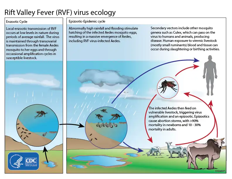
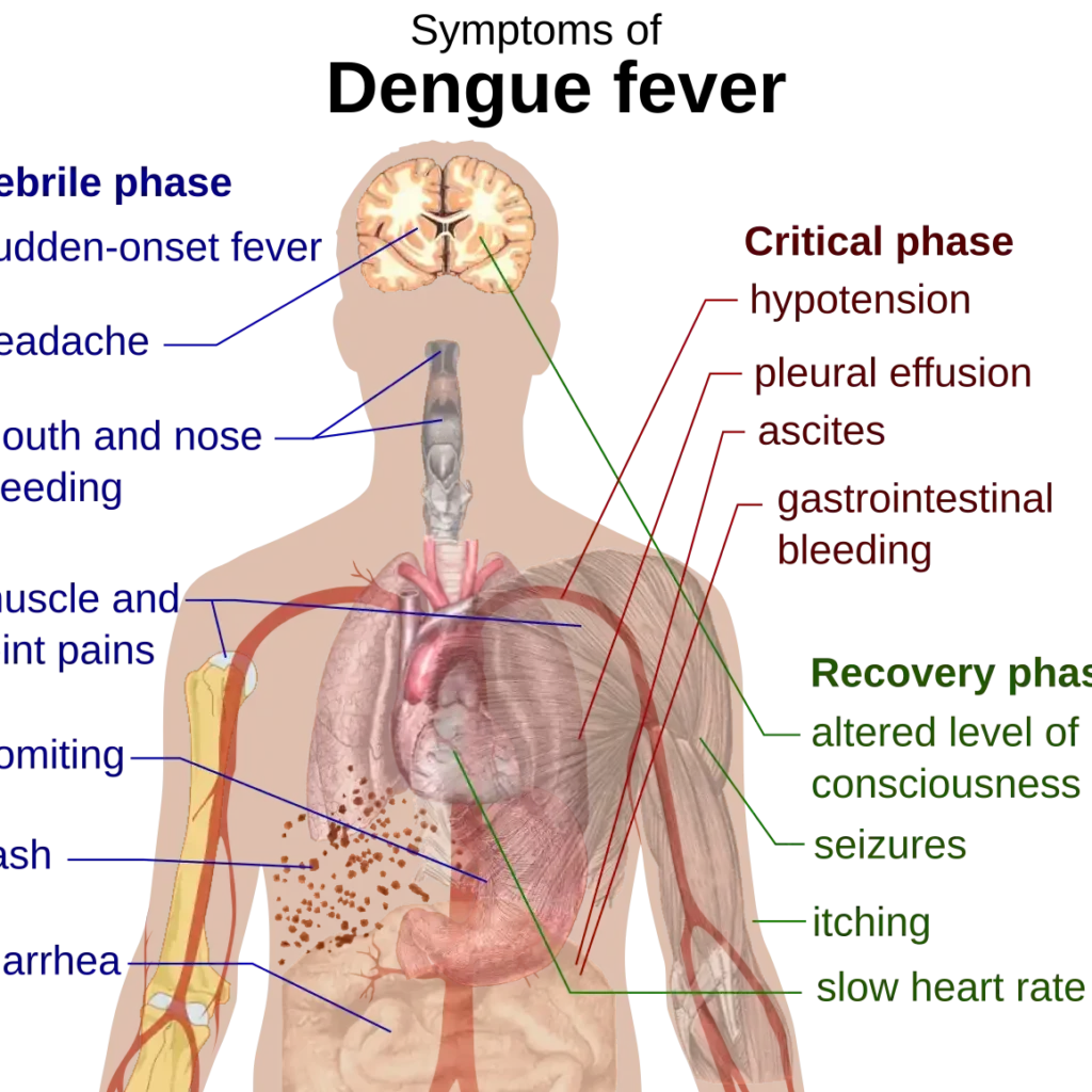
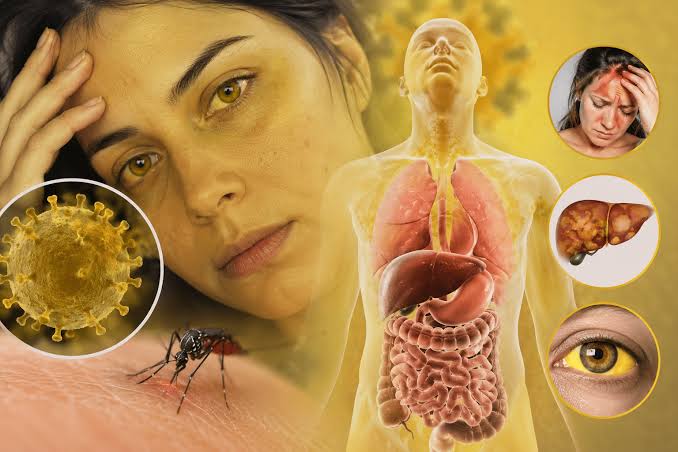
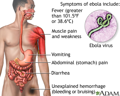
Ebola Virus Disease: Clinical Features, Diagnosis, Treatment and Outbreak Control Clinical Medicine Year 3 • a high-consequence viral haemorrhagic fever requiring early isolation, testing and supportive care Clinical safety note: A patient with fever or unexplained illness plus a compatible travel, outbreak, funeral, healthcare or animal-exposure history must be managed through the national viral-haemorrhagic-fever pathway. Call infection-prevention and public-health teams before collecting specimens or moving the patient. Do not perform unsafe injections, venepuncture or invasive procedures without appropriate PPE, trained staff and a plan for contaminated waste. Why Ebola matters Ebola virus disease (EVD) is a severe zoonotic infection caused by viruses in the genus Orthoebolavirus. It can begin as a nonspecific febrile illness and progress to profuse vomiting and diarrhoea, dehydration, shock, organ failure, coagulopathy, encephalopathy and death. The most important clinical intervention is not waiting for bleeding: early recognition, safe isolation, rapid testing, aggressive supportive care and contact tracing save lives. Ebola is not spread by casual contact before symptoms. Once symptoms begin, however, blood, vomit, stool, urine, breast milk, semen, vaginal fluids, saliva and other body fluids can transmit virus. Patients, caregivers, healthcare workers and people involved in funerals are at greatest risk when body fluids are handled without protection. Learning outcomes Describe Ebola virus structure, species, reservoirs and transmission. Explain the incubation period and the pathophysiology of endothelial, immune and hepatic injury. Recognise early, gastrointestinal, haemorrhagic and neurologic stages. Construct a safe diagnostic approach and distinguish EVD from malaria, typhoid, dengue, yellow fever and other VHF. Plan isolation, supportive care, approved species-specific therapeutics, survivor care and outbreak control. Definition and virology Ebola viruses are enveloped, filamentous, non-segmented, negative-sense single-stranded RNA viruses in the family Filoviridae. Important species include Zaire ebolavirus, Sudan ebolavirus, Bundibugyo ebolavirus, Taï Forest ebolavirus and Reston ebolavirus. Zaire ebolavirus is the species associated with many of the largest human outbreaks; Sudan and Bundibugyo viruses require separate clinical and vaccine considerations. The virion contains nucleoprotein, polymerase-associated proteins, matrix proteins and a surface glycoprotein. The glycoprotein enables attachment and entry into host cells and contributes to endothelial dysfunction and immune evasion. Mononuclear phagocytes, dendritic cells, endothelial cells, hepatocytes and adrenal cells become infected, leading to viraemia and multiorgan injury. Reservoir and transmission The exact natural reservoir is not fully established, but fruit bats are an important suspected reservoir. Human index cases may arise after exposure to infected wildlife or carcasses, followed by human-to-human transmission. Direct contact with blood or body fluids of a symptomatic patient. Contact with a deceased patient during preparation or burial. Unsafe injections, blood transfusion or contaminated instruments. Sexual exposure to survivors because Ebola virus can persist in semen for months. Breast milk and other secretions in selected clinical contexts. Animal exposure, including hunting, butchering or handling sick/dead wildlife. Incubation and communicability The incubation period is approximately 2–21 days, most often about one week. People are generally not infectious before symptoms begin. Infectivity rises with illness severity and is highest when the patient has vomiting, diarrhoea, bleeding or large amounts of virus-containing body fluid. Survivors require structured follow-up because viral persistence in semen, breast milk, ocular fluid or other immune-privileged sites can affect counselling and transmission prevention. Pathophysiology Virus enters through mucosa, broken skin, injection or inhalation of contaminated droplets/aerosols during high-risk procedures. It replicates in macrophages and dendritic cells and spreads through lymphatic tissue and blood. Innate immune signalling is disrupted; infected cells release inflammatory mediators while antigen presentation is impaired. Endothelial injury, cytokines and loss of vascular integrity cause capillary leak, hypotension and tissue oedema. Liver injury reduces clotting-factor synthesis; platelet and coagulation abnormalities produce coagulopathy. Vomiting, diarrhoea, fever and capillary leak cause profound dehydration, electrolyte disturbance, acidosis, kidney injury and shock. Clinical reasoning: Haemorrhage is dramatic but not required for diagnosis. A patient can be in life-threatening Ebola shock because of diarrhoeal fluid loss, capillary leak, acidosis and organ failure before visible bleeding appears. Clinical presentation 1. Early dry phase Early features are nonspecific: sudden fever, severe headache, fatigue, weakness, myalgia, arthralgia, backache, sore throat, conjunctival injection and anorexia. Malaria, typhoid, COVID-19 and other febrile infections may look identical at this stage. 2. Gastrointestinal/wet phase Nausea, repeated vomiting and watery diarrhoea. Abdominal pain, dysphagia, hiccups and profound weakness. Dehydration, postural dizziness, oliguria and electrolyte disturbance. Rash, red eyes and worsening transaminases. Confusion, agitation or reduced consciousness from shock, encephalitis or metabolic abnormalities. 3. Haemorrhagic and multiorgan disease Bleeding may include petechiae, ecchymoses, bleeding from venepuncture sites, gums, nose, gastrointestinal tract, vagina or urinary tract. Severe cases can develop shock, acute kidney injury, hepatitis, hypoglycaemia, respiratory distress, encephalopathy, seizures and multiorgan failure. The amount of external bleeding does not reliably predict severity. 4. Pregnancy and newborn considerations Pregnancy is associated with high maternal and fetal risk. Virus can be present in placenta, amniotic fluid, breast milk and other tissues. Pregnant patients require obstetric, infectious-disease and public-health coordination. Avoid unsafe obstetric procedures; use dedicated protocols for delivery, miscarriage, stillbirth and disposal of contaminated material. Case definition and exposure assessment Ask about Examples Why it changes risk Geography/time Residence or travel in an outbreak area within 21 days Links symptoms to an active transmission chain Contact Care for a suspected case, funeral attendance, blood/body-fluid contact Identifies high-risk exposure and contacts Healthcare Needlestick, unsafe injection, unprotected procedure May require urgent occupational-risk management Animal exposure Hunting, butchering or handling wildlife/primates/bats Possible index-case exposure Symptoms Fever, headache, vomiting, diarrhoea, rash, bleeding, weakness Determines triage and isolation urgency Investigations Routine severity assessment in a safe setting Capillary glucose, pulse oximetry, temperature, respiratory rate, blood pressure and mental state. Full blood count and platelets; leukopenia or thrombocytopenia may occur. Electrolytes, urea, creatinine, bicarbonate, liver tests, bilirubin and albumin. PT/INR, aPTT, fibrinogen and lactate where available. Malaria testing, blood cultures and other differential tests only through safe procedures and approved pathways. Specific Ebola tests RT-PCR: the main acute diagnostic test in blood, usually positive after viraemia develops. Antigen-detection tests: useful in approved field or reference-laboratory settings. Serology: IgM may support recent infection; IgG supports previous exposure or