
On this page
Overview
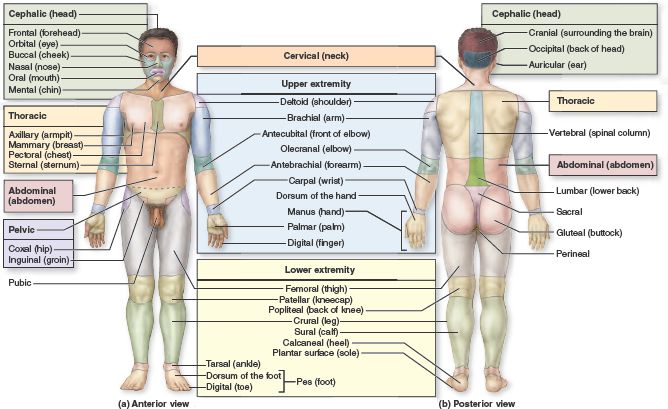
The Axial and Appendicular Skeleton
The human skeleton is divided into two major parts: the Axial Skeleton and the Appendicular Skeleton. Together, these two divisions provide the support, protection, and leverage necessary for movement.
The Axial Skeleton: The Body's Central Axis
The axial skeleton forms the longitudinal axis of the body. It consists of the bones of the skull, vertebral column (spine), and thoracic cage (ribs and sternum). In brief, it comprises the head and trunk.
Composition (approximately 80 bones):
- Skull (22 bones + 7 associated): Protects the brain and forms the face.
- Vertebral Column (26 bones): Protects the spinal cord and supports the head.
- Thoracic Cage (25 bones): Protects the heart and lungs.
The Skull
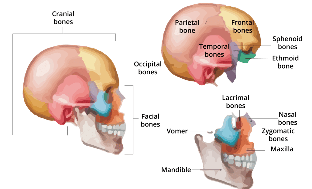
The skull is a bony structure that forms a protective cavity for the brain, provides the head with its shape, and is formed by 22 bones joined by fibrous joints called sutures. It consists of two main parts: the Cranium and the Face.
1. The Cranium (8 Bones)
The cranium is the bony box that houses and protects the brain.
Frontal Bone (1)
Forms the forehead and the superior part of the orbits.
Parietal Bones (2)
Form the superior and lateral walls of the cranium.
Temporal Bones (2)
Form the inferolateral aspects of the skull and parts of the cranial base; contain the organs of hearing.
Occipital Bone (1)
Forms the posterior wall and most of the base of the skull. The spinal cord passes through its foramen magnum.
Sphenoid Bone (1)
The central "keystone" bone of the cranium; articulates with all other cranial bones. Contains the sella turcica for the pituitary gland.
Ethmoid Bone (1)
Forms the anterior part of the cranial floor, the medial wall of the orbits, and the roof of the nasal cavity.
2. The Face (14 Bones)
These bones form the framework of the face, contain cavities for sensory organs, and provide attachment sites for facial muscles.
Mandible (1)
The lower jawbone; the largest and strongest bone of the face.
Maxillae (2)
The upper jawbones; they form the hard palate and hold the upper teeth.
Zygomatic Bones (2)
The cheekbones; they form the prominences of the cheeks.
Nasal Bones (2)
Form the bridge of the nose.
Lacrimal Bones (2)
Form part of the medial walls of the orbits; contain the lacrimal fossa for the tear ducts.
Palatine Bones (2)
Form the posterior part of the hard palate.
Vomer (1)
Forms the inferior part of the nasal septum.
Inferior Nasal Conchae (2)
Scroll-like bones forming part of the lateral walls of the nasal cavity.
B. The Vertebral Column (Spine)
The vertebral column serves as the main support of the body, protects the spinal cord, and provides attachment points for the ribs and muscles. It is a flexible, curved structure composed of 26 irregular bones in adults.
Functions of the Vertebral Column:
- Support: Transmits the weight of the head and trunk to the lower limbs.
- Protection: Surrounds and protects the delicate spinal cord.
- Movement: Provides attachment points for muscles, allowing trunk and neck movement.
- Shock Absorption: Intervertebral discs act as shock absorbers.
Regions and Curvatures
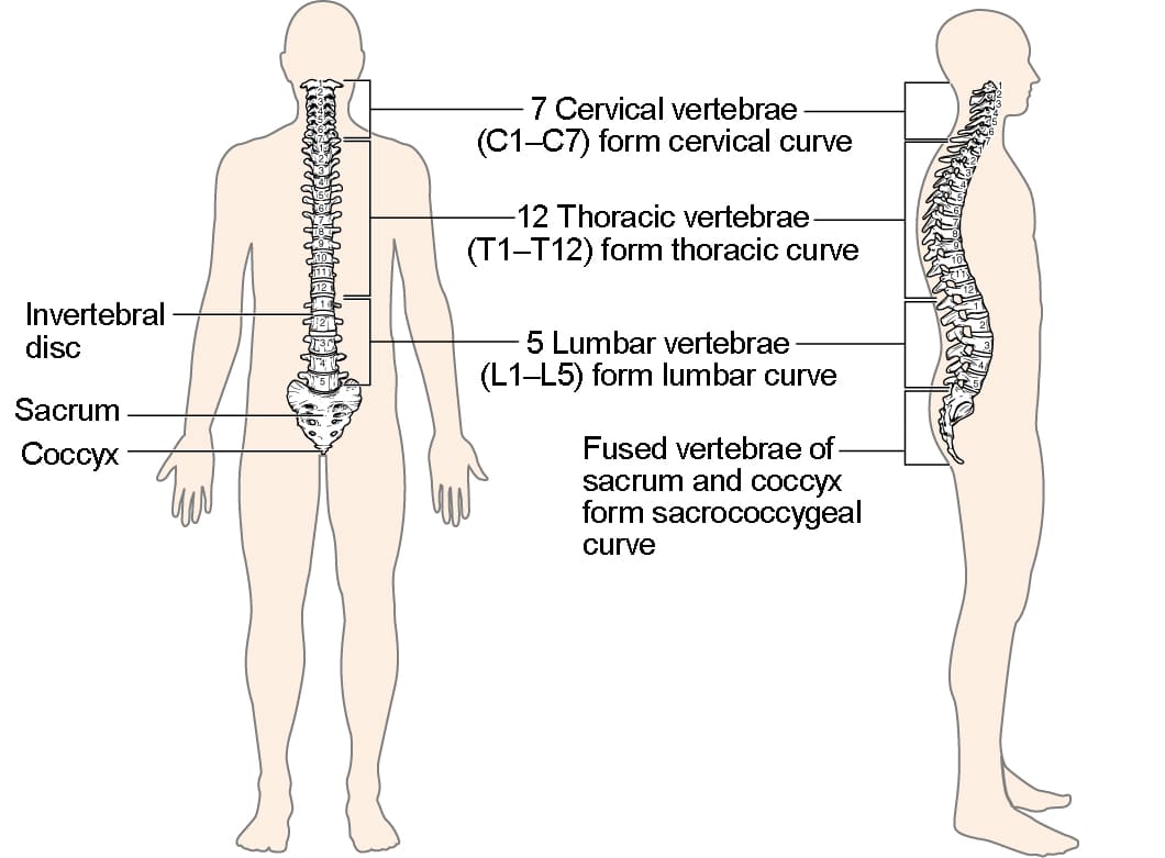
The spine is divided into five regions and has four natural curves that increase its resilience.
Vertebral Regions
Cervical (C1-C7): 7 vertebrae in the neck.
Thoracic (T1-T12): 12 vertebrae in the chest.
Lumbar (L1-L5): 5 vertebrae in the lower back.
Sacrum: 1 bone (5 fused vertebrae).
Coccyx: 1 bone (3-5 fused vertebrae).
Spinal Curvatures
Cervical & Lumbar: Concave posteriorly (secondary curves).
Thoracic & Sacral: Convex posteriorly (primary curves).
General Structure of a Vertebra
Most vertebrae share a common structural plan, consisting of a body, an arch, and various processes for muscle attachment and articulation.
- Vertebral Body (Centrum): The anterior, weight-bearing part.
- Vertebral Arch: Encloses the vertebral foramen, forming the vertebral canal for the spinal cord.
- Processes: Projections (spinous, transverse, articular) that serve as attachment and articulation points.
Intervertebral Discs
Located between adjacent vertebrae, these discs act as shock absorbers. Each is composed of an inner gelatinous nucleus pulposus and an outer collar of fibrocartilage called the anulus fibrosus.
Regional Characteristics of Vertebrae
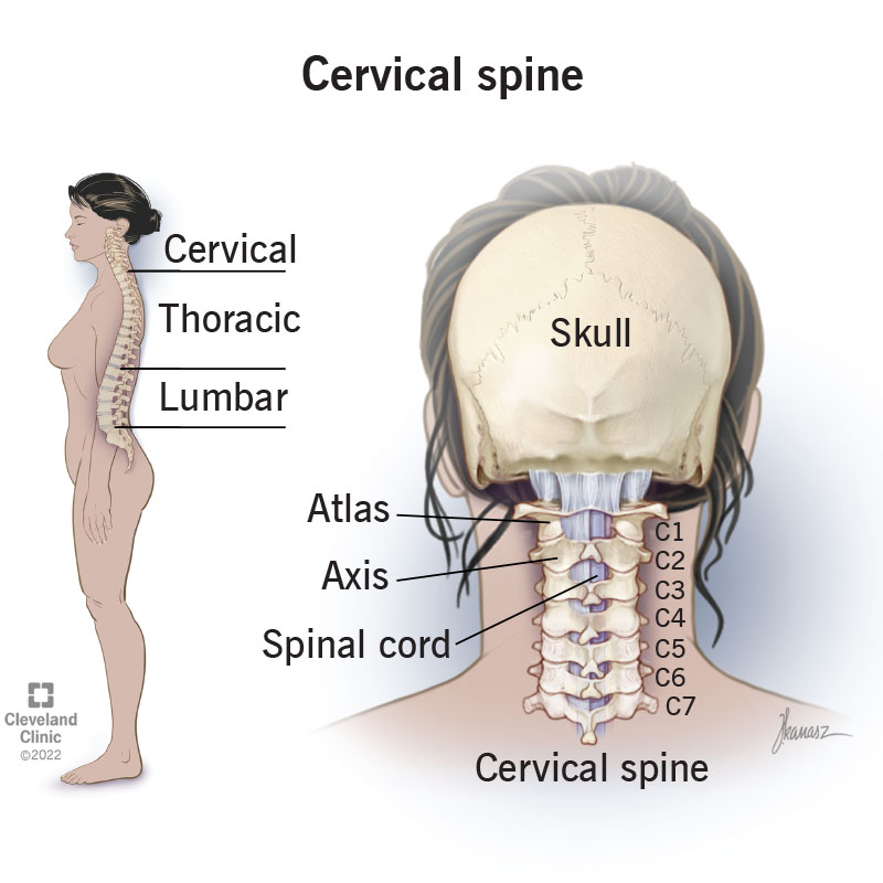
Cervical Vertebrae (C1-C7)
The smallest, lightest vertebrae. Their unique feature is the transverse foramina for vertebral arteries. C1 (Atlas) lacks a body and articulates with the skull ("yes" motion). C2 (Axis) has a dens that acts as a pivot for head rotation ("no" motion). Most have a bifid (split) spinous process.
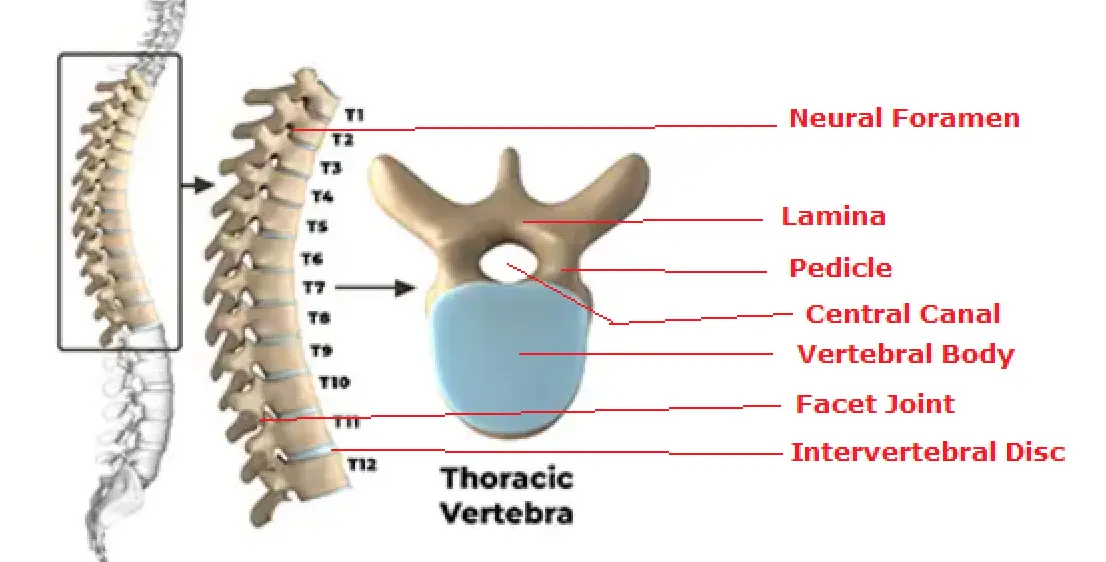
Thoracic Vertebrae (T1-T12)
Distinguished by their articulation with the ribs via costal facets on the vertebral bodies and transverse processes. They have a heart-shaped body and a long, slender spinous process that points sharply downward.
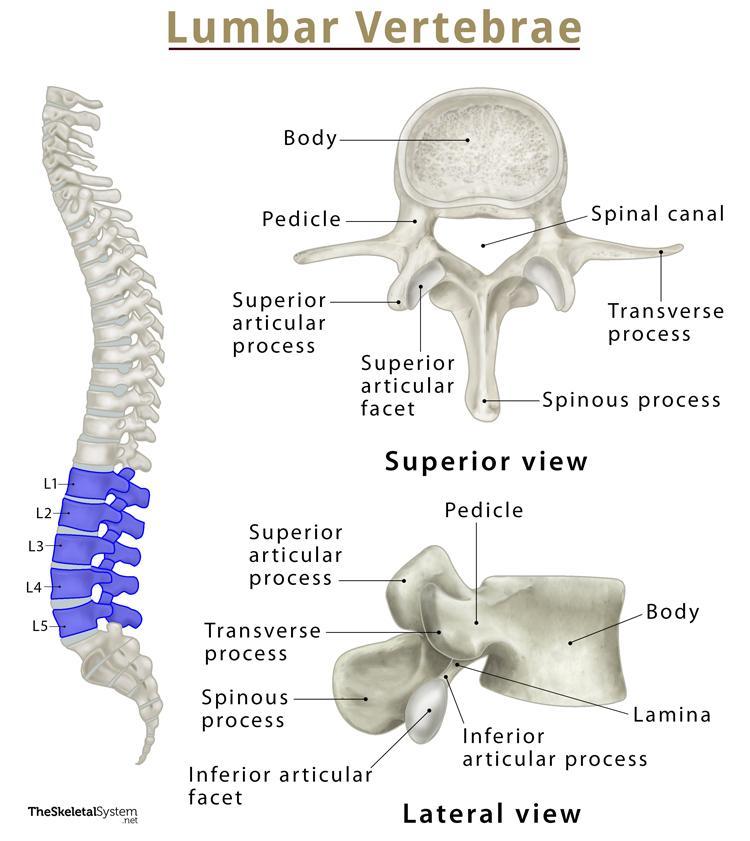
Lumbar Vertebrae (L1-L5)
The largest and strongest vertebrae, designed to bear the most body weight. They have a massive, kidney-shaped body and a short, thick, blunt spinous process that projects posteriorly.
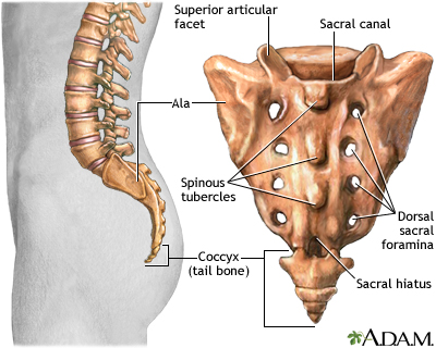
Sacrum and Coccyx
The Sacrum is a triangular bone formed by the fusion of 5 sacral vertebrae, forming the posterior wall of the pelvis. The Coccyx, or "tailbone," is a small triangular bone formed by the fusion of 3-5 coccygeal vertebrae.

C. The Thoracic Cage (Bony Thorax)
The thoracic cage forms the protective "rib cage" around the vital organs of the chest. It includes the sternum, ribs, and the twelve thoracic vertebrae.
Functions of the Thoracic Cage:
- Protection: Encloses and protects the heart, lungs, and major blood vessels.
- Support: Provides attachment points for the shoulder girdles and upper limbs.
- Respiration: Its ability to expand is crucial for ventilation, and it provides attachment for respiratory muscles.
Bones of the Thoracic Cage
The Sternum (Breastbone)
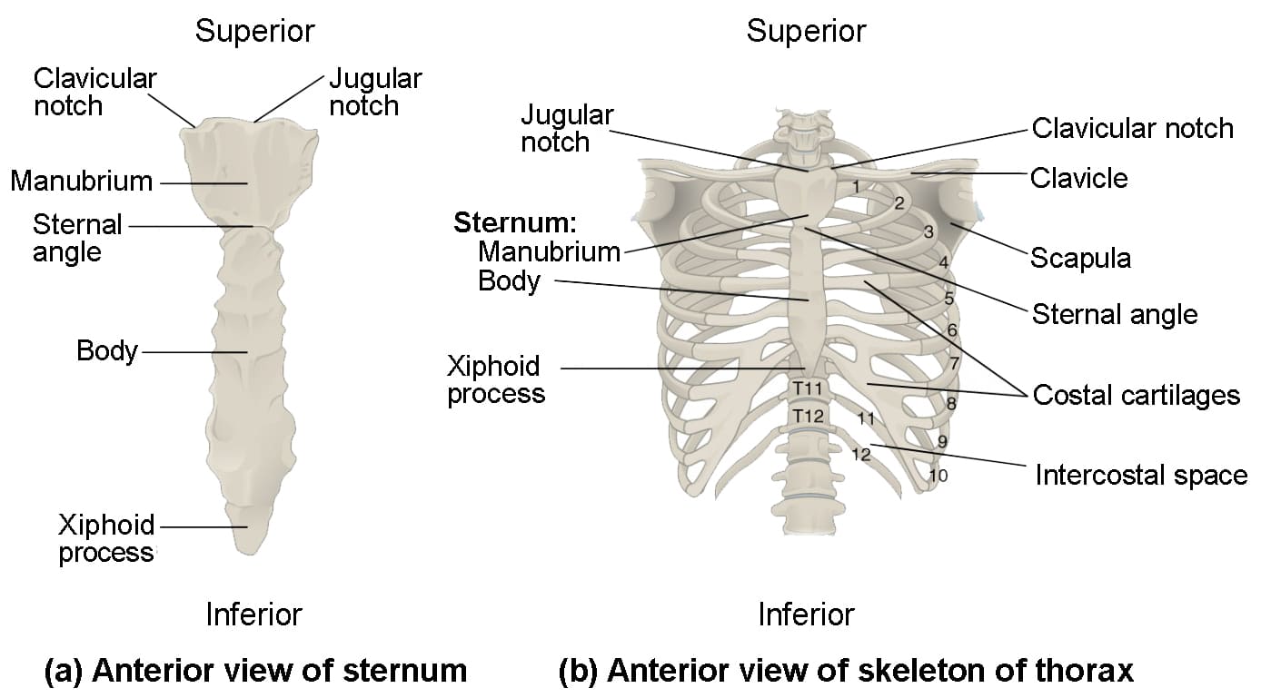
A flat bone in the anterior midline of the thorax, composed of three fused parts:
- Manubrium: The superior part, articulating with the clavicles and the first two pairs of ribs. Features the palpable jugular (suprasternal) notch.
- Body (Gladiolus): The middle and largest part, articulating with ribs 2-7.
- Xiphoid Process: The inferior-most, small projection that serves as an attachment point for some abdominal muscles.
The Ribs (12 pairs)
All ribs attach posteriorly to the thoracic vertebrae and generally curve inferiorly and anteriorly.
Types of Ribs (Based on Sternal Attachment)
- True Ribs (Pairs 1-7): Attach directly to the sternum via their own individual costal cartilages.
- False Ribs (Pairs 8-12):
- Pairs 8-10: Attach indirectly to the sternum by joining the costal cartilage of the rib above.
- Pairs 11-12 (Floating Ribs): Have no anterior attachment at all.
General Structure of a Rib
A typical rib consists of several key parts:
- Head: The posterior end, which articulates with the body of one or two thoracic vertebrae.
- Neck: The constricted region just lateral to the head.
- Tubercle: A knob-like projection that articulates with the transverse process of the corresponding vertebra.
- Shaft (Body): The main, curved portion of the rib.
- Costal Groove: A groove on the inferior border that protects the intercostal nerve and blood vessels.
- Costal Cartilage: The hyaline cartilage that connects the anterior end of the rib to the sternum.
Thoracic Vertebrae (T1-T12)
As previously discussed, these 12 vertebrae form the posterior boundary of the thoracic cage and provide the crucial articulation sites for all 12 pairs of ribs via their costal facets.
The Appendicular Skeleton
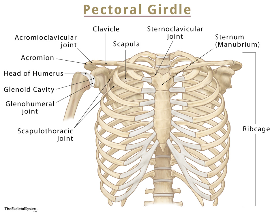
A. The Pectoral (Shoulder) Girdle
The pectoral girdle consists of two bones on each side of the body: the clavicle (collarbone) and the scapula (shoulder blade). These bones attach the upper limbs to the axial skeleton and provide attachment points for many muscles that move the upper limbs.
Functions of the Pectoral Girdle:
- Attachment: Connects the upper limb to the axial skeleton at the sternoclavicular joint (the only bony attachment).
- Mobility: Allows for a wide range of arm motion due to its loose attachment and the shallow glenoid cavity.
- Muscle Attachment: Provides sites for numerous muscles that move the shoulder and arm.
Bones of the Pectoral Girdle
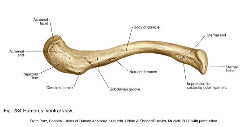
The Clavicle (Collarbone)
A slender, S-shaped bone that lies horizontally across the superior thorax. It acts as a brace, holding the scapula and arm away from the trunk, and transmits force from the upper limb to the axial skeleton.
- Sternal (medial) end: Articulates with the manubrium of the sternum, forming the sternoclavicular joint.
- Acromial (lateral) end: Articulates with the acromion of the scapula, forming the acromioclavicular joint.
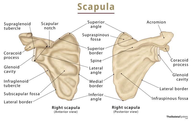
The Scapula (Shoulder Blade)
A thin, triangular flat bone on the posterior aspect of the rib cage. Its key features are crucial for muscle attachment and forming the shoulder joint.
Spine & Acromion
The Spine is a prominent posterior ridge that ends laterally in the Acromion, the palpable bony tip of the shoulder which articulates with the clavicle.
Glenoid Cavity (Fossa)
A shallow, pear-shaped depression on the lateral angle that articulates with the head of the humerus to form the highly mobile (but unstable) glenohumeral (shoulder) joint.
Coracoid Process
A hook-like process projecting anteriorly, serving as an attachment point for muscles and ligaments.
Fossae
Depressions for muscle attachment: the Supraspinous and Infraspinous Fossae (posterior), and the Subscapular Fossa (anterior).
B. The Upper Limbs
Each upper limb consists of 30 bones, specifically designed for mobility and manipulation. They are divided into three main segments: the arm, forearm, and hand.
1. The Arm (Brachium): Humerus
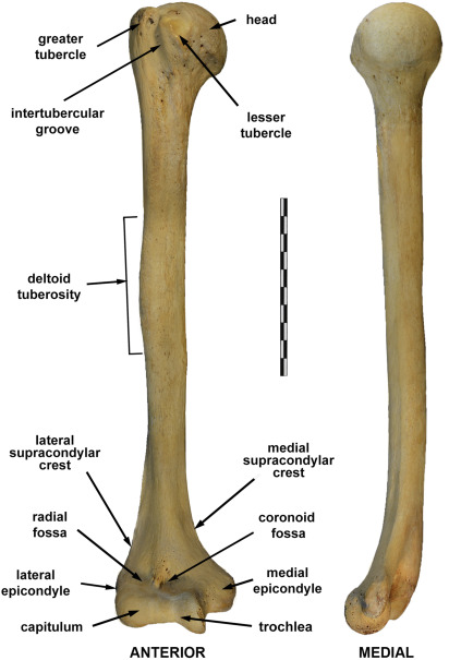
The humerus is the single bone of the arm, extending from the shoulder to the elbow. It is the longest and largest bone of the upper limb.
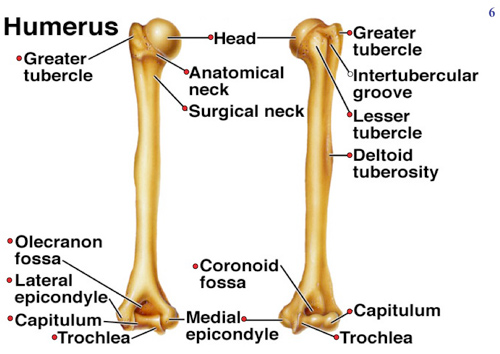
Key Features of the Humerus:
- Proximal End: Features the smooth Head (for the shoulder joint), the Greater and Lesser Tubercles for rotator cuff muscle attachment, and the Surgical Neck, a common fracture site.
- Shaft: Includes the Deltoid Tuberosity for deltoid muscle attachment and the posterior Radial Groove for the radial nerve.
- Distal End: Forms the elbow joint with the medial Trochlea (articulating with the ulna) and the lateral Capitulum (articulating with the radius). It also features the prominent Medial and Lateral Epicondyles and three fossae (Olecranon, Coronoid, Radial) that accommodate processes of the forearm bones during movement.
2. The Forearm (Antebrachium): Radius and Ulna
The forearm is formed by two parallel bones that allow for pronation and supination. They are connected by an Interosseous Membrane.

Ulna (Medial Bone)
The main bone forming the elbow joint. Its proximal end features the hook-like Olecranon Process (the "point" of the elbow) and the Coronoid Process, which together form the Trochlear Notch to grip the humerus. The distal end is small and features the Head and a pointed Styloid Process.
Radius (Lateral Bone)
The primary bone of the wrist joint. Its proximal end features a flat, disc-shaped Head that allows rotation against the humerus and ulna. The Radial Tuberosity serves as the attachment for the biceps brachii. The distal end is broad and features a pointed Styloid Process on the thumb side.
3. The Hand (Manus)
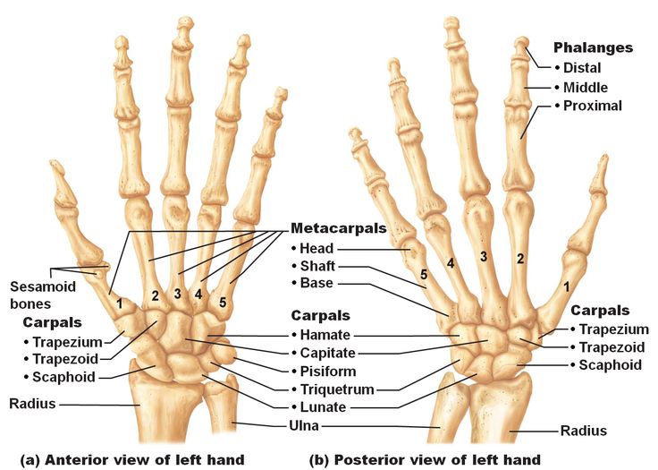
Each hand contains 27 bones adapted for dexterity and grip, divided into the carpals (wrist), metacarpals (palm), and phalanges (fingers).
a. Carpal Bones (8 Wrist Bones)
Eight small bones arranged in two rows that provide flexibility to the wrist.
- Proximal Row (lateral to medial): Scaphoid, Lunate, Triquetrum, Pisiform.
- Distal Row (lateral to medial): Trapezium, Trapezoid, Capitate, Hamate.
b. Metacarpal Bones (5 Palm Bones)
Five long bones that form the palm, numbered I to V from the thumb to the pinky finger. Their distal heads form the knuckles.
c. Phalanges (14 Finger Bones)
The bones of the digits.
- Thumb (Digit I): Has two phalanges (proximal and distal).
- Fingers (Digits II-V): Each has three phalanges (proximal, middle, and distal).
C. The Pelvic Girdle (Hip Girdle)
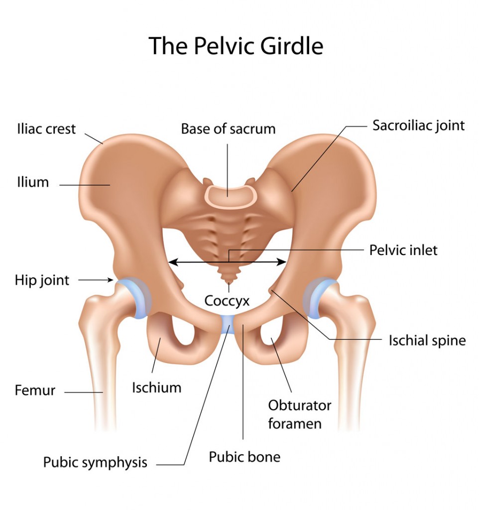
The pelvic girdle is a robust, basin-shaped structure formed by two ossa coxae (hip bones), which articulate with the sacrum posteriorly.
Functions of the Pelvic Girdle:
- Support: Transmits the weight of the upper body to the lower limbs.
- Protection: Encloses and protects the pelvic organs (bladder, reproductive organs, etc.).
- Attachment: Provides strong attachment points for muscles of the lower limbs and trunk.
- Articulation: Forms the hip joints by articulating with the heads of the femurs.
Bones of the Pelvic Girdle: The Os Coxa
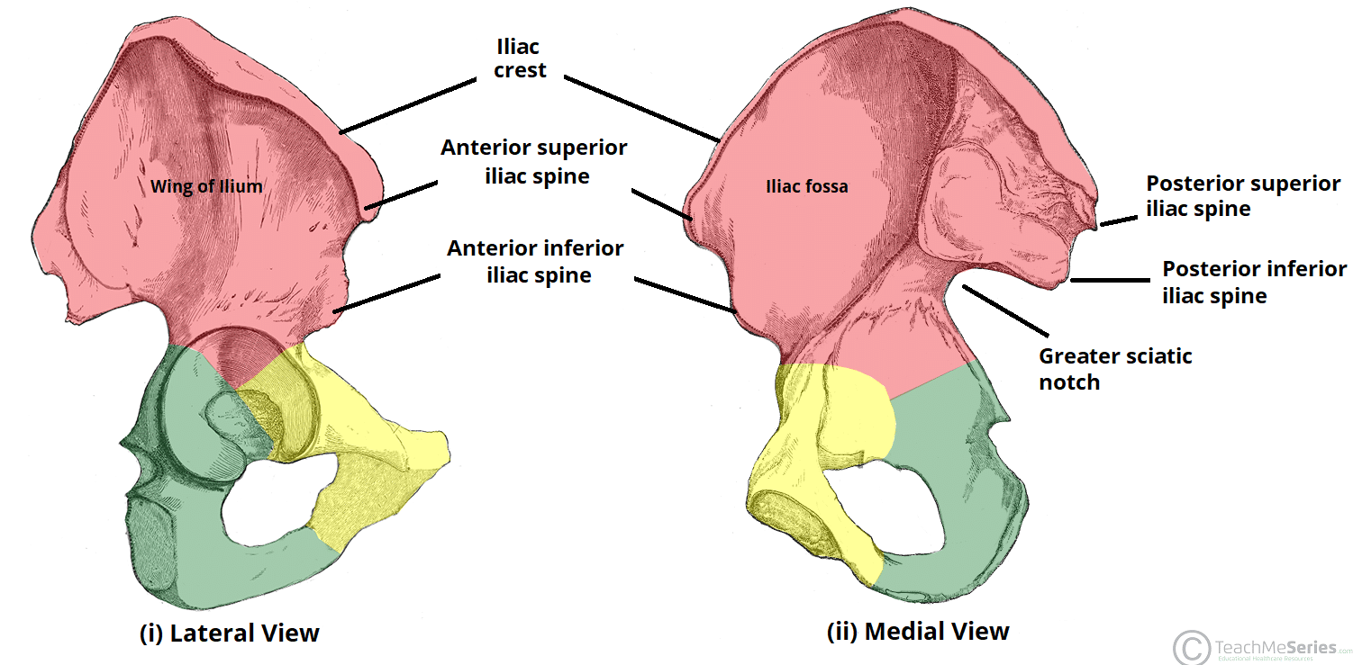
Each os coxa (hip bone) is a large, irregularly shaped bone formed by the fusion of three separate bones during adolescence: the ilium, ischium, and pubis. These three bones meet and fuse at the acetabulum, a deep, cup-shaped socket that articulates with the head of the femur.
a. Ilium
The largest and most superior part, forming the upper flank.
- Iliac Crest: The palpable superior border (the "hip bone").
- ASIS & PSIS: Anterior and Posterior Superior Iliac Spines, important anatomical landmarks.
- Greater Sciatic Notch: A large indentation for the sciatic nerve.
- Auricular Surface: Articulates with the sacrum to form the sacroiliac joint.
b. Ischium
Forms the posteroinferior part of the os coxa.
- Ischial Tuberosity: The large, roughened projection that supports body weight when sitting (the "sit bones").
- Ischial Spine: A sharp, pointed projection superior to the tuberosity.
c. Pubis
Forms the anteroinferior part of the os coxa.
- Pubic Symphysis: The fibrocartilaginous joint where the two pubic bones meet anteriorly.
- Pubic Arch: The angle formed by the inferior pubic rami, which differs between males and females.
Features of the Pelvis as a Whole
- Acetabulum: The deep, cup-shaped socket on the lateral surface of the os coxa where the ilium, ischium, and pubis fuse. It articulates with the head of the femur to form the hip joint.
- Obturator Foramen: A large opening inferior to the acetabulum, formed by the ischium and pubis, which is mostly closed by a membrane but allows passage for nerves and blood vessels.
- Pelvic Brim (Inlet): The boundary that separates the superior Greater (False) Pelvis from the inferior Lesser (True) Pelvis, which contains the pelvic organs.
D. The Lower Limbs
Each lower limb consists of 30 bones, specifically adapted for weight-bearing, locomotion, and maintaining balance. They are generally larger and stronger than the bones of the upper limbs and are divided into three main segments: the thigh, leg, and foot.
1. The Thigh: Femur and Patella
a. Femur (Thigh Bone)
The single bone of the thigh, extending from the hip to the knee. It is the longest, strongest, and heaviest bone in the body.
Key Features of the Femur:
- Proximal End: Features the spherical Head (with its Fovea Capitis) for the hip joint, the constricted Neck (a common fracture site), and the large Greater and Lesser Trochanters for muscle attachment.
- Shaft: Includes the prominent posterior ridge, the Linea Aspera, for attachment of many thigh muscles.
- Distal End: Forms the knee joint with the large Medial and Lateral Condyles. Also features the Medial and Lateral Epicondyles for ligament attachment and the anterior Patellar Surface where the kneecap glides.
b. Patella (Kneecap)
A small, triangular-shaped sesamoid bone located anterior to the knee joint. It protects the joint and increases the leverage of the quadriceps femoris muscle.
2. The Leg: Tibia and Fibula
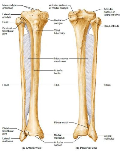
The leg is formed by two parallel bones connected by an Interosseous Membrane.
a. Tibia (Shin Bone)
The larger, medial, and primary weight-bearing bone of the leg. Its proximal end has flat Medial and Lateral Condyles to articulate with the femur. The anterior Tibial Tuberosity is the attachment site for the patellar ligament. The distal end forms the inner ankle bone, the Medial Malleolus.
b. Fibula (Lateral Bone)
The smaller, lateral bone that does not bear significant weight but serves for muscle attachment and ankle stability. The proximal Head articulates with the tibia. The distal end forms the outer ankle bone, the Lateral Malleolus, which provides important lateral stability to the ankle joint.
3. The Foot
Each foot contains 26 bones designed for supporting body weight and providing balance, divided into the tarsals (ankle), metatarsals (midfoot), and phalanges (toes).
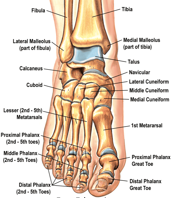
a. Tarsal Bones (7 Ankle Bones)
Seven irregularly shaped bones that form the posterior half of the foot. Key tarsals include:
- Talus: The uppermost tarsal, forming the ankle joint with the tibia and fibula. It receives the entire weight of the body.
- Calcaneus: The largest tarsal, forming the heel. It is the primary weight-bearing bone during standing and provides attachment for the Achilles tendon.
- Others: Navicular, Cuboid, and three Cuneiforms (Medial, Intermediate, Lateral).
b. Metatarsal Bones (5 Midfoot Bones)
Five long bones that form the midfoot, numbered I to V from the big toe to the pinky toe. They contribute to the arches of the foot.
c. Phalanges (14 Toe Bones)
The bones of the digits.
- Big Toe (Digit I / Hallux): Has two phalanges (proximal and distal).
- Other Toes (Digits II-V): Each has three phalanges (proximal, middle, and distal).
d. Arches of the Foot
The bones of the foot form three natural arches (two longitudinal, one transverse) that are supported by ligaments and tendons. They are crucial for shock absorption, providing springiness for locomotion, and adapting to uneven surfaces.
Lesson map
Use this map to move through the lesson in order:
Further reading
For deeper study, compare this lesson with current guidance and standard references from recognised medical and public-health organisations.