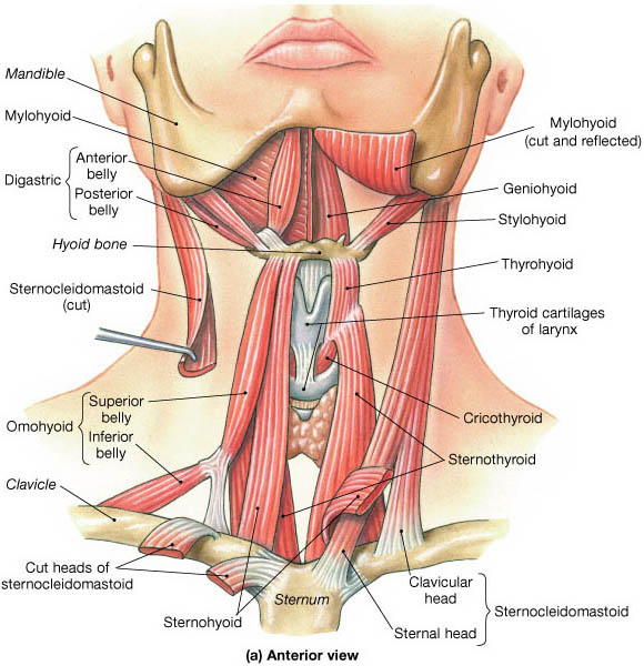
The Neck
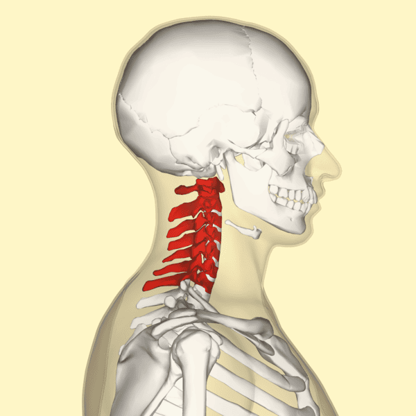
Anatomy of the Neck Module Learning Objectives By the conclusion of this exhaustive anatomical master guide, you will be deeply conversant with: The anatomical boundaries and the longitudinal compartmentalization of the neck. The detailed organization of the Anterior and Posterior Triangles, including their precise subdivisions and boundaries. The origins, insertions, innervations, and functions of the Suprahyoid and Infrahyoid (Strap) muscles. The complete Carotid Arterial System (Common, Internal, and External) along with the extensive branches of the External Carotid Artery. The major venous drainage of the neck, primarily focusing on the Internal Jugular Vein (IJV). The precise routing and innervation targets of the Cranial and Peripheral Nerves within the neck. The comprehensive anatomy, blood supply, and surgical relations of the Thyroid and Parathyroid Glands. I. Introduction and Boundaries of the Neck The neck is the vital, transitional anatomical tube providing critical continuity from the head to the trunk. It acts as a major conduit for the spinal cord, massive blood vessels supplying the brain, and the upper digestive and respiratory tracts. Defining the Anatomical Extent Anteriorly: The neck extends from the lower border of the mandible (jawbone) superiorly, down to the upper surface of the manubrium of the sternum inferiorly. Posteriorly: It extends from the superior nuchal line on the occipital bone of the skull superiorly, down to the intervertebral disc located between the C7 (Vertebra Prominens) and T1 vertebrae inferiorly. II. Longitudinal Organization: Compartments and Fascia Within this vital tube, the structures are highly organized into four distinct longitudinal compartments. These compartments are tightly bound by tough layers of deep cervical fascia, which serve not only to organize structures but to dictate the potential spread of deep neck infections. The Four Compartments The Visceral Compartment: Located anteriorly. It contains the vital tubular organs of the digestive and respiratory systems (Pharynx, Larynx, Trachea, and Esophagus), as well as several endocrine glands (Thyroid and Parathyroid glands). The Vertebral Compartment: Located posteriorly. It contains the rigid cervical vertebrae, the delicate spinal cord, exiting cervical nerves, and the postural muscles associated with the vertebral column. The Two Vascular Compartments (Left and Right): Located laterally on each side. They are encapsulated by the Carotid Sheath and contain the major blood vessels (Common/Internal Carotid Arteries and Internal Jugular Veins) as well as the Vagus nerve [CN X]. Fascial Layers of the Neck The neck is wrapped in layers of deep cervical fascia that enclose these compartments: Investing Fascia: Surrounds the entire neck like a collar, enclosing the SCM and Trapezius muscles. Pretracheal Fascia: Specifically encloses the anterior Visceral compartment (Thyroid, trachea, esophagus). Prevertebral Fascia: Encloses the posterior Vertebral compartment. Carotid Sheath: Encloses the lateral Vascular compartments. III. The Triangles of the Neck For descriptive, surgical, and diagnostic purposes, the neck is divided into two massive geometric regions separated by the diagonally placed Sternocleidomastoid (SCM) muscle: The Anterior Triangle and the Posterior Triangle. 1. The Posterior Triangle Located on the lateral aspect of the neck, behind the SCM. Anterior Boundary: The posterior border of the Sternocleidomastoid (SCM) muscle. Posterior Boundary: The anterior border of the Trapezius muscle. Inferior Boundary (Base): The middle one-third of the clavicle (collarbone). 2. The Anterior Triangle Located in the front of the neck, containing the most vital visceral and vascular structures. Lateral Boundary: The anterior border of the Sternocleidomastoid (SCM) muscle. Superior Boundary (Base): The inferior border of the mandible. Medial Boundary: The exact midline of the neck (from the chin down to the sternum). Subdivisions of the Anterior Triangle Because the anterior triangle is large and highly complex, anatomists further subdivide it into four smaller triangles using the Digastric and Omohyoid muscles as intersecting borders. The Submandibular Triangle: Outlined by the inferior border of the mandible superiorly, and the anterior and posterior bellies of the digastric muscle inferiorly. (Houses the submandibular salivary gland). The Submental Triangle: Outlined by the hyoid bone inferiorly, the anterior belly of the digastric muscle laterally, and the midline of the neck. (Located directly under the chin). The Muscular Triangle: Outlined by the hyoid bone superiorly, the superior belly of the omohyoid muscle and the anterior border of the SCM muscle laterally, and the midline of the neck. (Houses the infrahyoid strap muscles and thyroid gland). The Carotid Triangle: Outlined by the superior belly of the omohyoid muscle anteroinferiorly, the stylohyoid muscle and posterior belly of the digastric superiorly, and the anterior border of the SCM posteriorly. (Crucial surgical access point to the Carotid arterial system). IV. Musculature of the Anterior Triangle The muscles in the anterior triangle are primarily responsible for the complex movements of swallowing, speaking, and protecting the airway. They are logically grouped according to their physical location relative to the Hyoid bone (a U-shaped bone that does not articulate with any other bone). A. The Suprahyoid Muscles Located superior (above) the hyoid bone. They occupy the submental and submandibular triangles. They pass in a superior direction from the hyoid bone up to the skull or mandible. Primary Action: They raise/elevate the hyoid bone and the floor of the mouth, which is a critical action during swallowing. Muscle Innervation Action / Characteristics 1. Stylohyoid Facial nerve [CN VII] Pulls the hyoid bone posterosuperiorly (backward and upward) during swallowing. 2. Digastric Posterior belly: Facial nerve [CN VII]Anterior belly: Trigeminal nerve [CN V] Has two distinct bellies connected by an intermediate tendon which attaches to the body of the hyoid bone via a fibrous sling. 3. Mylohyoid Trigeminal nerve [CN V] Forms a muscular sling that supports and elevates the floor of the mouth and elevates the hyoid bone. 4. Geniohyoid Branch from anterior ramus of C1 (carried along the Hypoglossal nerve [CN XII]) Has two distinct functions depending on which bone is fixed:– If mandible is fixed: Elevates and pulls the hyoid forward.– If hyoid is fixed: Pulls the mandible downward and inward (opening the jaw). B. The Infrahyoid Muscles (Strap Muscles) Located inferior (below) the hyoid bone, occupying the muscular triangle. Because of their long, flat, ribbon-like appearance, they are widely referred to