
On this page
Overview
Blood groups are antigenic substances (glycoproteins and glycolipids) on the red cell surface, and they decide who can safely receive whose blood. Two systems dominate clinical practice: the ABO system, whose antibodies cause rapid intravascular haemolysis and acute transfusion reactions, and the Rh system, whose antibodies cross the placenta and cause haemolytic disease of the newborn. Over 300 other antigens exist, but competence in these two systems — plus correct typing and cross-matching — prevents the majority of transfusion deaths.
Learning objectives
- Explain the genetics and biochemistry of the ABO and Rh systems.
- Predict transfusion compatibility for any donor–recipient pair.
- Describe the pathophysiology and prevention of haemolytic disease of the newborn.
- Perform the logic of typing, antibody screening and cross-matching.
- Recognise and manage an acute haemolytic transfusion reaction.
Introduction to blood group systems
Historical anchors are worth remembering for exams: Karl Landsteiner discovered the ABO system in 1900 (Nobel Prize 1930) and, with Wiener, identified the Rh system in 1939. Landsteiner's insight was simple and immortal: mixing some people's blood makes it clot, and the pattern falls into groups. Every transfusion since has rested on that observation.
The ABO system: genetics and biochemistry
The ABO system is governed by three alleles on chromosome 9: IA, IB (codominant) and i (recessive).
Scroll the table sideways on a smaller screen.
| Genotype | Phenotype (group) | RBC antigens | Plasma antibodies |
|---|---|---|---|
| IAIA or IAi | A | A antigen | Anti-B |
| IBIB or IBi | B | B antigen | Anti-A |
| IAIB | AB | A and B antigens | None |
| ii | O | None (H only) | Anti-A and anti-B |
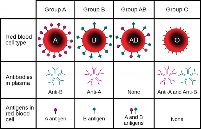
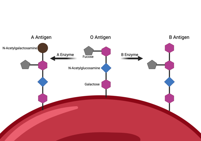
The biochemical basis. All red cells carry the H antigen precursor. The IA allele codes an enzyme adding N-acetylgalactosamine (creating the A antigen); the IB allele adds galactose (the B antigen); the i allele is non-functional, leaving H unmodified (group O).
Antibody character explains the danger. Anti-A and anti-B are naturally occurring (present by 3–6 months), IgM, complement-fixing — and therefore capable of rapid intravascular haemolysis. They are the principal cause of acute haemolytic transfusion reactions.
The Bombay phenotype (hh). Rare individuals lack the H antigen entirely (FUT1 mutations). They type as group O but carry anti-H, anti-A and anti-B — so they can only receive blood from other Bombay donors. Missing this converts a routine transfusion into a fatal one.
ABO compatibility for transfusion
Scroll the table sideways on a smaller screen.
| Recipient | Can receive from | Can donate to |
|---|---|---|
| A | A, O | A, AB |
| B | B, O | B, AB |
| AB | A, B, AB, O (universal recipient) | AB only |
| O | O only | A, B, AB, O (universal donor) |
The familiar shorthand: O negative is the universal donor (no A, B or D antigens); AB positive is the universal recipient (no anti-A, anti-B or anti-D antibodies). In emergencies, group O negative blood is given while the patient's own group is determined.
The Rh system
The Rh system — determined by RHD and RHCE genes on chromosome 1 — is the second most important clinically, and the most important in obstetrics. The D antigen is its most immunogenic member: Rh-positive means D present, Rh-negative means D absent. Roughly 15% of people of European ancestry are Rh-negative; the figure is lower in most African and East Asian populations, so Rh-negative blood is scarce where demand is highest.
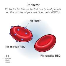
Rh antibodies differ from ABO antibodies in every way that matters: they are not naturally occurring — they form only after sensitisation (transfusion or pregnancy); they are IgG, not IgM; and IgG crosses the placenta. That last property is the entire clinical significance of the system.
Haemolytic disease of the newborn
The sequence, step by step:
- An Rh-negative mother carries an Rh-positive fetus (paternal D antigen).
- Fetal red cells cross the placenta during delivery, abdominal trauma or invasive procedures.
- The mother mounts a primary anti-D response — IgG, slow to appear, lasting for years.
- In the next Rh-positive pregnancy, maternal anti-D crosses the placenta and destroys fetal red cells.
- The fetus develops anaemia, jaundice, hydrops fetalis — or is stillborn. Each subsequent affected pregnancy tends to be worse.
The prevention is one of the great triumphs of immunology: anti-D immunoglobulin (RhIg) given to Rh-negative mothers at 28–30 weeks and within 72 hours after delivery (and after abortion or invasive procedures) mops up fetal D-positive cells before the maternal immune system notices them. Sensitisation never begins, and the next pregnancy is protected. Skipping the postnatal dose is one of the most costly omissions in obstetric care.
Other blood group systems
Scroll the table sideways on a smaller screen.
| System | Key antigens | Clinical relevance |
|---|---|---|
| Kell | K, k | Severe HDN and transfusion reactions |
| Duffy | Fya, Fyb | Fy(a−b−) confers resistance to P. vivax malaria — a natural-selection story written in blood groups |
| MNS | M, N, S, s | Transfusion reactions and HDN |
| Kidd | Jka, Jkb | Delayed haemolytic transfusion reactions |
Typing, cross-matching and the antiglobulin test
- Blood typing. The patient's red cells are mixed with known antisera (anti-A, anti-B, anti-D); agglutination reveals the group. Reverse (serum) typing confirms with known cells.
- Major cross-match. Donor red cells + recipient serum — detects recipient antibodies against donor cells. This is the critical compatibility test.
- Minor cross-match. Recipient red cells + donor serum — rarely performed in modern practice.
- Indirect antiglobulin test (IAT). Detects unexpected antibodies (e.g. anti-K, anti-D) in the patient's serum.
- Type and screen for patients who may need blood; type and cross-match when transfusion is anticipated — a distinction of time, cost and safety.
ABO versus Rh: the comparison
Scroll the table sideways on a smaller screen.
| Feature | ABO system | Rh system |
|---|---|---|
| Chromosome | 9 | 1 |
| Principal antigens | 2 (A, B) | 50+ (D most important) |
| Natural antibodies | Yes — anti-A, anti-B | No — only after sensitisation |
| Antibody class | IgM (complement-fixing) | IgG (crosses the placenta) |
| Major clinical danger | Acute haemolytic transfusion reaction | Haemolytic disease of the newborn |
Worked example: an acute transfusion reaction
Scenario. Fifteen minutes into a unit of blood, a patient develops fever, flank pain and red-brown urine. The clerical check shows the unit was labelled for another patient. Walk through the reasoning and the response.
- Recognise. Fever + flank pain + dark urine within minutes of transfusion = acute haemolytic reaction until proven otherwise. The likely mechanism: donor ABO-incompatible red cells meeting recipient IgM anti-A/anti-B — complement-mediated intravascular haemolysis.
- Act first. Stop the transfusion immediately, keep the line open with normal saline, and call for help. Check ABCs; the reaction can progress to shock, DIC and acute kidney injury.
- Verify and report. Clerical check of labels, return the unit and tubing to the blood bank, send post-reaction samples (blood and urine) for grouping, repeat cross-match and haemolysis markers (rising bilirubin, falling haptoglobin, haemoglobinuria).
- Treat. Aggressive intravenous fluids to protect the kidneys; treat shock; manage DIC if it develops; document and report to the transfusion committee.
- Prevent recurrence. Two identifiers checked at the bedside, every unit, every time — the reaction in this scenario was a clerical error before it was a biological one.
Clinical pearls
- Never skip the antibody screen before transfusion, even in "routine" surgery — Kidd and Kell antibodies announce themselves only through it.
- The Duffy–malaria link is a favourite exam crossover between transfusion science and parasitology: Fy(a−b−) red cells resist P. vivax invasion.
- In an emergency with no time to type: O negative blood; in an emergency with minutes to type but not cross-match: group-specific uncross-matched blood.
- Anti-D is for Rh-negative mothers of Rh-positive babies — and the dose must be given within 72 hours of the sensitising event.
Key points
- Blood groups are red-cell antigens; ABO and Rh systems dominate clinical transfusion practice.
- ABO: chromosome 9, three alleles, natural IgM antibodies that fix complement and cause acute intravascular haemolysis.
- Rh: chromosome 1, D antigen defines status, immune IgG antibodies that cross the placenta and cause HDN.
- O negative is the universal donor; AB positive the universal recipient; Bombay phenotype individuals are mislabelled as O and can only receive Bombay blood.
- Typing, screening and the major cross-match guard every transfusion; clerical checks guard them all.
- Anti-D immunoglobulin at 28–30 weeks and within 72 hours post-delivery prevents Rh sensitisation — a preventable-disease success story.
References and further reading
- Harmening DM. Modern Blood Banking & Transfusion Practices — serology, cross-matching and transfusion reactions. (Library reference.)
- Klein HG, Anstee DJ. Mollison's Blood Transfusion in Clinical Medicine — the definitive transfusion reference. (Library reference.)
- de Haas M, et al. Literature on the Rh system and haemolytic disease of the newborn. (Library reference.)
- Ministry of Health, Uganda. Uganda Clinical Guidelines — blood transfusion and obstetric haemorrhage protocols. (Library reference.)
For education only. Apply current Uganda Clinical Guidelines, local protocols and specialist advice in clinical practice.