Structure and Functions of Blood Vessels
Complete study notes covering the histology of vessel walls, classification of the vascular tree, hemodynamics, capillary exchange mechanisms, and major systemic vessels.
2.1 Introduction
Blood vessels form a closed system of tubes that transport blood between the heart and body tissues. There are five main types: arteries, arterioles, capillaries, venules, and veins. Together, they regulate blood flow, blood pressure, and the exchange of substances between blood and tissues.
2.2 General Structure of Blood Vessels
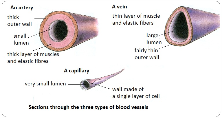
Most blood vessels share a common three-layered (tunica) wall structure:
| Layer | Name | Composition | Function |
|---|---|---|---|
| Tunica externa | Outer layer | Connective tissue (collagen) with elastic fibers | Protection, anchoring, nerve/vessel passage |
| Tunica media | Middle layer | Smooth muscle, elastic fibers (lamellae) | Vasoconstriction, vasodilation, pressure regulation |
| Tunica intima | Inner layer | Endothelium (simple squamous epithelium) on basement membrane | Smooth surface for blood flow; prevents clotting |
Capillaries consist only of a single layer of endothelium (tunica intima) and a basement membrane, allowing for efficient nutrient and gas exchange.
2.3 Arteries
Arteries carry blood away from the heart. They are classified by size and structure into three categories:
2.3.1 Elastic (Conducting) Arteries
- Largest arteries: Aorta, pulmonary trunk, brachiocephalic, common carotid, subclavian, common iliac arteries.
- Structure: Tunica media contains abundant elastic fibers (lamellae) with less smooth muscle.
- Function: Conduct blood from heart to medium-sized arteries; elastic recoil maintains continuous blood flow during diastole (Windkessel effect).
2.3.2 Muscular (Distributing) Arteries
- Medium-sized arteries: Radial, ulnar, femoral, tibial, coronary arteries.
- Structure: Tunica media contains more smooth muscle (40-60% of wall thickness) and less elastic tissue.
- Function: Distribute blood to specific organs and tissues; vasoconstriction and vasodilation regulate blood flow to organs.
2.3.3 Arterioles
- Smallest arteries: 10-100 um diameter.
- Structure: Tunica media has 1-3 layers of smooth muscle cells.
- Function: Major resistance vessels; regulate blood flow into capillary beds; primary site of peripheral vascular resistance.
- Precapillary sphincters: Rings of smooth muscle that control capillary blood flow.
- Systolic pressure: 90-120 mmHg (peak pressure during ventricular systole).
- Diastolic pressure: 60-80 mmHg (minimum pressure during ventricular diastole).
- Mean arterial pressure (MAP): ~93 mmHg = diastolic + 1/3 pulse pressure.
- Pulse pressure: systolic - diastolic (normally 30-40 mmHg).
2.4 Capillaries
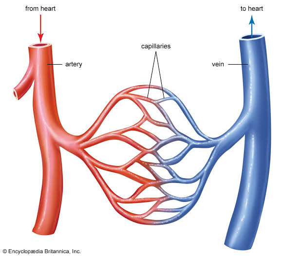
Capillaries are the smallest blood vessels (5-10 um diameter) and form extensive networks (capillary beds) between arterioles and venules. They are the primary site of exchange between blood and tissues.
2.4.1 Structure
- Wall composed of a single layer of endothelial cells on a basement membrane.
- No tunica media or tunica externa.
- Total surface area: Approximately 600 m² (enormous exchange capacity).
- Total length: Approximately 96,000 km if all capillaries were placed end-to-end.
2.4.2 Types of Capillaries
| Type | Structure | Location | Function |
|---|---|---|---|
| Continuous | Endothelial cells joined by tight junctions; complete basement membrane | Muscle, skin, lungs, brain (blood-brain barrier) | Most common; controlled exchange of water, ions, small molecules |
| Fenestrated | Endothelial cells have pores (fenestrae); thin/continuous basement membrane | Kidneys, intestines, endocrine glands | Rapid exchange of fluids, solutes; filtration, absorption |
| Sinusoidal | Large, irregular lumen; incomplete basement membrane; gaps between endothelial cells | Liver, spleen, bone marrow, lymph nodes | Allow passage of large molecules, cells (RBCs, WBCs) |
2.4.3 Capillary Exchange Mechanisms
- Diffusion: Primary mechanism for exchange of gases, nutrients, and waste products.
— Lipid-soluble: (O2, CO2, steroid hormones) pass directly through endothelial membranes.
— Water-soluble: (glucose, amino acids, ions) pass through intercellular clefts or fenestrae. - Transcytosis: Transport of large molecules (proteins, hormones) via vesicles (endocytosis + exocytosis).
- Bulk Flow (Filtration and Reabsorption): Driven by hydrostatic and osmotic pressure differences (Starling forces).
— At arterial end: net filtration (fluid moves out into interstitium).
— At venous end: net reabsorption (fluid moves back into capillary).
— Approximately 85% of filtered fluid is reabsorbed; 15% returns via lymphatic vessels.
Net filtration = Kf x [(Pc - Pi) - (πc - πi)]
- Pc = capillary hydrostatic pressure
- Pi = interstitial hydrostatic pressure
- πc = plasma colloid osmotic pressure
- πi = interstitial colloid osmotic pressure
2.5 Veins and Venules
Veins carry blood toward the heart. They have larger lumens and thinner walls than corresponding arteries.
2.5.1 Venules
- Smallest veins: 10-100 um; formed by union of capillaries.
- Postcapillary venules: Major site of leukocyte extravasation during inflammation.
- Tunica media is very thin (1-2 layers of smooth muscle).
2.5.2 Veins
- Medium and large veins have all three tunics, but tunica media is thinner than in arteries.
- Tunica externa is the thickest layer in large veins.
- Luminal diameter is larger than corresponding arteries.
- Blood pressure is low (approximately 10-15 mmHg in vena cava).
- Blood volume capacity is high (approximately 60% of total blood volume at rest).
Venous Return Mechanisms:
- Skeletal muscle pump: Contraction of surrounding muscles compresses veins, pushing blood toward the heart (one-way valves prevent backflow).
- Respiratory pump: During inspiration, thoracic pressure decreases and abdominal pressure increases, drawing blood toward the heart.
- One-way valves: Prevent backflow of blood, especially in lower limbs.
- Gravity: Aided by upright posture and venous tone.
Varicose Veins
Dilated, tortuous, elongated superficial veins (commonly saphenous veins). Caused by incompetent venous valves, increased venous pressure, or weakened vein walls.
Risk factors: prolonged standing, pregnancy, obesity, family history.
Complications: venous stasis ulcers, thrombophlebitis, bleeding.
2.6 Comparison of Blood Vessels
| Feature | Arteries | Capillaries | Veins |
|---|---|---|---|
| Wall thickness | Thick | Very thin (1 cell) | Thin |
| Tunica media | Thick (smooth muscle + elastic) | Absent | Thin |
| Lumen diameter | Smaller than vein | Smallest (5-10 um) | Larger than artery |
| Blood pressure | High (80-120 mmHg) | Low (20-40 mmHg) | Very low (5-15 mmHg) |
| Blood velocity | Fast | Very slow | Slow |
| Blood volume | Low (~15%) | Low (~5%) | High (~60%) |
| Valves | None (except near heart) | None | Present (especially in limbs) |
| Function | Transport blood away from heart | Exchange of gases, nutrients, wastes | Return blood to heart; blood reservoir |
2.7 Major Blood Vessels of the Body
2.7.1 Aorta and Major Arteries
- Ascending aorta: Gives rise to right and left coronary arteries.
- Aortic arch:
— Brachiocephalic trunk -> right subclavian + right common carotid
— Left common carotid artery
— Left subclavian artery - Thoracic aorta: Bronchial, esophageal, mediastinal, posterior intercostal arteries.
- Abdominal aorta:
— Celiac trunk (liver, stomach, spleen)
— Superior mesenteric artery (small intestine, proximal colon)
— Renal arteries (kidneys)
— Inferior mesenteric artery (distal colon, rectum)
— Common iliac arteries -> external iliac (lower limb) + internal iliac (pelvis)
2.7.2 Major Veins
- Superior vena cava: Drains head, neck, upper limbs, and thorax. Formed by union of right and left brachiocephalic veins.
- Inferior vena cava: Drains abdomen, pelvis, and lower limbs. Formed by union of right and left common iliac veins. Receives hepatic veins, renal veins, gonadal veins.
- Hepatic portal system: Unique system: veins -> capillaries -> veins. Hepatic portal vein carries nutrient-rich blood from GI tract, spleen, pancreas to liver for processing before entering systemic circulation.
2.8 Vascular Tone and Regulation
Vascular tone is the baseline contraction of smooth muscle in vessel walls, maintained by sympathetic nervous system activity.
Vasoconstriction:
- Mediated by alpha-1 adrenergic receptors on vascular smooth muscle.
- Increases peripheral resistance and blood pressure.
- Other mediators: endothelin-1, angiotensin II, vasopressin (ADH).
Vasodilation:
- Decreased sympathetic activity; mediated by beta-2 receptors (skeletal muscle).
- Local factors: decreased O2, increased CO2, increased H+, increased K+, increased adenosine, increased temperature (active hyperemia).
- Endothelial factors: nitric oxide (NO), prostacyclin (PGI2).
- Decreases peripheral resistance and blood pressure.
Visual Reference Summary
SUMMARY — STRUCTURE AND FUNCTIONS OF BLOOD VESSELS
Key Structures:
- All vessels have three layers (tunica intima, media, externa) except capillaries.
- Arteries: thick-walled, high pressure, elastic or muscular types.
- Capillaries: single-layer endothelium; site of exchange (continuous, fenestrated, sinusoidal).
- Veins: thin-walled, large lumen, low pressure, contain valves, capacity reservoirs.
Key Functions:
- Arteries: transport blood away from heart; regulate distribution via vasoconstriction/dilation.
- Capillaries: exchange of gases, nutrients, wastes, fluids between blood and tissues.
- Veins: return blood to heart; act as blood reservoirs; venous return aided by muscle pump, respiratory pump, and valves.
Understanding vessel structure explains why arteries are prone to atherosclerosis (high pressure, smooth muscle layer) while veins are prone to varicosities and thrombosis. Capillary permeability determines drug delivery and edema formation. Venous insufficiency leads to stasis dermatitis, ulcers, and deep vein thrombosis.
