Common Blood Disorders
Complete study notes covering the classification, pathophysiology, investigation, and management of Anaemia, Leukemia, and Hemophilia.
4.1 ANAEMIA
4.1.1 Definition
Anaemia is defined as a reduction in the oxygen-carrying capacity of blood due to decreased hemoglobin (Hb) concentration, reduced red blood cell (RBC) count, or low hematocrit levels relative to the physiological needs of the individual.
4.1.2 WHO 2024 Diagnostic Criteria
The World Health Organization updated hemoglobin cutoffs in 2024 using systematic review and GRADE methodology. These thresholds are critical for clinical diagnosis:
| Population | Hb Threshold (g/dL) |
|---|---|
| Children 6-59 months | <11.0 (revised to <10.5 for 6-23 months) |
| Children 5-12 years | <11.5 |
| Non-pregnant women | <12.0 |
| Pregnant women (all trimesters) | <11.0 |
| Pregnant women (2nd trimester) | <10.5 |
| Men | <13.0 |
Severity Classification (WHO):
- Mild: 10.0 — threshold
- Moderate: 7.0 — 9.9
- Severe: <7.0
4.1.3 Classification by Morphology (MCV)
| Type | MCV (fL) | MCH (pg) | Common Causes |
|---|---|---|---|
| Microcytic hypochromic | <80 fL | <27 pg | Iron deficiency, thalassemia, sideroblastic anemia, chronic disease |
| Normocytic normochromic | 80-100 fL | 27-33 pg | Acute blood loss, hemolytic anemia, chronic disease, aplastic anemia, renal failure |
| Macrocytic | >100 fL | >33 pg | B12 deficiency, folate deficiency, liver disease, alcoholism, hypothyroidism |

4.1.4 Classification by Pathophysiology
- Decreased production: Iron deficiency, B12/folate deficiency, aplastic anemia, chronic disease, renal failure.
- Increased destruction (hemolysis): Sickle cell disease, thalassemia, autoimmune hemolytic anemia, G6PD deficiency, malaria.
- Blood loss: Trauma, menorrhagia, GI bleeding (peptic ulcer, hookworm, colorectal cancer).
4.1.5 Iron Deficiency Anaemia (IDA)
Etiology: Chronic blood loss (menorrhagia, GI bleeding, hookworm infestation), inadequate dietary intake (vegetarian/vegan diets), malabsorption (celiac disease, gastrectomy), and increased demand (pregnancy, infancy, adolescence).
Clinical Features:
- General: Pallor (conjunctival, palmar, tongue), fatigue, weakness, dizziness.
- Cardiovascular: Tachycardia, palpitations, systolic flow murmur, heart failure (severe).
- Epithelial: Angular cheilitis, glossitis, koilonychia (spoon-shaped nails), pica.
- Neurological: Headache, irritability, reduced work capacity.
Investigations:
- CBC: Low Hb, low MCV, low MCH, low MCHC, high RDW.
- Peripheral smear: Microcytic, hypochromic RBCs; pencil cells; target cells.
- Iron studies: Low serum iron, low ferritin, high TIBC, low transferrin sat.
- Reticulocytes: Low or inappropriately normal.
Management:
- Oral iron: Ferrous sulfate 200 mg TDS (or 65 mg elemental iron).
- Dietary advice: Iron-rich foods (red meat, leafy greens); Vitamin C enhances absorption.
- Treat underlying cause: Control bleeding, treat hookworm, manage menorrhagia.
- Parenteral iron: For malabsorption, intolerance, or non-compliance.
- Transfusion: Reserved for severe anemia (Hb <7 g/dL) with hemodynamic compromise.
4.1.6 Megaloblastic Anaemia
Causes:
— B12 deficiency: Pernicious anemia (autoimmune), gastrectomy, ileal disease, vegan diet.
— Folate deficiency: Poor nutrition, alcoholism, malabsorption, pregnancy, hemolysis.
Clinical Features:
- General anemia symptoms plus:
- B12 deficiency: Neurological — subacute combined degeneration of spinal cord: loss of vibration/position sense, ataxia, spasticity, dementia; glossitis.
- Folate deficiency: No neurological symptoms; neural tube defects in pregnancy.
Investigations:
- CBC: Low Hb, high MCV (>100 fL), pancytopenia possible.
- Peripheral smear: Macrocytic RBCs, hypersegmented neutrophils (>5 lobes).
- B12/Folate level: Low (B12 <200 pg/mL; Folate <3 ng/mL).
- Anti-IF antibodies: Positive in pernicious anemia.
Never give folate alone if B12 deficiency is possible — it corrects the anemia but allows neurological damage to progress.
4.1.7 Sickle Cell Disease
- Autosomal recessive hemoglobinopathy (HbS).
- Point mutation in beta-globin gene (Glu -> Val at position 6).
- RBCs sickle under low oxygen, causing vaso-occlusion and hemolysis.
- Clinical: Painful crises, dactylitis, splenic sequestration, acute chest syndrome, stroke, priapism.
- Diagnosis: Hb electrophoresis.
- Management: Hydration, analgesia, oxygen, hydroxyurea, blood transfusion, penicillin prophylaxis in children.
- Always investigate the cause of iron deficiency in adult males and postmenopausal females — GI malignancy must be excluded.
- Never give folate alone if B12 deficiency is possible.
- Peripheral blood smear is essential for morphological classification.
4.2 LEUKEMIA
4.2.1 Definition
Leukemia is a malignant neoplasm of hematopoietic stem cells characterized by uncontrolled proliferation of abnormal white blood cells (blasts) in the bone marrow and peripheral blood, with subsequent suppression of normal hematopoiesis.
4.2.2 Classification
| Classification | Acute | Chronic |
|---|---|---|
| Myeloid | Acute Myeloid Leukemia (AML) | Chronic Myeloid Leukemia (CML) |
| Lymphoid | Acute Lymphoblastic Leukemia (ALL) | Chronic Lymphocytic Leukemia (CLL) |
— Acute leukemias: Rapid onset, immature blasts (>20% in bone marrow), fatal without treatment.
— Chronic leukemias: Insidious onset, more mature cells, slower progression.
4.2.3 Etiology and Risk Factors
- Genetic: Down syndrome (ALL risk 10-20x), Fanconi anemia, Bloom syndrome.
- Radiation: Atomic bomb survivors, therapeutic radiation.
- Chemicals: Benzene, alkylating agents, topoisomerase II inhibitors.
- Viruses: HTLV-1 — associated with adult T-cell leukemia/lymphoma.
- Prior chemo: Myelodysplastic syndrome progressing to AML.
4.2.4 Clinical Features
Result from bone marrow failure and organ infiltration:
| System | Manifestations |
|---|---|
| General | Fatigue, malaise, weight loss, fever, night sweats |
| Anemia | Pallor, dyspnea, tachycardia |
| Infections | Recurrent bacterial, viral, fungal infections (neutropenia) |
| Bleeding | Petechiae, ecchymoses, epistaxis, gingival bleeding (thrombocytopenia) |
| Organomegaly | Hepatomegaly, splenomegaly (especially CML, ALL) |
| Lymphadenopathy | Generalized lymph node enlargement (ALL, CLL) |
| Bone pain | Metaphyseal pain in children with ALL |
| CNS | Headache, cranial nerve palsies, meningismus (CNS involvement in ALL) |
| Other | Gum hypertrophy (AML M4/M5), skin infiltration (chloromas), testicular enlargement (ALL relapse) |
4.2.5 Investigations
- CBC: Anemia, thrombocytopenia, high/low WBC; blasts may be seen.
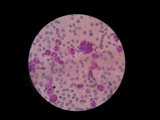
- Peripheral blood smear: Blasts (large, high N:C ratio, fine chromatin, prominent nucleoli, Auer rods in AML).
- Bone marrow aspirate/biopsy: Hypercellular marrow with >20% blasts (diagnostic gold standard).
- Flow cytometry (immunophenotyping): Identifies cell lineage (CD3, CD7 = T-cell; CD19, CD20 = B-cell; CD13, CD33 = myeloid).
- Cytogenetics/Karyotyping: Philadelphia chromosome t(9;22) in CML; t(8;21), inv(16) in AML.
- Lumbar puncture: CSF cytology for CNS involvement (ALL).
- Chest X-ray: Mediastinal mass (T-cell ALL).
4.2.6 Management
- Chemotherapy: Induction, consolidation, maintenance; combination regimens (e.g., 7+3 for AML: cytarabine + anthracycline).
- Targeted therapy: Tyrosine kinase inhibitors (imatinib, dasatinib) for BCR-ABL1+.
- Immunotherapy: CAR-T cell therapy, blinatumomab (CD19-directed) for B-ALL.
- Stem cell transplant: Allogeneic transplant for high-risk or relapsed disease.
- Supportive care: Blood transfusions, infection prophylaxis, growth factors (G-CSF), tumor lysis syndrome prevention.
Acute leukemia is a medical emergency. Tumor lysis syndrome can be fatal without prophylaxis (hydration, allopurinol, rasburicase).
4.3 HEMOPHILIA
4.3.1 Definition
Hemophilia is a group of inherited bleeding disorders caused by deficiency of specific coagulation factors, resulting in impaired secondary hemostasis and prolonged bleeding.
4.3.2 Classification
| Type | Deficient Factor | Inheritance | Incidence |
|---|---|---|---|
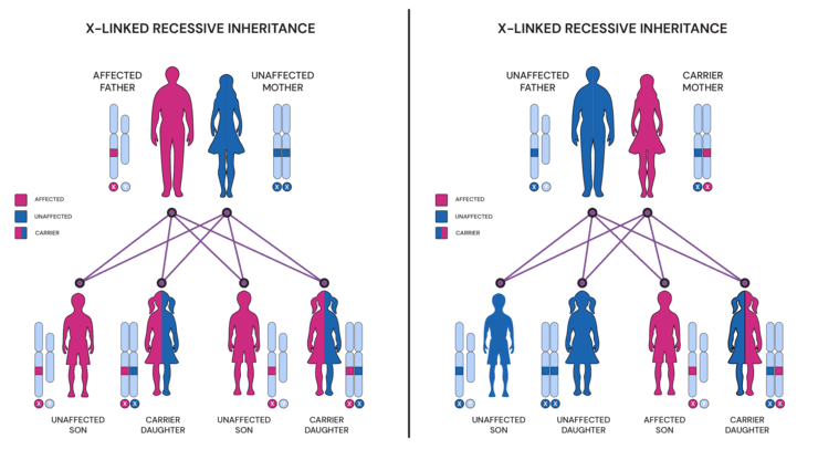
| Hemophilia A (Classic) | Factor VIII | X-linked recessive | 1 in 5,000-10,000 male births (80%) |
| Hemophilia B (Christmas Disease) | Factor IX | X-linked recessive | 1 in 30,000 male births (20%) |
| Hemophilia C | Factor XI | Autosomal recessive | Rare (Ashkenazi Jews) |
4.3.4 Pathophysiology
- Deficiency of Factor VIII or IX impairs the intrinsic coagulation pathway.
- Reduced generation of thrombin and fibrin.
- Inability to form stable clots results in prolonged bleeding, particularly after trauma or surgery.
- Spontaneous bleeding occurs in severe disease (<1% factor activity).
4.3.5 Severity Classification
| Severity | Factor Activity | Clinical Presentation |
|---|---|---|
| Severe | <1% | Spontaneous bleeding into joints (hemarthrosis), muscles, CNS; frequent episodes |
| Moderate | 1-5% | Bleeding after minor trauma; occasional spontaneous |
| Mild | >5-40% | Bleeding only after significant trauma, surgery, or dental extraction |
4.3.6 Clinical Features
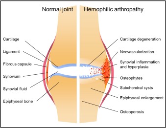
- Hemarthrosis: Bleeding into joints (knees, elbows, ankles, hips); causes pain, swelling, warmth, limited movement; repeated episodes lead to chronic synovitis and hemophilic arthropathy.
- Muscle hematomas: Intramuscular bleeding (iliopsoas, calf, forearm); can cause compartment syndrome.
- Mucocutaneous bleeding: Prolonged epistaxis, gum bleeding, prolonged bleeding after tooth extraction.
- GI bleeding: Melena, hematemesis.
- Hematuria: Painless hematuria from renal tract bleeding.
- Intracranial hemorrhage: Most serious complication; can be spontaneous or after minor head trauma; headache, vomiting, altered consciousness.
- Pseudotumors: Progressive cystic swelling from recurrent bleeding into muscle or bone; can compress nerves and destroy bone.
4.3.7 Investigations
Diagnosis is confirmed through laboratory testing using one-stage factor assays.
- aPTT: Prolonged (reflects intrinsic pathway defect).
- PT: Normal (extrinsic pathway intact).
- Bleeding time: Normal (primary hemostasis unaffected).
- Factor VIII assay: Reduced (Confirms Hemophilia A; severity by % activity).
- Factor IX assay: Reduced (Confirms Hemophilia B).
- Mixing study: Corrects with normal plasma (confirms factor deficiency vs. inhibitor).
- Inhibitor screen: Positive in 15-30% of severe Hemophilia A (Bethesda assay quantifies inhibitor titer).
4.3.8 Management
A. Factor Replacement Therapy
Factor Replacement Targets:
- Minor hemorrhage: 30-50% (Early joint bleed, minor muscle bleed, epistaxis).
- Major hemorrhage: 50-80% (Significant muscle bleed, GI bleed, post-dental).
- Life-threatening/CNS: 80-100% (Intracranial hemorrhage, major surgery, trauma).
- Prophylaxis: >1% (1-3% trough) in severe hemophilia to prevent joint disease.
Dose Calculation:
— Factor VIII: IU required = body weight (kg) x desired factor increase (%) x 0.5.
— Factor IX: IU required = body weight (kg) x desired factor increase (%) x 1.0.
B. Prophylaxis
Primary prophylaxis with factor replacement is recommended as first-line treatment for severe hemophilia to prevent joint bleeding and hemophilic arthropathy. Typically initiated before age 3 or after first joint bleed.
C. Management of Bleeding Episodes
- Joint (hemarthrosis): RICE (Rest, Ice, Compression, Elevation); immediate factor replacement; physiotherapy after resolution.
- Muscle: Factor replacement; physiotherapy; monitor compartment syndrome.
- CNS: Immediate factor replacement to 80-100%; CT scan; neurosurgical consultation if indicated.
- Dental extraction: Factor replacement to 50-80%; antifibrinolytics (tranexamic acid); local hemostatic measures.
D. Bypassing Agents (for Inhibitors)
- rFVIIa (NovoSeven): Directly activates Factor X; used in Hemophilia A/B with inhibitors.
- aPCC/FEIBA: Contains activated II, VII, IX, X; used in Hemophilia A with inhibitors; caution in Hemophilia B.
E. Adjunctive Therapies
- Desmopressin (DDAVP): Stimulates endogenous Factor VIII release; useful in mild Hemophilia A and von Willebrand disease.
- Antifibrinolytics: (Tranexamic acid, epsilon-aminocaproic acid); inhibit fibrinolysis; useful for mucosal bleeding and dental procedures.
- Gene therapy: Emerging treatment using AAV vectors; approved for Hemophilia B in some regions.
Intracranial Hemorrhage (Suspected or Confirmed):
- Immediate factor replacement to 80-100% (do NOT wait for imaging).
- Urgent CT scan of head.
- Neurosurgical consultation.
- Avoid IM injections, aspirin, and NSAIDs.
- Monitor neurological status closely.
**KEY CLINICAL POINT:** Any head trauma in a person with hemophilia, even minor, requires immediate factor replacement and urgent evaluation. Delay can be fatal.
4. COMMON BLOOD DISORDERS — SUMMARY
| Feature | Iron Deficiency Anaemia | Leukemia | Hemophilia A/B |
|---|---|---|---|
| Nature | Acquired nutritional/metabolic | Malignant neoplasm | Inherited coagulation disorder |
| Pathophysiology | Reduced Hb synthesis | Bone marrow failure + organ infiltration | Factor VIII/IX deficiency |
| Onset | Gradual (months) | Acute or chronic | Congenital (manifests with activity) |
| Key signs | Pallor, glossitis, koilonychia, tachycardia | Fatigue, infections, bleeding, organo-lymphadenopathy | Prolonged bleeding, hemarthrosis, muscle hematomas |
| Key lab finding | Low Hb, low MCV, low ferritin | Blasts in blood/marrow, pancytopenia | Prolonged aPTT, normal PT, low factor VIII/IX |
| Primary treatment | Iron supplement, treat cause | Chemotherapy, targeted therapy, transplant | Factor replacement, prophylaxis |
| Prognosis | Excellent with treatment | Variable; depends on type and risk factors | Good with modern therapy; normal life expectancy with prophylaxis |
- Anaemia workup: Always investigate the cause of iron deficiency — it is never normal in adult males or postmenopausal females. GI malignancy must be excluded.
- Blood transfusion safety: Always perform ABO and Rh typing plus antibody screen before transfusion. Cross-match donor blood with recipient serum.
- Hemophilia diagnosis: Prolonged aPTT with normal PT suggests intrinsic pathway deficiency. Factor assays distinguish Hemophilia A from B.
- Leukemia urgency: Acute leukemia is a medical emergency. Tumor lysis syndrome can be fatal without prophylaxis.
- Rh-negative pregnancy: Administer anti-D immunoglobulin at 28-30 weeks and postpartum to prevent alloimmunization and hemolytic disease of the newborn.
- WHO 2024 thresholds: Children 6-23 months now have Hb cutoff <10.5 g/dL; second-trimester pregnant women have specific threshold <10.5 g/dL.
