Doctors Revision

Author name: doctorsrevision@gmail.com

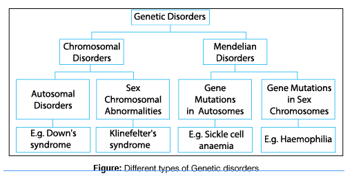
types of genetic disorders
Pathology

Genetic Disorders

Genetic Disorders Genetic Disorders Learning Objectives Genetics can feel overwhelming because it deals with the invisible instruction manuals of our bodies. We will break this down step-by-step so that by the end of this guide, you will be able to: Understand the general classification of genetic disorders and how they arise. Master the principal aspects of Mendelian disorders (the rules of inheritance). Confidently identify examples of different Mendelian disorders, as well as recognize the physical signs (phenotype) and genetic blueprints (genotype) needed to make a diagnosis. Impact of Genetic Disorders Genetic disorders are far more common than is widely appreciated by the general public. They are not rare anomalies; they are a fundamental part of human medicine. Lifetime Prevalence: The estimated lifetime prevalence of genetic diseases is 670 per 1000 individuals. This means that over the course of a lifetime, more than half the population will experience a disease that has a genetic component. Early Gestation: It is estimated that 50% of spontaneous abortions (miscarriages) during the early months of pregnancy occur because the embryo has a demonstrable chromosomal abnormality incompatible with life. Newborns and Youth: About 1% of all newborn infants possess a gross (large-scale, easily visible) chromosomal abnormality. Furthermore, approximately 5% of individuals under age 25 develop a serious disease with a significant genetic component. Genetics versus Genomics While these terms sound similar, they represent different scales of study: Genetics: The study of single genes or a few specific genes and their phenotypic effects (the physical traits they produce). Example: Studying the single mutated gene that causes Cystic Fibrosis. Genomics: The comprehensive study of all the genes in the entire genome and how they interact with each other. Example: DNA microarray analysis of tumors is an excellent example of genomics in current clinical use. It looks at thousands of genes at once to understand a cancer’s behavior. Analogy to make it stick: Genetics is like studying a single instrument in an orchestra to see if it is out of tune. Genomics is listening to the entire symphony to understand how all the instruments interact to create the overall sound. The most important contribution of genomics to human health will be identifying multifactorial diseases (like heart disease or diabetes) that arise from interactions among multiple genes and environmental factors. The Spectrum of Human Diseases Every human disease falls somewhere on a spectrum based on what causes it: Environmentally determined: Caused purely by outside factors (e.g., getting a sunburn or breaking a bone in a car accident). Genetically determined: Caused purely by DNA (e.g., Sickle cell anemia). Environmentally AND Genetically determined: A mixture of both. Someone might have a genetic predisposition to lung cancer, but smoking (environment) triggers it. Some Definitions Before looking at the specific diseases, we must clearly define the terminology used in medical genetics. Genetic Disorders: A heterogeneous (diverse) group of disorders caused by abnormalities in genes or whole chromosomes. Hereditary Disorders: These are derived from one’s parents and are transmitted through the germ line (sperm and egg cells) across generations. Therefore, these conditions are familial (they run in families). Congenital: Simply means “born with.” It is crucial to note that not all congenital diseases are genetic (e.g., a baby born with syphilis acquired it from the mother during birth, which is environmental), and not all genetic diseases are congenital (e.g., Huntington’s disease is genetic but symptoms don’t appear until age 40). Mutations: A permanent change in the DNA sequence. Germ Cells versus Somatic Cells Where a mutation happens dictates whether it can be passed on to children: Germ Cell Mutations: Mutations that affect sperm or egg cells. These are transmitted to the progeny (offspring) and give rise to inherited diseases. Somatic Cell Mutations: Mutations that arise in the regular cells of the body (like skin, liver, or lung cells) after birth. Understandably, these do not cause hereditary diseases because they are not in the sperm or egg. However, they are immensely important in the genesis of cancers and some congenital malformations. Classification of Mutations Mutations are classified by the “size” of the DNA mistake: Genome mutations: The largest errors. The loss or gain of whole chromosomes. This gives rise to monosomy (missing a chromosome) or trisomy (having an extra one, like Trisomy 21 causing Down Syndrome). Chromosome mutations: The rearrangement of genetic material. A whole chunk of a chromosome might break off and attach somewhere else. These give rise to visible structural changes in the chromosome under a microscope. Most of these are highly destructive and incompatible with survival. Gene mutations: The smallest, but most common, errors. These may result in partial or complete deletion of a specific gene, or more often, affect just a single base (a single “letter” in the DNA code). General Classification of Genetic Disorders Genetic disorders are grouped into three massive categories: Disorders related to mutant genes of large effect (Mendelian disorders). Diseases with multifactorial inheritance. Chromosomal disorders. Mendelian Disorders (Mutant Genes of Large Effect) Named after Gregor Mendel (the father of genetics), these disorders are the result of expressed mutations in single genes that have a very large, obvious effect on the body. An estimated 80% to 85% of these mutations are familial (inherited from parents). Most of these diseases are recessive, meaning a person needs two bad copies of the gene to show symptoms. Because of this, many people carry these mutations without having any serious phenotypic effect themselves. The Laws Governing Mendelian Inheritance To understand how these traits are passed down, we rely on Mendel’s fundamental laws, which were later proven by the discovery of meiosis (the cell division process that creates sperm and eggs). Law of Segregation (The “First Law”): States that when any individual produces gametes (sperm/eggs), the two copies of a gene separate, so that each gamete receives only one copy. A gamete will receive one allele or the other. In meiosis, the paternal and maternal chromosomes get physically separated, segregating the characters into two different gametes. Law of Independent Assortment (The “Second Law”):
Chronic Inflammation
Pathology

Chronic Inflammation

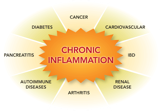
Chronic Inflammation Chronic Inflammation Learning Objectives By the end of this detailed study guide, you should be able to clearly understand and explain: The precise definition of chronic inflammation and identify the key cellular mediators involved. The specific mechanisms and different types of granulomatous inflammation (foreign body vs. immune). The various examples of giant cells, distinguishing between those that occur naturally (physiological) and those that indicate disease (pathological). Introduction to Chronic Inflammation Defining Chronic Inflammation Chronic inflammation is defined as a physiological response of prolonged duration—lasting for weeks or months (and sometimes even years). Unlike acute inflammation, which is a rapid, short-lived response characterized by fluid leakage and neutrophil infiltration, chronic inflammation is entirely different in its nature. The defining characteristic of chronic inflammation is that three distinct processes occur simultaneously at the site of the lesion: Active Inflammation: The immune system continues to fight. Tissue Injury: The ongoing battle causes collateral damage to the host’s own tissues. Attempts at Repair: The body simultaneously tries to heal the damage, usually through scarring (fibrosis) and the creation of new blood vessels. Onset and Progression Chronic inflammation does not always start the same way. It can initiate in two main ways: Following Acute Inflammation: If an initial acute inflammatory response fails to clear the offending agent (like a persistent bacterial infection), the body transitions the response into a chronic state. Insidious (Sneaky) Onset: It may begin as a low-grade, smoldering response from the very beginning. In these cases, there are absolutely no manifestations of a preceding acute reaction. The patient might not even realize it is happening until significant tissue damage has already occurred (e.g., in rheumatoid arthritis or atherosclerosis). Causes of Chronic Inflammation The immune system generally wants to clear a threat quickly and return to normal. Chronic inflammation only occurs when an offending agent is highly stubborn or when the immune system becomes misdirected. A. Persistent Infections Certain microorganisms are extremely difficult for the immune system to eradicate. They possess unique defense mechanisms that allow them to survive inside the body for long periods. Examples include: Mycobacteria: The bacteria responsible for Tuberculosis and Leprosy. They have waxy cell walls that resist being digested by immune cells. Treponema pallidum: The spiral-shaped bacterium that causes Syphilis. Certain Viruses, Fungi, and Parasites: These pathogens can hide within host cells or evade immune detection, leading to a constant, unresolved immune battle. B. Immune-Mediated Inflammatory Diseases (Hypersensitivity) Sometimes, the immune system inappropriately attacks the body’s own tissues or harmless environmental substances. Because the eliciting antigens (the targets of the attack) cannot be eliminated—since they are part of the body itself or constantly present in the environment—these disorders tend to be chronic and highly intractable (hard to treat). Autoimmune diseases: Conditions like Rheumatoid Arthritis or Systemic Lupus Erythematosus. Allergic diseases: Chronic asthma is a prime example of continuous inflammation driven by environmental antigens. C. Prolonged Exposure to Potentially Toxic Agents Continuous exposure to toxic substances over a long period forces the body into a state of chronic inflammation. These agents can be from outside the body (exogenous) or inside the body (endogenous). Exogenous Materials (Outside the body): An example is inhaled particulate silica. When inhaled, silica dust cannot be degraded by the lungs’ immune cells. The constant presence of these sharp, indestructible particles induces a chronic inflammatory response known as silicosis. Endogenous Agents (Inside the body): An example is chronically elevated plasma lipid components (high blood cholesterol). The persistent presence of toxic oxidized lipids in the blood vessel walls triggers a relentless inflammatory response that contributes heavily to the development of atherosclerosis (hardening of the arteries). Morphologic Features (What it looks like under a microscope) If a pathologist examines a tissue sample experiencing chronic inflammation under a microscope, they will observe three hallmark features that clearly distinguish it from acute inflammation. Infiltration with Mononuclear Cells: Acute inflammation is dominated by polymorphonuclear cells (neutrophils). In contrast, chronic inflammation is dominated by mononuclear cells—cells with a single, round, or un-lobed nucleus. These specifically include macrophages, lymphocytes, and plasma cells. Tissue Destruction: There is visible damage and destruction of the normal tissue architecture. This destruction is induced directly by the persistent offending agent (like a toxin or virus) or indirectly as collateral damage caused by the highly potent chemicals released by the accumulating inflammatory cells. Attempts at Healing: Because the tissue is being destroyed, the body desperately tries to patch the holes. This connective tissue replacement of damaged tissue is accomplished by two simultaneous processes: Angiogenesis: The sprouting and creation of new, fragile blood vessels to bring nutrients to the healing area. Fibrosis: The massive deposition of fibrous connective tissue (collagen) by fibroblasts, resulting in heavy scarring. Cells and Mediators of Chronic Inflammation The chronic inflammatory response is orchestrated by a specific cast of cellular characters. The major players include Macrophages, Lymphocytes, Plasma cells, Eosinophils, Neutrophils (in specific ongoing scenarios), and Mast cells. A. Macrophages: The Dominant Cells Macrophages are the undisputed heavyweights and dominant cells of chronic inflammation. They are large, highly capable tissue cells derived from circulating blood white blood cells called monocytes. After monocytes emigrate from the bloodstream into the tissues, they transform into macrophages. The Mononuclear Phagocyte System Macrophages are normally diffusely scattered in most connective tissues throughout the body to act as local guards. Together, these cells comprise the mononuclear phagocyte system (older name: reticuloendothelial system). Depending on the organ they reside in, they are given special names: Kupffer cells: Found in the Liver. Sinus histiocytes: Found in the Spleen and Lymph nodes. Microglial cells: Found in the Central Nervous System (Brain and Spinal Cord). Alveolar macrophages: Found in the Lungs. In all tissues, these cells act as essential filters (eating particulate matter, microbes, and dead/senescent cells) and as sentinels to alert the specific components of the adaptive immune system (T and B lymphocytes) to injurious stimuli. Lifecycle and Activation of Macrophages Migration: The half-life of circulating blood monocytes is very short, about 1 day. Under the influence of chemical signals (adhesion molecules

Classification and Nomenclature of Drugs
Pharmacology

Classification and Nomenclature of Drugs

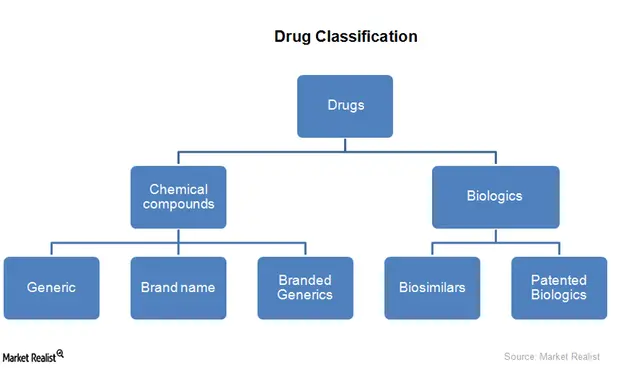
Classification & Nomenclature of Drugs Classification and Nomenclature of Drugs Introduction: Why do we classify drugs? With tens of thousands of individual drugs existing in modern medicine, studying them one by one is impossible. Drug classification refers to the systematic grouping of drugs based on shared characteristics. By grouping drugs logically, pharmacologists, physicians, and pharmacists can: Understand general drug actions and behaviors without memorizing every single drug. Predict therapeutic effects, potential side effects, and drug interactions. Guide rational, evidence-based drug therapy. Organize pharmacy inventories and hospital formularies efficiently. Part I: The Systems of Drug Classification There is no single “perfect” way to classify a drug. A single drug can fall into multiple categories depending on the system used. Below are the primary methods of classification used in pharmacology, including several advanced clinical classifications. A. Classification Based on Therapeutic Use (Clinical Indication) This is the most intuitive and user-friendly system, especially for clinicians and patients. It groups drugs strictly according to the disease, symptom, or condition they are intended to treat, regardless of their chemistry or how they work. Therapeutic Class Examples Indication (What it treats) Analgesics Paracetamol, Morphine, Ibuprofen Pain relief Antihypertensives Enalapril, Amlodipine, Losartan Hypertension (High Blood Pressure) Antidiabetics Metformin, Insulin, Glipizide Diabetes mellitus Antibiotics / Antimicrobials Amoxicillin, Ciprofloxacin, Azithromycin Bacterial infections Antimalarials Artemether, Quinine, Chloroquine Malaria Antipyretics Paracetamol, Aspirin Fever reduction Advantages and Limitations of Therapeutic Classification Advantage: It is highly practical in clinical practice. If a doctor diagnoses a patient with Malaria, they simply look at the “Antimalarial” group to choose a treatment. Limitation: It is scientifically imprecise because many drugs have multiple therapeutic uses, making strict classification difficult. Furthermore, two drugs in the same class (like Enalapril and Amlodipine for hypertension) work in entirely different ways. The Aspirin Conundrum Aspirin is a classic example of this limitation. It can be classified as an Analgesic (treats headache), an Antipyretic (treats fever), an Anti-inflammatory (treats arthritis), and an Antiplatelet (prevents heart attacks). Classifying it under just one therapeutic use ignores its other vital roles. B. Classification Based on Pharmacological Effect This system groups drugs according to their broad physiological or biochemical effects on the body’s systems. It bridges the gap between what the drug treats (therapeutic use) and exactly how it works at the molecular level (mechanism of action). Pharmacological Class Examples Physiological Effect Diuretics Furosemide, Hydrochlorothiazide Increase urine output (removes excess fluid) Sedatives / Hypnotics Diazepam, Phenobarbital Induce calmness, reduce anxiety, or induce sleep (CNS Depression) Vasodilators Nitroglycerin, Hydralazine Relax and dilate smooth muscle in blood vessels Bronchodilators Salbutamol, Albuterol Relax and dilate the bronchi/airways in the lungs CNS Stimulants Caffeine, Amphetamines Increase brain activity and alertness C. Classification Based on Mechanism of Action (MOA) This is the most specific and scientifically rigorous classification. It groups drugs according to how they produce their pharmacological effect at the molecular or cellular level. It looks at the specific receptors, enzymes, or ion channels the drug targets. Mechanism of Action Class Drug Example Specific Molecular Action ACE Inhibitors Enalapril, Lisinopril Blocks the Angiotensin-Converting Enzyme, preventing the formation of Angiotensin II. Beta-blockers (β-adrenergic antagonists) Propranolol, Atenolol Bind to and block β-adrenergic receptors in the heart, preventing adrenaline from binding. Proton Pump Inhibitors (PPIs) Omeprazole, Pantoprazole Irreversibly inhibit the gastric H⁺/K⁺ ATPase pump in the stomach lining, stopping acid secretion. DNA Gyrase Inhibitors Ciprofloxacin, Levofloxacin Inhibit bacterial DNA gyrase (topoisomerase II), physically halting bacterial DNA replication. Calcium Channel Blockers Amlodipine, Nifedipine Block voltage-gated calcium channels in blood vessels, preventing calcium influx and causing relaxation. Note: This classification is critical in modern pharmacology and rational drug design, as it allows scientists to predict exact drug-drug interactions and side effects based on molecular targets. D. Classification Based on Chemical Structure Drugs are grouped based on their chemical composition, molecular skeleton, or structural similarity. Drugs that share a chemical structure usually share similar pharmacological activities, mechanisms, and side-effect profiles. Chemical Class Examples Structural Characteristic Penicillins (Beta-Lactams) Penicillin G, Amoxicillin, Ampicillin Contain a four-membered Beta-Lactam ring essential for antibacterial activity. Benzodiazepines Diazepam, Lorazepam, Clonazepam Contain a benzene ring fused to a diazepine ring. Sulfonamides Sulfamethoxazole, Sulfasalazine Contain a sulfonamide (-SO2NH2) chemical group. Barbiturates Phenobarbital, Thiopental Derivatives of barbituric acid. Steroids Cortisol, Testosterone, Dexamethasone Contain a core of four fused carbon rings (cyclopentanoperhydrophenanthrene). Deep Dive Structure-Activity Relationship (SAR) Why do we care about chemical structure? Because of SAR. By understanding the chemical backbone of a drug, chemists can make tiny structural changes to improve the drug. For example, natural Penicillin G is destroyed by stomach acid and must be injected. By simply adding an amino (-NH2) group to its chemical structure, chemists created Amoxicillin, which survives stomach acid and can be taken as an oral pill. E. Classification Based on Source of Origin Historically, all drugs came from nature. Today, we classify them by where the raw materials originate. 1. Plant Sources (Natural) Many of our oldest and most powerful drugs are extracted directly from the leaves, roots, or sap of plants. Morphine: A potent painkiller extracted from the seed pods of the opium poppy (Papaver somniferum). Quinine: An antimalarial from the bark of the Cinchona tree. Digoxin: A heart failure medication from the Foxglove plant (Digitalis species). Atropine: From the Deadly Nightshade plant (Atropa belladonna). 2. Animal Sources (Natural) Extracts from animal tissues and glands. Insulin: Historically extracted from the pancreas of pigs (porcine) and cows (bovine). Heparin: A blood thinner extracted from porcine (pig) intestinal mucosa or bovine lungs. Premarin: Estrogen hormone replacements originally extracted from the urine of pregnant mares (horses). 3. Mineral Sources (Natural) Inorganic elements used therapeutically. Ferrous sulfate: Iron supplement for anemia. Magnesium sulfate: Used for eclampsia in pregnancy or as a laxative. Lithium: Used for bipolar disorder. Iodine: Used as an antiseptic and for thyroid function. 4. Microbial Sources (Natural) Drugs extracted from fungi or bacteria (often used to kill other competing bacteria). Penicillin: Discovered from the Penicillium mold/fungus. Streptomycin / Chloramphenicol: Extracted from soil bacteria of the Streptomyces species. 5. Synthetic and Semisynthetic Drugs The vast majority of

cell injury death
Pathology

Cell Injury & Death

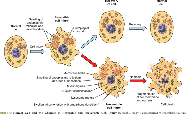
Cell Injury: Adaptation & Death Cell Injury, Death, and Adaptation Learning Objectives By the end of this comprehensive guide, you will be deeply conversant with: The different cellular responses to injury and the specific agents that cause them. The various types and stages of cell injury (Reversible vs. Irreversible). The underlying biochemical mechanisms and distinct morphological changes observed in cell injury. The defining characteristics of Cell Death (Necrosis vs. Apoptosis) and other emerging pathways. The different forms of cellular adaptations (Hypertrophy, Hyperplasia, Atrophy, Metaplasia) and cellular depositions, along with their clinicopathological relevance. Cellular Responses to Stress Cells are active, dynamic participants in their environment. They do not merely exist; they constantly adjust their internal structure and function to accommodate changing physiological demands and extracellular stresses. Under normal conditions, cells maintain a steady, balanced state called homeostasis, where the intracellular environment is kept within a highly regulated, narrow range of physiologic parameters. However, when a cell encounters stress or a pathologic stimulus, it follows a specific progression of responses: Adaptation: The cell’s first response is to adapt. This allows the cell to achieve a “new steady state,” preserving its viability and function in a hostile environment (e.g., a muscle cell growing larger to handle heavy lifting). Reversible Cell Injury: If the adaptive capability is exceeded, or if the external stress is inherently harmful but mild/transient, the cell sustains injury. However, up to a certain point, this injury is reversible, and the cell can return to a stable baseline if the stress is removed. Irreversible Cell Injury and Cell Death: If the injurious stimulus is severe, persistent, or rapid in onset, the cell passes a “point of no return.” It suffers irreversible injury and ultimately dies via Necrosis or Apoptosis. Clinical Example The Myocardium (Heart Muscle) Adaptation: In a patient with chronic high blood pressure, the heart must work harder to pump blood. In response to this increased hemodynamic load, the heart muscle cells enlarge (Hypertrophy). This adaptation helps short-term but increases the oxygen/metabolic demand of the heart. Reversible Injury: If a coronary artery narrows and the blood supply becomes inadequate (ischemia), the muscle first suffers reversible injury. The cells may swell and stop contracting efficiently, but they are not dead yet. Irreversible Injury: Unless the blood supply is rapidly restored (e.g., via a stent or clot-busting drug), the cells cross the threshold into irreversible injury, leading to cell death (Myocardial Infarction / Heart Attack). Causes of Cell Injury The agents that can injure cells range from the very large (physical trauma) to the submicroscopic (genetic mutations). They are generally grouped into seven categories: 1. Oxygen Deprivation (Hypoxia & Ischemia) Hypoxia is a deficiency of oxygen, which fundamentally injures cells by reducing aerobic oxidative respiration (halting ATP production). It is an extremely important and common cause of cell injury and death. Ischemia (Arterial obstruction): The most common cause of hypoxia. Ischemia is a loss of blood supply. Elaboration: Ischemia is actually worse than pure hypoxia because it not only deprives the tissue of oxygen but also deprives it of metabolic substrates (glucose) and fails to remove toxic metabolic waste products (like lactic acid). Inadequate oxygenation of the blood: Due to cardiorespiratory failure (e.g., severe pneumonia or heart failure). Decreased oxygen-carrying capacity: Seen in severe anemia, severe blood loss, or Carbon Monoxide (CO) poisoning (where CO irreversibly binds hemoglobin, blocking oxygen). 2. Physical Agents Physical forces can physically tear, burn, or irradiate cells. Examples include: Mechanical trauma (crush injuries, lacerations). Extremes of temperature (burns causing protein coagulation, deep cold causing ice crystal formation). Sudden changes in atmospheric pressure (decompression sickness). Radiation (ionizing radiation directly breaks DNA and creates free radicals). Electric shock (causes cardiac arrhythmias and severe tissue burning). 3. Chemical Agents and Drugs The list of chemicals that may produce cell injury defies compilation; almost any substance can be toxic in the wrong amount. Harmless substances in excess: Simple chemicals like glucose or salt in hypertonic concentrations can cause cell injury directly by deranging electrolyte and fluid balance, drawing water out of cells and killing them. Even pure oxygen at high concentrations is toxic (causing oxidative stress). Poisons: Strong acids and alkalis directly destroy cell membranes. Toxins and Drugs: Trace amounts of poisons (cyanide, arsenic), environmental pollutants (asbestos, carbon monoxide), social drugs (ethanol/alcohol, cigarette smoke), and even therapeutic drugs (chemotherapy) can injure cells. 4. Infectious Agents Biological pathogens attack cells either by directly invading them or by releasing toxins. These range from submicroscopic viruses (which hijack the cell’s DNA/RNA machinery) to bacteria, fungi, protozoa, and metazoa (tapeworms several feet in length). 5. Immunologic Reactions While the immune system defends against pathogens, friendly fire can cause massive tissue damage. Autoimmune diseases: Injurious reactions to endogenous self-antigens (the body attacks its own cells). Allergic reactions: Exaggerated immune responses to external agents (viruses, environmental substances, allergens) are important causes of tissue injury. 6. Genetic Abnormalities Errors in the DNA code can lead to cell death by preventing the creation of essential proteins. Deficient protein function: Such as enzyme defects leading to inborn errors of metabolism. Accumulation of damage: Misfolded proteins or damaged DNA trigger cell death (apoptosis) if they are beyond repair. Chromosomal abnormalities: Examples include congenital malformations associated with Down Syndrome (Trisomy 21). 7. Nutritional Imbalances A major global cause of cell injury. Deficiencies: Protein-calorie deficiencies cause an appalling number of deaths, chiefly among low-income populations. Specific vitamin deficiencies (e.g., Scurvy from Vitamin C deficiency) are not uncommon even in developed countries. Excesses: Nutritional excesses, such as obesity and high-fat diets, are significant factors in cellular injury leading to atherosclerosis, diabetes, and cancer. Factors Affecting Cell Response to Injury Why does one cell survive a stress while another dies? The outcome depends on two sets of variables: Variables of the Injurious Agent Nature of the injury: (Is it a mild toxin or a severe physical crush?) Duration of injury: (Did the ischemia last for 5 minutes or 50 minutes?) Severity: (A low dose vs. a massive overdose of a drug). Variables of the

mechanism of action
Pharmacology

Mechanism of Drug Action

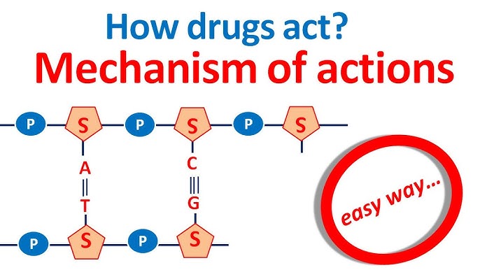
Pharmacodynamics: Mechanism of Drug Action Mechanism of Drug Action (Pharmacodynamics) Learning Objectives for this Exam Pharmacodynamics is the study of how drugs interact with the body at a molecular level. By the end of this guide, you will master: The four primary protein targets for drugs: Receptors, Ion Channels, Enzymes, and Transporters. The specific properties of receptors, including affinity, intrinsic activity, potency, and efficacy. The exact definitions of agonists (full, partial, inverse) and antagonists. The four major families of receptors and their specific operating speeds and mechanisms. A detailed understanding of G-Protein-Coupled Receptors (GPCRs) and their internal signaling pathways (cAMP and IP3/DAG). Introduction to Pharmacodynamics Pharmacodynamics is the branch of pharmacology concerned exclusively with the actions, interactions, and the specific mechanism (or mode) of action of drugs within the body. In simple terms, it studies exactly what the drug does to the body to produce a biological effect. When a drug enters the body, it must interact with something to cause a change. These interactions fall into two broad categories: Highly Specific Interactions: The drug precisely binds to a specific biological target (most commonly a pharmacological receptor) to exert its effect. Non-Specific Interactions: The drug produces an effect without binding to a specific receptor. For example, an antacid (like calcium carbonate) simply neutralizes stomach acid through basic chemistry, without needing a receptor. Molecular & Biochemical Mechanisms of Drug Action For drugs that act specifically, they must bind to certain proteins on or inside mammalian cells. These protein targets can be broadly divided into four fundamental categories: Receptors Ion Channels Enzymes Carrier Molecules (Transporters) Let us examine each of these four targets in deep detail, including the exact drugs that target them. Target I: Receptors Receptors are highly specialized protein structures located either on the surface of the mammalian cell membrane or entirely within the cell. They act as the sensing elements in the chemical communication system that coordinates the functions of all the different cells in the body. Natural chemical messengers (endogenous ligands) bind to these receptors to tell the cell what to do. These natural messengers include: Hormones (e.g., insulin, estrogen). Neurotransmitters (e.g., acetylcholine, dopamine). Other local mediators / Autocoids (e.g., Histamine, Serotonin / 5-HT). Many therapeutically useful drugs work by hijacking this system. They act either as agonists (mimicking the natural messenger) or antagonists (blocking the natural messenger) on these known endogenous receptors. Key Characteristics of Drugs Acting via Receptors Low Concentrations: Because receptors are highly sensitive, drugs targeting them can act effectively at very low concentrations in the blood. Structure–Activity Relationship (SAR): Receptors are extremely picky about shape. Very small modifications to a drug’s functional chemical groups, stereochemistry (3D arrangement), or molecular shape can significantly impact how tightly the drug binds (binding affinity) and how well it works (pharmacological activity). Specific Antagonism: Their effects can be precisely blocked by specific antagonists. Examples: Acetylcholine receptors can be blocked. Adrenaline receptors can be blocked. Histamine acts on specific H1, H2, H3, and H4 receptors (allergy medicines block H1). Dopamine acts on D1–D5 receptors (antipsychotic drugs block these). Morphine acts on specific opioid receptors named μ (mu), κ (kappa), and δ (delta). Target II: Ion Channels Cells use electrical charges to communicate, especially nerves and muscles. They do this by moving ions (like Sodium, Calcium, Potassium, and Chloride) in and out of the cell through specialized protein gates called Ion Channels. There are two main types of ion channels: Ligand-gated (ionotropic) channels: These are locked gates that only open when a specific chemical key (an agonist) binds directly to the receptor on the gate. Voltage-gated channels: These gates do not need a chemical key. Instead, they sense the electrical charge of the cell. They open or close in response to changes in the membrane potential (electrical voltage). How Drugs Act on Ion Channels: Direct Action: The drug physically binds directly to the channel protein itself, acting like a plug to block it, or locking it in an open position. Indirect Action: The drug binds to a separate receptor nearby, which then uses a messenger (like a G-protein) to tell the ion channel to open or close. Examples of Drugs Acting on Ion Channels: Voltage-gated sodium channels: These are blocked by local anesthetics (e.g., lidocaine). By blocking sodium from entering the nerve, the nerve cannot send a pain signal to the brain. L-type calcium channels: These are inhibited by dihydropyridines (a class of vasodilators, e.g., nifedipine). By blocking calcium from entering blood vessel muscles, the vessels relax, heavily lowering blood pressure. GABA receptor–chloride channel system: This is modulated by benzodiazepines (tranquillizers, e.g., diazepam). Diazepam binds to the channel, helping it open wider to let negatively charged chloride ions into the brain cell, severely calming and slowing down brain activity. ATP-sensitive potassium channels (KATP): Located in the pancreatic β-cells. These are blocked by sulfonylureas (diabetes medications). Blocking potassium from leaving the cell forces the pancreas to release stored insulin into the blood. Target III: Enzymes Enzymes are biological catalysts that speed up chemical reactions in the body (building things up or breaking them down). Many drugs act specifically as enzyme inhibitors. I. Competitive, Reversible Inhibition: The drug temporarily fights the natural substance for the active spot on the enzyme. If the drug wins, the enzyme halts. Because it is reversible, the effect wears off as the drug leaves the body. Example: Neostigmine. It reversibly inhibits the enzyme acetylcholinesterase (the enzyme that destroys acetylcholine). This allows acetylcholine to build up and help patients with severe muscle weakness. II. Irreversible, Non-Competitive Inhibition: The drug permanently binds to the enzyme, destroying its ability to function forever. The body must physically build entirely new enzymes to recover. Example: Aspirin. It permanently inhibits the cyclo-oxygenase (COX) enzyme, permanently stopping the production of chemicals that cause pain and inflammation. III. False Substrates: The drug tricks the enzyme. The enzyme thinks the drug is a normal building block and tries to process it, producing abnormal, broken metabolites that disrupt cell pathways. Example: Fluorouracil. This is an anticancer drug.

drug elimination
Pharmacology

Elimination & Clearance of Drugs

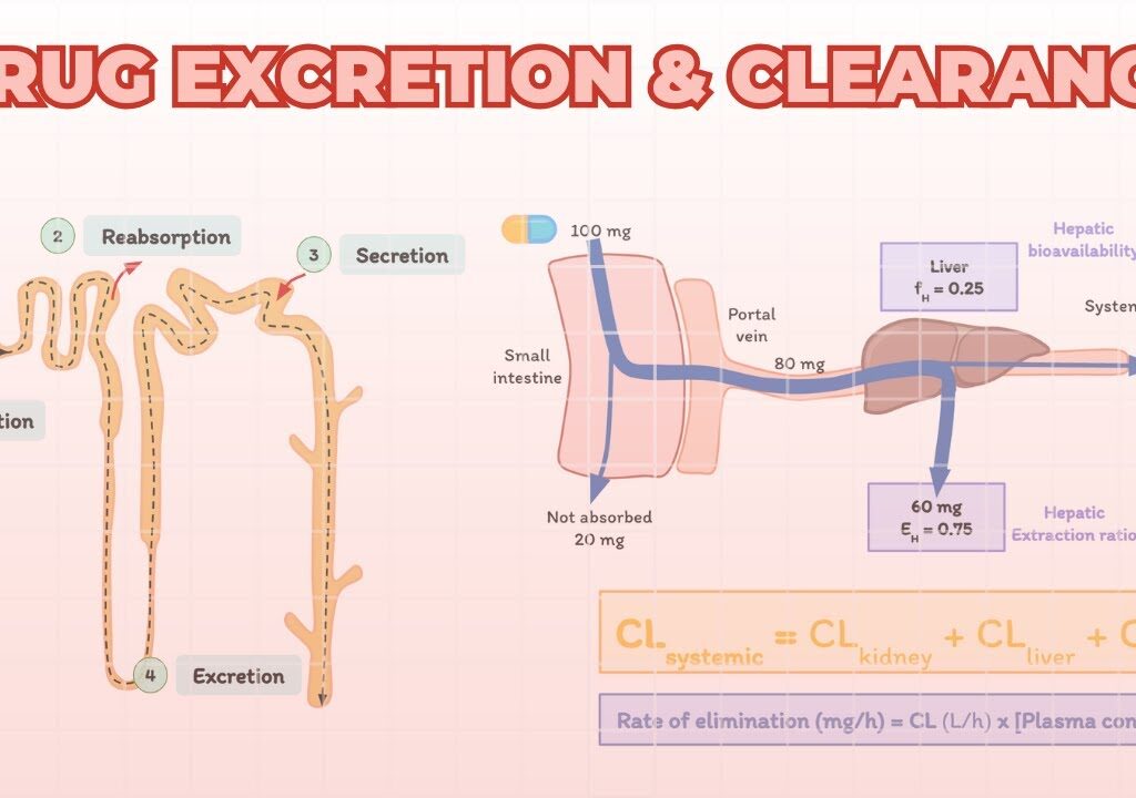
Pharmacokinetics: Drug Elimination & Clearance Pharmacokinetics of Elimination Module Overview This module covers the final stages of a drug’s journey through the body: Elimination and Excretion. You will learn not only how the body gets rid of drugs, but the mathematical principles (kinetics) that govern this removal. Mastering these concepts is crucial for determining how much of a drug to give (dosing) and how often to give it (dosing intervals) to maintain safe, steady, and therapeutic levels in a patient. The Fundamentals: Elimination vs. Excretion While often used interchangeably in casual conversation, in pharmacology, these two terms have different meanings: Elimination Elimination is the broad, overarching term. It concerns all the processes involved in the removal of active drugs from the body (and/or plasma) and their kinetic characteristics. If a drug is no longer active in the body, it has been eliminated. The major modes of drug elimination are: Biotransformation (Metabolism): The liver chemically alters the active drug into inactive metabolites. Even though the physical atoms of the drug are still in the body, the active drug has been eliminated. Excretion: The physical removal of the drug from the body. Excretion Excretion is a specific sub-process of elimination. It is the process by which drugs or their metabolites are irreversibly transferred from the internal environment to the external environment (i.e., passed out of the systemically absorbed body). Drugs and their metabolites can be excreted via several routes: Urine: The primary route (Renal Excretion). Feces: Biliary excretion via the bile duct into the intestines. Exhaled Air: Important for volatile anesthetics and alcohol. Saliva and Sweat: Minor routes. Breast Milk: Clinically crucial because excreted drugs can be unintentionally passed to a nursing infant. Renal Excretion: The Kidney’s Role The kidneys are the principal organs of excretion. For a drug to be efficiently excreted in the urine (renal excretion), it ideally needs to possess certain physical characteristics: Water-soluble (Hydrophilic): So it dissolves in urine. Small in molecular size: So it can be filtered. Slowly metabolized: If it is rapidly metabolized by the liver, the kidney only excretes the metabolites, not the parent drug. Non-volatile: Volatile gases are excreted by the lungs, not the kidneys. Net Renal Excretion = (Glomerular Filtration + Tubular Secretion) – Tubular Reabsorption To understand the equation above, we must break down the three distinct processes that occur inside the nephron (the functional unit of the kidney): A. Glomerular Filtration Blood enters the kidney’s glomerulus under high pressure. Glomerular filtration is a non-selective, unidirectional process. It acts like a simple sieve. What gets filtered? Water, small molecules, and unbound (free) drugs. What does NOT get filtered? Large proteins (like albumin) and any drug bound to those plasma proteins. Protein-bound drugs are simply too large to pass through the glomerular filter. Normal Rate: The normal Glomerular Filtration Rate (GFR) is approximately 120 ml/min. Clinical Note: GFR declines progressively after the age of 50 and drops drastically in patients with renal failure, requiring doctors to lower drug doses. B. Tubular Reabsorption As the filtered fluid travels down the renal tubules to become urine, the body realizes it has accidentally filtered out things it wants to keep. It reabsorbs them back into the blood. For drugs, this occurs mostly by passive diffusion. Lipid Soluble Drugs: If a drug is highly lipid-soluble, it will easily diffuse across the tubule walls back into the blood. In fact, 99% of the glomerular filtrate (mostly water) is reabsorbed, and lipid-soluble drugs follow this water back into the body. Non-Lipid Soluble & Ionized Drugs: These cannot cross the tubule membranes. They remain trapped in the urine and are excreted. Clinical Application The Role of Urinary pH (Ion Trapping) The pH of human urine can vary significantly (from 4.5 to 7.5). Because most drugs are weak acids or weak bases, the pH of the urine determines whether the drug becomes ionized (charged) or unionized (uncharged). Rule of thumb: Drugs become highly ionized in opposite-pH environments. Weak Bases: Ionize more in an acidic medium. If urine is acidic, basic drugs become ionized, cannot be reabsorbed, and are excreted. Weak Acids: Ionize more in a basic (alkaline) medium. If urine is alkaline, acidic drugs become ionized, are trapped in the tubule, and are excreted. Clinical Application: In an aspirin (weak acid) overdose, doctors administer sodium bicarbonate to alkalize the urine. This ionizes the aspirin in the kidney tubules, preventing its reabsorption and rapidly flushing it out of the body. C. Tubular Secretion This is the active transfer of organic acids and bases directly from the blood into the renal tubule, bypassing the glomerulus entirely. It is a carrier-mediated process that requires cellular energy because it pumps compounds against their concentration gradient. OATP (Organic Anion Transporting Polypeptide): Transports acidic drugs (anions). Examples include Penicillin, probenecid, uric acid, salicylates (aspirin), and furosemide. OCT (Organic Cation Transporter): Transports basic drugs (cations). Examples include Amiloride, quinine, procainamide, choline, and cimetidine. Competitive Inhibition: Because these transporters are limited in number, two drugs can compete for the same pump. For example, Probenecid competes with Penicillin for the OATP pump. Giving them together blocks Penicillin from being secreted, keeping it in the blood longer (historically used to prolong the effects of scarce penicillin). Elimination Kinetics: The Half-Life (t1/2) To mathematically model how fast a drug leaves the body, pharmacologists rely heavily on the concept of half-life. Definition: The Elimination Half-Life (t1/2) is the time required to eliminate 50% of a given amount of drug from the body, or specifically, the time it takes for the plasma concentration of a drug to fall to exactly half of its initial concentration. Plasma half-life: Time for plasma levels to drop by 50%. Whole body half-life: Time to eliminate 50% of the total drug content from the entire body. Why is Half-Life Important? It tells us the rate of decline of drug concentrations (though it does not necessarily dictate the duration of the biological effect). Most drugs are dosed according to their half-life. A drug with a 4-hour half-life might be

Metabolism of Drugs
Pharmacology

Metabolism of Drugs

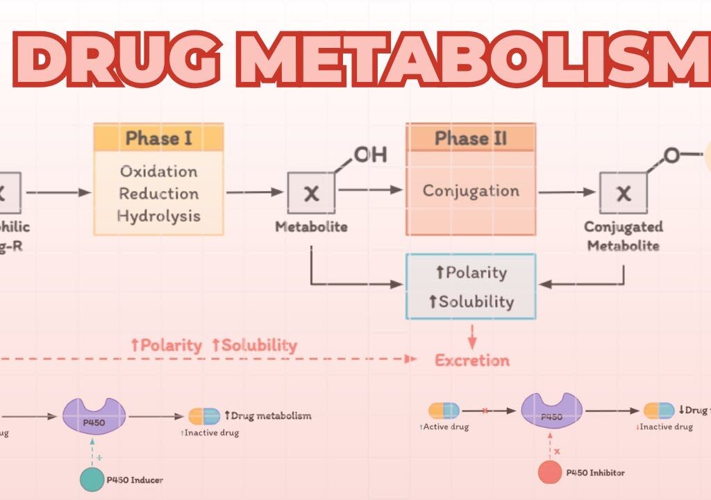
Pharmacodynamics: Mechanism of Action Drug Metabolism (Biotransformation) How to Approach This Topic Many students fear “Drug Metabolism” because of the heavy biochemistry and enzyme names. Do not panic. Think of drug metabolism simply as the body’s waste management system. The body wants to get rid of foreign chemicals (drugs). To do this, it must change their shape and properties so they can be flushed down the drain (kidneys). This guide will break down every mechanism, enzyme, and clinical scenario so you understand the “why” behind the science. Drug Metabolism Drug metabolism, also known as Biotransformation, is a core pillar of Pharmacokinetics. Remember the acronym ADME: Absorption, Distribution, Metabolism, and Excretion. Metabolism bridges the gap between a drug moving through your tissues and a drug leaving your body. The Journey of a Drug: Dose → Absorption → Blood Plasma (Free vs. Protein-Bound) → Distribution to Tissues/Receptors (Effect) → METABOLISM → Elimination (Renal Excretion). Why is Metabolism Absolutely Necessary? The Fundamental Problem: Most drugs that enter the body are designed to be lipophilic (fat-soluble). They need to be lipophilic so they can easily diffuse through the lipophilic cell membranes in your gut to be absorbed, and cross into tissues (like the brain) to exert their effects. The Catch-22: The kidneys (the body’s main filter) cannot efficiently excrete lipophilic drugs. When blood is filtered through the renal glomerulus, lipid-soluble drugs simply slide right back through the renal tubule membranes and are reabsorbed back into the systemic circulation. If we couldn’t metabolize them, lipophilic drugs would stay in the body forever, leading to massive accumulation and fatal toxicity. The Solution: The Definition of Metabolism Metabolism is the process where a drug is structurally altered (biotransformed) to become more polar and hydrophilic (water-soluble) so that it can be trapped in the urine and excreted. Analogy: Imagine trying to wash engine grease (a lipophilic drug) off your hands using only water (urine). It doesn’t work; the grease clings to your skin. You need soap (metabolism) to chemically alter the grease, making it mix with water so it can be rinsed down the drain. The Four Consequences of Drug Alteration When the body chemically alters a drug, four different clinical outcomes can occur. Metabolism doesn’t just mean destroying a drug; it means changing its pharmacological activity. 1. Most Common Active Drug → Inactive Metabolite The standard detoxifying process. The drug does its job, and the liver shuts it off. Examples: Paracetamol, Ibuprofen, Chloramphenicol. 2. Unpredictable Active Drug → Active or Toxic Metabolite Sometimes, the body’s attempt to alter a drug creates a byproduct that still has a therapeutic effect, or worse, is highly toxic. 3. Metabolic Activation Inactive Prodrug → Active Drug A Prodrug is a drug administered in an inactive form. It relies entirely on the body’s metabolism to activate it. We use prodrugs to improve absorption or bypass harsh stomach acid. 4. Clearance Unexcretable (Lipophilic) → Excretable (Hydrophilic) The structural conversion that allows final renal clearance and removal from the body. Consequence Tables Original Active Drug Active/Toxic Metabolite Formed (Outcome 2) Allopurinol (Gout medication) Alloxanthine (Also lowers uric acid) Digitoxin Digoxin (Active heart medication) Morphine Morphine-6-glucuronide (Highly active painkiller) Chloral hydrate Trichloroethanol Inactive Prodrug Active Metabolite (The functional drug) (Outcome 3) Levodopa (Crosses Blood-Brain Barrier) Dopamine (Treats Parkinson’s Disease) Sulindac Sulfide metabolite Prednisone Prednisolone (Active anti-inflammatory) Where Does Drug Metabolism Occur? While enzymes capable of biotransformation exist in almost every tissue (gut, lung, kidney, skin, placenta), the LIVER is the undisputed chief organ for drug metabolism. Nearly 90% of all drug metabolism happens here. Why the Liver? Blood Supply: The liver receives massive blood flow, specifically from the portal vein, which brings blood directly from the digestive tract. Enzyme Concentration: Liver cells (Hepatocytes) contain the body’s full complement of metabolizing enzymes in their smooth endoplasmic reticulum (ER), cytosol, and mitochondria. Crucial Concept The First-Pass Effect When you swallow a pill (PO – Per Os), it is absorbed in the intestines and goes into the portal vein. The portal vein goes straight to the liver before the drug reaches the rest of the body. The liver enzymes immediately metabolize a large portion of the drug. This “first-pass” can drastically reduce the amount of active drug that makes it to systemic circulation (bioavailability). If a drug has massive first-pass metabolism, it must be given via IV, sublingually, or transdermally to bypass the liver initially. Levels of Metabolism by Organ: High: Liver Medium: Lung, Kidney, Intestine Low: Skin, Testes, Placenta, Adrenals Very Low: Nervous System Reactions of Drug Metabolism: Phase I & Phase II To turn a stubborn, lipophilic drug into a water-soluble waste product, the liver uses a two-step process: Phase I and Phase II. (Note: Not all drugs go through both; some skip Phase I, some skip Phase II, and some go in reverse, but the standard sequence is I → II). Phase I Reactions: Modification (Functionalization) Goal: To modify the drug by unmasking or adding a small, polar “chemical hook” (like an -OH, -NH2, or -COOH group). This makes the drug slightly more water-soluble, but more importantly, it provides a handle for Phase II enzymes to grab onto. Types of Phase I Reactions: Oxidation: The most common. Involves the addition of oxygen or removal of hydrogen (e.g., converting a C-H bond to a C-OH bond: Hydroxylation, Dealkylation). Reduction: Addition of hydrogen. Hydrolysis: Breaking bonds using water. The Enzymes of Phase I Phase 1 is dominated by the Cytochrome P450 (CYP450) superfamily of enzymes, responsible for >95% of oxidative metabolism. They are located in the microsomes of the smooth endoplasmic reticulum (hence called microsomal monooxygenase enzymes). Understanding CYP450 Nomenclature: There are at least 18 different forms in humans. Take the most important one: CYP3A4 (which metabolizes ~50% of all drugs alone). CYP = Cytochrome P450 3 = Family (Families 1, 2, and 3 handle most drug metabolism) A = Subfamily 4 = Specific individual enzyme gene Overall, 60% of all drugs are metabolized primarily by the CYP450 family. Non-CYP Enzymes in Phase I (

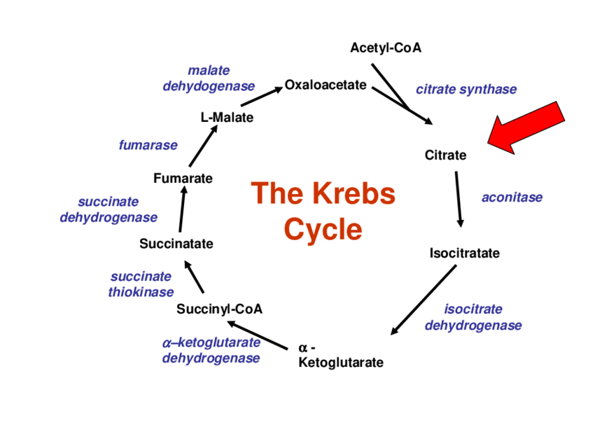
Tricarboxylic Acid (TCA) Cycle (Krebs Cycle / Citric Acid Cycle)
Biochemistry

Krebs / TCA Cycle Exam

Krebs Cycle Exam Biochemistry: TCA/Krebs Cycle Exam Biochemistry: TCA/Krebs Cycle Exam Test your knowledge with these 40 questions. Start Exam TCA/Krebs Cycle Exam Question 1/40 Submit Next Exam Complete! Here are your results, . Your Score 38/40 95% Download Full Report

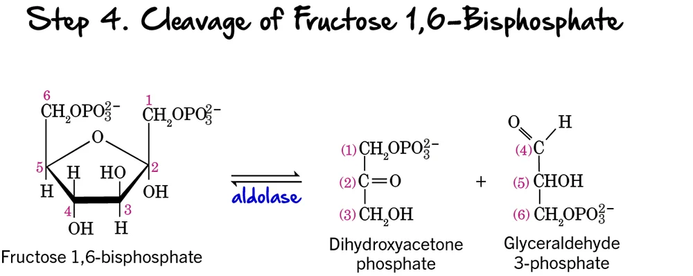
Glycolysis step 4 e1763166770749
Biochemistry

Glycolysis Exam

Glycolysis Exam Biochemistry: Glycolysis Exam Biochemistry: Glycolysis Exam Test your knowledge with these 40 questions. Start Exam Glycolysis Exam Question 1/40 Submit Next Exam Complete! Here are your results, . Your Score 38/40 95% Download Full Report

Drug Absorption & Distribution
Pharmacology

Drug Absorption & Distribution

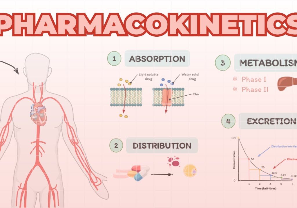
Pharmacokinetics: Drug Absorption & Distribution Learning Objectives By the end of this comprehensive lecture guide, you will be deeply conversant with: Fundamental definitions: What is a drug, and what is pharmacology? The subdivisions of pharmacology, focusing heavily on Pharmacokinetics. The mechanisms, factors, and clinical importance of drug Absorption and Bioavailability. The concept of Drug Distribution across various body fluid compartments. How to define, calculate, and interpret the Volume of Distribution (Vd). The critical role of Plasma Protein Binding and the dangers of drug displacement. Physiological factors affecting distribution (Blood Brain Barrier, tissue binding, pathology). What is a Drug? In pharmacology, a drug is defined as any chemical agent which affects any biological process. This is a very broad definition intentionally. It does not just mean medicine prescribed by a doctor; it includes naturally occurring substances, synthetic laboratory chemicals, recreational substances, and even everyday items like caffeine or alcohol, provided they cause a change in biological function when introduced to the body. What is Pharmacology? Pharmacology is the overarching scientific study of how drugs affect biological systems. To make this massive field manageable, it is divided into several specific sub-disciplines: Pharmacokinetics Simply put, this is what the body does to the drug. It covers how the drug moves through the body over time. Pharmacodynamics This is what the drug does to the body. It involves the molecular mechanisms, receptor binding, and physiological effects (e.g., lowering blood pressure). Pharmacotherapeutics The clinical study of the practical use of drugs to prevent, treat, or diagnose disease. Pharmacocognosy The highly specialized branch dealing with the identification of crude materials (like medicinal plants, herbs, or animal extracts) as potential drugs. Toxicology The study of the poisonous, adverse, or toxic effects of chemicals on living organisms. Introduction to Pharmacokinetics Pharmacokinetics is the journey of the drug through the body. It dictates how much of a dose actually reaches the target organ and how long it stays there. It is defined by four core processes (remembered by the acronym ADME): Absorption: The drug entering the blood. Distribution: The drug traveling via the blood to tissues. Metabolism (Biotransformation): The body (usually the liver) chemically altering the drug. Excretion: The body (usually the kidneys) removing the drug. Absorption of the Drug Absorption is the crucial first stage. It is defined as the process that involves the movement (transportation or passage) of a drug from its site or route of administration (e.g., the gut for an oral pill, the muscle for an injection) across biological membranes into the systemic blood stream. Mechanisms Used by Drugs to Cross Membranes Cell membranes are essentially biological barriers made of a lipid (fat) bilayer. Drugs must navigate this barrier using one of four primary mechanisms: 1. Simple (Passive) Diffusion This is the most common way drugs are absorbed. “Passive” means it happens naturally without the body spending any energy. Mechanism: Drugs move down a concentration gradient (from an area of high concentration, like the stomach, to an area of low concentration, like the blood). Energy: No energy (ATP) is required. Carriers: There is no use of special transport (carrier) proteins. Saturation: Because there are no carriers to get “full,” there is no saturation gradient required; as long as there is a concentration difference, diffusion continues. Types of Passive Diffusion: Via Aqueous Pores: Highly water-soluble (ionized or polar) drugs slip through tiny water-filled channels or pores in the membrane. Examples: Caffeine, ascorbic acid (Vitamin C), acetylsalicylic acid (Aspirin), nicotinamide. However, because these pores are very small, they play a limited role overall. Via the Lipid Layer: Highly lipid-soluble (non-ionized or non-polar) drugs dissolve directly into and pass through the fat of the cell membrane itself. Examples: Artemisinin, lumefantrine (antimalarials). The lipid layer plays the major role in simple diffusion. 2. Facilitated Diffusion While still a passive process (no energy used), this mechanism requires a “helper.” Mechanism: It occurs by the use of carrier proteins located within the cell membrane. The drug binds to the protein, the protein changes shape, and releases the drug on the other side. Gradient: The net flux is still from high concentration to low concentration. Energy: No energy is required. Saturation: Unlike simple diffusion, this requires a saturable gradient. Since there is a limited number of carrier proteins, if there is too much drug, all carriers become busy (saturated), and absorption maxes out. Examples: It is used for essential molecules that are too large or too polar for simple diffusion, such as amino acids, glucose, and folic acid. 3. Active Transport This mechanism forces drugs to move where they naturally wouldn’t go. Mechanism: Transportation acts against a concentration gradient or an electrochemical gradient (moving from low to high concentration). Energy: It strictly requires cellular energy (ATP) to pump the drug across. Carriers: It requires special transporter (carrier) proteins. Saturation: There is a “transport maximum” (T-max) for the substances; once all pumps are working, rate cannot increase. Rate: The rate of active transport heavily depends on the drug concentration in the environment competing for these pumps. 4. Pinocytosis Also known as “cell drinking,” this is reserved for massive molecules. Mechanism: Drugs with a Molecular Weight (MW) over 900 are transported this way. The drug molecule adheres to the cell membrane. The membrane then invaginates (folds inward), surrounds the drug, and pinches off to form a small intracellular vesicle. Energy: This is a highly active process and requires energy. Factors Affecting Drug Absorption Drug absorption is not uniform; it varies wildly based on the nature of the drug and the body itself. These factors are split into two categories. A. Drug-Related Factors Dosage Form: Solid drugs (like tablets) must break down before they can be absorbed. Disintegration: Breaking up of the large tablet into smaller granules/pieces after administration. Dissolution: The solid drug entering into a solvent (stomach fluid) to form a completely dissolved liquid solution. Rule of thumb: Solution forms (liquids, syrups) are absorbed much faster than unsolved (solid) forms because they skip the disintegration and dissolution steps. Chemical Nature: The physical chemistry

Scroll to Top