Doctors Revision

Author name: doctorsrevision@gmail.com

Autacoids Neuropeptides Ergot Alkaloids
Pharmacology

Autacoids, Neuropeptides & Ergot Alkaloids

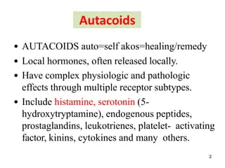
Autocoids Neuropeptides & Ergot Alkaloids Autacoids: 1. Introduction to Autacoids The word “Autacoid” comes from the Greek words Auto (meaning “self”) and Coids (meaning “healing/remedy”). They are frequently referred to as Local Hormones. Conceptual Check Autacoids vs. Classic Hormones Unlike classical hormones (like insulin or thyroid hormone) which are produced by a specific gland, secreted into the blood, and travel long distances to reach a target, Autacoids are produced locally by many different tissues, act locally near their site of synthesis, and have a very brief lifespan. Analogy: Think of them as the body’s “neighborhood watch” system. If a house is broken into (tissue trauma), you don’t wait for the national army (classical hormones) to arrive; the local neighborhood watch (autacoids) acts immediately at the exact site of injury to raise the alarm (inflammation/pain) and start repairs. Why are Autacoids Important? (Functions) Physiological: Regulate normal baseline organ functions (e.g., gastric acid secretion, local blood flow). Pathophysiological (Reaction to Injuries): They are the primary drivers of inflammation, pain, allergy, and the body’s response to tissue trauma. Transmission and Modulation: They act as mediators that fine-tune pain signals and nerve responses. Everyday Clinical Example: When you take an NSAID like Ibuprofen for a sprained ankle, you are specifically blocking the production of a lipid autacoid called a Prostaglandin. By shutting down this local autacoid, you stop the localized pain and swelling! Classification of Autacoids Autacoids are categorized by their chemical structure: Chemical Class Examples & Origin A. Amine Derivatives Histamine (derived from the amino acid Histidine) Serotonin (derived from the amino acid Tryptophan) B. Lipid Derivatives Eicosanoids: Prostaglandins, Thromboxane, Leukotrienes. Others: Interleukins, Platelet Activating Factor (PAF). C. Peptide Derivatives Kinins: Bradykinin. Renin-Angiotensin system. Neuropeptides. 2. Neuropeptides Neuropeptides are small, protein-like molecules (short chains of amino acids) used by neurons to communicate with each other. They act in an autocrine (acting on the cell that released it) or paracrine (acting on immediate neighboring cells) manner. Exam Trap: Neuropeptides vs. Classical Neurotransmitters Classical neurotransmitters (like dopamine, serotonin, glutamate) are fired into the synapse and then quickly sucked back up by reuptake pumps to be recycled and used again. NEUROPEPTIDES ARE NOT RECYCLED. Once they are secreted, they are broken down by specific enzymes (peptidases) and destroyed. The neuron must synthesize entirely new ones from the cell body (which takes time) and transport them down the axon. Do not forget this distinction! General Functions of Neuropeptides They are heavily responsible for higher-order brain functions and systemic regulation, including: Analgesia (pain regulation) Food intake (appetite stimulation/suppression) Learning & Memory Metabolism & Reproduction Social Behaviors Key Examples include: Neuropeptide Y (NPY), Cholecystokinin (CCK), Tachykinins (Substance P, Neurokinin), Arginine Vasopressin (AVP), and Corticotropin-Releasing Factor (CRF). Neuropeptide Y (NPY) NPY is a 36-amino acid peptide that acts as a potent neurotransmitter in both the Brain and the Autonomic Nervous System (ANS). Location Source Physiological Actions Brain (Central NPY) Produced mainly by the Hypothalamus. ↑ Food intake (Potent appetizer/orexigenic) ↑ Storage of energy as fat ↓ Anxiety and stress ↓ Voluntary alcohol intake ↓ Blood pressure and pain perception Regulates circadian rhythm and controls epileptic seizures. ANS (Peripheral NPY) Produced mainly by sympathetic neurons. Strong Vasoconstrictor Promotes the growth of fat tissue. NPY Receptors & Mechanisms NPY acts on G-Protein Coupled Receptors (GPCRs). Mammals have 5 types (Y1-Y5), but humans only express 4 functional types. Y1 (NPY1R) & Y5 (NPY5R): These are the Feeding Stimulators (Appetizers). Activation leads to massive hunger. Y2 (NPY2R) & Y4 (NPY4R): These act as Appetite Inhibitors (Anorectic). Mechanism of Action: NPY receptors are Gi-coupled (Inhibitory G-protein). When NPY binds, the Gi subunit is released, which inhibits the enzyme adenylate cyclase. This stops the conversion of ATP into the 2nd messenger cAMP. Clinical Scenario Anti-Obesity Drugs and NPY Because Y1 and Y5 receptors powerfully drive hunger and fat storage, pharmaceutical companies are actively researching Y1/Y5 Antagonists as therapeutic targets for obesity. Blocking these receptors could shut off the brain’s unnatural drive to overeat. Conversely, chronic stress increases NPY release in the periphery, which promotes the growth of visceral fat (explaining why chronic stress often leads to weight gain!). 3. Tachykinins (TAC) & Substance P Tachykinins form the largest family of neuropeptides. They get their name because they induce a rapid (“tachy”) contraction of gut tissues. Chemical Characteristic: All tachykinins share a common “C-terminal” sequence: “Phe-X-Gly-Leu-Met-NH2” (Where ‘X’ is either an aromatic or aliphatic amino acid, and COOH-terminus is the end of the protein chain). Synthesis Pathway: Preprotachykinin → Protachykinin → Tachykinin. Tachykinin Genes and Products TAC-1 Gene produces: Neurokinin A, Neurokinin K, Neuropeptide γ, and Substance P (SP). TAC-3 Gene produces: Neurokinin B. Tachykinin Receptors (GPCRs) Tachykinin receptors are Gq-coupled. Activation leads to the activation of Phospholipase C (PLC), which chops PIP2 into IP3 and DAG. This ultimately causes a massive release of intracellular Calcium. There are three main receptors, each with a preferred agonist: NK1R: Prefers Substance P. NK2R: Prefers Neurokinin A. NK3R: Prefers Neurokinin B. Substance P (SP) Substance P is an Undecapeptide (a chain of 11 amino acids). It is a highly potent mediator of pain signaling and inflammation. Receptor: Primarily binds to NK1R. The binding occurs via specific amino acid residues on the extracellular loops and transmembrane regions of the NK1 receptor. Physiological Roles: Promotes wound healing in humans (especially non-healing ulcers). Acts as a potent vasodilator. This vasodilation is entirely dependent on the release of Nitric Oxide (NO) from the endothelium. Transmits intense, burning pain signals to the brain (Neurogenic Inflammation). Clinical Application Substance P Antagonists (SPA) By blocking or depleting Substance P, we can block pain and severe nausea. Capsaicin: The active ingredient in chili peppers! Clinically used as a topical analgesic cream for arthritis and diabetic neuropathy. Mechanism: It initially causes a burning sensation (triggering SP release), but it eventually forces the nerve to release ALL of its Substance P. Because neuropeptides take a long time to synthesize (they aren’t recycled), the nerve is left empty of Substance P, rendering it completely unable to transmit pain signals for
sympatheic parasympathetic git neuro
Pharmacology

Autonomic Nervous System (ANS)

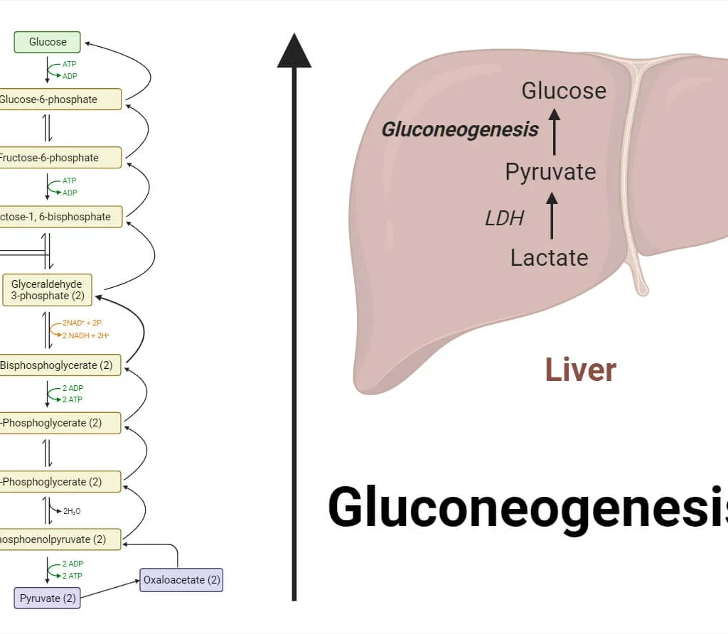
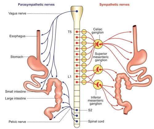
Autonomic Nervous System (ANS) Autonomic Nervous System (ANS): An Introduction to the Pharmacology Module Learning Outcomes This master guide is designed to make you deeply conversant with: The 4 Classes of Autonomic drugs. The role of Autonomic drugs in Clinical Practice (Cardiology, Respiratory, Psychiatry, etc.). Receptor and Non-receptor mechanisms of ANS drugs. Note on Adverse Effects (Type A-F) & ADME: While listed in the lecture’s opening slide, the provided slides focus exclusively on physiological effects and receptor dynamics. We will provide an emergency overview of Type A-F adverse effects at the end just in case it appears on your exam, but the bulk of this guide will strictly master the core ANS physiology and receptor profiles provided in the slides! 1. The Foundation: Why Autonomic Pharmacology? Before memorizing drugs, we must understand what we are treating. The nervous system (NS) is the ultimate communication system of the body. It acts as the critical LINK between the BODY and the ENVIRONMENT (both internal, like your sudden drop in blood pressure when you stand up, and external, like a lion chasing you). If this communication fails, HOMEOSTASIS (the stable, balanced state of the body) is violently disrupted. By understanding Autonomic Pharmacology, we can use drugs to artificially restore this communication and fix homeostasis. Autonomic pharmacology is highly LOGICAL (if you know the normal physiology, you know the drug’s effect) and incredibly CLINICALLY RELEVANT. It applies to: Psychiatric Medicine: Treating anxiety (e.g., using beta-blockers for stage fright). Respiratory Medicine: Treating asthma and COPD (e.g., inhalers that dilate airways). Cardiovascular Medicine: Treating hypertension, heart failure, and arrhythmias. GIT Medicine: Treating diarrhea, constipation, and stomach ulcers. Genitourinary Medicine: Treating overactive bladder or enlarged prostate issues. What is the Autonomic Nervous System (ANS)? The nervous system has two main outputs: Voluntary (Somatic – moving your arm to write a note) and Involuntary (Autonomic). The Autonomic Nervous System (ANS) is simply the “AUTOMATIC” part of the nervous system. It controls visceral organs (the “liquid-like” internal organs: heart, lungs, intestines, blood vessels) without you having to think about it. The ANS is divided into two competing branches. They are physiological antagonists (they do the exact opposite of each other to keep the body balanced): Sympathetic Nervous System (SNS): The “Accelerator.” Controls organs during STRESS (Fight, Flight, Fright). Parasympathetic Nervous System (PNS): The “Brakes.” Controls organs during REST (Rest and Digest / Breed and Feed). 2. The Sympathetic Nervous System: “Fight, Flight, Fright” The Scenario: You are walking in the bush and suddenly a lion jumps out at you. Your body instantly activates the Sympathetic Nervous System. Every single physiological change that happens next is designed to do one thing: Help you survive by fighting the lion or running away. The Chemical Messengers (Neurotransmitters) The sympathetic system communicates using three specific chemicals (Catecholamines). Because these are the messengers, drugs that mimic them are called Sympathomimetics (or Adrenergic drugs), and drugs that block them are called Sympatholytics. Noradrenaline (Norepinephrine): The primary neurotransmitter released directly at the nerve endings. Dopamine: A precursor and neurotransmitter, heavily involved in the kidneys and brain to maintain perfusion. Adrenaline (Epinephrine): This is a hormone, not a neurotransmitter. It is released by the Adrenal Gland directly into the blood. The adrenal gland output is 80% Adrenaline and 20% Noradrenaline. (This massive dump of adrenaline is what gives you that sudden “rush” in your chest when terrified). Sympathetic System Effects by Organ (Think deeply: “How does this help me run from the lion?”) Organ System Sympathetic Effect Why? (The Logical Reason) Cardiovascular (Heart) Heart Races: Increased Heart Rate (Chronotropy), increased Force of Contraction (Inotropy), and increased Conduction speed (Dromotropy). To rapidly pump massive amounts of oxygenated blood to the vital organs and legs for running. Increased force means a higher stroke volume per beat. Cardiovascular (Vessels) Blood is Diverted: ALL non-essential blood vessels (like those in the skin and gut) CONSTRICT. Blood vessels specifically going to Skeletal Muscles and the Brain DILATE. You don’t need blood in your stomach right now. You need maximum blood (oxygen) in your brain to think fast, and in your muscles to run. (This is why people turn “pale as a ghost” when terrified—skin blood vessels clamp shut!). Respiratory Bronchial Smooth Muscle RELAXES (Bronchodilation). Bronchial secretions DECREASE. Respiratory rate INCREASES. Relaxes the airways to open them up as wide as possible. Clears out mucus. This maximizes Oxygen (O2) uptake to fuel the skeletal muscles for sprinting. Gastrointestinal (GIT) Digestion Shuts Down: Motility DECREASES, Secretions DECREASE (causing Anorexia/lack of appetite), Sphincters TIGHTEN. Digesting food wastes massive amounts of energy and blood. Constipation and delayed gastric emptying occur to save energy for survival. You won’t feel hungry while running for your life. Genitourinary Urine Output DECREASES: The bladder wall (Detrusor muscle) relaxes, but the exit door (Sphincters/Trigone) TIGHTENS. Renin-Angiotensin System is ACTIVATED. Stopping to pee while running from a lion is a bad idea. It wastes energy and time. Activating Renin reabsorbs Sodium and Water in the kidneys, raising blood volume and blood pressure to sustain the “fight.” Reproductive Penile Erection INHIBITED. Uterine smooth muscle RELAXES. Genital secretions DECREASE. Blood is diverted to skeletal muscles. Reproduction is a waste of energy during a life-or-death crisis. (Sympathetic system specifically triggers ejaculation, but inhibits the erection phase). Central Nervous System Alertness INCREASES (can cause anxiety). Concentration INCREASES. Memory INCREASES. You need ultimate focus on the threat (the lion) to survive, dodging obstacles instantly. Skin Sweating INCREASES. Body temperature RISES (due to high metabolism). Body hairs ERECT (Piloerection). Sweating cools the rapidly overheating engine (your body). Raised hairs attempt to make you look larger and more intimidating to predators. Metabolism (CATABOLIC) Glucose goes UP: Glycogenolysis & Gluconeogenesis increase. Fat breaks down: Lipolysis increases. Proteins break down. Catabolism means breaking things down for energy. Your muscles need massive amounts of instant glucose and fatty acids to fuel the sprint, so the liver dumps its sugar reserves into the blood. Exocrine Glands DECREASE in salivation (causing a dry mouth and difficulty speaking). Decrease in tearing (dry eyes). Decrease

Preclinical Testing
Pharmacology

Preclinical Testing

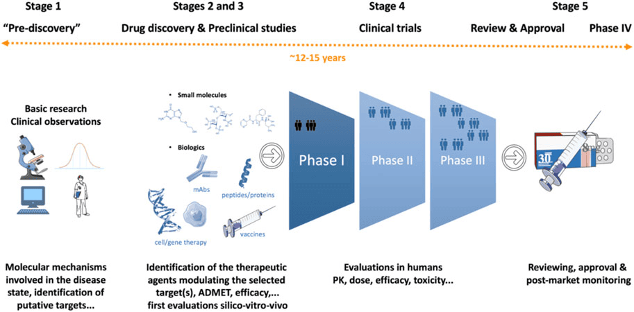
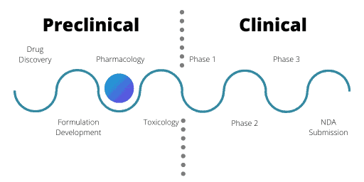
Preclinical Testing Preclinical Testing How to Approach This Topic Before a medicine can ever be prescribed to a sick patient, or a new medical device can be implanted in a human body, it must undergo rigorous, exhaustive testing. You cannot simply invent a chemical and give it to a human being. This lecture covers the entire phase that happens before humans are involved. We will break down every single test, why we use animals, what documents must be filed, and the extreme ethical and scientific importance of this process. 1. The Drug Development Process (The Big Picture) To understand where preclinical trials fit in, you must memorize the timeline of how a drug is born and brought to the pharmacy shelves. The process follows a strict, sequential pipeline: Basic Research: This is the purely academic stage. Scientists study biology at the most fundamental level. They look at Molecular biology, understand the Pathophysiology (how a disease harms the body), and study Genetics. They are not making drugs yet; they are just trying to understand the disease. R&D (Research and Development): Target Identification: Finding the exact enzyme, receptor, or cell part causing the disease. Compound Screening: Testing thousands of raw chemicals to see if any interact with that target. Lead Identification & Optimization: Finding the “lead” (the best chemical candidate) and tweaking its chemistry to make it stronger. Pre-clinical Studies: (Our Focus!) The phase where the optimized chemical is tested in the laboratory and on living animals. We test for In Vitro efficacy (in glass test tubes), In Vivo efficacy (in living animals), the exact Mechanism of Action / Proof of Concept, and we conduct IND-enabling studies (gathering all the safety data needed to get permission to test on humans). Clinical Trials: Testing on actual human beings. Phase 1: Testing on a small group of healthy volunteers just to see if it is safe in humans. Phase 2: Testing on a larger group of sick patients to see if it actually cures the disease. Phase 3: Massive testing on thousands of sick patients across many hospitals to confirm efficacy and monitor rare side effects. Review & Approval: Filing a massive application called an NDA (New Drug Application). The regulatory body (like the FDA or the National Drug Authority) heavily evaluates the data, approves the drug for sale, and conducts Post-release monitoring (sometimes called Phase 4, watching the drug once millions of people are buying it). 2. What Are Preclinical Trials? Preclinical studies, routinely known as nonclinical trials, are extensive laboratory tests of novel drugs (new medications), gene therapy solutions, or medical devices. They are universally conducted on animal subjects before any human testing is allowed. The Primary Objective While we absolutely want to know if the drug cures the disease (efficacy), the absolute primary objective of pre-clinical investigations is determining the eventual safety profile of a product. In medicine, the golden rule is “First, do no harm.” A drug that cures a headache but destroys the liver will never be allowed into human trials. We use animals to find these deadly side effects early. The ultimate goal of all this testing is strictly bureaucratic: to gather the sufficient information needed to file an IND. What is an IND? An IND (Investigational New Drug) application is a massive dossier submitted to a regulatory agency (like the FDA in the USA, or the NDA in Uganda). It is essentially a request asking for legal permission to test the drug on humans. The agency will only say “yes” if the preclinical animal data proves the drug is reasonably safe to administer to humans. The Animal Testing Funnel (Attrition Rate) After identifying a potential compound, it is given to animals to expose its whole pharmacological profile (what it does from head to toe). This follows a strict, stepping-stone approach: Step 1: Small Rodents. Experiments almost always begin with mice, rats, guinea pigs, hamsters, and rabbits. Why? They are mammals (sharing similar organ systems to humans), they breed rapidly, and they are inexpensive to house in large numbers. Step 2: Larger Animals. Following a favorable, safe outcome in rodents, the studies are escalated to larger mammals whose biology is much closer to humans, such as dogs, cats, and monkeys (non-human primates). Scenario The Brutal Rejection Process Imagine a pharmaceutical company creates 10,000 different chemicals to cure hypertension. They test them in glass tubes, and 500 show promise. They give those 500 to mice. 400 of those chemicals kill the mice. Those are rejected. The remaining 100 are given to dogs. 95 of them cause liver failure in dogs. Those are rejected. As the evaluation progresses, unfavorable compounds get rejected at each step. Ultimately, only a very few (perhaps 5 out of the original 10,000) will ever reach the stage where administration to man is even considered. This massive failure rate is why developing drugs is incredibly expensive. 3. The 10 Specific Types of Preclinical Studies When a drug is in the preclinical phase, it is subjected to an exhaustive battery of ten distinct types of tests. You must know what each one aims to discover. a) Screening Test These are extremely quick and easy assays designed to determine a simple “yes or no” question: Is a specific pharmacodynamic activity present or absent? We do not care how it works yet; we just want to know if it works. Example 1: Analgesic (pain-killing) action. A mouse is placed on a warm Hot Plate. A normal mouse will lift and lick its paws after 5 seconds due to the heat. We give the mouse the new drug. If the mouse now waits 15 seconds to lick its paws, the drug successfully blocked the pain! We have screened for analgesic activity. Example 2: Hypoglycemic action. We inject the drug into a rat and measure its blood sugar an hour later. Did the blood sugar drop? Yes or no. b) Tests on Isolated Organs and Bacterial Cultures Before putting a drug into a whole, living, breathing animal, we often test

The Drug Development Process
Pharmacology

The Drug Development Process

The Drug Development Process The Drug Development Process Overview Bringing a new drug to the pharmacy shelf is not a simple laboratory experiment; it is a massive, highly regulated journey. This guide will break down the entire process from a simple idea in a lab to post-marketing surveillance. Examiners love to test your knowledge on the differences between the Clinical Trial Phases (I, II, III, and IV), the definition of a new drug, and the “Pyramid of Uncertainty.” Pay close attention to the scenarios provided, as they will help you remember the dry facts. 1. Introduction: The Pyramid of Uncertainty The development of a new drug is an incredibly time-consuming and extremely expensive process. During the last 50 years, hundreds of new drugs have been introduced to save lives, while many older drugs have been entirely deleted (withdrawn) from the market due to newly discovered toxicities or better alternatives. We call this the “Pyramid of Uncertainty” because the failure rate is exceptionally high. Less than 1% (<1%) of compounds that go into testing eventually become licensed, usable medicines. The Timeline and Attrition Rate To successfully bring just one single new drug to the market, it requires a deep understanding of both the development process and the integral role that preclinical (animal/lab) testing plays. Let’s look at the numbers: Time: It takes 10 to 12 years (sometimes up to 24 years from the initial idea) on average for an experimental drug to travel from the laboratory bench to the patient’s medicine cabinet. Success Rate: Out of 5,000 to 10,000 compounds screened during initial discovery, only about 250 will make it to preclinical (animal) testing. From those 250, only FIVE (5) compounds will be deemed safe enough to enter human clinical trials (Phase I). Out of those 5 compounds tested in humans, only ONE (1) is finally approved by regulatory bodies. Stage of Development Number of Compounds Surviving Failure Rate at this Stage Discovery / Idea 5,000 – 10,000 N/A Preclinical Testing 250 50% fail here Phase I (Clinical) 5 30% fail here Phase II (Clinical) 1 (sometimes 2) 50% fail here Phase III (Clinical) 1 – FDA Review & Approval 1 Product Licensed – 2. What is a “New Drug” and Who Regulates It? Definition of a NEW DRUG In pharmacology and law, a “new drug” does not just mean a chemical that was invented yesterday. The legal definition encompasses three specific scenarios: A Completely New Substance: A chemical entity which, except during local clinical trials, has never been used before in the country. An Already Approved Drug with NEW CLAIMS: If a drug is already on the market, but the manufacturer wants to market it with modified or new claims. This includes a new indication (what disease it treats), a new dosage, a new dosage form (changing from a tablet to an IV injection), or a new route of administration. Clinical Scenario Minoxidil was originally approved as an oral tablet to treat high blood pressure. Later, researchers discovered it caused hair growth. When the company wanted to sell it as a topical lotion for baldness (new indication, new route, new dosage form), it had to go through the approval process again as a legally “New Drug.” Fixed-Dose Combination (FDC): Two or more already known and approved drugs proposed to be combined for the very first time in a single pill at a fixed ratio. Clinical Scenario Drug A (Artemether) and Drug B (Lumefantrine) are both known malaria drugs. If a company decides to combine them into one single tablet (Coartem), that combination is legally considered a “New Drug” and must be tested to ensure the two chemicals don’t react toxically with each other inside the pill. Regulatory Authorities and Guidelines Every country has a strict police force for medicines to protect the public. They issue guidelines on clinical trials that are required to be carried out before a drug can be imported or manufactured. Uganda: NDA (National Drug Authority). The power to grant permission for a new drug to be tested and marketed in Uganda rests solely with the NDA, governed by the NDA Act. The NDA Act details exactly what preclinical (animal) data is required before human tests begin. United States: US-FDA (Food and Drug Administration). Europe: EMEA (European Medicines Agency). United Kingdom: MHRA (Medicines and Healthcare products Regulatory Agency). Japan: MHLW (Ministry of Health, Labour and Welfare). Australia: TGA (Therapeutic Goods Administration). 3. The 8 Steps in New Drug Development The journey follows a strict chronological order: Idea or Basic Research New Drug Discovery Screening Preclinical Studies Formulation Development IND (Investigational New Drug) Application Clinical Studies (Human Trials) Official License / Regulations / Marketing 4. Step A & B: Basic Research and New Drug Discovery A. Basic Research Before you can invent a drug, you must thoroughly understand the disease. Start by studying normal and abnormal body functions. Investigate each component of the disease (its pathophysiology). Ask questions: What are the symptoms? What is the root cause? Which is the target organ? What are the biochemical pathways involved? Look up information obtained in previous research and publications. Find out at exactly which stage we can stop the disease progression. This becomes OUR TARGET! Search for a targeted drug, isolate the index compound, perform early animal testing for safety, and eventually seek approval to test in humans. B. New Drug Discovery (4 Sub-steps) Once the research is done, the actual discovery phase begins, taking roughly 5 to 6 years. Target Identification: Choosing a specific cellular or genetic chemical within our body (the “target”) that is associated with the disease. Target Validation: Checking and confirming that interacting with this specific target actually changes the disease condition. (Analogy: Making sure you have found the correct lock before you start building keys.) Lead Identification: Finding a “Lead compound.” A lead is a substance believed to have the potential to treat the disease. Scientists use massive collections (libraries) of up to 5,000-10,000 molecules. Each molecule is rigorously tested to confirm its effect

Pharmacogenomics & Pharmacogenetics
Pharmacology

Pharmacogenomics & Pharmacogenetics

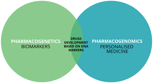
Pharmacogenomics & Pharmacogenetics Pharmacogenomics & Pharmacogenetics Learning Objectives & Module Roadmap This is a challenging topic because it merges genetics, biochemistry, and clinical medicine.By the end, you will be able to: Define and distinguish between pharmacogenetics and pharmacogenomics. Explain the “Why” behind differential drug responses in patients sharing the exact same disease. Identify all determinants (both genetic and non-genetic) of drug efficacy and toxicity. Master the “Hall of Fame” clinical examples (Warfarin, Codeine, Clopidogrel, Abacavir, etc.) of how genetic variations directly influence drug responses. Appreciate the ultimate goal: The transition from trial-and-error medicine to Precision/Personalized Medicine. 1. Introduction: The Problem with Traditional Pharmacology In traditional medicine, a “one-size-fits-all” approach is used. Ten patients come in with identical symptoms, identical lab findings, and the exact same disease. The doctor gives all ten patients the exact same drug at the exact same dose. What actually happens? Normal/Expected Response: Some patients experience excellent therapeutic effects and get better. Lack of Response: Some patients show absolutely no improvement. It’s as if they took a sugar pill. Exaggerated/Toxic Response: Some patients get dangerously sick from a standard dose (overdose effect). Idiosyncratic/Unexpected Response: Some patients develop bizarre, completely unpredictable side effects that have nothing to do with the drug’s primary mechanism. Traditional pharmacology cannot fully explain this massive variability. This is where Pharmacogenetics and Pharmacogenomics step in. They provide the missing puzzle piece: understanding how underlying genetic differences shape our response to drugs. Defining the Terms: Genetics vs. Genomics These terms are often used interchangeably in clinical practice, but technically, they have a subtle difference in scope: Pharmacogenetics: The “Micro” view. This is the study of how a single gene (or a few specific genes) influences an individual’s response to drugs. Example: Looking only at the CYP2C9 gene to see how a patient metabolizes Warfarin. Pharmacogenomics: The “Macro” view. This is the broader, system-wide study of how the entire genome (all the genes, their interactions, and multiple biological pathways) influences drug response. Example: Using a massive multi-gene testing panel to predict a patient’s overall toxicity risk before starting complex chemotherapy. Analogy: Pharmacogenetics is like inspecting the spark plugs on a car to see why it won’t start. Pharmacogenomics is plugging the car into a massive computer diagnostic system that checks the entire electrical grid, fuel system, and engine simultaneously. 2. Determinants of Drug Efficacy and Toxicity Why do drugs work differently in different people? The answer lies in a combination of factors. Drug response depends on a complex interplay of: Environmental & Physiological Factors: Age (infants and the elderly metabolize drugs much slower). Sex (hormonal differences affect drug processing). Diet (e.g., grapefruit juice famously blocks certain liver enzymes). Liver/Kidney function (if the organs that clear drugs are broken, toxicity occurs). Co-morbidities (having other diseases). Drug-related Factors: Formulation (tablet vs. IV), route of administration, and dangerous drug-drug interactions. Genetic Factors: Inherited variations in the DNA that code for drug-metabolizing enzymes, transport proteins, or the actual cellular targets (receptors) the drug binds to. Epigenetics and Gene Regulation: Changes that don’t alter the DNA code, but change how it is read. Mechanisms like DNA methylation, histone modification, and microRNAs act as “light switches” that can silence or activate specific genes, thereby influencing drug response. Ethnic and Population Differences: Certain genetic variants naturally cluster in specific populations due to evolutionary history. Clinical Example: Differential Drug Efficacy by Ethnicity Beta-blockers (blood pressure medications) are a classic example. Statistically, beta-blockers work less effectively for hypertension in Black populations compared to other drugs like ACE inhibitors or Calcium-Channel Blockers. This emphasizes that drug efficacy is not solely about the chemical molecule; it is heavily dependent on the patient’s unique population biology. 3. Types of Genetic Variation Genetic variation refers to differences in the DNA sequence among individuals. These differences can drastically influence Pharmacokinetics (ADME: Absorption, Distribution, Metabolism, Excretion) or Pharmacodynamics (what the drug does to its target receptors). There are six major types of genetic variations relevant to pharmacology: 1. Single Nucleotide Polymorphisms (SNPs) Definition: A change in just a single base pair (e.g., an Adenine ‘A’ is swapped for a Guanine ‘G’, or a Cytosine ‘C’ is swapped for a Thymine ‘T’). Prevalence: These are by far the most common type of genetic variation in humans. Impact: This single letter change can alter the entire amino acid sequence, drastically change an enzyme’s activity, alter receptor binding, or it might just be “silent” (doing nothing at all). Classic Examples: CYP2C19 SNPs: Affects the activation of Clopidogrel (poor metabolizers = treatment failure). VKORC1 SNPs: Increases sensitivity to Warfarin (causing a high bleeding risk). ABCB1 SNPs: Alters the activity of P-glycoprotein (a “bouncer” protein that kicks drugs out of cells), influencing the absorption and efflux of drugs like Digoxin. 2. Insertions and Deletions (Indels) Definition: The addition (insertion) or loss (deletion) of small DNA fragments in a gene. Impact: If you add or remove letters, you can cause a frameshift mutation, completely altering the reading frame of the DNA. This usually destroys the resulting protein structure or activity. Examples: Indel in UGT1A1 promoter: Causes reduced glucuronidation (breakdown) of the chemotherapy drug Irinotecan, leading to severe neutropenia and diarrhea. Indels in DPYD gene: Causes reduced breakdown of 5-Fluorouracil (5-FU), leading to severe, often fatal toxicity. 3. Copy Number Variations (CNVs) Definition: The duplication or deletion of entire genes or massive gene segments. Impact: Think of this as “dosage.” If you have 4 copies of a gene instead of 2, you make way more of that enzyme. It can drastically increase or decrease enzyme expression. Examples: CYP2D6 gene duplication: Creates “ultra-rapid metabolizers” who convert Codeine to Morphine too quickly, causing morphine toxicity. Deletion of the GSTT1 gene: Results in a complete lack of certain detoxification enzymes, making the patient highly vulnerable to carcinogens and certain drugs. 4. Variable Number Tandem Repeats (VNTRs) / Microsatellites Definition: Repeated short DNA sequences (like a molecular stutter, e.g., CACACA repeats) located in regulatory or coding regions. Impact: They act like a dimmer switch, affecting gene transcription, stability, or how much

Adverse Drug Reactions
Pharmacology

Adverse Drug Reactions

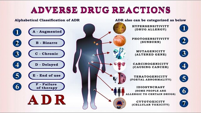
Adverse Drug & Reactions Adverse Drug Reactions (ADRs) Before we memorize definitions, understand the real-world impact of Adverse Drug Reactions. In the least developing countries, people die and are buried without anyone ever knowing whether they died from the actual disease they were fighting, or from the medication they used to treat it. Medications are powerful chemical tools. When used incorrectly, or when the body reacts unexpectedly, they can be lethal. Your goal as a medical professional is to foresee, identify, and manage these reactions. 1. Terms In medical pharmacology, words have very strict meanings. You must be able to distinguish between an ADR, an ADE, and a Side Effect. What is an Adverse Drug Reaction (ADR)? An Adverse Drug Reaction is formally defined as any unintended or noxious (harmful) effect which meets the following strict criteria: It is suspected to be due to a drug. It occurs at doses normally used in man (this is crucial—if someone takes 50 pills on purpose, the resulting liver failure is an overdose, not a standard ADR according to older definitions, though the FDA includes it now). It is severe enough that it may require treatment, a decrease in the dose, or total withdrawal of the drug. It dictates caution in the future use of the same drug for that patient. Adverse Drug Event (ADE) vs. ADR An Adverse Drug Event (ADE) is a broader umbrella term. It is any untoward (unlucky/bad) occurrence that may present during medical treatment. The Difference: An ADE does not necessarily have a causal relationship with the treatment. Scenario You give a patient a blood pressure pill. An hour later, they trip on a rug, fall, and break their arm. The broken arm happened during medical treatment (making it an Adverse Drug Event), but the pill didn’t cause the rug to be there. However, if the blood pressure pill caused severe dizziness, causing them to fall, that is an Adverse Drug Reaction (ADR) because there is a causal link. What is a Side Effect? A Side Effect is an extended pharmacological action of a drug. It is entirely predictable based on how the drug works in the body. Clinical Example Atropine is given as an anticholinergic drug to dry up secretions in the lungs before surgery or to treat a slow heart rate. Because we know it blocks acetylcholine (the “rest and digest” chemical), we completely expect it to cause dryness of the mouth. The dry mouth is a side effect—a direct extension of its normal pharmacological action. 2. Regulatory Perspectives: WHO vs. FDA Different health organizations define ADRs slightly differently, which affects how statistics are reported globally. The WHO Definition The World Health Organization (WHO) describes an ADR as: “The noxious and unintended drug effect which occurs at doses employed in man for prophylaxis (prevention), diagnosis, or therapy.” The Limitation: This definition only encompasses part of the problem. It strictly says “at doses normally employed.” The use of this highly restrictive definition hinders the reporting of ADRs because it ignores human error, addiction, and accidental poisonings. The FDA Definition The US Food and Drug Administration (FDA) uses a much broader, more realistic definition. The FDA defines an ADR as: “An undesirable effect, reasonably associated with the use of the drug, that may occur as a part of the pharmacological action of a drug OR may be unpredictable in its occurrence.” Reporting Purposes: To capture the full scope of drug harm, the FDA strictly includes incidents of overdose (whether accidental, suicidal, or criminal) and incidents due to drug dependence or withdrawal after the cessation of drug administration. 3. Grading the Severity of Adverse Drug Reactions When an ADR happens, it is categorized into one of four grades based on how aggressively the medical team must respond: Minor: No therapy, no specific antidote, and no prolongation of hospitalization is required. (Example: A mild, temporary headache after taking a medication that resolves on its own). Moderate: Requires a change in drug therapy, specific medical treatment, or prolongs the patient’s hospital stay. (Example: A drug causes a severe rash that requires prescribing antihistamines and keeping the patient overnight for observation). Severe: Potentially life-threatening, causes permanent damage, or requires intensive medical treatment (ICU). (Example: A drug causes severe anaphylactic shock restricting breathing, requiring intubation). Lethal: The drug directly or indirectly contributes to the death of the patient. 4. Classification Systems: Rawlins and Thompson There are several ways to classify ADRs. The simplest, most foundational method was proposed by Rawlins and Thompson. They divided all drug reactions into two major classes: Type A and Type B (also known as Type 1 and Type 2). Feature Type A (Type 1) – Augmented Type B (Type 2) – Bizarre Synonyms Predictable, toxic, quantitative, dose-related. Unpredictable, allergic, idiosyncratic, qualitative, dose-independent. Mechanism Predictable and clearly understood based on the drug’s normal mechanism of action. Usually poorly understood. It has nothing to do with the drug’s intended action. Site of Action 1. Same site of primary drug action.2. Another site for primary and secondary actions. Unrelated to the normal site of action. Incidence (Frequency) High (70% to 75% of all ADRs). Very common. Low (around 30%). Comparatively rare. Morbidity (Sickness) Generally High (many people feel mild to moderate sickness). Severe illness is common when it strikes. Mortality (Death rate) Low. Rarely kills the patient. High. Often causes serious illness and death. Reproducibility Reproducible (If you give the high dose again, it will happen again). Not reproducible reliably in laboratory settings. Often not observed during conventional pharmacological and toxicological screening programs. Treatment strategy Adjust (Decrease) the dose. Stop treatment immediately. Withdraw the drug completely. Deep Dive: Causes of Type A (Type 1) Reactions Type A reactions happen because there is simply too much active drug in the body, or the body is too sensitive to it. The causes are broken down into three areas: Pharmaceutical Causes: Increased availability at the site of absorption. (e.g., A manufacturing error makes a pill release its contents too quickly). Pharmacokinetic (PK) Causes:

Drug Interactions & Loss of Effect
Pharmacology

Drug Interactions & Loss of Effect

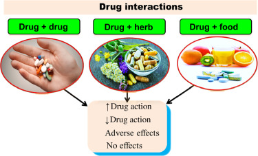
Drug Interactions & Loss of Effect Drug Interactions & Loss of Effect Core Learning Objectives By the time you finish studying this guide, you will be absolute masters of the following concepts: Describing Drug Combinations: You will know exactly what happens when doctors prescribe two drugs at the same time. You will understand the mathematical and biological differences between additive, synergistic, potentiation, and antagonistic interactions. Explaining the Loss of Drug Effect: You will understand the exact biological reasons why a drug might stop working in a patient over time. You will be able to clearly differentiate between tachyphylaxis, tolerance, refractoriness, and drug resistance. Part 1: Combining Drugs with Similar or Related Effects In clinical medicine, patients rarely take just one medication. When two or more drugs are given concurrently (at the same time), they interact. These interactions can be highly beneficial (helping the patient heal faster) or highly dangerous. We classify these outcomes into four specific mathematical categories based on how their dose-response curves interact. 1. The Additive Effect Math Rule: 1 + 1 = 2 An additive effect occurs when the combined effect of two drugs equals the exact sum of their individual effects. Neither drug boosts the other; they just work side-by-side doing their own job, often acting on similar receptors or pathways. Clinical Example: Aspirin + Paracetamol (Acetaminophen). Both of these medications relieve pain and reduce fever. If you take 50% of a full dose of Aspirin and 50% of a full dose of Paracetamol together, you get exactly 100% pain relief. Expanded Mechanism: Aspirin works predominantly in the peripheral tissues by irreversibly inhibiting COX-1 and COX-2 enzymes. Paracetamol works more centrally in the brain (possibly via COX-3 or peroxidase sites). Their actions simply add up. Clinical Knowledge: Why do doctors do this? By combining two drugs, the doctor can use a lower dose of each individual drug. This drastically reduces the risk of dose-dependent side effects (like severe stomach bleeding from too much Aspirin, or fatal liver toxicity from too much Paracetamol) while still achieving perfect pain relief. 2. Potentiation Math Rule: ½ + 1 = 2 (or 0 + 1 = 2) Potentiation happens when you mix a drug that has an active therapeutic effect with a substance that has little to zero therapeutic effect on its own. However, this seemingly useless second substance massively amplifies the power of the first drug. Clinical Example: Amoxicillin + Clavulanic Acid (sold together as Augmentin). The Problem: Many bacteria have evolved a defense shield—an enzyme called beta-lactamase, which chemically breaks open and destroys the antibiotic Amoxicillin before it can kill the bacteria. The Solution: Clavulanic acid has almost zero ability to kill bacteria on its own. However, it is an expert “suicide inhibitor” that irreversibly binds to and destroys the bacteria’s beta-lactamase shield. The Result: By giving Clavulanic acid (which does nothing alone), it completely opens the door for Amoxicillin to rush in, bind to penicillin-binding proteins (PBPs), and kill the bacteria. The antibiotic activity is highly potentiated. 3. Synergism Math Rule: 1 + 1 = 3 (or more) Synergism is a stronger, more powerful interaction than potentiation. The total effect produced by combining two active drugs is much greater than the simple sum of their individual effects. They team up to create a massive, multiplied response, often by blocking sequential steps in a single metabolic pathway. Clinical Example: Sulfamethoxazole + Trimethoprim (sold as Co-trimoxazole). Expanded Mechanism: Bacteria must synthesize their own folic acid from scratch to create DNA and survive. This requires a multi-step assembly line. Step 1: Sulfamethoxazole acts as a competitive inhibitor of the enzyme dihydropteroate synthase. Step 2: Trimethoprim blocks the very next enzyme in the chain, dihydrofolate reductase (DHFR). The Result: Used alone, each drug merely slows the bacteria down (they are bacteriostatic). But used together, they completely and sequentially shut down the folic acid factory, turning a weak antibacterial effect into a highly lethal, synergistic bacterial wipeout (bactericidal effect). 4. Antagonism Math Rule: 1 + 1 < 2 (or 1 + 1 = 0) Antagonism occurs when one drug actively opposes, reduces, or completely blocks the effect of another drug. The combined effect is actually less than expected. Clinical Example: Opioids (Morphine/Heroin) + Naloxone. Expanded Mechanism: If a patient takes an opioid, it powerfully binds to mu-opioid receptors in the brain to slow breathing, induce euphoria, and stop pain. If the dose is too high, breathing stops entirely (fatal overdose). The Result: If you give Naloxone intravenously, it acts as a perfect competitive antagonist with a much higher affinity for the receptor than the opioid. It aggressively rips the opioid off the receptor and blocks it without activating it. The effect of the opioid drops to zero instantly, waking the patient up and saving them from an overdose. Part 2: The Five Mechanisms of Drug Antagonism When one drug blocks another (Antagonism), it can happen in five entirely different biological ways. Understanding exactly how the blockade happens—whether in the blood, in the liver, or at the receptor level—is critical for pharmacology exams. 1. Chemical Antagonism This is the most basic physical interaction. Chemical antagonism occurs when two substances react directly with each other in the body fluids (like the blood, the stomach, or the gut lumen) before they even reach a cell receptor. They physically bind together to form an inactive complex, leading to the inactivation of one or both substances. Real-World Examples of Chemical Antagonism Chelating Agents (e.g., Dimercaprol or EDTA): If a patient has heavy metal poisoning (like swallowing lead, arsenic, or mercury), doctors inject a chelating agent. This drug acts like a chemical claw, physically grabbing the heavy metal molecules floating in the blood and forming highly stable, inactive pairings (chelates) that are water-soluble and safely peed out by the kidneys. Antacids (e.g., Aluminium hydroxide): If a patient has severe heartburn or a peptic ulcer, they take an antacid. The basic aluminium hydroxide directly collides with the acidic gastric hydrochloric acid (HCl) in the stomach. They undergo a

signaling
Pharmacology

Signaling Mechanisms

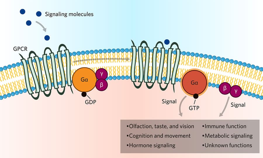
Signaling Mechanisms Signaling Mechanisms How to Approach This Topic Pharmacodynamics (how drugs act on the body) is all about communication. Cells are blind and deaf; they rely entirely on chemical messages. When you take a drug, it acts as a messenger. This entire lecture focuses on how the message gets from the outside of the cell to the inside, forcing the cell to change its behavior. We will break down every single pathway so you can easily understand Introduction to Drug-Responsive Signaling Mechanisms Before we examine the specific pathways, we must understand the basic sequence of events: An agonist drug (the messenger or “key”) binds to its specific receptor (the “lock”). This binding event directly activates an effector or signaling mechanism. The effector causes a biological change inside the cell. There are several different categories of these signaling mechanisms known in pharmacology. We classify them based on where the receptor is located and how it translates the message. The Major Categories of Signaling Mechanisms: Intracellular receptors: The receptor is hidden deep inside the cell (in the cytoplasm or nucleus). Membrane receptors directly coupled to ion channels: The receptor is on the surface and acts as a direct physical gate for ions. Receptors linked via coupling proteins to intracellular effectors: The receptor is on the surface and uses a middleman (like a G-protein) to send a message inside. Receptors that function as enzymes or transporters: The receptor itself performs a chemical reaction or moves molecules. Intracellular Receptors Most drugs stop at the cell surface. However, some drugs are highly lipid-soluble (fat-soluble), allowing them to melt right through the cell membrane and enter the interior of the cell. Once inside, they find intracellular receptors. The Process Step-by-Step: The hormone or drug crosses the cell membrane. It binds to the intracellular receptor. This binding releases regulatory proteins (which were holding the receptor in an inactive state). The receptor is now activated. In many cases, two activated receptors will pair up and join together (a process called dimerization). This new hormone-receptor complex travels (translocates) directly into the cell’s nucleus. Inside the nucleus, the complex physically attaches to specific regions of DNA called response elements in spacer DNA. This interaction forces the DNA to either increase or decrease gene expression (the manufacturing of new proteins). The Analogy Think of the cell like a factory. Most drugs are delivery drivers who drop a package at the front desk (membrane receptors). But intracellular drugs are like VIP executives. They walk right past the front door, go straight into the manager’s office (the nucleus), and rewrite the factory’s rulebook (DNA) to change what the factory produces. Key Characteristics and Examples Because these drugs require the cell to read DNA and build entirely new proteins from scratch, the pharmacologic responses elicited via modification of gene expression have two absolute rules: They are slower in onset (it takes hours to days to build new proteins). They are longer in duration than many other drugs (even after the drug leaves the body, the newly built proteins stick around and keep working for days). Examples of ligands that use Intracellular Receptors: Steroids (Glucocorticoids): Drugs interacting with glucocorticoid receptors lead to the gene expression of proteins that heavily inhibit the production of inflammatory mediators. Thyroid hormones. Gonadal steroids (Estrogen, Testosterone). Vitamin D. Clinical Scenario: Asthma Attack If a patient arrives at the hospital having a severe asthma attack, giving them an inhaled steroid (an intracellular drug) will not save them immediately because steroids take hours to change gene expression and reduce inflammation. Instead, you must give them Albuterol (a fast-acting membrane receptor drug) to open the airways instantly. The steroid is given to prevent attacks over the next few days. Membrane Receptors Directly Coupled to Ion Channels These are the fastest receptors in the human body. They work in milliseconds. The receptor and the effector are the exact same physical structure. Mechanism: Endogenous ligands (the body’s natural chemicals) regulate the flow of ions through excitable membranes by activating receptors that are directly coupled to ion channels. There are no second messengers involved. It is a simple gate. Many drugs act by either mimicking (agonist) or antagonizing (blocker) the actions of these natural ligands. Example 1 The Nicotinic Receptor Ligand: Acetylcholine (ACh). Ion Channel: Coupled directly to a Sodium/Potassium (Na+/K+) ion channel. Locations: Present in the Autonomic Nervous System (ANS) ganglia, the skeletal myoneural junction (where nerves tell muscles to move), and the Central Nervous System (CNS). Pharmacology: This receptor is a prime target for many drugs, including: Nicotine (acts as an agonist). Choline esters. Ganglion blockers. Skeletal muscle relaxants (used during surgery to paralyze muscles by blocking this receptor). Example 2 The GABA-A Receptor Ligand: Gamma-aminobutyric acid (GABA). Ion Channel: Coupled directly to a Chloride (Cl-) ion channel. When chloride flows into a nerve cell, it makes the cell highly negative and puts it to sleep (inhibition). Locations: Central Nervous System (CNS). Pharmacology: This receptor can be heavily modulated (enhanced) by drugs that calm the brain down: Anticonvulsants (anti-seizure medications). Benzodiazepines (anti-anxiety medications like Valium or Xanax). Barbiturates (heavy sedatives). Receptors Linked Via Coupling Proteins (G-Proteins) This is the largest and most famous family of receptors in pharmacology. They are often called “serpentine” receptors because they are made of a single protein chain that snakes back and forth across the cell membrane exactly seven times (seven transmembrane spanning domains). The third loop on the inside of the cell is physically coupled to the G-protein effector mechanism. The G-protein is a middleman. It binds GTP (Guanosine Triphosphate) to become active. The Relay Race Analogy: The Drug (Runner 1) passes the baton to the Receptor on the outside. The Receptor passes the baton to the G-Protein (Runner 2) on the inside. The G-Protein passes the baton to an Enzyme (Runner 3). The Enzyme creates Second Messengers (Runner 4) which flood the cell and finish the race. There are three main types of G-proteins you must memorize: Gs, Gi, and Gq. A. The Gs Pathway

GLUCONEOGENESIS
Biochemistry

Gluconeogenesis Exam

Gluconeogenesis Exam Biochemistry: Gluconeogenesis Exam Biochemistry: Gluconeogenesis Exam Test your knowledge with these 30 questions. Start Exam Gluconeogenesis Exam Question 1/30 Submit Next Exam Complete! Here are your results, . Your Score 28/30 93% Download Full Report

Scroll to Top