Doctors Revision

Pharmacology

Pharmacology

Pharmacology Mid-Term Examination

Pharmacology Mid-Term Examination — MBChB Year 1 Mid-Term Examination Paper Pharmacology Bachelor of Medicine & Bachelor of Surgery (MBChB) • Year 1 • Semester 1, 2026 General, Autonomic & Autacoid Pharmacology (PHA 121)  |  Chemotherapy of Infections & Malignancies (PHA 222) 2 HrsDuration 100Total Marks A · B · CSections General Principles PK/PD Autonomic NS ANS Autacoids Local Hormones Anti-Infectives Bacterial Antifungal & Antiviral Antiprotozoal & Anthelmintic Cancer Chemotherapy Instructions to Candidates Answer ALL questions in Section A (Objectives & Fill-ins). Answer any THREE questions from Section B. Answer any TWO questions from Section C. Write clearly and legibly. Generic drug names are preferred over brand names. Do not write anything in the margins. Section A 40 MARKS Part I — Objectives (20 Marks). Answer ALL questions. Choose the most appropriate answer. 1. The time required for the plasma concentration of a drug to fall by 50% is known as: A. BioavailabilityB. Volume of distribution C. Half-life (t½)D. Clearance Show Answer Answer: C. Half-life (t½) — Determines dosing interval; after ~4-5 half-lives a drug is considered eliminated. 2. A drug that binds to a receptor and produces a submaximal response even at full occupancy is called a: A. Full agonistB. Partial agonist C. AntagonistD. Inverse agonist Show Answer Answer: B. Partial agonist — Has affinity but lower intrinsic efficacy than a full agonist. 3. Atropine exerts its effect by acting as a: A. Muscarinic receptor agonistB. Muscarinic receptor antagonist C. Nicotinic receptor antagonistD. Cholinesterase inhibitor Show Answer Answer: B. Muscarinic receptor antagonist — Used for bradycardia, organophosphate poisoning, and preanesthetic drying of secretions. 4. Which receptor subtype mediates bronchodilation when stimulated by salbutamol? A. α1B. β1 C. β2D. M3 Show Answer Answer: C. β2 — β2-adrenoceptor stimulation relaxes bronchial smooth muscle, hence its use in asthma. 5. Which autacoid is responsible for the classic triple response (redness, wheal, flare) in skin injury? A. Prostaglandin E2B. Histamine C. SerotoninD. Bradykinin Show Answer Answer: B. Histamine — Released from mast cells; acts via H1 receptors on vasculature. 6. Penicillins exert their bactericidal effect primarily by: A. Inhibiting protein synthesis at the 30S ribosomeB. Inhibiting bacterial cell wall (peptidoglycan) synthesis C. Inhibiting DNA gyraseD. Disrupting folic acid synthesis Show Answer Answer: B. Inhibiting bacterial cell wall (peptidoglycan) synthesis — By binding penicillin-binding proteins (transpeptidases). 7. Which class of antibiotics is classically associated with tendon rupture and QT prolongation as adverse effects? A. MacrolidesB. Aminoglycosides C. FluoroquinolonesD. Tetracyclines Show Answer Answer: C. Fluoroquinolones — e.g. ciprofloxacin; also associated with cartilage damage in children. 8. Amphotericin B exerts its antifungal action by: A. Inhibiting ergosterol synthesisB. Binding ergosterol and forming membrane pores C. Inhibiting fungal DNA synthesisD. Inhibiting beta-glucan synthase Show Answer Answer: B. Binding ergosterol and forming membrane pores — Causes leakage of intracellular contents; nephrotoxicity is a key adverse effect. 9. The drug of choice for uncomplicated Plasmodium falciparum malaria in most endemic African settings is: A. ChloroquineB. Artemether-lumefantrine C. DoxycyclineD. Mefloquine alone Show Answer Answer: B. Artemether-lumefantrine — An artemisinin-based combination therapy (ACT), the WHO-recommended first-line regimen. 10. Methotrexate exerts its anticancer effect by inhibiting: A. Topoisomerase IIB. Dihydrofolate reductase C. Ribonucleotide reductaseD. Microtubule assembly Show Answer Answer: B. Dihydrofolate reductase — Blocks folate-dependent synthesis of purines and thymidylate, halting DNA synthesis. 11. Which second messenger is generated when noradrenaline acts on α1-adrenoceptors? A. cAMPB. IP3/DAG C. cGMPD. Direct ion channel opening Show Answer Answer: B. IP3/DAG — α1 receptors are Gq-coupled, activating phospholipase C. 12. Organophosphate poisoning is treated with atropine and which other agent? A. NeostigmineB. Pralidoxime C. PhysostigmineD. Pyridostigmine Show Answer Answer: B. Pralidoxime — Reactivates acetylcholinesterase by removing the phosphate group, if given before “aging” occurs. 13. Which route of administration avoids first-pass hepatic metabolism? A. OralB. Sublingual C. Rectal (upper)D. All of the above Show Answer Answer: B. Sublingual — Drug is absorbed directly into systemic circulation, bypassing the portal system. 14. Aspirin’s antiplatelet effect is due to irreversible inhibition of: A. LipoxygenaseB. Cyclooxygenase-1 (COX-1) C. Phospholipase A2D. Thromboxane synthase only Show Answer Answer: B. Cyclooxygenase-1 (COX-1) — Reduces thromboxane A2 production in platelets for the lifespan of the platelet (~7-10 days). 15. Which anti-tuberculosis drug is most associated with peripheral neuropathy, prevented by co-administration of pyridoxine (Vitamin B6)? A. RifampicinB. Isoniazid C. EthambutolD. Pyrazinamide Show Answer Answer: B. Isoniazid — Depletes pyridoxine, causing peripheral neuropathy if unsupplemented. 16. Metronidazole is particularly effective against which class of organisms? A. Aerobic gram-positive cocciB. Anaerobic bacteria and protozoa C. Atypical bacteria (Mycoplasma)D. Fungi Show Answer Answer: B. Anaerobic bacteria and protozoa — Effective against organisms like Giardia, Entamoeba, Trichomonas, and anaerobes such as Bacteroides. 17. Acyclovir selectively targets virus-infected cells because it requires activation by: A. Host cell kinases onlyB. Viral thymidine kinase C. Viral proteaseD. Viral reverse transcriptase Show Answer Answer: B. Viral thymidine kinase — Phosphorylates acyclovir to its active form, giving it selectivity for HSV/VZV-infected cells. 18. Vincristine, a vinca alkaloid, exerts its anticancer effect by: A. Stabilizing microtubulesB. Inhibiting microtubule polymerization C. Alkylating DNAD. Intercalating DNA Show Answer Answer: B. Inhibiting microtubule polymerization — Arrests cell division in metaphase; notable adverse effect is peripheral neuropathy. 19. The “cheese reaction” (hypertensive crisis with tyramine-containing foods) is a classic risk with which drug class? A. Beta-blockersB. Monoamine oxidase inhibitors (MAOIs) C. Calcium channel blockersD. ACE inhibitors Show Answer Answer: B. Monoamine oxidase inhibitors (MAOIs) — MAO inhibition prevents tyramine breakdown, causing excess norepinephrine release. 20. Albendazole and mebendazole act against helminths primarily by: A. Paralyzing the worm via GABA agonismB. Inhibiting microtubule formation by binding tubulin C. Increasing cell membrane permeability to calciumD. Inhibiting acetylcholinesterase Show Answer Answer: B. Inhibiting microtubule formation by binding tubulin — Disrupts glucose uptake and energy metabolism in helminths, leading to their death. Part II — Fill in the Blanks (20 Marks). Answer ALL questions in this part. 21.The study of what the body does to a drug (absorption, distribution, metabolism, excretion) is called click to reveal. 22.The dose of a drug required to produce a therapeutic effect in 50% of
NSAIDs & Prostanoids
Pharmacology

NSAIDs & Prostanoids

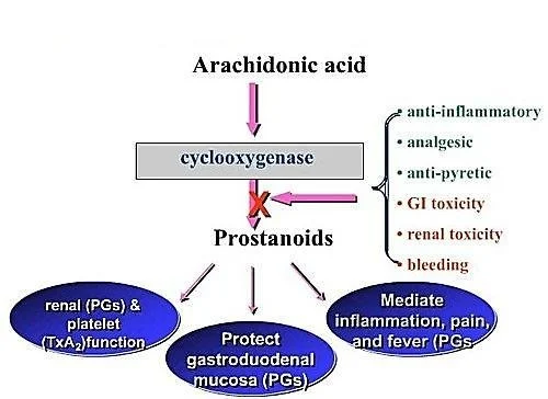
NSAIDS & Prostanoids NSAIDs & Prostanoids Pharmacology Module Overview This master guide covers the pharmacology of Non-Steroidal Anti-Inflammatory Drugs (NSAIDs) and Prostanoids. We will explore the Arachidonic Acid pathway, the profound differences between COX-1 and COX-2, specific drug classifications, and the synthetic prostanoids used to manipulate everything from childbirth to glaucoma. Enhanced with clinical scenarios and deep-dive explanations to guarantee exam success. 1. The Foundation: Prostanoids and the MOA of NSAIDs Before understanding the drugs, you must understand the assembly line that makes the molecules these drugs block. This is the Arachidonic Acid Pathway. Think of this pathway as a factory that takes raw materials from the cell wall and turns them into highly active chemical messengers. MEMBRANE PHOSPHOLIPIDS ↓Enzyme: Phospholipase A2 (BLOCKED by Corticosteroids)↓ ARACHIDONIC ACID ↙ Lipoxygenase ↓ Leukotrienes(Cause Bronchospasm/Asthma) ↘ Cyclooxygenase (COX)(BLOCKED by NSAIDs) ↓ PGG2 → PGH2(Endoperoxides) ↓ PROSTANOIDS:Prostaglandins (PGE2, PGF2α, PGD2)Thromboxane (TXA2)Prostacyclin (PGI2) Clinical Pearl Steroids vs. NSAIDs & The “Shunt” Phenomenon Notice that Corticosteroids block the pathway at the very top (Phospholipase). Therefore, steroids stop BOTH Leukotrienes (which cause asthma) and Prostanoids. NSAIDs only block the COX enzyme lower down. This means NSAIDs stop pain and fever (Prostanoids) but do nothing to stop Leukotrienes. In fact, in some asthma patients, giving an NSAID creates a “Leukotriene Shunt”. Because the COX pathway is blocked, all the built-up Arachidonic acid is violently pushed down the Lipoxygenase pathway, causing a massive overproduction of Leukotrienes. This triggers a severe, life-threatening asthma attack (a condition clinically known as Aspirin-Exacerbated Respiratory Disease or AERD). The Cyclooxygenase (COX) Isozymes: The “Housekeeper” vs. The “Fire Alarm” The COX enzyme comes in different versions (isoforms). Knowing the difference is the absolute key to understanding NSAID side effects and why pharmaceutical companies spent billions inventing specific COX-2 inhibitors. COX-1 (The Housekeeper) COX-2 (The Fire Alarm) COX-3 (The Mystery) Constitutive: Always active, working in the background 24/7. Responsible for physiologic production of prostanoids to regulate normal cellular processes. Gastric Cytoprotection: Makes protective stomach mucus and neutralizes stomach acid. Vascular Homeostasis & Platelet Aggregation: Balances blood flow and clotting. Kidney Function: Regulates and maintains renal blood flow. Inducible: Normally absent, but ramps up massively during emergencies (trauma, infection). Responsible for elevated production of prostanoids in disease states. Expression at sites greatly increases to cause Pain, Inflammation, and Fever. (Also expressed normally in brain, kidney, and bone). Predominantly has effects in the Central Nervous System (CNS). Often theorized to be the exact target of Acetaminophen (Paracetamol) which beautifully explains why it reduces fever/pain centrally in the brain but has absolutely no anti-inflammatory effect in the body’s tissues. 2. Classification of NSAIDs NSAIDs are classified either by their chemical structure/efficacy or by how selectively they block the COX enzymes. Classification by COX Selectivity (The Slide 4 Breakdown) Note on exam preparation: Some drugs straddle the line of selectivity based on dose. For example, Aspirin is selective for COX-1 at low doses, but non-selective at high doses. Selective COX-1 Inhibitors (Usually low doses): Low dose Aspirin, Ketoprofen, Flurbiprofen, Indomethacin, and Ketorolac (sometimes spelled ‘Ketoloid’ on older slides). Non-Selective COX Inhibitors (Traditional NSAIDs): Piroxicam, Tenoxicam, Ibuprofen, Naproxen, Diclofenac. These hit both COX-1 and COX-2 equally, killing pain but ruining the stomach. Selective COX-2 Inhibitors (The “-coxibs” & friends): Celecoxib, Etoricoxib, Meloxicam (preferential), Nimesulide. Designed to kill pain without giving you a stomach ulcer. Classification by Efficacy and Chemical Class (The Slide 5 Breakdown) Why do we care about chemical classes? Because if a patient is highly allergic or fails to respond to an NSAID from the “Propionic Acid” class, a wise doctor will switch them to a completely different chemical class, like an “Oxicam”. 1. Analgesic & Marked Anti-inflammatory Non-Selective COX Inhibitors (Traditional) Salicylic Acid Derivatives: Aspirin Propionic Acid Derivatives: Naproxen, Ibuprofen, Ketoprofen Pyrazolon Derivatives: Phenylbutazone Acetic Acid Derivatives: Diclofenac, Aceclofenac, Nebumetone, Sulindac Pyrrolo-pyrrole Derivatives: Ketorolac Indole Derivatives: Indomethacin Oxicams: Piroxicam, Tenoxicam 2. Analgesic & Moderate Anti-inflammatory Fenamates: Meclofenamic acid, Tolfenamic acid, Flufenamic acid Anthranilic acid: Mefenamic acid 3. Preferential & Selective COX-2 Preferential COX-2 Inhibitors: Meloxicam, Nimesulide. Selective COX-2 Inhibitors: Celecoxib, Etoricoxib. 4. Analgesics with POOR/NO Anti-inflammatory Para-aminophenol Derivatives: Acetaminophen/Paracetamol. 3. Mechanism of Action (MOA) and General Adverse Effects Primary MOA: NSAIDs inhibit the cyclooxygenase (COX) enzyme, resulting in the reduced biosynthesis of Prostanoids (Prostaglandins, Prostacyclin, and Thromboxane A2). Why do Traditional NSAIDs cause side effects? Aspirin and older, non-selective NSAIDs block BOTH COX-1 and COX-2. By blocking COX-2, they brilliantly stop inflammation, pain, and fever. BUT, by blocking COX-1, the release of PGs required for homeostatic (housekeeping) function is totally disrupted. The Mechanisms of Toxicity The Stomach: PGE2 and PGI2 normally stimulate the production of thick, protective gastric mucus and bicarbonate. They also maintain rich blood flow to the stomach wall. NSAIDs stop this synthesis. Result: The stomach acid literally burns through the unprotected stomach wall, causing Gastric and Duodenal Ulcers, and severe GI Bleeding. The Kidneys: PGE2 and PGI2 are responsible for actively dilating the afferent renal arteriole (the blood vessel bringing blood INTO the kidney filter), which maintains the Glomerular Filtration Rate (GFR). If you block this (especially in elderly patients with already impaired kidneys or low blood volume), blood flow to the kidney drops sharply.Result: Serious kidney damage, acute renal failure, and severe fluid retention. General Adverse Reactions of NSAIDs (System by System) Gastrointestinal Tract (Most Common): Nausea, vomiting, diarrhea, constipation, epigastric pain, indigestion, abdominal distress, intestinal ulceration, stomatitis, jaundice, bloating, anorexia, and dry mouth. Central Nervous System (CNS): Dizziness, headache, drowsiness, insomnia. Cardiovascular: Decrease or increase in blood pressure (often increasing it due to fluid retention), and cardiac arrhythmias. Renal: Hematuria (blood in urine) and acute renal failure (in those with pre-existing impaired function). Special Senses: Visual disturbances, blurred or diminished vision. Hematologic: Anemia (often secondary to chronic microscopic GI bleeding over months of daily NSAID use). 4. Deep Dive: Aspirin, Acetaminophen, and Selective COX-2s A. ASPIRIN (Acetylsalicylic Acid) Aspirin is completely unique among all NSAIDs. It irreversibly acetylates both isoforms of the COX enzyme. This means it covalently binds to the enzyme

Eicosanoids Pharmacology
Pharmacology

Eicosanoids Pharmacology

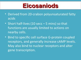
Autocoids — Eicosanoids Eicosanoids Pharmacology 1. Introduction to Eicosanoids Definition: Eicosanoids are biological signaling molecules (local hormones/autacoids) that are products of polyunsaturated long-chain fatty acids. The prefix “Eicosa-“ means 20 in Greek, because these molecules are almost entirely derived from 20-carbon essential fatty acids, most commonly Arachidonic Acid. Hormones vs. Eicosanoids (The “Global Email” vs. “Sticky Note” Analogy) Unlike regular hormones (like insulin) which are stored in glands and travel globally through the blood, eicosanoids are not stored. They are highly unstable and have a half-life of seconds to minutes. Therefore, they are synthesized on demand from cell membrane lipids and act locally right where they are made (paracrine action on neighbors, or autocrine action on themselves). Major Classifications Eicosanoids are divided into families based on the specific enzyme that creates them from the raw material: a) Cyclooxygenase (COX) derivatives: These include the Prostaglandins (PGs) and Thromboxane (TXA2). b) Lipoxygenase (LOX) products: These include the Leukotrienes (LTs) and Lipoxins. c) Cytochrome P450 (CYP) Epoxyoxygenase pathway: Produces EETs (Epoxyeicosatrienoic acids). 2. The Synthesis Cascade (The Arachidonic Acid Pathway) To understand the drugs, you MUST understand how eicosanoids are made. Picture a cell membrane. The lipids in that membrane hold the raw material (Arachidonic Acid) locked away safely. STEP 1: THE RELEASE Cell Membrane Phospholipids (Diacylglycerol or Phospholipid) ↓ Enzyme: Phospholipase A2 (PLA2) (or Phospholipase C) Arachidonic Acid (Free and active) Exam Gold: The Corticosteroid Blockade Exam Note: Corticosteroids (like Prednisone or Dexamethasone) stimulate the production of a protein called Annexin A1 (also known as Lipocortin-1), which completely blocks Phospholipase A2. This shuts down the ENTIRE cascade right at the top. No Arachidonic Acid means no prostaglandins and no leukotrienes. This is exactly why steroids are such incredibly powerful, broad-spectrum anti-inflammatories compared to NSAIDs! Once Arachidonic Acid is free, it acts as a crossroads and can go down one of three enzymatic paths: Path A: The COX Pathway Arachidonic Acid + COX-1 or COX-2 (PGH2 Synthase / Peroxidase) → PGG2 → Prostaglandin H2 (PGH2). PGH2 is the unstable “parent” molecule. Depending on the specific tissue enzymes present, PGH2 becomes: Prostaglandins: PGE2, PGF2α, PGD2. Prostacyclin (PGI2): Synthesized via Prostacyclin synthase (primarily in vascular endothelium). Thromboxane (TXA2): Synthesized via Thromboxane synthase (primarily in platelets). Path B: The LOX Pathway Arachidonic Acid + 5-LOX (Lipooxygenase + FLAP protein) → 5-HPETE. 5-HPETE becomes: Leukotrienes: LTA4 → LTB4, LTC4, LTD4, LTE4. HETEs: (e.g., 8-HETE, 12-HETE, 15-HETE) – play crucial roles in inflammation and immune cell recruitment. Path C: Cytochrome P450 Pathway Arachidonic Acid + CYP Epoxygenases → EETs. These play a role in maintaining vascular tone (vasodilation), renal function, and overall cardiovascular protection. 3. Mechanism of Action and Receptors Eicosanoids do not enter cells. They bind to cell surface receptors that are all coupled to G-proteins (GPCRs). Crucial Second Messenger Mechanisms You must know whether they cause relaxation or contraction at the cellular level (tying back to your signaling lectures!): Relaxers (PGI2 and PGE2): Link to Gs proteins. Increase Adenylyl Cyclase → Increases cAMP → Decreases intracellular Calcium (Ca++). Result: Smooth muscle relaxation and Vasodilation. Contractors (TXA2, PGF2α): Link to Gq proteins. Activate Phospholipase C → Increases IP3 → Increases intracellular Calcium (Ca++). Result: Smooth muscle contraction, Vasoconstriction, and Platelet Aggregation. 4. Physiological & Pharmacologic Effects by System This is where the exam will test your clinical application. Memorize these specific receptor actions: A. The Vasculature (Blood Vessels) PGEs (PGE1, PGE2): Potent vasodilators. Prostacyclin (PGI2): Potent vasodilator. Can produce profound hypotension (low blood pressure). Thromboxane A2 (TXA2): Potent vasoconstrictor. Leukotrienes (LTC4, LTD4): Cause massive capillary leakiness (vascular permeability), contributing heavily to the swelling (edema) seen in severe inflammation. **Alprostadil (PGE1): Specifically dilates the ductus arteriosus in neonates. B. Platelets (The Blood Clotting Tug-of-War) There is a constant balance (a “see-saw”) in your blood between two eicosanoids to prevent you from bleeding out or forming fatal clots: Prostacyclin (PGI2): Produced by healthy blood vessel walls. It INHIBITS platelet aggregation. (Mnemonic: Prostacyclin keeps blood CYCLING smoothly). Thromboxane A2 (TXA2): Produced by platelets. It is a massive platelet activator/aggregator. (Mnemonic: Thromboxane causes THROMBI / clots). Inflammation (Leukocytes): LTB4 is a powerful chemotactic agent (it acts as a chemical beacon, attracting eosinophils, monocytes, and neutrophils to the site of injury). Conversely, prostaglandins generally inhibit cellular and humoral immunity to keep the immune system from overreacting. C. The Lungs (Bronchial Tone) Prostaglandins: Have mixed effects on bronchial muscle (PGE1/PGE2 cause bronchodilation, PGD2/PGF2α cause constriction). TXA2: Causes bronchoconstriction. Inhibitors of thromboxane will therefore reduce the bronchoconstrictive response. Leukotrienes (LTC4, LTD4): Extremely potent bronchoconstrictors. These are the main culprits in deadly asthma attacks! D. The Uterus (Obstetrics) PGE2 and PGF2α: Cause powerful uterine contractions, especially in a pregnant uterus. Clinical Tie-In (Dysmenorrhea): Overproduction of PGE2 and PGF2α during menstruation causes severe uterine cramping (primary dysmenorrhea). This is why taking an NSAID (which blocks these prostaglandins) cures menstrual cramps! Clinically, synthetic versions are used as abortifacients (to induce medical abortions) or to induce labor at term. E. Gastrointestinal Tract (GIT) PGEs and PGI2: Inhibit gastric acid secretion (which is normally stimulated by feeding, histamine, or gastrin). They act as a shield, promoting the maintenance of the gastric mucosa by stimulating heavy mucus and bicarbonate secretion. Clinical Tie-In: This is exactly why taking NSAIDs (which block PGE production) causes stomach ulcers! You strip away the stomach’s protective mucus shield. F. The Kidneys PGE2 and PGI2: Cause renal vasodilation (specifically of the afferent arteriole), increase Renal Blood Flow (RBF), increase GFR, and promote diuresis (water excretion). (If a patient takes too many NSAIDs, they lose this vasodilation, the kidney starves of blood, leading to Acute Kidney Injury). TXA2: Causes renal vasoconstriction and has an ADH-like action (retains water). G. Central Nervous System (CNS) & Eye CNS: PGE2 is the primary mediator of Fever, Pain perception, and Sleep. When a virus attacks you, the brain generates PGE2 to reset the hypothalamus thermostat, causing fever. Eye: PGF2α regulates the outflow of aqueous humor. 5. Clinical Pharmacology: Uses of Prostanoids and Analogues In pharmacology, we create synthetic versions (analogs)

Serotonin Pharmacology
Pharmacology

Serotonin Pharmacology

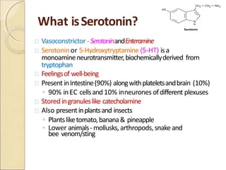
Autocoids — Serotonin Serotonin & Migraine Pharmacology 1. Brief Recap: What are Autacoids? Before diving into Serotonin, remember the baseline definition from the start of the lecture. Autacoids are the body’s local communication network. Definition: Endogenous substances (made in the body) that act as biological factors or “local hormones”. (Greek: Autos = self, Akos = remedy). Characteristics: Present in very small amounts, have distinct biological activity, are short-living with a short duration of action, and act at or very close to their site of release. Systemic Effect: Although they are “local”, if produced in massive amounts, they can enter the circulation and cause whole-body (systemic) effects. Functions: They regulate physiological baselines, mediate pathophysiological reactions to injuries (like inflammation), and modulate nerve transmission. Analogy Endocrine Hormones vs. Autacoids Think of standard Endocrine Hormones (like insulin or thyroid hormone) as a company-wide email broadcast. They travel through the main server (the bloodstream) to reach every department in the body. In contrast, Autacoids are like sticky notes left on a coworker’s desk. They are meant only for the immediate neighbor (local action) and are thrown away quickly (short duration of action). Chemical Classification of Autacoids Autacoids are classified into four main families based on their chemical structure: 1. Amines Histamine, Serotonin (5-HT). 2. Polypeptides Kinins, Oxytocin, Angiotensin, Vasopressin, Endothelins. 3. Fatty Acids Prostaglandins, Leukotrienes, Thromboxanes, PAF (Platelet Activating Factor). 4. Others Nitric Oxide (NO), Cytokines. 2. Serotonin (5-HT): Synthesis and Metabolism Serotonin, chemically known as 5-hydroxytryptamine (5-HT), is an indoleethylamine. It is widely distributed in nature—found in plants (like bananas and pineapples), animal tissues, venoms, and insect stings. A. The Synthesis Pathway Serotonin is built from the amino acid L-tryptophan. This is a critical two-step process: L-Tryptophan ↓ (Enzyme: Tryptophan Hydroxylase) — *Rate Limiting Step* 5-Hydroxytryptophan (5-HTP) ↓ (Enzyme: Decarboxylase) 5-Hydroxytryptamine (Serotonin / 5-HT) The Rate-Limiting Step: Hydroxylation at the C5 position is the bottleneck of the whole process. The body can only make Serotonin as fast as Tryptophan Hydroxylase works. Experimental Blockers: You can chemically block this rate-limiting step using drugs like p-chlorophenylalanine (PCPA / fenclonine) and p-chloroamphetamine. Experimentally, these were used to reduce serotonin in carcinoid syndrome, but they are too toxic for clinical human use. B. Inactivation and Metabolism Once Serotonin does its job, it must be rapidly inactivated so it doesn’t continuously overstimulate the body. It is metabolized primarily by the enzyme Monoamine Oxidase (MAO). Serotonin (5-HT) ↓ (Enzyme: MAO) 5-hydroxyindoleacetaldehyde ↓ (Enzyme: Aldehyde Dehydrogenase) 5-HIAA (5-hydroxyindoleacetic acid) — *The Principal Metabolite* Exam Trap! The Carcinoid Tumor Diagnostic Test Clinical Scenario: A patient presents with severe flushing, severe diarrhea, and right-sided heart valve issues. You suspect a Carcinoid Tumor (a rare gut tumor that secretes massive amounts of serotonin). The Test: You measure the 24-hour urinary excretion of 5-HIAA (the final breakdown product). High 5-HIAA confirms massive serotonin synthesis. The Trap: Before the test, you MUST prohibit the patient from eating foods rich in serotonin or tryptophan (e.g., Bananas, Pineapples, Plums). If they eat a bunch of bananas before the test, their body will metabolize that dietary serotonin, their urine 5-HIAA will skyrocket, giving a false positive for a tumor! Clinical Scenario: MAO Inhibitors & Serotonin Syndrome If a patient is taking a drug that blocks Monoamine Oxidase (an MAOI antidepressant like Phenelzine), the serotonin cannot be broken down. If this patient then takes another drug that increases serotonin (like an SSRI or MDMA/Ecstasy), serotonin builds up to lethal levels. This causes Serotonin Syndrome: hyperthermia, muscle rigidity, tremors, and potentially death. 3. Storage, Release, and Locations of 5-HT Where is Serotonin found in Mammals? The Gut (90%): Over 90% of all serotonin in the human body is located in the enterochromaffin cells of the gastrointestinal tract. (Deep Explanation: This is why SSRI antidepressants, which increase active serotonin everywhere, almost always cause GI upset, nausea, and diarrhea in the first week of use! The gut has far more serotonin receptors than the brain). The Blood (Platelets): Serotonin floats in the blood stored safely inside platelets. Platelets don’t make serotonin; they suck it up from the plasma using an active Serotonin Transporter (SERT). (Why? When you get cut, platelets clump together and release serotonin to cause local vasoconstriction, stopping the bleeding!). The Central Nervous System (Nerve Endings): Found heavily in the raphe nuclei of the brainstem. These neurons synthesize, store, and release 5-HT as a true neurotransmitter controlling mood and sleep. The Pineal Gland: Here, serotonin serves as a precursor. An enzyme (Hydroxyindole-O-methyltransferase) converts serotonin into Melatonin, the hormone that induces sleep. How is it Stored? Whether in a nerve ending or a platelet, serotonin is pumped into protective storage vesicles by a pump called the Vesicle-Associated Transporter (VAT). Pharmacological Blockade: The drug Reserpine completely blocks VAT. If serotonin cannot get into the protective vesicle, it is left out in the open and is destroyed by MAO in the cytoplasm. Therefore, Reserpine severely depletes stored serotonin (just like it depletes catecholamines), which historically caused severe, suicidal depression in patients taking it for high blood pressure. 4. Physiological Actions of Serotonin System Specific Actions of 5-HT Central Nervous System (CNS) Affects mood, sleep, appetite, temperature regulation, pain perception, blood pressure, and vomiting. Deficiency: Causes depression, anxiety, migraines. Neuroendocrine: Controls hypothalamic cells releasing anterior pituitary hormones. Gastrointestinal (GI) Causes intense rhythmic contractions of the small intestines (via 5-HT4). Stimulates vomiting via the 5-HT3 receptors on vagal nerves. Cardiovascular System Potent contraction of smooth muscle (via 5-HT2), causing constriction of veins. Exception: It does not contract skeletal muscle or heart muscle. Triggers Platelet aggregation (clotting) via 5-HT2. Respiratory System Causes mild stimulation in healthy lungs, but triggers severe bronchoconstriction in asthmatics (via 5-HT2 in smooth muscles). (Explanation: Asthmatic airways are hyper-reactive to autacoids. Even a tiny bit of serotonin can trigger an asthma attack). 5. Serotonin Receptors (The Pharmacology Targets) There are at least 15 types and subtypes of serotonin receptors. You must memorize the mechanisms of the main ones: Crucial Mechanism Trap Receptors 1 through 6 are all G-protein coupled receptors (GPCRs).

Histamine Pharmacology
Pharmacology

Histamine Pharmacology

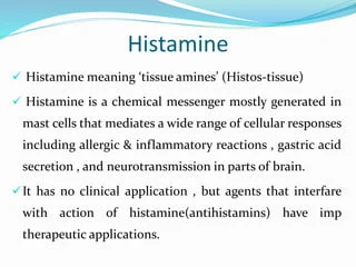
Autocoids — Histamine Histamine Pharmacology 1. Introduction to Autacoids What is an Autacoid? The term comes from the Greek words Autos (meaning “self”) and Akos (meaning “medicinal agent” or “remedy”). Therefore, an autacoid is literally a “self-remedy.” By definition, Autacoids are endogenous substances (made naturally inside the body) that act as biological factors or “local hormones”. Exam Trap: Autacoids vs. Classic Hormones A classic hormone (like insulin or thyroid hormone) is produced in a specific, centralized gland, dumped into the systemic bloodstream, and travels a long distance to reach its target organ. Autacoids are DIFFERENT: They are produced by widely distributed tissues all over the body, not a single gland. They act locally (at or very close to their exact site of synthesis and release). They are present in very small amounts. They have a short lifespan with a very short duration of action (they are rapidly destroyed to prevent them from causing systemic chaos). Note: However, if produced in massive, pathological amounts (like during severe anaphylactic shock), they can overcome local destruction, enter the systemic circulation, and have life-threatening systemic effects. Classification & Examples of Autacoids You must know the chemical classification of the different autacoids. Exam questions frequently mix these up: Chemical Class Examples Amines Histamine, Serotonin (5-HT) Polypeptides (Proteins) Kinins (Bradykinin), Oxytocin, Angiotensin, Vasopressin, Endothelins Fatty Acids (Eicosanoids) Prostaglandins, Leukotrienes, Thromboxanes, Platelet Activating Factor (PAF) Others Nitric Oxide (NO – Endothelium-derived relaxing factor), Cytokines 2. Histamine: Synthesis, Storage, and Metabolism Histamine is a ubiquitous molecule. It is present everywhere: in bacteria, plants, animals, and notably in venoms and stinging fluids (like bee stings, wasp venom, or stinging nettle plants). Chemistry & Synthesis Chemistry: It is a basic amine, specifically a β-aminoethylimidazole. Synthesis: The amino acid L-Histidine undergoes decarboxylation (the chemical removal of a CO2 molecule) to become Histamine. The specific enzyme that performs this action is L-Histidine decarboxylase. Inactivation & Metabolism Because histamine is so incredibly potent, it must be deactivated rapidly if it isn’t safely stored away. There are two major metabolic pathways the body uses to break it down and excrete it in the urine: Pathway 1 (Methylation): Conversion to N-methylhistamine (via the enzyme N-methyl transferase), which is then oxidized by MAO (Monoamine Oxidase) / DAO into methylimidazoleacetic acid. Pathway 2 (Oxidation): Direct conversion by the enzyme Diamine Oxidase (DAO) into imidazoleacetic acid (IAA). 3. Histamine Storage and Release Mechanisms Where is histamine kept? In humans, it is mostly stored inside Mast Cells (found abundantly in tissues interfacing with the outside world like Skin, Lungs, and GI tract) and Basophils (circulating in the blood). Inside these cells, histamine is locked up in granules, tightly bound to a heparin-protein complex so it doesn’t leak out. Histamine can be released in two distinct ways: Immunologic (Antigen-mediated) and Non-Immunologic. A. Immunologic Release (Antigen-Mediated) This is the classic Type I Hypersensitivity (Immediate Allergic Reaction). The Process: A person is exposed to an allergen (e.g., pollen, peanuts). Their immune system mistakenly creates IgE antibodies against it. These IgE antibodies attach to the surface of mast cells (a process called sensitizing the cell). Upon a second exposure to the same pollen, the allergen physically bridges and cross-links the IgE antibodies on the mast cell surface. The Result: The mast cell degranulates “explosively”, dumping massive amounts of histamine into the tissue. This specific process is energy-dependent (requires ATP) and requires calcium. Crucial Physiological Concept Negative Feedback & The Lung Exception In skin mast cells and blood basophils, the released histamine eventually binds back onto its own H2 receptors located on the mast cell’s own surface. This acts as a biological “brakes” system, inhibiting further histamine release (Negative Feedback). EXAM EXCEPTION: This feedback inhibition does NOT occur in lung mast cells! This is exactly why allergic asthma attacks in the lungs can spiral out of control so rapidly and become fatal; there are no built-in brakes to stop the continuous histamine release in the bronchioles. B. Non-Antigen Mediated Release This release mechanism does not require the immune system to be sensitized with IgE. It happens through direct physical or chemical interaction. Chemical Release: Certain drugs and chemicals can physically enter the mast cell and displace histamine from its heparin complex, forcing it out. Examples: Morphine, Tubocurarine (neuromuscular blocker), radiocontrast media (used in CT scans), amides, alkaloids, and basic polypeptides (like wasp/bee venoms). Mechanical Release: Physical trauma forces the mast cells to burst open. Examples: Vigorous scratching of the skin, severe burns, or crushing injuries. Cellular Proliferation: Pathological overgrowth of cells naturally increases total body histamine levels simply because there are more cells making it. Examples: Leukemia, Gastric Carcinoid Tumors. Physical Stimuli: Extreme cold, excessive heat, or exposure to bacterial toxins. Clinical Scenario “Red Man Syndrome” & IV Morphine The Event: If a nurse pushes an intravenous dose of Morphine too fast, the patient may suddenly flush bright red, feel intensely hot, become incredibly itchy, and their blood pressure might drop precipitously. The Mechanism: This is frequently mistaken for an allergy. It is not a true allergy (no IgE is involved). The rapid bolus of morphine chemically displaced histamine from the patient’s mast cells all at once, causing sudden, massive vasodilation. This is a classic example of Non-Antigen Mediated Chemical Release. The Fix: Stop the infusion, administer an antihistamine (like Diphenhydramine), and when restarting, push the morphine much slower. 4. Sites of Histamine Action Histamine regulates multiple physiological systems beyond just making you sneeze: Mast Cells & Basophils: Triggers standard inflammation and allergy symptoms (Skin itching, Lung wheezing, GIT cramping). Central Nervous System (CNS): Acts as a critical neurotransmitter, keeping the brain awake and alert. Neuroendocrine: Regulates hormones. It stimulates the release of ACTH, Prolactin (PRL), Vasopressin (VP), Oxytocin, and LH. It inhibits the release of GH and TSH. Thermal & Cardio: Causes hyperthermia (feverish feeling) via H1/H3 receptors located in the preoptic nucleus of the hypothalamus. Body Weight & Sleep: Acts as a powerful appetite suppressant (via H1), potentiates the hormone leptin (causing weight loss signaling), accelerates lipolysis (fat breakdown), and regulates

Pharmacology

Autacoids, Neuropeptides & Ergot Alkaloids

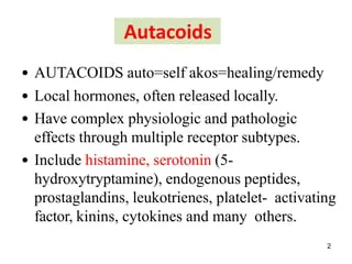
Autocoids Neuropeptides & Ergot Alkaloids Autacoids: 1. Introduction to Autacoids The word “Autacoid” comes from the Greek words Auto (meaning “self”) and Coids (meaning “healing/remedy”). They are frequently referred to as Local Hormones. Conceptual Check Autacoids vs. Classic Hormones Unlike classical hormones (like insulin or thyroid hormone) which are produced by a specific gland, secreted into the blood, and travel long distances to reach a target, Autacoids are produced locally by many different tissues, act locally near their site of synthesis, and have a very brief lifespan. Analogy: Think of them as the body’s “neighborhood watch” system. If a house is broken into (tissue trauma), you don’t wait for the national army (classical hormones) to arrive; the local neighborhood watch (autacoids) acts immediately at the exact site of injury to raise the alarm (inflammation/pain) and start repairs. Why are Autacoids Important? (Functions) Physiological: Regulate normal baseline organ functions (e.g., gastric acid secretion, local blood flow). Pathophysiological (Reaction to Injuries): They are the primary drivers of inflammation, pain, allergy, and the body’s response to tissue trauma. Transmission and Modulation: They act as mediators that fine-tune pain signals and nerve responses. Everyday Clinical Example: When you take an NSAID like Ibuprofen for a sprained ankle, you are specifically blocking the production of a lipid autacoid called a Prostaglandin. By shutting down this local autacoid, you stop the localized pain and swelling! Classification of Autacoids Autacoids are categorized by their chemical structure: Chemical Class Examples & Origin A. Amine Derivatives Histamine (derived from the amino acid Histidine) Serotonin (derived from the amino acid Tryptophan) B. Lipid Derivatives Eicosanoids: Prostaglandins, Thromboxane, Leukotrienes. Others: Interleukins, Platelet Activating Factor (PAF). C. Peptide Derivatives Kinins: Bradykinin. Renin-Angiotensin system. Neuropeptides. 2. Neuropeptides Neuropeptides are small, protein-like molecules (short chains of amino acids) used by neurons to communicate with each other. They act in an autocrine (acting on the cell that released it) or paracrine (acting on immediate neighboring cells) manner. Exam Trap: Neuropeptides vs. Classical Neurotransmitters Classical neurotransmitters (like dopamine, serotonin, glutamate) are fired into the synapse and then quickly sucked back up by reuptake pumps to be recycled and used again. NEUROPEPTIDES ARE NOT RECYCLED. Once they are secreted, they are broken down by specific enzymes (peptidases) and destroyed. The neuron must synthesize entirely new ones from the cell body (which takes time) and transport them down the axon. Do not forget this distinction! General Functions of Neuropeptides They are heavily responsible for higher-order brain functions and systemic regulation, including: Analgesia (pain regulation) Food intake (appetite stimulation/suppression) Learning & Memory Metabolism & Reproduction Social Behaviors Key Examples include: Neuropeptide Y (NPY), Cholecystokinin (CCK), Tachykinins (Substance P, Neurokinin), Arginine Vasopressin (AVP), and Corticotropin-Releasing Factor (CRF). Neuropeptide Y (NPY) NPY is a 36-amino acid peptide that acts as a potent neurotransmitter in both the Brain and the Autonomic Nervous System (ANS). Location Source Physiological Actions Brain (Central NPY) Produced mainly by the Hypothalamus. ↑ Food intake (Potent appetizer/orexigenic) ↑ Storage of energy as fat ↓ Anxiety and stress ↓ Voluntary alcohol intake ↓ Blood pressure and pain perception Regulates circadian rhythm and controls epileptic seizures. ANS (Peripheral NPY) Produced mainly by sympathetic neurons. Strong Vasoconstrictor Promotes the growth of fat tissue. NPY Receptors & Mechanisms NPY acts on G-Protein Coupled Receptors (GPCRs). Mammals have 5 types (Y1-Y5), but humans only express 4 functional types. Y1 (NPY1R) & Y5 (NPY5R): These are the Feeding Stimulators (Appetizers). Activation leads to massive hunger. Y2 (NPY2R) & Y4 (NPY4R): These act as Appetite Inhibitors (Anorectic). Mechanism of Action: NPY receptors are Gi-coupled (Inhibitory G-protein). When NPY binds, the Gi subunit is released, which inhibits the enzyme adenylate cyclase. This stops the conversion of ATP into the 2nd messenger cAMP. Clinical Scenario Anti-Obesity Drugs and NPY Because Y1 and Y5 receptors powerfully drive hunger and fat storage, pharmaceutical companies are actively researching Y1/Y5 Antagonists as therapeutic targets for obesity. Blocking these receptors could shut off the brain’s unnatural drive to overeat. Conversely, chronic stress increases NPY release in the periphery, which promotes the growth of visceral fat (explaining why chronic stress often leads to weight gain!). 3. Tachykinins (TAC) & Substance P Tachykinins form the largest family of neuropeptides. They get their name because they induce a rapid (“tachy”) contraction of gut tissues. Chemical Characteristic: All tachykinins share a common “C-terminal” sequence: “Phe-X-Gly-Leu-Met-NH2” (Where ‘X’ is either an aromatic or aliphatic amino acid, and COOH-terminus is the end of the protein chain). Synthesis Pathway: Preprotachykinin → Protachykinin → Tachykinin. Tachykinin Genes and Products TAC-1 Gene produces: Neurokinin A, Neurokinin K, Neuropeptide γ, and Substance P (SP). TAC-3 Gene produces: Neurokinin B. Tachykinin Receptors (GPCRs) Tachykinin receptors are Gq-coupled. Activation leads to the activation of Phospholipase C (PLC), which chops PIP2 into IP3 and DAG. This ultimately causes a massive release of intracellular Calcium. There are three main receptors, each with a preferred agonist: NK1R: Prefers Substance P. NK2R: Prefers Neurokinin A. NK3R: Prefers Neurokinin B. Substance P (SP) Substance P is an Undecapeptide (a chain of 11 amino acids). It is a highly potent mediator of pain signaling and inflammation. Receptor: Primarily binds to NK1R. The binding occurs via specific amino acid residues on the extracellular loops and transmembrane regions of the NK1 receptor. Physiological Roles: Promotes wound healing in humans (especially non-healing ulcers). Acts as a potent vasodilator. This vasodilation is entirely dependent on the release of Nitric Oxide (NO) from the endothelium. Transmits intense, burning pain signals to the brain (Neurogenic Inflammation). Clinical Application Substance P Antagonists (SPA) By blocking or depleting Substance P, we can block pain and severe nausea. Capsaicin: The active ingredient in chili peppers! Clinically used as a topical analgesic cream for arthritis and diabetic neuropathy. Mechanism: It initially causes a burning sensation (triggering SP release), but it eventually forces the nerve to release ALL of its Substance P. Because neuropeptides take a long time to synthesize (they aren’t recycled), the nerve is left empty of Substance P, rendering it completely unable to transmit pain signals for

Pharmacology

Autonomic Nervous System (ANS)

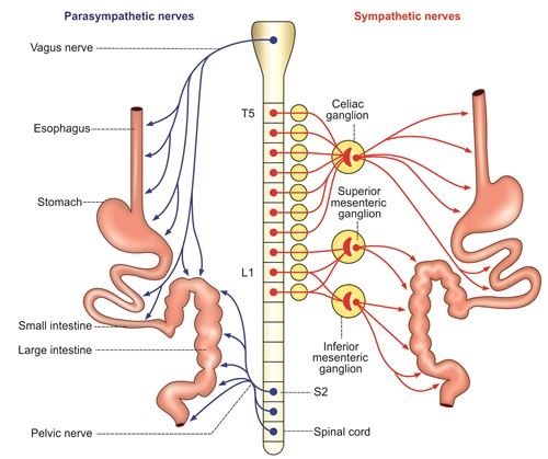
Autonomic Nervous System (ANS) Autonomic Nervous System (ANS): An Introduction to the Pharmacology Module Learning Outcomes This master guide is designed to make you deeply conversant with: The 4 Classes of Autonomic drugs. The role of Autonomic drugs in Clinical Practice (Cardiology, Respiratory, Psychiatry, etc.). Receptor and Non-receptor mechanisms of ANS drugs. Note on Adverse Effects (Type A-F) & ADME: While listed in the lecture’s opening slide, the provided slides focus exclusively on physiological effects and receptor dynamics. We will provide an emergency overview of Type A-F adverse effects at the end just in case it appears on your exam, but the bulk of this guide will strictly master the core ANS physiology and receptor profiles provided in the slides! 1. The Foundation: Why Autonomic Pharmacology? Before memorizing drugs, we must understand what we are treating. The nervous system (NS) is the ultimate communication system of the body. It acts as the critical LINK between the BODY and the ENVIRONMENT (both internal, like your sudden drop in blood pressure when you stand up, and external, like a lion chasing you). If this communication fails, HOMEOSTASIS (the stable, balanced state of the body) is violently disrupted. By understanding Autonomic Pharmacology, we can use drugs to artificially restore this communication and fix homeostasis. Autonomic pharmacology is highly LOGICAL (if you know the normal physiology, you know the drug’s effect) and incredibly CLINICALLY RELEVANT. It applies to: Psychiatric Medicine: Treating anxiety (e.g., using beta-blockers for stage fright). Respiratory Medicine: Treating asthma and COPD (e.g., inhalers that dilate airways). Cardiovascular Medicine: Treating hypertension, heart failure, and arrhythmias. GIT Medicine: Treating diarrhea, constipation, and stomach ulcers. Genitourinary Medicine: Treating overactive bladder or enlarged prostate issues. What is the Autonomic Nervous System (ANS)? The nervous system has two main outputs: Voluntary (Somatic – moving your arm to write a note) and Involuntary (Autonomic). The Autonomic Nervous System (ANS) is simply the “AUTOMATIC” part of the nervous system. It controls visceral organs (the “liquid-like” internal organs: heart, lungs, intestines, blood vessels) without you having to think about it. The ANS is divided into two competing branches. They are physiological antagonists (they do the exact opposite of each other to keep the body balanced): Sympathetic Nervous System (SNS): The “Accelerator.” Controls organs during STRESS (Fight, Flight, Fright). Parasympathetic Nervous System (PNS): The “Brakes.” Controls organs during REST (Rest and Digest / Breed and Feed). 2. The Sympathetic Nervous System: “Fight, Flight, Fright” The Scenario: You are walking in the bush and suddenly a lion jumps out at you. Your body instantly activates the Sympathetic Nervous System. Every single physiological change that happens next is designed to do one thing: Help you survive by fighting the lion or running away. The Chemical Messengers (Neurotransmitters) The sympathetic system communicates using three specific chemicals (Catecholamines). Because these are the messengers, drugs that mimic them are called Sympathomimetics (or Adrenergic drugs), and drugs that block them are called Sympatholytics. Noradrenaline (Norepinephrine): The primary neurotransmitter released directly at the nerve endings. Dopamine: A precursor and neurotransmitter, heavily involved in the kidneys and brain to maintain perfusion. Adrenaline (Epinephrine): This is a hormone, not a neurotransmitter. It is released by the Adrenal Gland directly into the blood. The adrenal gland output is 80% Adrenaline and 20% Noradrenaline. (This massive dump of adrenaline is what gives you that sudden “rush” in your chest when terrified). Sympathetic System Effects by Organ (Think deeply: “How does this help me run from the lion?”) Organ System Sympathetic Effect Why? (The Logical Reason) Cardiovascular (Heart) Heart Races: Increased Heart Rate (Chronotropy), increased Force of Contraction (Inotropy), and increased Conduction speed (Dromotropy). To rapidly pump massive amounts of oxygenated blood to the vital organs and legs for running. Increased force means a higher stroke volume per beat. Cardiovascular (Vessels) Blood is Diverted: ALL non-essential blood vessels (like those in the skin and gut) CONSTRICT. Blood vessels specifically going to Skeletal Muscles and the Brain DILATE. You don’t need blood in your stomach right now. You need maximum blood (oxygen) in your brain to think fast, and in your muscles to run. (This is why people turn “pale as a ghost” when terrified—skin blood vessels clamp shut!). Respiratory Bronchial Smooth Muscle RELAXES (Bronchodilation). Bronchial secretions DECREASE. Respiratory rate INCREASES. Relaxes the airways to open them up as wide as possible. Clears out mucus. This maximizes Oxygen (O2) uptake to fuel the skeletal muscles for sprinting. Gastrointestinal (GIT) Digestion Shuts Down: Motility DECREASES, Secretions DECREASE (causing Anorexia/lack of appetite), Sphincters TIGHTEN. Digesting food wastes massive amounts of energy and blood. Constipation and delayed gastric emptying occur to save energy for survival. You won’t feel hungry while running for your life. Genitourinary Urine Output DECREASES: The bladder wall (Detrusor muscle) relaxes, but the exit door (Sphincters/Trigone) TIGHTENS. Renin-Angiotensin System is ACTIVATED. Stopping to pee while running from a lion is a bad idea. It wastes energy and time. Activating Renin reabsorbs Sodium and Water in the kidneys, raising blood volume and blood pressure to sustain the “fight.” Reproductive Penile Erection INHIBITED. Uterine smooth muscle RELAXES. Genital secretions DECREASE. Blood is diverted to skeletal muscles. Reproduction is a waste of energy during a life-or-death crisis. (Sympathetic system specifically triggers ejaculation, but inhibits the erection phase). Central Nervous System Alertness INCREASES (can cause anxiety). Concentration INCREASES. Memory INCREASES. You need ultimate focus on the threat (the lion) to survive, dodging obstacles instantly. Skin Sweating INCREASES. Body temperature RISES (due to high metabolism). Body hairs ERECT (Piloerection). Sweating cools the rapidly overheating engine (your body). Raised hairs attempt to make you look larger and more intimidating to predators. Metabolism (CATABOLIC) Glucose goes UP: Glycogenolysis & Gluconeogenesis increase. Fat breaks down: Lipolysis increases. Proteins break down. Catabolism means breaking things down for energy. Your muscles need massive amounts of instant glucose and fatty acids to fuel the sprint, so the liver dumps its sugar reserves into the blood. Exocrine Glands DECREASE in salivation (causing a dry mouth and difficulty speaking). Decrease in tearing (dry eyes). Decrease

Preclinical Testing
Pharmacology

Preclinical Testing

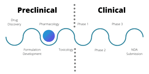
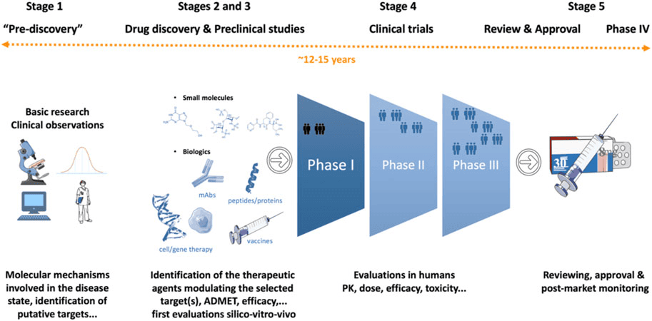
Preclinical Testing Preclinical Testing How to Approach This Topic Before a medicine can ever be prescribed to a sick patient, or a new medical device can be implanted in a human body, it must undergo rigorous, exhaustive testing. You cannot simply invent a chemical and give it to a human being. This lecture covers the entire phase that happens before humans are involved. We will break down every single test, why we use animals, what documents must be filed, and the extreme ethical and scientific importance of this process. 1. The Drug Development Process (The Big Picture) To understand where preclinical trials fit in, you must memorize the timeline of how a drug is born and brought to the pharmacy shelves. The process follows a strict, sequential pipeline: Basic Research: This is the purely academic stage. Scientists study biology at the most fundamental level. They look at Molecular biology, understand the Pathophysiology (how a disease harms the body), and study Genetics. They are not making drugs yet; they are just trying to understand the disease. R&D (Research and Development): Target Identification: Finding the exact enzyme, receptor, or cell part causing the disease. Compound Screening: Testing thousands of raw chemicals to see if any interact with that target. Lead Identification & Optimization: Finding the “lead” (the best chemical candidate) and tweaking its chemistry to make it stronger. Pre-clinical Studies: (Our Focus!) The phase where the optimized chemical is tested in the laboratory and on living animals. We test for In Vitro efficacy (in glass test tubes), In Vivo efficacy (in living animals), the exact Mechanism of Action / Proof of Concept, and we conduct IND-enabling studies (gathering all the safety data needed to get permission to test on humans). Clinical Trials: Testing on actual human beings. Phase 1: Testing on a small group of healthy volunteers just to see if it is safe in humans. Phase 2: Testing on a larger group of sick patients to see if it actually cures the disease. Phase 3: Massive testing on thousands of sick patients across many hospitals to confirm efficacy and monitor rare side effects. Review & Approval: Filing a massive application called an NDA (New Drug Application). The regulatory body (like the FDA or the National Drug Authority) heavily evaluates the data, approves the drug for sale, and conducts Post-release monitoring (sometimes called Phase 4, watching the drug once millions of people are buying it). 2. What Are Preclinical Trials? Preclinical studies, routinely known as nonclinical trials, are extensive laboratory tests of novel drugs (new medications), gene therapy solutions, or medical devices. They are universally conducted on animal subjects before any human testing is allowed. The Primary Objective While we absolutely want to know if the drug cures the disease (efficacy), the absolute primary objective of pre-clinical investigations is determining the eventual safety profile of a product. In medicine, the golden rule is “First, do no harm.” A drug that cures a headache but destroys the liver will never be allowed into human trials. We use animals to find these deadly side effects early. The ultimate goal of all this testing is strictly bureaucratic: to gather the sufficient information needed to file an IND. What is an IND? An IND (Investigational New Drug) application is a massive dossier submitted to a regulatory agency (like the FDA in the USA, or the NDA in Uganda). It is essentially a request asking for legal permission to test the drug on humans. The agency will only say “yes” if the preclinical animal data proves the drug is reasonably safe to administer to humans. The Animal Testing Funnel (Attrition Rate) After identifying a potential compound, it is given to animals to expose its whole pharmacological profile (what it does from head to toe). This follows a strict, stepping-stone approach: Step 1: Small Rodents. Experiments almost always begin with mice, rats, guinea pigs, hamsters, and rabbits. Why? They are mammals (sharing similar organ systems to humans), they breed rapidly, and they are inexpensive to house in large numbers. Step 2: Larger Animals. Following a favorable, safe outcome in rodents, the studies are escalated to larger mammals whose biology is much closer to humans, such as dogs, cats, and monkeys (non-human primates). Scenario The Brutal Rejection Process Imagine a pharmaceutical company creates 10,000 different chemicals to cure hypertension. They test them in glass tubes, and 500 show promise. They give those 500 to mice. 400 of those chemicals kill the mice. Those are rejected. The remaining 100 are given to dogs. 95 of them cause liver failure in dogs. Those are rejected. As the evaluation progresses, unfavorable compounds get rejected at each step. Ultimately, only a very few (perhaps 5 out of the original 10,000) will ever reach the stage where administration to man is even considered. This massive failure rate is why developing drugs is incredibly expensive. 3. The 10 Specific Types of Preclinical Studies When a drug is in the preclinical phase, it is subjected to an exhaustive battery of ten distinct types of tests. You must know what each one aims to discover. a) Screening Test These are extremely quick and easy assays designed to determine a simple “yes or no” question: Is a specific pharmacodynamic activity present or absent? We do not care how it works yet; we just want to know if it works. Example 1: Analgesic (pain-killing) action. A mouse is placed on a warm Hot Plate. A normal mouse will lift and lick its paws after 5 seconds due to the heat. We give the mouse the new drug. If the mouse now waits 15 seconds to lick its paws, the drug successfully blocked the pain! We have screened for analgesic activity. Example 2: Hypoglycemic action. We inject the drug into a rat and measure its blood sugar an hour later. Did the blood sugar drop? Yes or no. b) Tests on Isolated Organs and Bacterial Cultures Before putting a drug into a whole, living, breathing animal, we often test

The Drug Development Process
Pharmacology

The Drug Development Process

The Drug Development Process The Drug Development Process Overview Bringing a new drug to the pharmacy shelf is not a simple laboratory experiment; it is a massive, highly regulated journey. This guide will break down the entire process from a simple idea in a lab to post-marketing surveillance. Examiners love to test your knowledge on the differences between the Clinical Trial Phases (I, II, III, and IV), the definition of a new drug, and the “Pyramid of Uncertainty.” Pay close attention to the scenarios provided, as they will help you remember the dry facts. 1. Introduction: The Pyramid of Uncertainty The development of a new drug is an incredibly time-consuming and extremely expensive process. During the last 50 years, hundreds of new drugs have been introduced to save lives, while many older drugs have been entirely deleted (withdrawn) from the market due to newly discovered toxicities or better alternatives. We call this the “Pyramid of Uncertainty” because the failure rate is exceptionally high. Less than 1% (<1%) of compounds that go into testing eventually become licensed, usable medicines. The Timeline and Attrition Rate To successfully bring just one single new drug to the market, it requires a deep understanding of both the development process and the integral role that preclinical (animal/lab) testing plays. Let’s look at the numbers: Time: It takes 10 to 12 years (sometimes up to 24 years from the initial idea) on average for an experimental drug to travel from the laboratory bench to the patient’s medicine cabinet. Success Rate: Out of 5,000 to 10,000 compounds screened during initial discovery, only about 250 will make it to preclinical (animal) testing. From those 250, only FIVE (5) compounds will be deemed safe enough to enter human clinical trials (Phase I). Out of those 5 compounds tested in humans, only ONE (1) is finally approved by regulatory bodies. Stage of Development Number of Compounds Surviving Failure Rate at this Stage Discovery / Idea 5,000 – 10,000 N/A Preclinical Testing 250 50% fail here Phase I (Clinical) 5 30% fail here Phase II (Clinical) 1 (sometimes 2) 50% fail here Phase III (Clinical) 1 – FDA Review & Approval 1 Product Licensed – 2. What is a “New Drug” and Who Regulates It? Definition of a NEW DRUG In pharmacology and law, a “new drug” does not just mean a chemical that was invented yesterday. The legal definition encompasses three specific scenarios: A Completely New Substance: A chemical entity which, except during local clinical trials, has never been used before in the country. An Already Approved Drug with NEW CLAIMS: If a drug is already on the market, but the manufacturer wants to market it with modified or new claims. This includes a new indication (what disease it treats), a new dosage, a new dosage form (changing from a tablet to an IV injection), or a new route of administration. Clinical Scenario Minoxidil was originally approved as an oral tablet to treat high blood pressure. Later, researchers discovered it caused hair growth. When the company wanted to sell it as a topical lotion for baldness (new indication, new route, new dosage form), it had to go through the approval process again as a legally “New Drug.” Fixed-Dose Combination (FDC): Two or more already known and approved drugs proposed to be combined for the very first time in a single pill at a fixed ratio. Clinical Scenario Drug A (Artemether) and Drug B (Lumefantrine) are both known malaria drugs. If a company decides to combine them into one single tablet (Coartem), that combination is legally considered a “New Drug” and must be tested to ensure the two chemicals don’t react toxically with each other inside the pill. Regulatory Authorities and Guidelines Every country has a strict police force for medicines to protect the public. They issue guidelines on clinical trials that are required to be carried out before a drug can be imported or manufactured. Uganda: NDA (National Drug Authority). The power to grant permission for a new drug to be tested and marketed in Uganda rests solely with the NDA, governed by the NDA Act. The NDA Act details exactly what preclinical (animal) data is required before human tests begin. United States: US-FDA (Food and Drug Administration). Europe: EMEA (European Medicines Agency). United Kingdom: MHRA (Medicines and Healthcare products Regulatory Agency). Japan: MHLW (Ministry of Health, Labour and Welfare). Australia: TGA (Therapeutic Goods Administration). 3. The 8 Steps in New Drug Development The journey follows a strict chronological order: Idea or Basic Research New Drug Discovery Screening Preclinical Studies Formulation Development IND (Investigational New Drug) Application Clinical Studies (Human Trials) Official License / Regulations / Marketing 4. Step A & B: Basic Research and New Drug Discovery A. Basic Research Before you can invent a drug, you must thoroughly understand the disease. Start by studying normal and abnormal body functions. Investigate each component of the disease (its pathophysiology). Ask questions: What are the symptoms? What is the root cause? Which is the target organ? What are the biochemical pathways involved? Look up information obtained in previous research and publications. Find out at exactly which stage we can stop the disease progression. This becomes OUR TARGET! Search for a targeted drug, isolate the index compound, perform early animal testing for safety, and eventually seek approval to test in humans. B. New Drug Discovery (4 Sub-steps) Once the research is done, the actual discovery phase begins, taking roughly 5 to 6 years. Target Identification: Choosing a specific cellular or genetic chemical within our body (the “target”) that is associated with the disease. Target Validation: Checking and confirming that interacting with this specific target actually changes the disease condition. (Analogy: Making sure you have found the correct lock before you start building keys.) Lead Identification: Finding a “Lead compound.” A lead is a substance believed to have the potential to treat the disease. Scientists use massive collections (libraries) of up to 5,000-10,000 molecules. Each molecule is rigorously tested to confirm its effect

Pharmacogenomics & Pharmacogenetics
Pharmacology

Pharmacogenomics & Pharmacogenetics

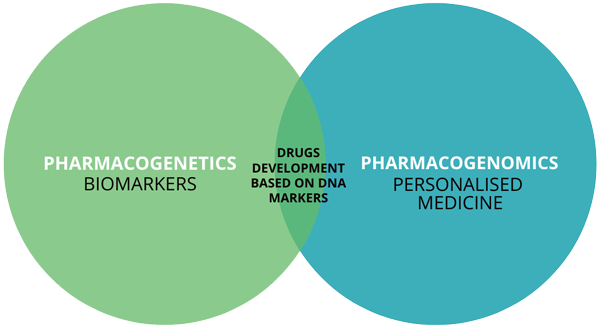
Pharmacogenomics & Pharmacogenetics Pharmacogenomics & Pharmacogenetics Learning Objectives & Module Roadmap This is a challenging topic because it merges genetics, biochemistry, and clinical medicine.By the end, you will be able to: Define and distinguish between pharmacogenetics and pharmacogenomics. Explain the “Why” behind differential drug responses in patients sharing the exact same disease. Identify all determinants (both genetic and non-genetic) of drug efficacy and toxicity. Master the “Hall of Fame” clinical examples (Warfarin, Codeine, Clopidogrel, Abacavir, etc.) of how genetic variations directly influence drug responses. Appreciate the ultimate goal: The transition from trial-and-error medicine to Precision/Personalized Medicine. 1. Introduction: The Problem with Traditional Pharmacology In traditional medicine, a “one-size-fits-all” approach is used. Ten patients come in with identical symptoms, identical lab findings, and the exact same disease. The doctor gives all ten patients the exact same drug at the exact same dose. What actually happens? Normal/Expected Response: Some patients experience excellent therapeutic effects and get better. Lack of Response: Some patients show absolutely no improvement. It’s as if they took a sugar pill. Exaggerated/Toxic Response: Some patients get dangerously sick from a standard dose (overdose effect). Idiosyncratic/Unexpected Response: Some patients develop bizarre, completely unpredictable side effects that have nothing to do with the drug’s primary mechanism. Traditional pharmacology cannot fully explain this massive variability. This is where Pharmacogenetics and Pharmacogenomics step in. They provide the missing puzzle piece: understanding how underlying genetic differences shape our response to drugs. Defining the Terms: Genetics vs. Genomics These terms are often used interchangeably in clinical practice, but technically, they have a subtle difference in scope: Pharmacogenetics: The “Micro” view. This is the study of how a single gene (or a few specific genes) influences an individual’s response to drugs. Example: Looking only at the CYP2C9 gene to see how a patient metabolizes Warfarin. Pharmacogenomics: The “Macro” view. This is the broader, system-wide study of how the entire genome (all the genes, their interactions, and multiple biological pathways) influences drug response. Example: Using a massive multi-gene testing panel to predict a patient’s overall toxicity risk before starting complex chemotherapy. Analogy: Pharmacogenetics is like inspecting the spark plugs on a car to see why it won’t start. Pharmacogenomics is plugging the car into a massive computer diagnostic system that checks the entire electrical grid, fuel system, and engine simultaneously. 2. Determinants of Drug Efficacy and Toxicity Why do drugs work differently in different people? The answer lies in a combination of factors. Drug response depends on a complex interplay of: Environmental & Physiological Factors: Age (infants and the elderly metabolize drugs much slower). Sex (hormonal differences affect drug processing). Diet (e.g., grapefruit juice famously blocks certain liver enzymes). Liver/Kidney function (if the organs that clear drugs are broken, toxicity occurs). Co-morbidities (having other diseases). Drug-related Factors: Formulation (tablet vs. IV), route of administration, and dangerous drug-drug interactions. Genetic Factors: Inherited variations in the DNA that code for drug-metabolizing enzymes, transport proteins, or the actual cellular targets (receptors) the drug binds to. Epigenetics and Gene Regulation: Changes that don’t alter the DNA code, but change how it is read. Mechanisms like DNA methylation, histone modification, and microRNAs act as “light switches” that can silence or activate specific genes, thereby influencing drug response. Ethnic and Population Differences: Certain genetic variants naturally cluster in specific populations due to evolutionary history. Clinical Example: Differential Drug Efficacy by Ethnicity Beta-blockers (blood pressure medications) are a classic example. Statistically, beta-blockers work less effectively for hypertension in Black populations compared to other drugs like ACE inhibitors or Calcium-Channel Blockers. This emphasizes that drug efficacy is not solely about the chemical molecule; it is heavily dependent on the patient’s unique population biology. 3. Types of Genetic Variation Genetic variation refers to differences in the DNA sequence among individuals. These differences can drastically influence Pharmacokinetics (ADME: Absorption, Distribution, Metabolism, Excretion) or Pharmacodynamics (what the drug does to its target receptors). There are six major types of genetic variations relevant to pharmacology: 1. Single Nucleotide Polymorphisms (SNPs) Definition: A change in just a single base pair (e.g., an Adenine ‘A’ is swapped for a Guanine ‘G’, or a Cytosine ‘C’ is swapped for a Thymine ‘T’). Prevalence: These are by far the most common type of genetic variation in humans. Impact: This single letter change can alter the entire amino acid sequence, drastically change an enzyme’s activity, alter receptor binding, or it might just be “silent” (doing nothing at all). Classic Examples: CYP2C19 SNPs: Affects the activation of Clopidogrel (poor metabolizers = treatment failure). VKORC1 SNPs: Increases sensitivity to Warfarin (causing a high bleeding risk). ABCB1 SNPs: Alters the activity of P-glycoprotein (a “bouncer” protein that kicks drugs out of cells), influencing the absorption and efflux of drugs like Digoxin. 2. Insertions and Deletions (Indels) Definition: The addition (insertion) or loss (deletion) of small DNA fragments in a gene. Impact: If you add or remove letters, you can cause a frameshift mutation, completely altering the reading frame of the DNA. This usually destroys the resulting protein structure or activity. Examples: Indel in UGT1A1 promoter: Causes reduced glucuronidation (breakdown) of the chemotherapy drug Irinotecan, leading to severe neutropenia and diarrhea. Indels in DPYD gene: Causes reduced breakdown of 5-Fluorouracil (5-FU), leading to severe, often fatal toxicity. 3. Copy Number Variations (CNVs) Definition: The duplication or deletion of entire genes or massive gene segments. Impact: Think of this as “dosage.” If you have 4 copies of a gene instead of 2, you make way more of that enzyme. It can drastically increase or decrease enzyme expression. Examples: CYP2D6 gene duplication: Creates “ultra-rapid metabolizers” who convert Codeine to Morphine too quickly, causing morphine toxicity. Deletion of the GSTT1 gene: Results in a complete lack of certain detoxification enzymes, making the patient highly vulnerable to carcinogens and certain drugs. 4. Variable Number Tandem Repeats (VNTRs) / Microsatellites Definition: Repeated short DNA sequences (like a molecular stutter, e.g., CACACA repeats) located in regulatory or coding regions. Impact: They act like a dimmer switch, affecting gene transcription, stability, or how much

Scroll to Top