Doctors Revision

Author name: doctorsrevision@gmail.com

Urinary Tract Infections (UTIs)
Microbiology

Urinary Tract Infections (UTIs)

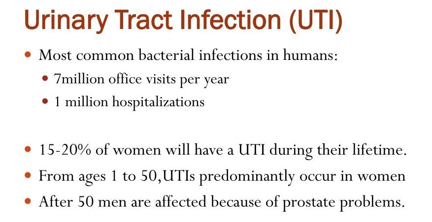
Urinary Tract Infections (UTIs) Urinary Tract Infections (UTIs) Module Overview Welcome to the comprehensive master guide on Urinary Tract Infections (UTIs). This guide covers everything from the microscopic battleground between bacterial virulence factors and host defenses, to step-by-step diagnostic workups and evidence-based treatment guidelines. 1. The Definitions Before diving into pathology, you must master the precise terminology used to describe urinary infections. Bacteriuria: Simply means the presence of bacteria in the urine. Significant Bacteriuria: The number of bacteria in voided urine exceeds what would be expected from normal contamination by the anterior urethra. Cutoff: ≥ 105 bacteria/mL. If you see this, infection must be seriously considered. Asymptomatic Bacteriuria: Significant bacteriuria (≥ 105) in a patient with absolutely ZERO symptoms. (We will discuss later who gets treated for this and who does not!) Location: UTIs can be confined to the lower tract (bladder/urethra) or involve both the upper (kidneys) and lower tracts. Cystitis (Lower UTI): A clinical syndrome involving dysuria (painful urination), frequency, urgency, and occasionally suprapubic (lower abdominal) tenderness. Acute Pyelonephritis (Upper UTI): A more severe clinical syndrome characterized by flank pain or tenderness (costovertebral angle), fever, often associated with the lower tract symptoms (dysuria, urgency, frequency). Uncomplicated UTI: Infection in a structurally and neurologically normal urinary tract. Complicated UTI: Infection in a urinary tract with functional or structural abnormalities (e.g., indwelling catheters, neurogenic bladder, or kidney stones/calculi). Clinical Scenario Uncomplicated vs. Complicated Patient A: A healthy 22-year-old female presents with painful urination and urgency for 2 days. She has no medical history. This is an Uncomplicated UTI (Cystitis). Patient B: A 65-year-old male with an enlarged prostate (BPH) and a history of kidney stones presents with the same symptoms. Because his urinary tract has structural blockages that prevent normal urine flushing, this is a Complicated UTI and requires much more aggressive management. Exam Trap: Urosepsis Criteria Urosepsis is the sepsis syndrome caused by a UTI. It is a life-threatening medical emergency. To diagnose it, you need clinical evidence of a UTI PLUS two or more of the following SIRS (Systemic Inflammatory Response Syndrome) criteria: Temperature: > 38°C (Fever) OR < 36°C (Hypothermia) Heart Rate: > 90 beats per minute (Tachycardia) Respiratory Rate: > 20 breaths/minute, OR PaCO2 < 32 mm Hg (Tachypnea/hyperventilation) White Blood Cell Count: > 12,000/mm3 (Leukocytosis), OR < 4,000/mm3 (Leukopenia), OR > 10% band forms (immature neutrophils). 2. Epidemiology & Common Bugs Females: UTI is much more common in women. 1-2% of young, non-pregnant women have it at any given time. 40% of all females will have a symptomatic UTI in their lifetime. Males: Extremely rare in young men (prevalence is only 0.04%). Clinical Pearl: If a young man gets a UTI, look for a structural defect or a Sexually Transmitted Disease (STD)! Older Age: Incidence skyrockets in the elderly (10% of men, 20% of women) due to functional impairments, prostate enlargement, and estrogen loss. The “Ojambo 2008” Ugandan Data: Over 95% of UTIs are caused by a single bacterial species. According to Ojambo 2008, the predominant organisms are: Escherichia coli (45%) – The undisputed king of UTIs. Klebsiella species (17%) Staphylococcus species (8%) – Most common Gram-positive. Enterococcus (5%) Other common offenders: Proteus, Pseudomonas, Enterobacter, and Candida (fungus, usually seen in diabetics or patients with chronic indwelling catheters). 3. Pathogenesis: A UTI is an epic battle between bacterial virulence factors and host defense mechanisms. The Routes of Invasion Ascending Route (Most Common): Bacteria from the gut colonize the perineum/urethra and climb up into the bladder. Why women? The female urethra is short and anatomically very close to the warm, moist vulvar and perianal areas, making fecal contamination highly likely. Hematogenous Route (Blood-borne): Infection of the kidney tissue by organisms traveling in the blood. Clinical Scenario: A patient with Staphylococcus aureus endocarditis (heart valve infection) throws infected blood clots into the kidneys, causing renal abscesses. Lymphatic Route: Rare, spread via lymphatic channels. Parasite Virulence Factors Not all E. coli cause UTIs. The ones that do are called Uropathogenic E. coli (UPEC) clones (Serogroups O1, O2, O4, O6, O7, O8, O75, O150, and O18ab). They possess specific genetic superpowers: Adhesins (Fimbriae/Pili): Prevent the bacteria from being washed away by urine. P fimbriae: Bind to Gal-α 1-4 (P blood group antigen). Strongly associated with Pyelonephritis and bacteremia. Type 1 fimbriae: Bind to mannosylated proteins (uroplakin Ia) on bladder cells. Associated with cystitis. Resistance to serum bactericidal activity. K Antigen (Capsules): High quantities of K1, K5, K12 capsular antigens physically protect bacteria from leukocyte phagocytosis. Aerobactin: An iron-scavenging protein (siderophore). Iron is scarce in urine; aerobactin steals it for the bacteria to grow. Hemolysin & Cytotoxic Necrotizing Factor type 1 (CNF-1) & Sat Toxin: Toxins that facilitate tissue invasion, cause severe renal tubular damage, and lyse red blood cells to make even more iron available to the invading E. coli. Urease (Specifically in Proteus species): Proteus produces urease, which splits urea into ammonia. This strongly correlates with its ability to cause severe pyelonephritis and struvite kidney stones. Deep Dive: Ammonia makes the urine highly alkaline. This change in pH causes magnesium, ammonium, and phosphate to crystallize, forming massive “staghorn” struvite stones that fill the entire renal pelvis! Master Table: Uropathogenic E. coli Adhesins Adhesin Genetic Sequence Receptor Target Clinical Comments Type 1 fimbriae (MS) Pil, fimH Mannosylated proteins on epithelial cells (uroplakin Ia) & PMNs Binds to Tamm-Horsfall protein (THP) and SIgA. P fimbriae (MR) papG (class Ia, II, III) Gal-α 1-4 (P blood group antigen) Class II: Strongly associated with pyelonephritis & bacteremia. Class III: Cystitis in patients with urinary tract abnormalities. S/F1C fimbriae (MR) Sfa/fac Sialyl-(α-2-3) galactoside Adherence is inhibited by THP. Type 1C (MR) Fac Undetermined Possibly associated with pyelonephritis. G fimbriae (MR) — Terminal N-acetyl-D-glucosamine — M fimbriae (MR) — Galactose-N-acetyl-galactosamine / Blood group M (glycophorin A) — Dr family Drb operon, Afa E1-5, Afa F Dr blood group antigen (decay accelerating factor – DAF) & type IV collagen Found in 16% of first-time cystitis isolates. The Host’s Defenses (Why we don’t always have
Skin and Soft Tissue Infections (SSTIs)
Microbiology

Skin and Soft Tissue Infections (SSTIs)

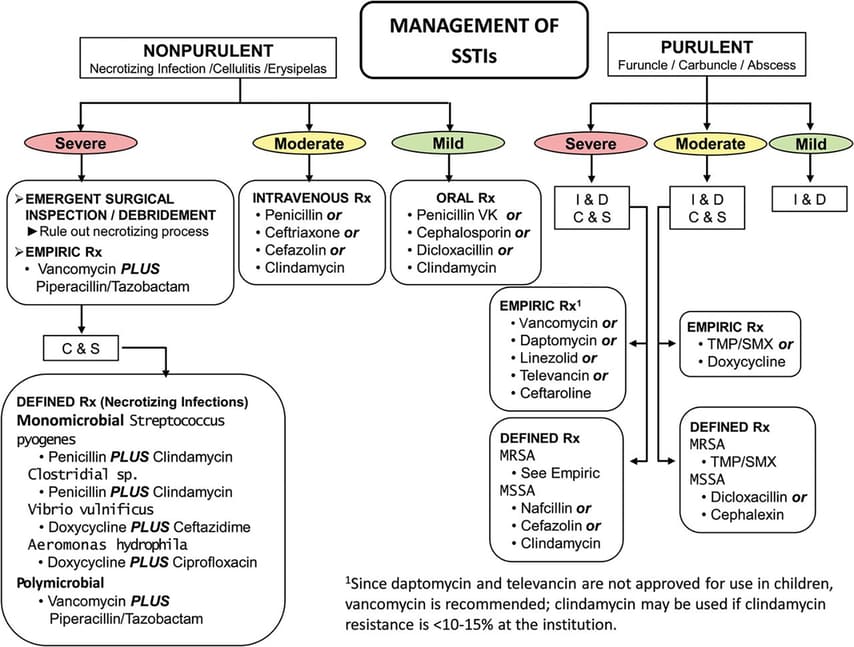
Skin and Soft Tissue Infections (SSTIs) Skin and Soft Tissue Infections (SSTIs) 1. Introduction to Skin & Soft Tissue Infections (SSTIs) General Overview: SSTIs range from minor superficial infections (like a tiny pimple) to rapidly spreading, life-threatening emergencies (like flesh-eating bacteria). The skin normally acts as an impenetrable physical and immunological barrier; infections usually require a breach (such as trauma, an insect bite, surgery, or maceration from prolonged moisture). Classic General Clinical Presentation: Local Signs: Accumulation of pus (purulence), intense redness (erythema), pain/tenderness, swelling (edema) due to increased vascular permeability. Systemic Signs: Fever, chills, malaise (as cytokines like TNF and IL-1 enter the bloodstream). Severe Complication: Bacteremia (bacteria entering the bloodstream, potentially leading to widespread sepsis and septic shock). General Diagnostic Approach: Specimen Collection and Processing Exam Trap: Never just swab a dry, intact crust or a superficial ulcer base. You must get to the active, deep infection! Swabbing dry crusts only yields dead bacteria or environmental contaminants. Collection History & Prep: The site MUST be heavily decontaminated first with soap and 70% isopropyl alcohol. Why? To avoid culturing normal, harmless skin flora (like Staphylococcus epidermidis) which will confuse the lab results and lead to the prescription of unnecessary antibiotics. The Procedure (Aspiration > Swabs): Use a sterile needle and syringe to aspirate (pull out) the loculated fluid or pus from the absolute depths of pustular/vesicle wounds or abscesses. Fluid is always vastly superior to a dry swab. Transport: Use the aspirating syringe itself as the transport container (safely capped). If there is a delay in processing, the sample MUST go into an anaerobic transport container. Clinical Reason: Deep tissues, especially in diabetics or deep bite wounds, often harbor strict anaerobes (like Bacteroides). Room air (oxygen) is toxic to them and will kill them before they reach the lab, giving you a false negative! Swabs: If a swab must be used, it should be placed in an anaerobic transport medium or inoculated directly onto culture media right at the patient’s bedside. Laboratory Processing: Gram Stain: Done first! It acts as a rapid guide for the clinician to select early empiric antibiotics (e.g., seeing Gram-positive cocci in clusters immediately suggests Staph, prompting the use of Flucloxacillin or Vancomycin) and tells the lab which specific culture media to use. Culture: The lab uses both selective and enriched non-selective media. You must know these three: 5% Sheep Blood Agar: Detects hemolysis patterns (Alpha, Beta, Gamma) crucial for identifying Streptococcus and Staphylococcus. MacConkey Agar: Selects specifically for Gram-negatives (like E. coli or Pseudomonas), inhibiting Gram-positives. Chocolate Agar: Cooked blood agar that releases internal cell nutrients, used for fastidious (picky) organisms like Haemophilus influenzae. 2. Superficial Infections (The Pyodermas) Pyoderma literally means “pus in the skin.” These are highly contagious, superficial infections predominantly affecting the epidermis. A. Impetigo (Non-Bullous) Pathophysiology & Etiology: A superficial, intraepidermal (top layer of skin), unilocular vesicopustule. It frequently occurs after minor trauma like insect bites or scratches which break the skin barrier, allowing surface bacteria to invade. Causative Agents: Group A Streptococci (GAS) (Specifically M-serotypes 2, 49, 52, 55, 57, 59, 60, 61), Group C and G Streptococci, and Staphylococcus aureus. Clinical Scenario The Honey-Crusted Child A 6-year-old boy presents to the pediatric clinic with a cluster of sores around his mouth and nose. His mother mentions he had mosquito bites there a few days ago and kept scratching them. The sores have burst, leaving a classic “honey-colored crust.” This golden crust is the absolute hallmark of non-bullous impetigo, formed by dried serum and bacterial proteins. Diagnostics & Exam Gold! The Serology Trap Gram Stain: Reveals Gram-positive cocci. Culture: Take exudate from beneath an unroofed crust. It will grow S. aureus, GAS, or a mixture of both. Serology (Exam Gold!): If caused by Streptococcus, the Anti-Streptolysin O (ASO) titer will be SCANT (negative). Why? Because the skin lipids (cholesterol in the skin) locally bind to, inhibit, and destroy Streptolysin O! Therefore, no ASO antibodies are made. Instead, you must look for an anti-DNase B response, which readily occurs and proves a recent skin Strep infection (vital if the patient later develops Post-Streptococcal Glomerulonephritis). Treatment: Topical antibiotics (like Mupirocin) for mild, localized cases. Systemic ampicillin, penicillin, erythromycin, or cephalosporins for widespread cases or immunocompromised hosts. B. Bullous Impetigo Pathophysiology & Etiology: Caused specifically by S. aureus of Phage Group II (usually type 71). This specific strain produces ETA toxin (Exfoliative Toxin A). Mechanism: The ETA toxin acts as highly specific molecular scissors. It specifically cleaves desmoglein 1 (a transmembrane glycoprotein of desmosomes that acts like velcro to hold skin cells together). This causes subcorneal separation of the epidermis, creating a pocket that fills with fluid. Clinical History & Presentation: Seen almost exclusively in newborns and young children. Lesions begin as vesicles that quickly turn into large, flaccid bullae (blisters) containing clear yellow fluid. The bullae lack a surrounding ring of redness. They quickly rupture, leaving a moist, raw red surface. C. Staphylococcal Scalded Skin Syndrome (SSSS) Pathophysiology: Similar to bullous impetigo, but instead of the toxin acting locally, the S. aureus exfoliative exotoxin enters the bloodstream and acts systemically across the entire body. Clinical History & Presentation: Begins abruptly. The patient develops a fever, intense skin tenderness, and a scarlatiniform (sandpaper-like red) rash. Large, flaccid, clear bullae form, promptly rupture, and result in the separation of massive sheets of skin. Visual Note: The child looks exactly like they have suffered a severe, widespread boiling water burn. (Unlike Toxic Epidermal Necrolysis/TEN, which involves the deeper dermal-epidermal junction and mucous membranes, SSSS is highly superficial and usually spares the mucous membranes). D. Staphylococcal Scarlet Fever & Toxic Shock Syndrome (TSS) Staphylococcal Scarlet Fever: Caused by S. aureus enterotoxins (A through D) and Toxic Shock Syndrome Toxin 1 (TSST-1). Presents with a scarlatiniform rash and skin desquamation (peeling), particularly on the palms and soles. Toxic Shock Syndrome (TSS): A severe, life-threatening acute febrile illness driven by “superantigens” that massively hyper-activate T-cells, causing a “cytokine storm.” Clinical Presentation: Generalized scarlatiniform eruption, intense desquamation, severe

History & Diagnostics in Microbiology
Microbiology

History & Diagnostics in Microbiology

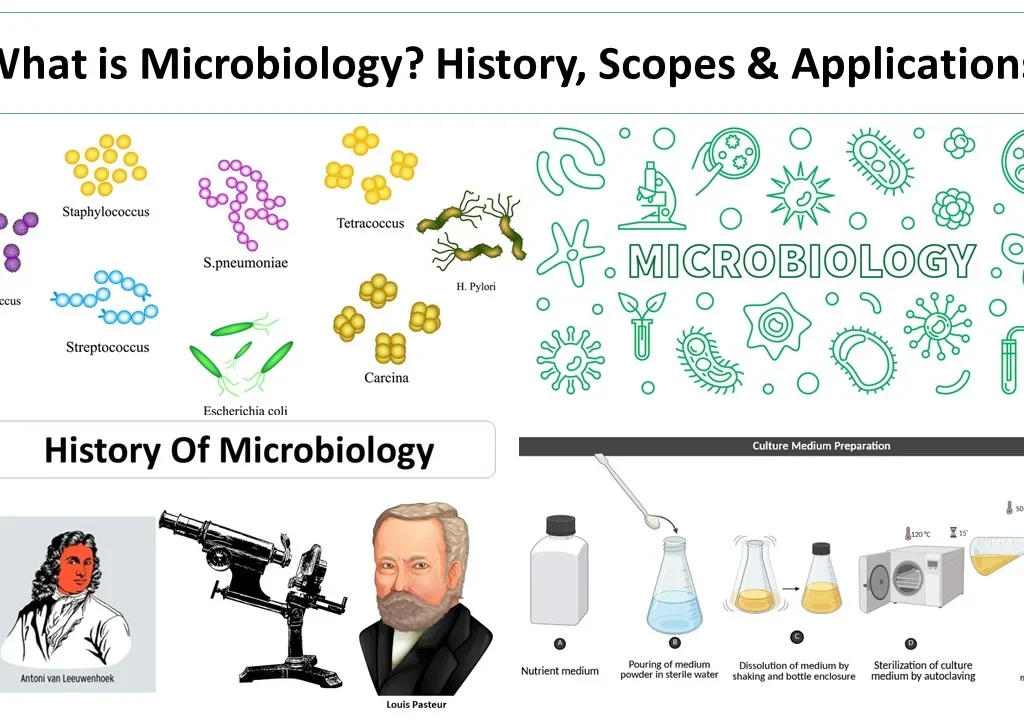
History & Diagnostics in Microbiology History & Diagnostics in Microbiology PART 1: HISTORY OF MICROBIOLOGY 1. The Dark Ages of Disease Before the invention of microscopes, humans were completely blind to the microscopic world. Diseases were attributed to supernatural causes (curses, angry gods) or “miasmas” (bad, foul-smelling air from rotting organic matter). Slowly, the concept of contagion (disease spreading by touch, clothing, or proximity) began to emerge, but the actual physical agents of disease remained a complete mystery. Historical Context: During the bubonic plague (Black Death), “plague doctors” wore bird-like masks stuffed with sweet-smelling flowers. Why? Because they genuinely believed the disease was caused by inhaling the foul “miasma” smell of death, rather than being bitten by infected flea vectors! 2. The Pioneers of Microscopy (The Lens Makers) We couldn’t study bacteria until we could see them. Three men made this possible: Zacharias Janssen (1570-1638) A Dutch spectacle maker who invented the concept of compounding lenses. He placed two lenses inside a single sliding tube, creating the first rudimentary compound microscope, allowing for enlarged images of microscopic forms. Robert Hooke (1635-1703) In the 1660s, he modified the microscope (using a 6-inch tube and two convex lenses). He famously observed cork, seaweed, and sponges. He coined the term “cell” because the tiny rectangular structural boxes in cork reminded him of the bare, empty monastery rooms (cells) where monks lived. In 1665, he published his spectacular findings in his famous book, Micrographia. He was the first to describe fungi, detailing a bluish mold on leather and a white mold (which his detailed descriptions allow us to classify today as Mucor). Antony van Leeuwenhoek (1632-1723) Known forever as the Father of Microbiology. A brilliant, self-made scientist from Delft, Holland. He made 419 lenses and over 250 single-lens microscopes, achieving a staggering, crystal-clear magnification of 200-300x. He observed sperm, blood cells, and most famously, the scrapings from his own teeth (which we now know were massive bacterial biofilms!). He wrote extensively detailed letters to the British Royal Society describing tiny, moving unicellular creatures he affectionately called ‘animalcules’. In 1683, he published the very first sketches of the three principle bacterial shapes: rods (bacilli), cocci (spheres), and spirals. 3. The Great Debate: Abiogenesis vs. Biogenesis For centuries, scientists fought a bitter war over where life actually came from. Did it magically appear from non-living matter (Abiogenesis / Spontaneous Generation), or did life only come from pre-existing life (Biogenesis)? Scientist Experiment & Conclusion Stance Van Helmont (1580-1644) Placed dirty clothes and wheat/cheese in a dark stable for 21 days. Found mice. Concluded the dirt/wheat magically “created” mice. (He ignored the fact that mice simply walked in to eat the cheese!). Supported Abiogenesis Francesco Redi (1626-1697) The 3-Jar Meat Experiment. One open jar (maggots grew), one covered in parchment (no maggots), one covered in gauze (eggs laid on top of gauze, no maggots on meat). Proved flies MUST lay eggs to make maggots. Opposed Abiogenesis Louis Joblot (1645-1723) Boiled hay infusion and divided it. Covered vessel = no growth. Uncovered = microbial growth. Opening the covered one later allowed growth. Supported Biogenesis Lazzaro Spallanzani (1729-1799) Boiled meat broth for a long time to destroy heat-resistant spores and completely sealed the flask in flame. Result: No growth. Opponents stubbornly claimed he destroyed the “vital air” needed for magic generation. Opposed Abiogenesis The Final Nail in the Coffin: Louis Pasteur (1862) Louis Pasteur (1822-1895) permanently ended the spontaneous generation debate with a stroke of genius. He designed a special ‘Swan-necked’ (S-shaped) flask. He boiled nutrient broth inside it to sterilize it. Because the flask was completely open at the very end, “vital air” could easily enter, satisfying his stubborn critics. However, gravity and the S-curve of the neck physically trapped all heavy dust particles and bacteria from the air, preventing them from falling into the broth. Result: NO GROWTH. The broth remained sterile indefinitely. When he deliberately broke the neck off, allowing dust to fall directly in, microbial growth appeared immediately. Biogenesis was proven forever! 4. The Golden Age of Microbiology The late 1800s saw an explosion of life-saving discoveries, primarily led by two bitter international rivals: Pasteur (France) and Koch (Germany). Louis Pasteur (The Innovator) Discovered anaerobic bacteria (1877) during studies on butyric acid fermentation (bacteria that live without oxygen). Discovered that Yeast is the microorganism responsible for converting sugar into alcohol. Solved the massive economic crisis of souring French wine by inventing Pasteurization (mildly boiling fruit juices/milk to kill specific spoilage contaminants without ruining the taste). Vaccines & Immunology (1880): Discovered active immunization by a happy accident. While studying chicken cholera (Pasteurella spp.), he found that leaving cultures out on the bench to age made them lose their pathogenicity (virulence). Injecting these “attenuated” (weakened) older cultures didn’t kill the chickens, but amazingly protected them from future deadly doses! Created the first attenuated rabies vaccine and famously saved a young boy (Joseph Meister) who had been savagely bitten by a rabid dog. Robert Koch (1843-1910) (The Methodical Bacteriologist) A German scientist who gave us the strict laboratory techniques we still use today. Isolated the exact microorganisms causing Anthrax and Tuberculosis. Developed solid media (using agar instead of liquid broths or potatoes) for culturing bacteria and invented the streak plate technique to physically isolate pure, single colonies. Exam Trap: Koch’s Postulates Koch created 4 strict guidelines/rules to definitively prove that a specific microbe causes a specific disease. To pass the test: The microorganism must be found in abundance in ALL organisms suffering from the disease, but NOT found in healthy organisms. The microorganism must be isolated from the diseased animal and grown in pure culture in the lab. The cultured microorganism must cause the exact same disease when introduced into a healthy lab animal. The microorganism must be re-isolated from the newly diseased animal and identified as completely identical to the original specific causative agent. The Exception / Caveat (Highly Testable!): We now know there are major exceptions to Koch’s rules! – Asymptomatic carriers (like Typhoid Mary) violate

Bacterial Growth, Genetics, and Structure
Microbiology

Bacterial Growth, Genetics, and Structure

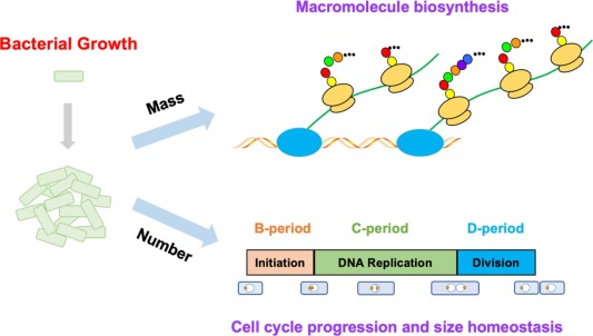
Bacterial Growth, Genetics, and Structure Microbiology Foundations: Bacterial Growth, Genetics, and Structure Module Overview Before a bacterium can divide and cause an infection, it needs the right fuel and environment. Think of bacteria as microscopic factories; they need raw materials (nutrition) and ideal factory conditions (environment). Understanding these mechanisms is the fundamental basis of targeted antibiotic therapy and infectious disease management. 1. Bacterial Growth & Nutritional Requirements A. Nutrient Requirements (The “Raw Materials”) Just like human cells, bacterial cells are highly complex and require specific building blocks to construct their membranes, DNA, and proteins. Water: Essential for all biochemical reactions. It is the universal solvent in which all intracellular metabolic processes occur. Carbon Source (C): The backbone of all living molecules (carbohydrates, lipids, proteins). Bacteria are often classified by how they get carbon (e.g., heterotrophs get it from organic compounds like glucose; autotrophs get it from CO2). Nitrogen Source (N): Crucial for building amino acids (which make up proteins) and nucleic acids (which make up DNA/RNA). Inorganic Salts, Sulfur (S), & Phosphorus (P): Phosphorus is needed to synthesize ATP (energy currency) and the phospholipid bilayer of the cell membrane. Sulfur is strictly needed for certain sulfur-containing amino acids (like cysteine and methionine) which hold proteins together via disulfide bonds. Growth Factors: Essential vitamins and amino acids that the bacteria cannot synthesize on their own. If the environment lacks these, the bacteria cannot survive. B. Environmental Factors (The “Factory Conditions”) Temperature: Most human pathogens grow best at 37°C (normal human body temperature). These are known as mesophiles. (Clinical Note: This is why the human body generates a fever—it raises the temperature above 37°C to make the environment uncomfortably hot and hostile for the invading bacteria!) Gas (Oxygen): Determines if they can breathe in air or if air is toxic to them (detailed in the next section). pH: Most bacteria prefer a neutral pH (around 7.0), though some have adapted to survive extreme acid. Example: Helicobacter pylori in the stomach survives the highly acidic gastric juice (pH ~2.0) by secreting an enzyme called urease, which creates a neutralizing “cloud” of ammonia around the bacteria. Osmotic Pressure: Salt and sugar concentrations in the environment. Clinical Example: High salt environments usually pull water out of bacteria, killing them (which is why curing meat with salt prevents rotting). However, Staphylococcus aureus is a “halophile” (salt-lover) and can easily survive on the salty surface of human skin, making it a major cause of surgical wound infections. 2. Oxygen Requirements: Aerobic vs. Anaerobic Bacteria Clinical Scenario: The Rusty Nail A patient steps on a rusty nail. The deep puncture wound closes over quickly, trapping bacteria inside with no oxygen. This is the perfect, deadly environment for an obligate anaerobe (like Clostridium tetani) to thrive and cause tetanus. Understanding oxygen requirements tells you exactly where an infection can survive in the human body! Oxygen is highly reactive. When metabolized, it creates deadly byproducts called Reactive Oxygen Species (ROS), such as superoxide radicals (O2–) and hydrogen peroxide (H2O2). To survive in oxygen, a bacteria MUST have specific enzyme “shields” (Catalase and Superoxide Dismutase – SOD) to neutralize these toxins. Type of Bacterium Effect of Oxygen Growth Pattern in a Tube Enzyme Status (The “Shields”) Classic Clinical Examples Obligate Aerobes Only aerobic growth; O2 is strictly required. Growth occurs only at the very top of the tube where O2 is highest. Have Catalase and Superoxide Dismutase (SOD) to neutralize toxic oxygen radicals. Mycobacterium tuberculosis (This is why TB infections classically target the APEX of the lungs, where oxygen concentration is highest!) Facultative Anaerobes Adaptable! Both aerobic and anaerobic growth. Greater growth with O2, but can survive without it. Growth is best at the top, but occurs throughout the entire tube. Have Catalase and SOD to neutralize toxic oxygen. Escherichia coli (E. coli) and Staphylococcus aureus. Obligate Anaerobes Only anaerobic growth. Oxygen is highly toxic/lethal. Growth occurs only at the very bottom of the tube where there is zero O2. Lacks enzymes to neutralize harmful forms of O2. Clostridium tetani, Bacteroides fragilis (Found deep in the gut). Aerotolerant Anaerobes Only anaerobic growth, but it can continue growing in the presence of O2. Growth occurs evenly throughout the entire tube; O2 has no effect. Presence of one enzyme (SOD) allows them to partially tolerate O2. Lactobacillus and Streptococcus pyogenes. Microaerophiles Only aerobic growth, but strictly requires LOW concentrations of oxygen. Growth occurs right in the middle of the tube (where O2 is low but not zero). Produce lethal amounts of toxic oxygen if exposed to normal atmospheric air. Helicobacter pylori and Campylobacter jejuni. 3. The Bacterial Growth Curve When bacteria invade a host or are put in a culture tube, they follow a predictable, 4-stage life cycle. Exam Trap: Know exactly what happens in the Log phase vs. Stationary phase! 1. Lag Phase The “Prep” Phase Bacteria are sensing their environment, gathering nutrients, and turning on specific enzymes needed to digest local food sources. There is NO increase in the number of living bacterial cells during this phase. 2. Log Phase (Exponential Phase) The “Population Boom” There is an exponential increase in the number of living cells. The bacteria are replicating at maximum speed. Clinical Pearl: This is when bacteria are rapidly building new cell walls and dividing. Therefore, this is the exact phase where antibiotics that target cell wall synthesis (like Penicillin or Cephalosporins) are most spectacularly effective! 3. Stationary Phase The “Plateau” Nutrients are running out, and toxic metabolic waste is building up. The rate of cell division exactly equals the rate of cell death. (Deep Dive: In this phase, bacteria like Clostridium and Bacillus realize they are starving and will trigger the formation of Endospores to survive the upcoming famine). Because cell wall synthesis slows down drastically here, Penicillin becomes much less effective against bacteria in an abscess (which are usually in the stationary phase). 4. Death (Decline) Phase The “Collapse” There is an exponential decrease in the number of living cells due to complete nutrient depletion and

Classification & Taxonomy bacteria (1)
Microbiology

Classification & Taxonomy of Bacteria

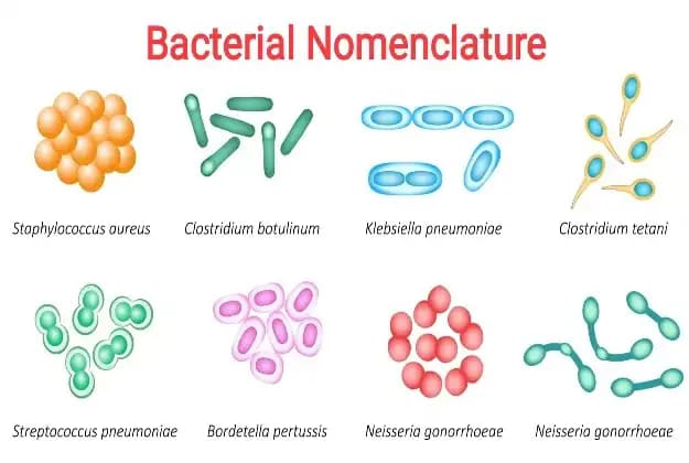
Classification & Taxonomy of Bacteria Cell Biology & Bacterial Taxonomy Why This Matters In clinical medicine, you cannot treat an invisible enemy without knowing exactly what it is. Bacterial taxonomy isn’t just memorizing names; it is the roadmap to prescribing the correct life-saving antibiotics. By understanding a bacterium’s shape, oxygen requirements, and cell wall structure, a doctor can predict exactly how a disease will progress and which drug will destroy the pathogen without harming the patient. 1. Taxonomical Hierarchies and Nomenclature Taxonomy is the science of classifying organisms. In microbiology, we classify bacteria into a strict hierarchy to understand their relationships and pathogenic behaviors. The Hierarchy of Bacteria Kingdom: Prokaryotes (organisms lacking a true nucleus). Order: The name always ends with the suffix ‘-ales’. Example: Enterobacteriales. Family: Many families exist in one order. The name always ends with the suffix ‘-eae’. Examples: Enterobacteriaceae, Pseudomonodaceae. Genus: Each family is divided into genera. Example: Within Enterobacteriaceae, you have Escherichia, Klebsiella, Enterobacter. Species: The most specific group. Example: Escherichia coli. Sub-species: Further divisions based on tiny genetic/antigenic differences (e.g., Salmonella enterica subsp. enterica). Clinical Extension: This level is vital for tracking outbreaks! For example, tracking the deadly food-poisoning strain E. coli O157:H7 distinguishes it from the harmless E. coli living normally in your gut. Rules for Bacterial Nomenclature (Naming) Medical professionals must communicate without ambiguity. Only one correct name exists, determined by the International Journal of Systematic Bacteriology (IJSB). Confusing names are rejected. Every bacterium must have a Genus and a Species name. The Genus name must start with a Capital letter. The species name must start with a small (lowercase) letter. The entire name MUST be italicized (if typing) or underlined (if handwriting). Example: Staphylococcus aureus or Escherichia coli. All names are written in Latin. 2. Classification of Medically Important Bacteria (Based on Cell Wall) This is the most critical flowchart in clinical microbiology. Bacteria are primarily classified by their cell wall structure, which dictates what antibiotics will work against them. A. Lacking Cell Wall Genera: Mycoplasma & Ureaplasma Clinical Pearl & Exam Trap The Mycoplasma Exception Because they have NO cell wall, they are naturally resistant to all beta-lactam antibiotics (like Penicillin, Amoxicillin, Cephalosporins). Why? Because beta-lactams work exclusively by destroying the cell wall (peptidoglycan). You cannot destroy a wall that doesn’t exist! Example: Mycoplasma pneumoniae causes “walking pneumonia.” You must treat it with drugs that target ribosomes (inside the cell), like Macrolides (Azithromycin) or Tetracyclines. B. Rigid Cell Wall (The vast majority of bacteria) Divided based on how they live and their shape/staining: Obligate Intracellular Bacteria: Must live inside a host cell to survive (they cannot make their own ATP—they are “energy parasites”). Genera: Chlamydia, Rickettsia, Coxiella, Ehrlichia. Filamentous Bacteria (Branching, fungus-like): Genera: Actinomyces, Nocardia, Mycobacteria. Clinical Pearl: Mycobacteria (which causes Tuberculosis) has a rigid wall heavily loaded with mycolic acid (waxy lipids), making it “Acid-Fast” instead of truly Gram-positive or negative. Regular Gram stain bounces right off this wax. We must use the Ziehl-Neelsen (Acid-Fast) stain, where TB appears as bright red snappers. Free-Living, Simple Unicellular (Gram Positive vs Gram Negative): Gram Positive (Purple/Blue Stain) Gram Negative (Pink/Red Stain) Cocci (Spheres): Staphylococcus (Clusters – looks like grapes. Common on skin). Streptococcus (Chains – like a pearl necklace. Causes strep throat). Enterococcus Peptostreptococcus Rods (Bacilli): Bacillus (Spore former – Anthrax) Clostridium (Spore former – Tetanus/Botulism) Corynebacterium (Diphtheria) Listeria (Food poisoning in pregnant women) Erysipelothrix Lactobacillus Propionibacterium Cocci: Neisseria (e.g., N. gonorrhoeae and N. meningitidis) Moraxella Acinetobacter Enteric Rods (Gut bugs): Escherichia, Klebsiella, Proteus, Salmonella, Shigella, Vibrio, Helicobacter, Campylobacter, Bacteroides. Non-Enteric Rods (Respiratory/Zoonotic): Pseudomonas, Haemophilus, Brucella, Bordetella, Legionella, Pasteurella. C. Flexible Cell Wall (Spirochetes) Corkscrew-shaped bacteria that move using axial filaments (internal flagella that twist the entire cell like a drill). Genera: Treponema (Causes Syphilis), Borrelia (Causes Lyme Disease), Leptospira. Diagnostic Note: They are so incredibly thin that they cannot be seen with a normal light microscope. Doctors must use a special “Darkfield Microscope” to see Treponema pallidum swimming in fluid from a syphilis sore. 3. Morphology: Size and Shape of Bacteria Size and Its Importance Bacterial cell size ranges from 0.1 to 5µm in diameter. Cyanobacteria: 5 × 40µm (Huge for bacteria) Escherichia coli: 1.3 × 3µm (Average) Haemophilus influenzae: 0.25 × 1.2µm (Very small) Exam Favorite Why is being small a massive advantage? The rate at which nutrients enter and waste products exit the cell is inversely proportional to cell size. This is because smaller cells have a massively larger Surface Area-to-Volume ratio. Result: The smaller the cell, the faster the metabolic rate, and the incredibly faster the growth/replication rate. This explains why a few E. coli bacteria on a piece of chicken can double their population every 20 minutes, leading to millions of bacteria and massive food poisoning overnight! Shapes of Bacteria The shape of the cell directly affects its ecology (how it survives in its environment). Coccus (Spheres): Can exist as single cocci, diplococci (pairs, e.g., Neisseria gonorrhoeae looks exactly like two kidney beans facing each other), streptococci (chains), staphylococci (grape-like clusters), tetrads (groups of 4), or sarcina (groups of 8). Bacillus (Rods): Coccobacillus (very short, plump rods that look almost like cocci), standard rods (e.g., Bacillus anthracis looks like long boxcars). Vibrio: Comma-shaped, curved rods (e.g., Vibrio cholerae, shaped like a comma to rapidly dart through thick intestinal mucus). Spirillum / Spirochete: Helical, corkscrew-shaped. Filamentous: Long, branching threads (mimicking fungal hyphae to spread through tissue). 4. Bacterial Cell Structures (Anatomy of a Prokaryote) A. Cytoplasm Structures (Inside the cell) Nuclear Region (Nucleoid): Prokaryotes do NOT have a true nucleus or a nuclear membrane. Their DNA is a single, long, circular strand that is heavily supercoiled (twisted up tight like a rubber band) to fit inside the tiny cell. Ribosomes: Small particles made of protein and rRNA, essential for translation (protein synthesis). Bacterial ribosomes are 70S in size (composed of 50S and 30S subunits). Granules (Inclusion bodies): Used to store energy (like Glycogen) or serve as structural building blocks when nutrients are plentiful. Plasmids: Extrachromosomal circular

Neoplasia & Oncology
Pathology

Neoplasia & Oncology

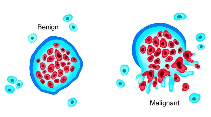
Neoplasia & Oncology Neoplasia & Oncology Exam Focus & Objectives Neoplasia is one of the most heavily tested topics in medical exams. To master this, you must shift your thinking from normal physiology to pathological, rogue biology. By the end of this exhaustive guide, you will master: The precise nomenclature (naming rules) of tumors, including the famous “exceptions” that appear on every exam. The distinct morphological and behavioral differences between Benign vs. Malignant tumors. The intricate genetic mechanisms (Oncogenes vs. Tumor Suppressor Genes) and viral etiologies of cancer. The systemic effects of cancer, especially the high-yield Paraneoplastic Syndromes. The crucial difference between Grading and Staging. 1. Definitions & Anatomy of a Tumor Before we classify tumors, we must define exactly what we are dealing with. The terminology is precise and highly testable. Cancer: A genetic disorder caused by DNA mutations. It is not a single disease, but a collection of disorders driven by corrupted genetic code. Neoplasia: Literally translates to “new growth.” A neoplasm is an abnormal mass of tissue whose growth exceeds and is uncoordinated with that of normal tissues, and persists even after the stimuli that evoked the change is removed. Tumor: Literally means “Swelling.” While originally a sign of inflammation, in modern medicine, “tumor” is used interchangeably with “neoplasm.” Tumors can be Benign (innocent, localized) or Malignant (cancerous, spreading). Oncology: The clinical and scientific study of tumors (from the Greek oncos, meaning tumor). The Two Basic Components of ALL Tumors Whether a tumor is benign or malignant, it is constructed of two main parts. Think of a tumor like a rogue city: The Parenchyma: These are the transformed neoplastic cells. These are the actual mutated “bad guys.” The parenchyma determines the biological behavior of the tumor and is what we use to name the tumor. The Stroma: This is the supporting, host-derived, non-neoplastic tissue. It is made up of connective tissue, blood vessels, and host-derived inflammatory cells. The Rogue Army Analogy: The parenchyma is the rogue army; the stroma is the supply lines (blood vessels) and infrastructure (connective tissue) the army forces the host to build for them so they can survive. Without a blood supply (stromal angiogenesis), a tumor cannot physically grow larger than 1-2 millimeters! 2. Nomenclature: How We Name Tumors Tumor nomenclature is based entirely on the parenchyma (the cell of origin). This is a heavily tested area where suffixes give away the diagnosis. A. Benign Tumors General Rule: Benign tumors are designated by attaching the suffix “-oma” to the cell type from which the tumor arises. Fibroblast + oma = Fibroma (Benign tumor of fibrous tissue). Chondrocyte (cartilage) + oma = Chondroma. Lipocyte (fat) + oma = Lipoma (e.g., benign lipoma of the small intestine or under the skin). Osteocyte (bone) + oma = Osteoma. Smooth Muscle + oma = Leiomyoma (e.g., uterine fibroids are actually benign leiomyomas!). Special Benign Epithelial Tumors: Papillomas: Benign epithelial neoplasms growing on any surface that produce microscopic or macroscopic finger-like fronds (e.g., Squamous cell papilloma). Polyp: A mass that projects above a mucosal surface (like in the gut/colon) to form a macroscopically visible structure. (Note: A polyp is a descriptive macroscopic term. It can technically be benign or malignant, but most are benign adenomatous polyps). Cystadenomas: Hollow, cystic masses that typically arise in the ovary. Adenoma: A benign epithelial tumor that either arises from glands or forms a glandular pattern. B. Malignant Tumors (CANCERS) Malignant tumors are named based on their embryological origin (Mesenchymal vs. Epithelial). SARCOMAS Mesenchymal / Connective Tissue Origin Malignant tumors arising in solid mesenchymal tissues (bone, cartilage, fat, muscle, blood vessels). Fibrosarcoma Liposarcoma Chondrosarcoma Osteogenic sarcoma (Osteosarcoma) Angiosarcoma (blood vessels) Leiomyosarcoma (smooth muscle) Rhabdomyosarcoma (skeletal/striated muscle) Liquid Mesenchymal: Malignancies arising from blood-forming cells are called Leukemias or Lymphomas. CARCINOMAS Epithelial Origin Malignant neoplasms of epithelial cell origin (regardless of which of the 3 germ layers the epithelium came from). Carcinomas are the most common cancers in adults. Adenocarcinoma: Carcinomas that grow in a glandular pattern (e.g., Colon adenocarcinoma, Prostate adenocarcinoma). Squamous cell carcinoma: Carcinomas that produce squamous cells (often arising in the skin, cervix, or lung). Microscopically, these often show “pink keratin pearls” or “intercellular bridges”. CRITICAL EXAM TRAPS: The Malignant “-omas” The suffix “-oma” usually means benign. However, examiners LOVE to test the famous exceptions that sound benign but are absolutely, lethally MALIGNANT. Memorize these: Melanoma: Malignant tumor of melanocytes (skin). Lymphoma: Malignant tumor of lymphoid tissue. Mesothelioma: Malignant tumor of the mesothelium (pleura of lung, strongly linked to asbestos exposure). Seminoma: Malignant tumor of testicular germ cells. C. Mixed Tumors & Teratomas Mixed Tumors: Arise from a single clone of cells capable of differentiating into more than one cell type (e.g., Pleomorphic adenoma of the salivary gland, containing both epithelial tissue and cartilage-like stroma). Teratoma: A special type of mixed tumor containing recognizable mature or immature cells/tissues derived from more than one germ cell layer (endoderm, mesoderm, ectoderm), and sometimes all three! Origin: They originate from totipotential germ cells (cells with the capacity to turn into ANY tissue in the body). These normally reside in the ovary and testis, or abnormally in midline embryonic rests. Pathology: Because germ cells can differentiate into anything, a teratoma might contain hair, bone, epithelium, muscle, fat, and teeth all thrown together in a disorganized “helter-skelter” fashion! (e.g., Ovarian cystic teratoma / dermoid cyst). D. The “Fake Tumors” (Non-Neoplastic Lesions) These two are often tested to confuse you. They sound like tumors, but they are congenital anomalies or disorganized normal tissue. Hamartoma A mass of disorganized tissue indigenous (native) to that particular site. It is the right tissue, just messy. Example: A disorganized mass of normal lung cartilage and respiratory epithelium in the lung, or bile ducts inside the liver. Note: Newer evidence shows some have clonal mutations and are now considered benign neoplasms, but classically they are disorganized native tissue. Choristoma A congenital anomaly consisting of a heterotopic (out of place) nest of normal cells. It is perfectly normal tissue, completely lost. Example: A

NSAIDs & Prostanoids
Pharmacology

NSAIDs & Prostanoids

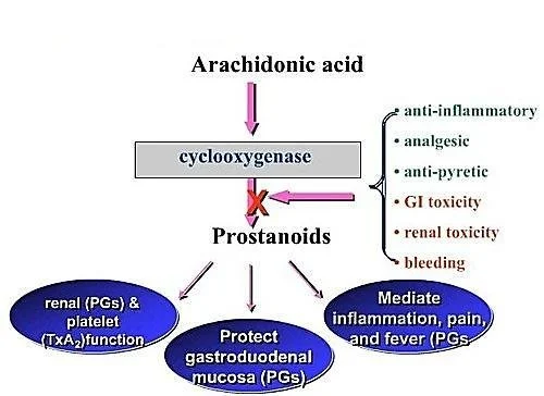
NSAIDS & Prostanoids NSAIDs & Prostanoids Pharmacology Module Overview This master guide covers the pharmacology of Non-Steroidal Anti-Inflammatory Drugs (NSAIDs) and Prostanoids. We will explore the Arachidonic Acid pathway, the profound differences between COX-1 and COX-2, specific drug classifications, and the synthetic prostanoids used to manipulate everything from childbirth to glaucoma. Enhanced with clinical scenarios and deep-dive explanations to guarantee exam success. 1. The Foundation: Prostanoids and the MOA of NSAIDs Before understanding the drugs, you must understand the assembly line that makes the molecules these drugs block. This is the Arachidonic Acid Pathway. Think of this pathway as a factory that takes raw materials from the cell wall and turns them into highly active chemical messengers. MEMBRANE PHOSPHOLIPIDS ↓Enzyme: Phospholipase A2 (BLOCKED by Corticosteroids)↓ ARACHIDONIC ACID ↙ Lipoxygenase ↓ Leukotrienes(Cause Bronchospasm/Asthma) ↘ Cyclooxygenase (COX)(BLOCKED by NSAIDs) ↓ PGG2 → PGH2(Endoperoxides) ↓ PROSTANOIDS:Prostaglandins (PGE2, PGF2α, PGD2)Thromboxane (TXA2)Prostacyclin (PGI2) Clinical Pearl Steroids vs. NSAIDs & The “Shunt” Phenomenon Notice that Corticosteroids block the pathway at the very top (Phospholipase). Therefore, steroids stop BOTH Leukotrienes (which cause asthma) and Prostanoids. NSAIDs only block the COX enzyme lower down. This means NSAIDs stop pain and fever (Prostanoids) but do nothing to stop Leukotrienes. In fact, in some asthma patients, giving an NSAID creates a “Leukotriene Shunt”. Because the COX pathway is blocked, all the built-up Arachidonic acid is violently pushed down the Lipoxygenase pathway, causing a massive overproduction of Leukotrienes. This triggers a severe, life-threatening asthma attack (a condition clinically known as Aspirin-Exacerbated Respiratory Disease or AERD). The Cyclooxygenase (COX) Isozymes: The “Housekeeper” vs. The “Fire Alarm” The COX enzyme comes in different versions (isoforms). Knowing the difference is the absolute key to understanding NSAID side effects and why pharmaceutical companies spent billions inventing specific COX-2 inhibitors. COX-1 (The Housekeeper) COX-2 (The Fire Alarm) COX-3 (The Mystery) Constitutive: Always active, working in the background 24/7. Responsible for physiologic production of prostanoids to regulate normal cellular processes. Gastric Cytoprotection: Makes protective stomach mucus and neutralizes stomach acid. Vascular Homeostasis & Platelet Aggregation: Balances blood flow and clotting. Kidney Function: Regulates and maintains renal blood flow. Inducible: Normally absent, but ramps up massively during emergencies (trauma, infection). Responsible for elevated production of prostanoids in disease states. Expression at sites greatly increases to cause Pain, Inflammation, and Fever. (Also expressed normally in brain, kidney, and bone). Predominantly has effects in the Central Nervous System (CNS). Often theorized to be the exact target of Acetaminophen (Paracetamol) which beautifully explains why it reduces fever/pain centrally in the brain but has absolutely no anti-inflammatory effect in the body’s tissues. 2. Classification of NSAIDs NSAIDs are classified either by their chemical structure/efficacy or by how selectively they block the COX enzymes. Classification by COX Selectivity (The Slide 4 Breakdown) Note on exam preparation: Some drugs straddle the line of selectivity based on dose. For example, Aspirin is selective for COX-1 at low doses, but non-selective at high doses. Selective COX-1 Inhibitors (Usually low doses): Low dose Aspirin, Ketoprofen, Flurbiprofen, Indomethacin, and Ketorolac (sometimes spelled ‘Ketoloid’ on older slides). Non-Selective COX Inhibitors (Traditional NSAIDs): Piroxicam, Tenoxicam, Ibuprofen, Naproxen, Diclofenac. These hit both COX-1 and COX-2 equally, killing pain but ruining the stomach. Selective COX-2 Inhibitors (The “-coxibs” & friends): Celecoxib, Etoricoxib, Meloxicam (preferential), Nimesulide. Designed to kill pain without giving you a stomach ulcer. Classification by Efficacy and Chemical Class (The Slide 5 Breakdown) Why do we care about chemical classes? Because if a patient is highly allergic or fails to respond to an NSAID from the “Propionic Acid” class, a wise doctor will switch them to a completely different chemical class, like an “Oxicam”. 1. Analgesic & Marked Anti-inflammatory Non-Selective COX Inhibitors (Traditional) Salicylic Acid Derivatives: Aspirin Propionic Acid Derivatives: Naproxen, Ibuprofen, Ketoprofen Pyrazolon Derivatives: Phenylbutazone Acetic Acid Derivatives: Diclofenac, Aceclofenac, Nebumetone, Sulindac Pyrrolo-pyrrole Derivatives: Ketorolac Indole Derivatives: Indomethacin Oxicams: Piroxicam, Tenoxicam 2. Analgesic & Moderate Anti-inflammatory Fenamates: Meclofenamic acid, Tolfenamic acid, Flufenamic acid Anthranilic acid: Mefenamic acid 3. Preferential & Selective COX-2 Preferential COX-2 Inhibitors: Meloxicam, Nimesulide. Selective COX-2 Inhibitors: Celecoxib, Etoricoxib. 4. Analgesics with POOR/NO Anti-inflammatory Para-aminophenol Derivatives: Acetaminophen/Paracetamol. 3. Mechanism of Action (MOA) and General Adverse Effects Primary MOA: NSAIDs inhibit the cyclooxygenase (COX) enzyme, resulting in the reduced biosynthesis of Prostanoids (Prostaglandins, Prostacyclin, and Thromboxane A2). Why do Traditional NSAIDs cause side effects? Aspirin and older, non-selective NSAIDs block BOTH COX-1 and COX-2. By blocking COX-2, they brilliantly stop inflammation, pain, and fever. BUT, by blocking COX-1, the release of PGs required for homeostatic (housekeeping) function is totally disrupted. The Mechanisms of Toxicity The Stomach: PGE2 and PGI2 normally stimulate the production of thick, protective gastric mucus and bicarbonate. They also maintain rich blood flow to the stomach wall. NSAIDs stop this synthesis. Result: The stomach acid literally burns through the unprotected stomach wall, causing Gastric and Duodenal Ulcers, and severe GI Bleeding. The Kidneys: PGE2 and PGI2 are responsible for actively dilating the afferent renal arteriole (the blood vessel bringing blood INTO the kidney filter), which maintains the Glomerular Filtration Rate (GFR). If you block this (especially in elderly patients with already impaired kidneys or low blood volume), blood flow to the kidney drops sharply.Result: Serious kidney damage, acute renal failure, and severe fluid retention. General Adverse Reactions of NSAIDs (System by System) Gastrointestinal Tract (Most Common): Nausea, vomiting, diarrhea, constipation, epigastric pain, indigestion, abdominal distress, intestinal ulceration, stomatitis, jaundice, bloating, anorexia, and dry mouth. Central Nervous System (CNS): Dizziness, headache, drowsiness, insomnia. Cardiovascular: Decrease or increase in blood pressure (often increasing it due to fluid retention), and cardiac arrhythmias. Renal: Hematuria (blood in urine) and acute renal failure (in those with pre-existing impaired function). Special Senses: Visual disturbances, blurred or diminished vision. Hematologic: Anemia (often secondary to chronic microscopic GI bleeding over months of daily NSAID use). 4. Deep Dive: Aspirin, Acetaminophen, and Selective COX-2s A. ASPIRIN (Acetylsalicylic Acid) Aspirin is completely unique among all NSAIDs. It irreversibly acetylates both isoforms of the COX enzyme. This means it covalently binds to the enzyme

Eicosanoids Pharmacology
Pharmacology

Eicosanoids Pharmacology

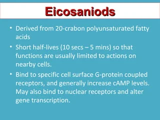
Autocoids — Eicosanoids Eicosanoids Pharmacology 1. Introduction to Eicosanoids Definition: Eicosanoids are biological signaling molecules (local hormones/autacoids) that are products of polyunsaturated long-chain fatty acids. The prefix “Eicosa-“ means 20 in Greek, because these molecules are almost entirely derived from 20-carbon essential fatty acids, most commonly Arachidonic Acid. Hormones vs. Eicosanoids (The “Global Email” vs. “Sticky Note” Analogy) Unlike regular hormones (like insulin) which are stored in glands and travel globally through the blood, eicosanoids are not stored. They are highly unstable and have a half-life of seconds to minutes. Therefore, they are synthesized on demand from cell membrane lipids and act locally right where they are made (paracrine action on neighbors, or autocrine action on themselves). Major Classifications Eicosanoids are divided into families based on the specific enzyme that creates them from the raw material: a) Cyclooxygenase (COX) derivatives: These include the Prostaglandins (PGs) and Thromboxane (TXA2). b) Lipoxygenase (LOX) products: These include the Leukotrienes (LTs) and Lipoxins. c) Cytochrome P450 (CYP) Epoxyoxygenase pathway: Produces EETs (Epoxyeicosatrienoic acids). 2. The Synthesis Cascade (The Arachidonic Acid Pathway) To understand the drugs, you MUST understand how eicosanoids are made. Picture a cell membrane. The lipids in that membrane hold the raw material (Arachidonic Acid) locked away safely. STEP 1: THE RELEASE Cell Membrane Phospholipids (Diacylglycerol or Phospholipid) ↓ Enzyme: Phospholipase A2 (PLA2) (or Phospholipase C) Arachidonic Acid (Free and active) Exam Gold: The Corticosteroid Blockade Exam Note: Corticosteroids (like Prednisone or Dexamethasone) stimulate the production of a protein called Annexin A1 (also known as Lipocortin-1), which completely blocks Phospholipase A2. This shuts down the ENTIRE cascade right at the top. No Arachidonic Acid means no prostaglandins and no leukotrienes. This is exactly why steroids are such incredibly powerful, broad-spectrum anti-inflammatories compared to NSAIDs! Once Arachidonic Acid is free, it acts as a crossroads and can go down one of three enzymatic paths: Path A: The COX Pathway Arachidonic Acid + COX-1 or COX-2 (PGH2 Synthase / Peroxidase) → PGG2 → Prostaglandin H2 (PGH2). PGH2 is the unstable “parent” molecule. Depending on the specific tissue enzymes present, PGH2 becomes: Prostaglandins: PGE2, PGF2α, PGD2. Prostacyclin (PGI2): Synthesized via Prostacyclin synthase (primarily in vascular endothelium). Thromboxane (TXA2): Synthesized via Thromboxane synthase (primarily in platelets). Path B: The LOX Pathway Arachidonic Acid + 5-LOX (Lipooxygenase + FLAP protein) → 5-HPETE. 5-HPETE becomes: Leukotrienes: LTA4 → LTB4, LTC4, LTD4, LTE4. HETEs: (e.g., 8-HETE, 12-HETE, 15-HETE) – play crucial roles in inflammation and immune cell recruitment. Path C: Cytochrome P450 Pathway Arachidonic Acid + CYP Epoxygenases → EETs. These play a role in maintaining vascular tone (vasodilation), renal function, and overall cardiovascular protection. 3. Mechanism of Action and Receptors Eicosanoids do not enter cells. They bind to cell surface receptors that are all coupled to G-proteins (GPCRs). Crucial Second Messenger Mechanisms You must know whether they cause relaxation or contraction at the cellular level (tying back to your signaling lectures!): Relaxers (PGI2 and PGE2): Link to Gs proteins. Increase Adenylyl Cyclase → Increases cAMP → Decreases intracellular Calcium (Ca++). Result: Smooth muscle relaxation and Vasodilation. Contractors (TXA2, PGF2α): Link to Gq proteins. Activate Phospholipase C → Increases IP3 → Increases intracellular Calcium (Ca++). Result: Smooth muscle contraction, Vasoconstriction, and Platelet Aggregation. 4. Physiological & Pharmacologic Effects by System This is where the exam will test your clinical application. Memorize these specific receptor actions: A. The Vasculature (Blood Vessels) PGEs (PGE1, PGE2): Potent vasodilators. Prostacyclin (PGI2): Potent vasodilator. Can produce profound hypotension (low blood pressure). Thromboxane A2 (TXA2): Potent vasoconstrictor. Leukotrienes (LTC4, LTD4): Cause massive capillary leakiness (vascular permeability), contributing heavily to the swelling (edema) seen in severe inflammation. **Alprostadil (PGE1): Specifically dilates the ductus arteriosus in neonates. B. Platelets (The Blood Clotting Tug-of-War) There is a constant balance (a “see-saw”) in your blood between two eicosanoids to prevent you from bleeding out or forming fatal clots: Prostacyclin (PGI2): Produced by healthy blood vessel walls. It INHIBITS platelet aggregation. (Mnemonic: Prostacyclin keeps blood CYCLING smoothly). Thromboxane A2 (TXA2): Produced by platelets. It is a massive platelet activator/aggregator. (Mnemonic: Thromboxane causes THROMBI / clots). Inflammation (Leukocytes): LTB4 is a powerful chemotactic agent (it acts as a chemical beacon, attracting eosinophils, monocytes, and neutrophils to the site of injury). Conversely, prostaglandins generally inhibit cellular and humoral immunity to keep the immune system from overreacting. C. The Lungs (Bronchial Tone) Prostaglandins: Have mixed effects on bronchial muscle (PGE1/PGE2 cause bronchodilation, PGD2/PGF2α cause constriction). TXA2: Causes bronchoconstriction. Inhibitors of thromboxane will therefore reduce the bronchoconstrictive response. Leukotrienes (LTC4, LTD4): Extremely potent bronchoconstrictors. These are the main culprits in deadly asthma attacks! D. The Uterus (Obstetrics) PGE2 and PGF2α: Cause powerful uterine contractions, especially in a pregnant uterus. Clinical Tie-In (Dysmenorrhea): Overproduction of PGE2 and PGF2α during menstruation causes severe uterine cramping (primary dysmenorrhea). This is why taking an NSAID (which blocks these prostaglandins) cures menstrual cramps! Clinically, synthetic versions are used as abortifacients (to induce medical abortions) or to induce labor at term. E. Gastrointestinal Tract (GIT) PGEs and PGI2: Inhibit gastric acid secretion (which is normally stimulated by feeding, histamine, or gastrin). They act as a shield, promoting the maintenance of the gastric mucosa by stimulating heavy mucus and bicarbonate secretion. Clinical Tie-In: This is exactly why taking NSAIDs (which block PGE production) causes stomach ulcers! You strip away the stomach’s protective mucus shield. F. The Kidneys PGE2 and PGI2: Cause renal vasodilation (specifically of the afferent arteriole), increase Renal Blood Flow (RBF), increase GFR, and promote diuresis (water excretion). (If a patient takes too many NSAIDs, they lose this vasodilation, the kidney starves of blood, leading to Acute Kidney Injury). TXA2: Causes renal vasoconstriction and has an ADH-like action (retains water). G. Central Nervous System (CNS) & Eye CNS: PGE2 is the primary mediator of Fever, Pain perception, and Sleep. When a virus attacks you, the brain generates PGE2 to reset the hypothalamus thermostat, causing fever. Eye: PGF2α regulates the outflow of aqueous humor. 5. Clinical Pharmacology: Uses of Prostanoids and Analogues In pharmacology, we create synthetic versions (analogs)

Serotonin Pharmacology
Pharmacology

Serotonin Pharmacology

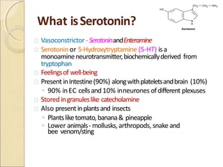
Autocoids — Serotonin Serotonin & Migraine Pharmacology 1. Brief Recap: What are Autacoids? Before diving into Serotonin, remember the baseline definition from the start of the lecture. Autacoids are the body’s local communication network. Definition: Endogenous substances (made in the body) that act as biological factors or “local hormones”. (Greek: Autos = self, Akos = remedy). Characteristics: Present in very small amounts, have distinct biological activity, are short-living with a short duration of action, and act at or very close to their site of release. Systemic Effect: Although they are “local”, if produced in massive amounts, they can enter the circulation and cause whole-body (systemic) effects. Functions: They regulate physiological baselines, mediate pathophysiological reactions to injuries (like inflammation), and modulate nerve transmission. Analogy Endocrine Hormones vs. Autacoids Think of standard Endocrine Hormones (like insulin or thyroid hormone) as a company-wide email broadcast. They travel through the main server (the bloodstream) to reach every department in the body. In contrast, Autacoids are like sticky notes left on a coworker’s desk. They are meant only for the immediate neighbor (local action) and are thrown away quickly (short duration of action). Chemical Classification of Autacoids Autacoids are classified into four main families based on their chemical structure: 1. Amines Histamine, Serotonin (5-HT). 2. Polypeptides Kinins, Oxytocin, Angiotensin, Vasopressin, Endothelins. 3. Fatty Acids Prostaglandins, Leukotrienes, Thromboxanes, PAF (Platelet Activating Factor). 4. Others Nitric Oxide (NO), Cytokines. 2. Serotonin (5-HT): Synthesis and Metabolism Serotonin, chemically known as 5-hydroxytryptamine (5-HT), is an indoleethylamine. It is widely distributed in nature—found in plants (like bananas and pineapples), animal tissues, venoms, and insect stings. A. The Synthesis Pathway Serotonin is built from the amino acid L-tryptophan. This is a critical two-step process: L-Tryptophan &downarrow; (Enzyme: Tryptophan Hydroxylase) — *Rate Limiting Step* 5-Hydroxytryptophan (5-HTP) &downarrow; (Enzyme: Decarboxylase) 5-Hydroxytryptamine (Serotonin / 5-HT) The Rate-Limiting Step: Hydroxylation at the C5 position is the bottleneck of the whole process. The body can only make Serotonin as fast as Tryptophan Hydroxylase works. Experimental Blockers: You can chemically block this rate-limiting step using drugs like p-chlorophenylalanine (PCPA / fenclonine) and p-chloroamphetamine. Experimentally, these were used to reduce serotonin in carcinoid syndrome, but they are too toxic for clinical human use. B. Inactivation and Metabolism Once Serotonin does its job, it must be rapidly inactivated so it doesn’t continuously overstimulate the body. It is metabolized primarily by the enzyme Monoamine Oxidase (MAO). Serotonin (5-HT) &downarrow; (Enzyme: MAO) 5-hydroxyindoleacetaldehyde &downarrow; (Enzyme: Aldehyde Dehydrogenase) 5-HIAA (5-hydroxyindoleacetic acid) — *The Principal Metabolite* Exam Trap! The Carcinoid Tumor Diagnostic Test Clinical Scenario: A patient presents with severe flushing, severe diarrhea, and right-sided heart valve issues. You suspect a Carcinoid Tumor (a rare gut tumor that secretes massive amounts of serotonin). The Test: You measure the 24-hour urinary excretion of 5-HIAA (the final breakdown product). High 5-HIAA confirms massive serotonin synthesis. The Trap: Before the test, you MUST prohibit the patient from eating foods rich in serotonin or tryptophan (e.g., Bananas, Pineapples, Plums). If they eat a bunch of bananas before the test, their body will metabolize that dietary serotonin, their urine 5-HIAA will skyrocket, giving a false positive for a tumor! Clinical Scenario: MAO Inhibitors & Serotonin Syndrome If a patient is taking a drug that blocks Monoamine Oxidase (an MAOI antidepressant like Phenelzine), the serotonin cannot be broken down. If this patient then takes another drug that increases serotonin (like an SSRI or MDMA/Ecstasy), serotonin builds up to lethal levels. This causes Serotonin Syndrome: hyperthermia, muscle rigidity, tremors, and potentially death. 3. Storage, Release, and Locations of 5-HT Where is Serotonin found in Mammals? The Gut (90%): Over 90% of all serotonin in the human body is located in the enterochromaffin cells of the gastrointestinal tract. (Deep Explanation: This is why SSRI antidepressants, which increase active serotonin everywhere, almost always cause GI upset, nausea, and diarrhea in the first week of use! The gut has far more serotonin receptors than the brain). The Blood (Platelets): Serotonin floats in the blood stored safely inside platelets. Platelets don’t make serotonin; they suck it up from the plasma using an active Serotonin Transporter (SERT). (Why? When you get cut, platelets clump together and release serotonin to cause local vasoconstriction, stopping the bleeding!). The Central Nervous System (Nerve Endings): Found heavily in the raphe nuclei of the brainstem. These neurons synthesize, store, and release 5-HT as a true neurotransmitter controlling mood and sleep. The Pineal Gland: Here, serotonin serves as a precursor. An enzyme (Hydroxyindole-O-methyltransferase) converts serotonin into Melatonin, the hormone that induces sleep. How is it Stored? Whether in a nerve ending or a platelet, serotonin is pumped into protective storage vesicles by a pump called the Vesicle-Associated Transporter (VAT). Pharmacological Blockade: The drug Reserpine completely blocks VAT. If serotonin cannot get into the protective vesicle, it is left out in the open and is destroyed by MAO in the cytoplasm. Therefore, Reserpine severely depletes stored serotonin (just like it depletes catecholamines), which historically caused severe, suicidal depression in patients taking it for high blood pressure. 4. Physiological Actions of Serotonin System Specific Actions of 5-HT Central Nervous System (CNS) Affects mood, sleep, appetite, temperature regulation, pain perception, blood pressure, and vomiting. Deficiency: Causes depression, anxiety, migraines. Neuroendocrine: Controls hypothalamic cells releasing anterior pituitary hormones. Gastrointestinal (GI) Causes intense rhythmic contractions of the small intestines (via 5-HT4). Stimulates vomiting via the 5-HT3 receptors on vagal nerves. Cardiovascular System Potent contraction of smooth muscle (via 5-HT2), causing constriction of veins. Exception: It does not contract skeletal muscle or heart muscle. Triggers Platelet aggregation (clotting) via 5-HT2. Respiratory System Causes mild stimulation in healthy lungs, but triggers severe bronchoconstriction in asthmatics (via 5-HT2 in smooth muscles). (Explanation: Asthmatic airways are hyper-reactive to autacoids. Even a tiny bit of serotonin can trigger an asthma attack). 5. Serotonin Receptors (The Pharmacology Targets) There are at least 15 types and subtypes of serotonin receptors. You must memorize the mechanisms of the main ones: Crucial Mechanism Trap Receptors 1 through 6 are all G-protein coupled receptors (GPCRs).

Histamine Pharmacology
Pharmacology

Histamine Pharmacology

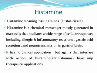
Autocoids — Histamine Histamine Pharmacology 1. Introduction to Autacoids What is an Autacoid? The term comes from the Greek words Autos (meaning “self”) and Akos (meaning “medicinal agent” or “remedy”). Therefore, an autacoid is literally a “self-remedy.” By definition, Autacoids are endogenous substances (made naturally inside the body) that act as biological factors or “local hormones”. Exam Trap: Autacoids vs. Classic Hormones A classic hormone (like insulin or thyroid hormone) is produced in a specific, centralized gland, dumped into the systemic bloodstream, and travels a long distance to reach its target organ. Autacoids are DIFFERENT: They are produced by widely distributed tissues all over the body, not a single gland. They act locally (at or very close to their exact site of synthesis and release). They are present in very small amounts. They have a short lifespan with a very short duration of action (they are rapidly destroyed to prevent them from causing systemic chaos). Note: However, if produced in massive, pathological amounts (like during severe anaphylactic shock), they can overcome local destruction, enter the systemic circulation, and have life-threatening systemic effects. Classification & Examples of Autacoids You must know the chemical classification of the different autacoids. Exam questions frequently mix these up: Chemical Class Examples Amines Histamine, Serotonin (5-HT) Polypeptides (Proteins) Kinins (Bradykinin), Oxytocin, Angiotensin, Vasopressin, Endothelins Fatty Acids (Eicosanoids) Prostaglandins, Leukotrienes, Thromboxanes, Platelet Activating Factor (PAF) Others Nitric Oxide (NO – Endothelium-derived relaxing factor), Cytokines 2. Histamine: Synthesis, Storage, and Metabolism Histamine is a ubiquitous molecule. It is present everywhere: in bacteria, plants, animals, and notably in venoms and stinging fluids (like bee stings, wasp venom, or stinging nettle plants). Chemistry & Synthesis Chemistry: It is a basic amine, specifically a β-aminoethylimidazole. Synthesis: The amino acid L-Histidine undergoes decarboxylation (the chemical removal of a CO2 molecule) to become Histamine. The specific enzyme that performs this action is L-Histidine decarboxylase. Inactivation & Metabolism Because histamine is so incredibly potent, it must be deactivated rapidly if it isn’t safely stored away. There are two major metabolic pathways the body uses to break it down and excrete it in the urine: Pathway 1 (Methylation): Conversion to N-methylhistamine (via the enzyme N-methyl transferase), which is then oxidized by MAO (Monoamine Oxidase) / DAO into methylimidazoleacetic acid. Pathway 2 (Oxidation): Direct conversion by the enzyme Diamine Oxidase (DAO) into imidazoleacetic acid (IAA). 3. Histamine Storage and Release Mechanisms Where is histamine kept? In humans, it is mostly stored inside Mast Cells (found abundantly in tissues interfacing with the outside world like Skin, Lungs, and GI tract) and Basophils (circulating in the blood). Inside these cells, histamine is locked up in granules, tightly bound to a heparin-protein complex so it doesn’t leak out. Histamine can be released in two distinct ways: Immunologic (Antigen-mediated) and Non-Immunologic. A. Immunologic Release (Antigen-Mediated) This is the classic Type I Hypersensitivity (Immediate Allergic Reaction). The Process: A person is exposed to an allergen (e.g., pollen, peanuts). Their immune system mistakenly creates IgE antibodies against it. These IgE antibodies attach to the surface of mast cells (a process called sensitizing the cell). Upon a second exposure to the same pollen, the allergen physically bridges and cross-links the IgE antibodies on the mast cell surface. The Result: The mast cell degranulates “explosively”, dumping massive amounts of histamine into the tissue. This specific process is energy-dependent (requires ATP) and requires calcium. Crucial Physiological Concept Negative Feedback & The Lung Exception In skin mast cells and blood basophils, the released histamine eventually binds back onto its own H2 receptors located on the mast cell’s own surface. This acts as a biological “brakes” system, inhibiting further histamine release (Negative Feedback). EXAM EXCEPTION: This feedback inhibition does NOT occur in lung mast cells! This is exactly why allergic asthma attacks in the lungs can spiral out of control so rapidly and become fatal; there are no built-in brakes to stop the continuous histamine release in the bronchioles. B. Non-Antigen Mediated Release This release mechanism does not require the immune system to be sensitized with IgE. It happens through direct physical or chemical interaction. Chemical Release: Certain drugs and chemicals can physically enter the mast cell and displace histamine from its heparin complex, forcing it out. Examples: Morphine, Tubocurarine (neuromuscular blocker), radiocontrast media (used in CT scans), amides, alkaloids, and basic polypeptides (like wasp/bee venoms). Mechanical Release: Physical trauma forces the mast cells to burst open. Examples: Vigorous scratching of the skin, severe burns, or crushing injuries. Cellular Proliferation: Pathological overgrowth of cells naturally increases total body histamine levels simply because there are more cells making it. Examples: Leukemia, Gastric Carcinoid Tumors. Physical Stimuli: Extreme cold, excessive heat, or exposure to bacterial toxins. Clinical Scenario “Red Man Syndrome” & IV Morphine The Event: If a nurse pushes an intravenous dose of Morphine too fast, the patient may suddenly flush bright red, feel intensely hot, become incredibly itchy, and their blood pressure might drop precipitously. The Mechanism: This is frequently mistaken for an allergy. It is not a true allergy (no IgE is involved). The rapid bolus of morphine chemically displaced histamine from the patient’s mast cells all at once, causing sudden, massive vasodilation. This is a classic example of Non-Antigen Mediated Chemical Release. The Fix: Stop the infusion, administer an antihistamine (like Diphenhydramine), and when restarting, push the morphine much slower. 4. Sites of Histamine Action Histamine regulates multiple physiological systems beyond just making you sneeze: Mast Cells & Basophils: Triggers standard inflammation and allergy symptoms (Skin itching, Lung wheezing, GIT cramping). Central Nervous System (CNS): Acts as a critical neurotransmitter, keeping the brain awake and alert. Neuroendocrine: Regulates hormones. It stimulates the release of ACTH, Prolactin (PRL), Vasopressin (VP), Oxytocin, and LH. It inhibits the release of GH and TSH. Thermal & Cardio: Causes hyperthermia (feverish feeling) via H1/H3 receptors located in the preoptic nucleus of the hypothalamus. Body Weight & Sleep: Acts as a powerful appetite suppressant (via H1), potentiates the hormone leptin (causing weight loss signaling), accelerates lipolysis (fat breakdown), and regulates

Scroll to Top