Doctors Revision

Microbiology

Infective Endocarditis (IE)
Microbiology

Infective Endocarditis (IE)

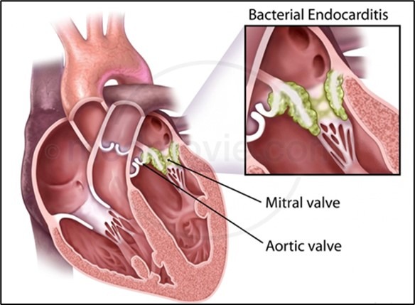
Infective Endocarditis (IE) Module Overview This master guide covers the complete pathophysiology, microbiology, clinical presentation, and management of Infective Endocarditis. As requested, the core lecture material is completely intact, heavily expanded with deeper clinical correlations, vivid analogies, and practical exam scenarios to ensure absolute mastery of the topic. 1. What is Infective Endocarditis? (The Basics) Definition: Infective endocarditis (IE) is an infection of the endocardial surface of the heart. The endocardium is the smooth, innermost lining of the heart chambers and the heart valves. The term “infective” implies the physical, active presence of microorganisms (bacteria or fungi) invading this lining, resulting in a physical mass called a vegetation or lesion. Deep Dive: What exactly is a vegetation? It is a bulky, deadly clump made of three things: host platelets, host fibrin (clotting material), and massive colonies of living microorganisms. Because it is physically attached to the heart, it moves with every heartbeat. Where does it happen? Heart Valves: This is the most common site. The valves (Mitral, Aortic, Tricuspid, Pulmonary) are constantly opening and closing, dealing with high-pressure blood flow. If they get damaged, they are prime targets for infection. Septal Defects: If a patient has a hole in their heart (like a Ventricular Septal Defect – VSD), the blood aggressively shoots through that hole. This high-velocity “jet stream” damages the surrounding tissue on the opposite side of the hole, creating a rough, scarred patch where circulating bacteria can easily latch on. Mural Endocardium: This refers to the flat muscular walls of the heart chambers. Though less common than valves, infections can happen here if the wall is damaged by turbulent blood flow or foreign devices (like pacemaker wires rubbing against the tissue). 2. Anatomy Recap: The Valves of the Heart To understand IE, you must deeply understand your valves and the path of blood flow. The heart has four main doors (valves) that keep blood flowing in one direction. Right Side of the Heart Pumps deoxygenated blood to the Lungs. Tricuspid Valve: Between the right atrium and right ventricle. Clinical Pearl: This valve is most commonly infected in Intravenous (IV) Drug Users. Why? Because when dirty needles inject bacteria into a vein, the venous blood travels directly into the right side of the heart first. The tricuspid valve is the first door the bacteria hit. Pulmonary Valve: Between the right ventricle and the pulmonary trunk. (Rarely infected). Left Side of the Heart Pumps oxygenated blood to the entire Body. Mitral Valve: Between the left atrium and left ventricle. Aortic Valve: Between the left ventricle and the aorta. Note: Left-sided valves are under much higher pressure (working against systemic blood pressure) and experience more wear and tear. Therefore, they are the most common sites for IE in the general, non-IV-drug-using population. 3. Classification of IE: Acute vs. Subacute/Chronic Historically, IE was divided based on how fast it killed the patient if left untreated. This is a crucial distinction for exams as it dictates both the causative bug and the clinical presentation. A. Acute Infective Endocarditis Analogy: Think of this as a rapid, violent home invasion. It happens incredibly fast and causes massive destruction. Progression: Follows a fulminant (severe and sudden) course. Death can occur in several days to less than 6 weeks without treatment. Clinical Picture: The patient looks incredibly sick. They will have a high-spiking fever, systemic toxicity (sepsis, dangerously low blood pressure, rapid heart rate), and severe leukocytosis (massively elevated white blood cell count). Valve Status: Because the bugs are so aggressive, Acute IE can attack and completely destroy perfectly normal, healthy heart valves. Causative Agents (The Aggressive Bugs): Staphylococcus aureus (The #1 cause of acute IE. Known for being highly destructive, producing enzymes that liquefy tissue). Streptococcus pyogenes (Group A Strep). Streptococcus pneumoniae. Neisseria gonorrhoeae (Rare today due to antibiotics, but a classic board-exam historical cause). Clinical Scenario – Acute IE A 28-year-old male with a history of heroin use is brought to the ER by his friends. He has a fever of 104°F (40°C), shaking chills, and looks extremely toxic and confused. When you listen to his heart, you hear a loud, brand-new systolic murmur that wasn’t there last week. His right-sided tricuspid valve is being rapidly chewed apart by Staphylococcus aureus introduced via a dirty needle. B. Subacute and Chronic Infective Endocarditis Analogy: Think of this as termites slowly eating away at a house over months. It is insidious, sneaky, and gradual. Progression: Subacute leads to death in 6 weeks to 3 months. Chronic takes longer than 3 months. Valve Status: These bugs are weak. They usually CANNOT infect a healthy valve. They almost always occur in the setting of prior valvular disease (e.g., a patient with a history of rheumatic fever, a congenital bicuspid aortic valve, or mitral valve prolapse). Clinical Picture: Slow, indolent (lazy/painless) course. Symptoms are vague: low-grade fever (maybe 99.5°F – 100.5°F), heavy night sweats, unexplained weight loss, generalized fatigue, and “vague systemic complaints” (the patient just feels “off” for weeks). Causative Agents (The Sneaky Bugs): Viridans streptococci (These naturally live in your mouth and throat. They are less aggressive but will happily settle on a previously damaged valve because they produce dextrans that bind perfectly to scarred tissue). Clinical Scenario – Subacute IE A 65-year-old woman with a known childhood history of rheumatic fever (which permanently scarred her mitral valve) visits her dentist for a deep tooth extraction. Two months later, she goes to her primary care doctor complaining of severe fatigue, losing 10 pounds without trying, and waking up drenched in night sweats. Blood cultures reveal Viridans streptococci. The bacteria entered her bloodstream during the dental work and slowly grew on her old, scarred heart valve over the last 8 weeks. 4. Pathogenesis (How exactly does the vegetation form?) This is a step-by-step process of how bacteria build a fortress on a heart valve. Examiners love testing these steps because it explains why treatment is so difficult. Endothelial Damage (The Scratch): The smooth valve surface must first be
Genital Tract Infections (GTIs)
Microbiology

Genital Tract Infections (GTIs)

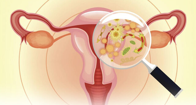
Genital Tract Infections (GTIs) Introduction & Broad Overview Genital Tract Infections (GTIs) encompass a wide variety of diseases affecting the reproductive organs of both males and females. Because the reproductive tract is deeply connected to the urinary system (especially in males) and the abdominal cavity (in females via the fallopian tubes), these infections can range from mild local irritations to life-threatening systemic diseases. Public Health Context: GTIs are heavily tested in exams because of their massive global burden. If poorly managed, they lead to devastating consequences such as irreversible infertility, chronic pelvic pain, ectopic pregnancies, and a severely increased risk of transmitting/acquiring HIV (due to open genital ulcers and mucosal inflammation). The Three Main Sources of Genital Infections (Aetiology) To diagnose and treat a GTI, you must first know where it came from. Infections generally fall into three specific categories: Category Where They Come From How They Spread (Pathogenesis) Common Clinical Examples 1. Endogenous Infections Organisms that are normally found in the vagina (Normal Flora/Microbiome). Usually not spread from person to person. They occur when the normal environment (pH or bacterial balance) is disrupted, allowing naturally occurring microbes to overgrow. (Exam tip: Broad-spectrum antibiotic use kills the “good” Lactobacilli, causing the vaginal pH to rise. This creates a perfect environment for dormant yeast or bad bacteria to suddenly multiply and attack). Yeast infections (Candidiasis), Bacterial Vaginosis (BV). 2. Sexually Transmitted Infections (STIs) Acquired from an infected sexual partner. Direct sexual contact (vaginal, anal, or oral) with infected mucosal surfaces or fluids. (Note: Many individuals act as “asymptomatic carriers,” meaning they spread the disease without ever showing visible signs). Gonorrhoea, Chlamydia, Syphilis, Chancroid, Trichomoniasis, Genital Herpes (HSV), Genital Warts (HPV), HIV. 3. Iatrogenic Infections Introduced from outside the body, or normal flora pushed into sterile areas by a medical procedure. Occurs during medical interventions. Examples: Pushing vaginal bacteria into the sterile uterus during IUD insertion, childbirth, abortions, or using unsterile/contaminated instruments (like uterine sounds or speculums). Poor infection control in the clinic is the primary culprit. Pelvic Inflammatory Disease (PID) following a procedure, postpartum sepsis, post-abortion infections. General Pathogenesis: How Do Pathogens Invade? Pathogens have different strategies for attacking the host. In an exam, you may be asked to classify an organism by its mode of spread: Local Invasion: The pathogen attacks the exact spot it touches, invading the skin and mucous membranes to cause local sores or ulcers. Examples: Treponema pallidum (causes chancre), Haemophilus ducreyi (causes chancroid), Herpes Simplex Virus (causes vesicles). Scenario: A patient notices a painful, fluid-filled blister on their genitalia a few days after unprotected sex. The virus hasn’t gone to the brain or heart; it is locally destroying the skin cells. Systemic Dissemination (Bloodstream): The pathogen enters locally but uses the bloodstream to travel to distant organs. Examples: Treponema pallidum (Secondary and Tertiary Syphilis), HIV. Scenario: Months after an initial painless genital ulcer healed, a patient suddenly develops a full-body rash, including on the palms of their hands and soles of their feet. The bacteria (Treponema) have used the blood to travel everywhere. Ascending Infection: The pathogen enters at the bottom (urethra or cervix) and physically climbs up the mucosal lining into the sterile upper tract (uterus, tubes). Examples: Neisseria gonorrhoeae, Chlamydia trachomatis. Mechanism: Bacteria use physical structures like “pili” (grappling hooks) to pull themselves up the mucosal walls against the downward flow of gravity and mucus. Vertical Transmission (Maternal-Fetal): Infants born through an infected vaginal canal can swallow or get pathogens in their eyes. Examples: Neonatal conjunctivitis from Gonorrhea or Chlamydia. Clinical Decision: If a mother has an active Genital Herpes (HSV) outbreak with visible blisters during labor, a Cesarean section (C-section) is heavily indicated to prevent the baby from catching a fatal brain infection (HSV encephalitis) on the way out. Anatomy of GTIs: Lower vs. Upper Tract A. Lower Reproductive Tract Infections These affect the vulva, vagina, and cervix. If left untreated, they act as the gateway to the upper tract. 1. Vaginitis (Vulvo-vaginitis) This is inflammation of the vagina, often presenting with painful irritation, itching, and abnormal discharge. It is commonly facilitated by disruptive events like abortions, IUD insertions, or menstrual regulation. 🔥 HIGH YIELD EXAM COMPARISON The Causes of Vaginitis Memorize this table. You will almost certainly be tested on how to differentiate these three under a microscope, via pH, and via clinical symptoms. Condition Clinical Signs (Discharge) Microscopy / Lab Tests Vaginal pH (Normal is 3.0 – 4.5) Candida (Yeast)Endogenous Abnormal, thick, white curd-like (cottage cheese) discharge. Extreme itching (pruritus). Fungi (hyphae/spores) visible on a wet prep slide treated with 10% Potassium Hydroxide (KOH). (KOH dissolves the human cells but leaves the tough fungal walls intact for easy viewing). pH is normal (< 4.5) Bacterial Vaginosis (BV)Endogenous Thin, grayish, homogeneous discharge. The Amsel Criteria (Need 3 of 4 for diagnosis): 1. Homogeneous discharge. 2. “Clue cells” on microscopy (>20%). (These are vaginal epithelial cells completely coated in tiny bacteria, making them look like a fried egg covered in black pepper). 3. pH > 4.5. 4. Positive “Whiff Test”: A sharp “fishy” amine odor is produced when 10% KOH is added to the secretions. pH is elevated (> 4.5) Trichomonas InfectionSTI (Protozoa) Frothy, greenish-yellow, purulent discharge. Often accompanied by a “Strawberry Cervix” (tiny red hemorrhages on the cervix). Motile, bi-flagellated trichomonads darting around on a wet mount microscopy. pH is elevated (> 4.5) 2. Cervical Infections (Cervicitis) The cervix is the neck of the uterus. Infections here are most classically caused by the two major ascending STIs: Chlamydia and Gonorrhea. Cervicitis is often completely asymptomatic in women, making it a highly dangerous silent carrier of disease that can be unknowingly passed to partners or ascending to the uterus. B. Upper Reproductive Tract Infections These affect the normally sterile environments of the Uterus, Fallopian tubes, and Ovaries. Pathogenesis: Usually occurs as a direct complication of untreated lower tract STIs (Gonorrhea and Chlamydia ascending past the cervix). Pelvic Inflammatory Disease (PID): A severe, painful inflammation of the upper pelvic organs. Patients present with

Gastro-intestinal Infections (1)
Microbiology

Gastro-intestinal Infections

Gastro-intestinal Infections (GIs) Gastrointestinal Infections 1. Introduction to Gastrointestinal Infections Gastrointestinal infections are diseases that primarily affect the stomach and intestines. When we talk about these infections, we usually use the term Gastroenteritis. Definition Definition of Gastroenteritis: It is a syndrome of diarrhea and/or vomiting that involves the upper small bowel or the colon. The Exception: Helicobacter pylori (which causes gastritis and stomach ulcers) is NOT classified under gastroenteritis. This is a common trick question on exams! Why is this important? These are among the most debilitating infectious diseases across all age groups. In heavily populated (often developing) areas, the number of deaths from diarrheal diseases exceeds deaths from almost all other causes. How do we know it’s infectious? Even before doctors find the exact bacteria or virus under a microscope, they suspect an infectious cause because of three epidemiological clues: Case clustering: Many people in the same area get sick at the same time. Group spread: It spreads rapidly within families, daycares, or dormitories. Traveler’s Diarrhea: People get sick after traveling to new regions. The Global Scope and Burden Childhood Mortality: Globally, diarrheal diseases are a leading cause of death in children. Long-term Morbidity (Illness): Repeated GI infections impact a child’s growth and development because they cause malabsorption (the gut cannot absorb nutrients) and malnutrition. The Vicious Cycle: Acute infectious diarrhea makes nutritional deficiencies much worse. Why? Because being sick increases the body’s caloric demands and causes the breakdown of structural proteins in the body. Conversely, a child who is already undernourished has lower resistance and is more likely to catch acute infectious diarrhea. Persistent Diarrhea: If diarrhea lasts more than 14 days, it is classified as persistent and is strongly associated with poor nutrition. Community Impact: Acute gastroenteritis is the second most common illness in the community (right behind respiratory infections like the common cold), leading to frequent doctor visits and medication use. 2. Epidemiologic and Environmental Factors (Who, Where, When) The frequency, type, and severity of an enteric (gut) infection depend on three main things: WHO you are (Host Risk): Risk varies greatly based on age (infants and elderly are most vulnerable), living conditions (sanitation, crowding), personal and cultural habits (handwashing, food preparation), and group exposures (eating at a buffet). WHERE you are (Geography & Climate): The types of bugs that cause illness vary by climate. Tropics (Developing nations): ETEC (Enterotoxigenic E. coli), EPEC (Enteropathogenic E. coli), and heavy burdens of parasites are the main culprits. Temperate Zones (Developed nations like Japan, N. America, Europe): EHEC (Enterohemorrhagic E. coli) is a major problem here. Viral causes (like Rotavirus/Norovirus) are universal and affect young children in both temperate and tropical climates. WHEN you are there (Seasonality): Temperate climates: Enteric illnesses peak during the winter months (mostly viral). Tropical climates: Illnesses peak during the summer months (mostly bacterial, as bacteria multiply rapidly in warm weather). 3. Host vs. Microbial Factors A. HOST FACTORS (What protects us or makes us vulnerable?) Your body has several defense mechanisms. When these fail, infection occurs. Species, Genotype, and Age: Some people are genetically more susceptible. Very young and very old people have weaker immune systems. Personal Hygiene: Handwashing is critical. Infective Dose: This is how many bacteria you need to swallow to actually get sick. Shigella: Highly virulent! You only need to ingest 10 to 100 organisms to get dysentery. Salmonella: Less virulent. You need to ingest 100,000 or more organisms to get sick. Gastric Acidity (The Stomach Acid Barrier): This is your first line of defense. A normal stomach pH of less than 4 will kill most swallowed organisms within 30 minutes. If a patient is taking antacids (like Omeprazole), their pH goes up, making them highly susceptible to infections! Intestinal Motility: Normal bowel movements constantly “flush” bacteria out. If motility is slow, bacteria can overgrow. Enteric Microflora: Your “good bacteria” compete with bad bacteria for space and food, preventing infection. Immunity: Phagocytic (white blood cells eating bugs), Humoral (antibodies like IgA in the gut), and Cell-mediated immunity. Human Milk: Breast milk contains non-specific protective factors and maternal antibodies that protect infants. Intestinal Receptors: Some bugs only infect you if you have the specific cellular receptors they need to attach to. B. MICROBIAL FACTORS (How the bugs attack us) 1. TOXINS Many bacteria don’t even need to invade your gut wall to make you sick; they just spit out toxic chemicals. Toxins alter GI structure or function in the absence of the organism itself. i. Neurotoxins: Usually ingested as preformed toxins in food (meaning the bacteria made the poison in the food before you ate it). This causes rapid-onset food poisoning (vomiting within 1-6 hours). Examples: Staphylococcal food poisoning, Bacillus cereus (from reheated fried rice), and Botulinum toxins. Mechanisms: Staph enterotoxin acts as a “super-antigen” on the Central Nervous System (triggering massive vomiting). Botulinum toxin attacks the Neuromuscular Junction (NMJ) by preventing the release of acetylcholine (Ach) from pre-synaptic vesicles, leading to flaccid paralysis. ii. Enterotoxins: These directly affect the intestinal mucosa to cause massive fluid secretion (watery diarrhea). The Classic Example – Cholera Toxin: EXAM FOCUS How Cholera works (Step-by-step): The toxin has an “A” (active) and “B” (binding) subunit. The B subunit binds to a specific receptor on the gut cell called a ganglioside. This allows the A2 subunit to be released inside the cell. The A subunit activates an enzyme called basolateral epithelial adenylate cyclase. It does this via a process called adenosine diphosphate (ADP)-ribosylation of Gs-alpha (Gsα). This causes a massive increase in cyclic AMP (cAMP) inside the cell. The result: High cAMP opens ion channels, causing chloride and water to flood out of the cell into the gut lumen, causing severe “rice water” diarrhea. Note: Prostaglandins, platelet-activating factor, and serotonin might also play a role in the gut’s secretory response to cholera. iii. Cytotoxins & Mixed Toxins: “Cyto” means cell. These toxins physically destroy the mucosal cells, resulting in inflammatory colitis and bloody dysentery. The Prototype: Shiga toxin from Shigella dysenteriae type 1. It causes severe

Upper Respiratory Tract Infections (URTIs) (1)
Microbiology

Lower Respiratory SystemInfections (LRTI)

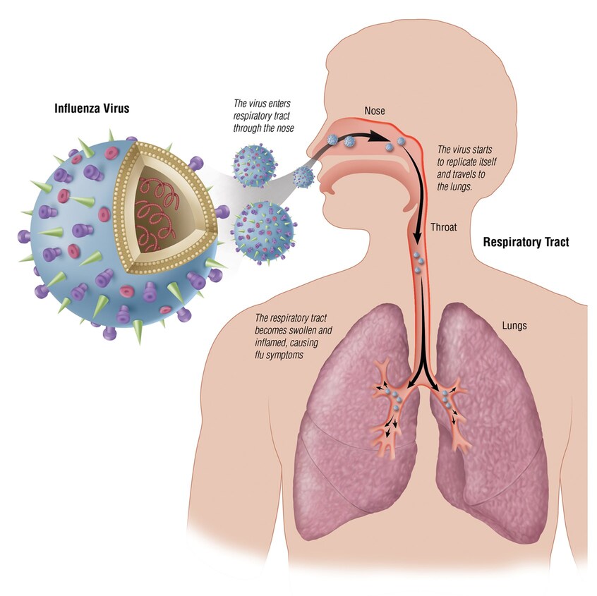
Lower Respiratory Tract Infections (LRTIs) Respiratory Tract Infections (RTI) Module Overview This master guide provides an exhaustive look into Respiratory Tract Infections. It covers everything from the foundational anatomy and natural defenses of the lungs, to the specific clinical syndromes of the upper and lower respiratory tracts, and finally the rigorous laboratory protocols required to accurately diagnose these potentially life-threatening diseases. 1. Anatomy of the Respiratory System To understand respiratory infections, we must first divide the respiratory tract into two main anatomical and functional compartments. The vocal cords roughly serve as the dividing line between the two. A. Upper Respiratory System (URTI) Structures: Nose, pharynx (throat), and associated structures (middle ear, sinuses, tonsils). Primary Purpose: To take in environmental air, and then warm, filter, and moisten it before it reaches the delicate lungs. It acts as the body’s natural HVAC (Heating, Ventilation, and Air Conditioning) system. Clinical Significance: This is the most common site of infections in the human body. Because it is the first point of contact with the outside world, it constantly encounters viruses and bacteria. B. Lower Respiratory System (LRTI) Structures: Larynx (voice box), trachea (windpipe), bronchi, bronchioles, and alveoli (air sacs). Primary Purpose: Ventilation (moving air in and out) and true gas exchange (swapping oxygen for carbon dioxide in the blood). Clinical Significance: Infections here are generally much more severe, potentially life-threatening, and harder to clear than URTIs because any inflammation here directly compromises oxygenation. Clinical Insight Sites of Infection & Pathogen Preference Specific pathogens love specific anatomical sites due to distinct cellular receptors and temperature preferences. For example: Pharynx: Adenoviral pharyngitis, Strep throat, Diphtheria. Larynx/Epiglottis: Laryngitis, Epiglottitis. Lungs/Alveoli: Pneumonia, Tuberculosis, Histoplasmosis, Coccidioidomycosis, RSV, Legionnaire’s disease. Triage Application Why the Divide Matters When a patient presents to the ER with a cough, the doctor’s immediate goal is to determine if it’s an URTI or an LRTI. URTIs are usually viral, benign, and sent home with supportive care. LRTIs (like pneumonia) often require chest X-rays, blood work, IV antibiotics, and hospital admission. Differentiating the two saves lives and resources. 2. Upper Respiratory Tract Infection (URTI) Syndromes A. The Common Cold (Infectious Rhinitis) The common cold is a mild, self-limiting viral infection of the upper respiratory mucosa. Causative Agents: Rhinovirus (most common, accounting for 30-50%), Coronaviruses, RSV (Respiratory Syncytial Virus), and Parainfluenza virus. Epidemiology: Highly common in the cooler, winter months in temperate climates, and during the rainy season in tropical areas (like Uganda). Presentation: Rhinitis (runny, stuffy nose), mild headache, and conjunctival suffusion (red, watery eyes). Clinical Pearl – The Danger of Antibiotic Misuse: Because these are exclusively viral, antibiotics are completely useless. Treatment is purely symptomatic (decongestants, rest, hydration). Overprescribing antibiotics for the common cold is the leading driver of global antibiotic resistance. Educating the patient is the most important treatment! B. Pharyngitis / Tonsillitis An inflammatory syndrome of the pharynx (sore throat) caused by various microorganisms. Causes: The vast majority are viral (Rhinovirus, Coronavirus, Adenovirus, Herpes Simplex Virus, Parainfluenza, Influenza, Coxsackievirus, Epstein-Barr virus, Cytomegalovirus). It often occurs as part of a broader common cold or flu syndrome. Bacterial Causes: The most significant bacterial cause is Group A Streptococcus (Streptococcus pyogenes), accounting for 5% to 20% of cases. Other rare bacterial causes include Neisseria gonorrhoeae (from oral sex) and Corynebacterium spp. (Diphtheria). Clinical Scenario Strep Throat & The Centor Criteria A 10-year-old presents with a sudden, severe sore throat, fever, and swollen neck lymph nodes, but NO cough. Looking in the mouth, you see white exudates (pus) on the tonsils. The Centor Criteria is used by doctors to score the likelihood of Bacterial Strep Throat vs a Viral sore throat: Absence of cough (+1 point) Swollen, tender anterior cervical lymph nodes (+1 point) Temperature > 38°C / 100.4°F (+1 point) Tonsillar exudate or swelling (+1 point) Age 3-14 (+1 point) A high score justifies a rapid strep test or empirical antibiotics. This is classic Group A Strep. We must treat this with Penicillin not just to cure the throat, but to prevent a dangerous autoimmune complication later known as Rheumatic Fever, which can permanently damage heart valves! C. Epiglottitis A severe, life-threatening inflammation of the epiglottis (the flap that covers the windpipe during swallowing). If it swells too much, it completely blocks the airway, suffocating the patient. Epidemiology: Usually occurs in cooler months. Historically affected young children (ages 2-7). Causative Organisms: Haemophilus influenzae type b (now rare due to the highly successful Hib vaccine!), Streptococcus pyogenes, and Pneumococcus. Clinical Presentation: The child will appear highly toxic, drooling (because it hurts too much to swallow their own saliva), and leaning forward in a “Tripod Position” to keep their airway open. A lateral neck X-ray will reveal the classic “Thumbprint Sign” (the swollen epiglottis looks like a thumb pressing into the airway). Diagnostic Rule (Life or Death): Blood culture is the gold standard. NEVER stick a throat swab or tongue depressor into the mouth of a child suspected of having epiglottitis! Doing so can trigger a reflex spasm that snaps the airway completely shut, killing the child instantly in the clinic. Secure the airway first (often in the OR) before any examination. D. Otitis Media (Middle Ear Infection) Inflammation of the middle ear space, located right behind the eardrum (tympanic membrane or TM). Anatomical Deep Dive: Why Kids Get It More: Children are far more prone to Otitis Media than adults because a child’s Eustachian tube (the tube connecting the middle ear to the throat) is shorter, narrower, and more horizontal. This makes it incredibly easy for bacteria from the throat to crawl up into the ear, and very difficult for the ear to drain fluid out. Clinical Confirmation: Requires an acute onset of symptoms. Signs of Effusion (fluid build-up): Using a pneumatic otoscope, a doctor will see a bulging Tympanic Membrane, limited mobility of the eardrum when puffing air at it, an air-fluid level, or otorrhoea (pus draining out if the eardrum ruptures). Symptoms: Erythema (redness) of the TM, and distinct, severe otalgia (ear pain) that

Upper Respiratory Tract Infections (URTIs) (1)
Microbiology

Upper Respiratory Tract Infections (URTIs)

Upper Respiratory Tract Infections (URTIs) Upper Respiratory Tract Infections (URTIs) 1. Overview and Magnitude of the Problem An Upper Respiratory Tract Infection (URTI), commonly referred to as “the common cold”, is a symptom complex primarily caused by viruses, occasionally bacteria, and very rarely fungi. EXAM TRIVIA The term “URTI” is actually considered a misnomer (inaccurate name). Why? Because it incorrectly implies that there are absolutely no lower respiratory tract symptoms (like deep chest coughs or bronchial irritation), which isn’t always true. Viral URTIs often trigger lower respiratory reactivity, meaning a “head cold” frequently causes chest symptoms. The Magnitude (How common is it?) Global/USA: The “Coryza syndrome” (common cold) is the most common condition seen in Outpatient Departments (OPD). Acute pharyngitis accounts for 7 million annual visits in adults (1-2% of all visits). Acute sinusitis hits 20 million people annually. Uganda : The prevalence of URTIs among children in rural Uganda was recorded at 37.4% (Mbonye, 2004), and 18.33% among under-fives (UDHS 2000/01). Regional Vulnerability: In Uganda, the highest percentage of cases were in the Northern region, followed by the Eastern region. Children aged 6-35 months are far more susceptible than infants <5 months (who still have maternal antibodies) or children >35 months (who have built their own immunity through repeated exposure). Socioeconomic Impact: URTIs carry a massive cost to society, causing missed work days, missed school classes, and unnecessary medical expenses (especially when parents demand unnecessary antibiotics). Risk Factors for URTIs Why do some people get sick while others don’t? It comes down to environmental and host factors: Climate: Cold winter months in temperate zones; rainy seasons in the tropics. Elaboration: The cold weather itself doesn’t cause the virus. Rather, bad weather forces people to stay indoors, keeping windows closed, breathing recycled air, and sharing germs in close proximity. Environment: Indoor overcrowding (homes, schools, daycare centers) and indoor air pollution (like wood-burning stoves). Overcrowding in crisis/refugee-affected areas is a massive risk due to poor ventilation and shared living spaces. Host Factors: Lack of immunization, congenital (birth) or acquired (e.g., HIV) immunodeficiency, and anatomical disorders (like a cleft palate or a severely deviated septum which impairs normal nasal drainage). Transmission: Spread via aerosols (fine mist that hangs in the air), droplets (heavy sneezes that fall on surfaces), or direct hand-to-hand contact with infected secretions, which are then passed to the nares (nose) or eyes. Example: Rubbing your eye after touching an infected doorknob is a primary route of infection! 2. Anatomy and Innate Immunity of the URT Anatomical Relevance The URT consists of the nasal cavity, paranasal sinuses, pharynx, and larynx. The critical exam concept here is anatomical continuity. The nasopharynx is directly connected to the middle ear via the Eustachian tube, and directly connected to the paranasal sinuses via small openings called ostia. Therefore, a simple nose infection can easily travel up the tubes into the ears or sinuses. Innate Immunity (How the body protects itself) The URT is not defenseless. It has a robust, multi-layered defense system: 1. Protective/Structural Measures Pseudostratified Columnar Ciliated Epithelium: This is the dominant tissue lining the URT. It acts like an escalator. The cilia (tiny hairs) constantly beat in a coordinated manner to sweep trapped harmful agents downward towards the pharynx to be swallowed and destroyed by stomach acid. Clinical Note: Cigarette smoking literally paralyzes these cilia, which is why smokers get frequent chest and sinus infections! Mucosal Secretions: Goblet cells secrete mucus. Mucus is a sticky macromolecular polysaccharide. It is *not* nutritious for bacteria, meaning bacteria can’t eat it to survive. It traps foreign particles, and as it sloughs off, the pathogens are removed with it. Saprophytic Microorganisms (Normal Flora): These are “good” bacteria living in your nose and throat. They offer protection via competitive inhibition—they eat up the local resources and take up physical space, preventing “bad” pathogenic bacteria from taking root. 2. Humoral Factors (Chemical) Lysozyme (Muramidase): A crucial hydrolytic enzyme found in secretions. Mechanism: It specifically breaks the bond between N-acetylglucosamine (GlcNac) and N-acetylmuramic acid (MurNac) in bacterial cell walls, essentially popping the bacteria like a balloon. Collectins (SP-A and SP-D): Surfactant Proteins. SP-A binds to the Lipopolysaccharide (LPS) of Gram-negative bacteria, acting as a flag (opsonization) to induce macrophages to eat them. SP-D acts in the humid phase of airways but does not induce phagocytosis directly. Other Factors: Complement system, Interferons (IFNs – fight viruses), lactoferrin (steals iron from bacteria to starve them), and Acute Phase Proteins (LBP). 3. Cellular Defenses Non-specific immune cells jump into action: Airway epithelial cells. Phagocytes: Neutrophils/PMNs, eosinophils, monocytes, macrophages. Natural Killer (NK) cells: Seek out and destroy your own cells that have been hijacked by viruses. Basophils/Mast cells: Release histamine to trigger beneficial inflammation. Dendritic Cells: Antigen Presenting Cells (APCs) that show the virus to the T-cells. 3. Specific URTI Syndromes A. The Common Cold (Coryza) A self-limiting viral infection of the upper respiratory tract, lasting about 7-10 days. Aetiology (Causes): Rhinovirus is the undisputed king (up to 60% of cases). Others include Coronavirus, Parainfluenza, RSV (Respiratory Syncytial Virus), Adenovirus, Influenza, and Enterovirus/Coxsackievirus. Exam Note: These viruses evade the immune system by constantly undergoing antigenic variation (mutating their surface proteins so your memory cells don’t recognize them next time). Pathogenesis: Virus invades the epithelium → triggers massive inflammation → sloughing off of columnar epithelial cells. Symptoms are driven by chemical mediators (Bradykinins, Prostaglandins, Histamine, Interleukins IL-1, IL-6, IL-8) and parasympathetic/alpha-adrenergic nerve reflexes. Clinical Features: Incubation is short (12-72 hrs). Cardinal signs: Nasal discharge, nasal obstruction, sneezing, scratchy/sore throat, cough. Mild fever (high fever is uncommon and suggests something worse, like the Flu or a bacterial infection). Can have facial pressure/ear fullness. Complications: Mucosal damage from the virus alters the normal flora. This, combined with aggressive nose blowing, physically pushes bacteria into sterile areas (sinuses/middle ear), causing secondary bacterial infections. Treatment: Purely symptomatic! Antihistamines, NSAIDs (for pain/fever), warm saline gargles. Antibiotics are useless against viruses and only cause harm by promoting resistant bacterial colonization. *Note: Even if nasal discharge becomes thick and greenish/yellowish,

Infections of the Central Nervous System (CNS)
Microbiology

Infections of the Central Nervous System (CNS)

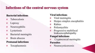
Infections of the Central Nervous System (CNS) Infections of the Central Nervous System (CNS) Exam Prep Focus Welcome to CNS Infections! This section is highly tested on exams because recognizing a CNS infection quickly is a matter of life and death. 1. Introduction to CNS Infections The Central Nervous System (brain and spinal cord) is a highly protected fortress. However, when invaders breach the walls, the results are devastating. Why? Because the cranium (skull) and vertebrae are rigid bones. When infection causes inflammation and swelling, there is nowhere for the tissue to expand. This leads to increased pressure, crushing vital brain structures, resulting in significant morbidity (disability) and mortality (death). Deep The Monro-Kellie Doctrine To understand why brain swelling is so lethal, think of the skull as a rigid, closed box containing three things: Brain Tissue (80%), Blood (10%), and CSF (10%). If a bacterial infection causes the brain tissue to swell with edema, it takes up more space. Because the skull cannot expand, the body must squeeze out the blood and CSF to make room. This leads to brain ischemia (lack of blood flow) and eventually pushes the brain out the bottom of the skull (herniation), which is fatal. Agents: Viruses, bacteria, fungi, protozoa, and helminths (parasites). The Mimics: Not everything that looks like an infection is one. Tumors, medications, and systemic illnesses can present with identical symptoms. Timeline of Infection: Acute: Hours to days (highly virulent organisms, e.g., Bacterial Meningitis). Subacute: Days to weeks. Chronic: Weeks to months (e.g., Tuberculosis, Fungal infections). Meningitis vs. Encephalitis The distinction between these syndromes is technically artificial (since etiology and pathology often overlap—e.g., Tuberculous meningitis can be subacute or chronic), but it is crucial for guiding clinical management. Acute Meningitis: Inflammation of the meninges (the protective layers covering the brain). Characterized by the onset of meningeal symptoms over hours to days. Headache is the prominent early symptom, followed later by confusion, stupor, or coma if untreated. Chronic Meningitis: Symptoms, signs, and abnormal Cerebrospinal Fluid (CSF) findings last for at least 4 weeks. Encephalitis: Infection/inflammation of the brain tissue itself (parenchyma). Distinguished by decreased mentation (confusion, stupor, altered mental status) or seizures EARLY in the course of the disease, with minimal meningeal signs (stiff neck). Clinical Pearl & Classic Presentation Most patients with CNS infections present with a classic triad/tetrad: Fever, Headache, Altered Mental Status, and Focal Neurologic Deficits. However, be careful on exams! These are nonspecific, and not every patient will have all of them. Clinical Scenario 1 The ER Triage: Meningitis A 20-year-old college student presents to the ER with a severe, pounding headache, a fever of 103°F, and severe photophobia (light hurts his eyes). When you ask him to touch his chin to his chest, he screams in pain (nuchal rigidity). He knows his name, location, and the date. Diagnosis: Meningitis. The infection is currently localized to the meningeal wrappers; his actual brain tissue is intact, so his mental status is completely normal right now. Clinical Scenario 2 The ER Triage: Encephalitis A 45-year-old man is brought in by his wife. He has a mild fever. She says he has been acting “bizarrely,” talking to people who aren’t there, and earlier he had a grand mal seizure. His neck is completely soft and pain-free when bent. Diagnosis: Encephalitis. The infection has directly attacked the brain tissue (parenchyma), immediately altering his personality and triggering electrical storms (seizures), without inflaming the meninges. 2. Epidemiology and Etiology (The “Who” and “What”) A. Bacterial Meningitis Bacterial meningitis remains a major global threat. Historically, Haemophilus influenzae type B (HiB) was a leading cause in children, but thanks to the HiB vaccine, its incidence has drastically declined. The “Big Three” (Account for >80% of cases): Haemophilus influenzae (45% historically, capsular type B strains) Streptococcus pneumoniae (47%, 18 pneumococcal serotypes) Neisseria meningitidis (Serogroups B, C, and Y) Other important causes: Streptococcus agalactiae (Group B Strep – 52% incidence in its specific demographic). Most common cause in neonates! Listeria monocytogenes (8%, serotypes 1/2b and 4b). Affects the very young, very old, and pregnant/immunocompromised. Aerobic Gram-Negative Bacilli (Klebsiella, E. coli, Serratia, Pseudomonas, Salmonella). Staphylococci (S. aureus, S. epidermidis). Exam High-Yield: Bacteria by Age & Predisposing Factor Age / Risk Factor Bacterial Pathogens to Suspect Clinical Logic (Why?) < 1 month (Neonate) S. agalactiae, E. coli, L. monocytogenes, Klebsiella pneumoniae Baby catches these passing through the mother’s birth canal or from maternal blood. 1 – 23 months S. agalactiae, E. coli, H. influenzae, S. pneumoniae, N. meningitidis Maternal antibodies wane; baby is exposed to respiratory droplets in daycare. 2 – 50 years (Adults) S. pneumoniae, N. meningitidis Standard community-acquired respiratory transmission. (Close quarters like dorms/military barracks highly favor N. meningitidis). > 50 years (Elderly) S. pneumoniae, N. meningitidis, L. monocytogenes, Gram-negative bacilli Aging immune system allows Listeria (from unpasteurized foods) and gut bacteria to invade. Immunocompromised S. pneumoniae, N. meningitidis, L. monocytogenes, Gram-negatives (incl. P. aeruginosa) Lack of T-cell/B-cell function allows opportunistic bugs to thrive. Basilar Skull Fracture S. pneumoniae, H. influenzae, Group A Strep Fracture connects the nasopharynx directly to the brain, allowing respiratory flora to leak in. Head Trauma / Neurosurgery S. aureus, S. epidermidis, P. aeruginosa Skin flora and resistant hospital bugs get pushed directly into the skull. Exam Pearl: The Listeria Threat Listeria monocytogenes is unique because it grows extremely well in cold temperatures (like inside a refrigerator). This is why pregnant women, the elderly, and immunocompromised patients are explicitly warned to avoid unpasteurized soft cheeses, cold deli meats, and hot dogs. Eating these can introduce Listeria into the gut, which then crosses into the blood and preferentially attacks the meninges. B. Viral Meningitis Viruses are the major cause of “Aseptic Meningitis”. “Aseptic” means the patient has meningitis symptoms and lymphocytic pleocytosis (high lymphocyte white blood cells in CSF), but routine bacterial cultures come back negative. Enteroviruses: The most common cause overall. Herpesviruses: HSV-1, HSV-2, VZV (Chickenpox/Shingles), CMV, EBV, HHV-6/7/8. (Exam note: HSV-1 is the most common cause of fatal, sporadic viral encephalitis, notoriously destroying

Urinary Tract Infections (UTIs)
Microbiology

Urinary Tract Infections (UTIs)

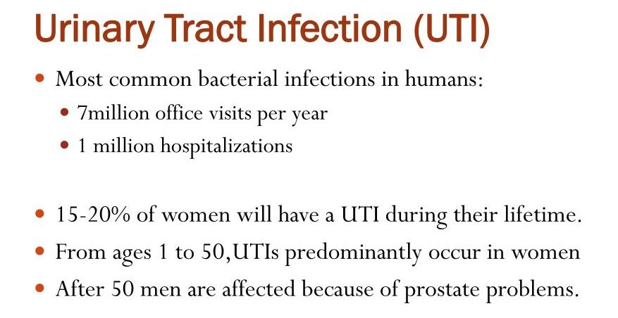
Urinary Tract Infections (UTIs) Urinary Tract Infections (UTIs) Module Overview Welcome to the comprehensive master guide on Urinary Tract Infections (UTIs). This guide covers everything from the microscopic battleground between bacterial virulence factors and host defenses, to step-by-step diagnostic workups and evidence-based treatment guidelines. 1. The Definitions Before diving into pathology, you must master the precise terminology used to describe urinary infections. Bacteriuria: Simply means the presence of bacteria in the urine. Significant Bacteriuria: The number of bacteria in voided urine exceeds what would be expected from normal contamination by the anterior urethra. Cutoff: ≥ 105 bacteria/mL. If you see this, infection must be seriously considered. Asymptomatic Bacteriuria: Significant bacteriuria (≥ 105) in a patient with absolutely ZERO symptoms. (We will discuss later who gets treated for this and who does not!) Location: UTIs can be confined to the lower tract (bladder/urethra) or involve both the upper (kidneys) and lower tracts. Cystitis (Lower UTI): A clinical syndrome involving dysuria (painful urination), frequency, urgency, and occasionally suprapubic (lower abdominal) tenderness. Acute Pyelonephritis (Upper UTI): A more severe clinical syndrome characterized by flank pain or tenderness (costovertebral angle), fever, often associated with the lower tract symptoms (dysuria, urgency, frequency). Uncomplicated UTI: Infection in a structurally and neurologically normal urinary tract. Complicated UTI: Infection in a urinary tract with functional or structural abnormalities (e.g., indwelling catheters, neurogenic bladder, or kidney stones/calculi). Clinical Scenario Uncomplicated vs. Complicated Patient A: A healthy 22-year-old female presents with painful urination and urgency for 2 days. She has no medical history. This is an Uncomplicated UTI (Cystitis). Patient B: A 65-year-old male with an enlarged prostate (BPH) and a history of kidney stones presents with the same symptoms. Because his urinary tract has structural blockages that prevent normal urine flushing, this is a Complicated UTI and requires much more aggressive management. Exam Trap: Urosepsis Criteria Urosepsis is the sepsis syndrome caused by a UTI. It is a life-threatening medical emergency. To diagnose it, you need clinical evidence of a UTI PLUS two or more of the following SIRS (Systemic Inflammatory Response Syndrome) criteria: Temperature: > 38°C (Fever) OR < 36°C (Hypothermia) Heart Rate: > 90 beats per minute (Tachycardia) Respiratory Rate: > 20 breaths/minute, OR PaCO2 < 32 mm Hg (Tachypnea/hyperventilation) White Blood Cell Count: > 12,000/mm3 (Leukocytosis), OR < 4,000/mm3 (Leukopenia), OR > 10% band forms (immature neutrophils). 2. Epidemiology & Common Bugs Females: UTI is much more common in women. 1-2% of young, non-pregnant women have it at any given time. 40% of all females will have a symptomatic UTI in their lifetime. Males: Extremely rare in young men (prevalence is only 0.04%). Clinical Pearl: If a young man gets a UTI, look for a structural defect or a Sexually Transmitted Disease (STD)! Older Age: Incidence skyrockets in the elderly (10% of men, 20% of women) due to functional impairments, prostate enlargement, and estrogen loss. The “Ojambo 2008” Ugandan Data: Over 95% of UTIs are caused by a single bacterial species. According to Ojambo 2008, the predominant organisms are: Escherichia coli (45%) – The undisputed king of UTIs. Klebsiella species (17%) Staphylococcus species (8%) – Most common Gram-positive. Enterococcus (5%) Other common offenders: Proteus, Pseudomonas, Enterobacter, and Candida (fungus, usually seen in diabetics or patients with chronic indwelling catheters). 3. Pathogenesis: A UTI is an epic battle between bacterial virulence factors and host defense mechanisms. The Routes of Invasion Ascending Route (Most Common): Bacteria from the gut colonize the perineum/urethra and climb up into the bladder. Why women? The female urethra is short and anatomically very close to the warm, moist vulvar and perianal areas, making fecal contamination highly likely. Hematogenous Route (Blood-borne): Infection of the kidney tissue by organisms traveling in the blood. Clinical Scenario: A patient with Staphylococcus aureus endocarditis (heart valve infection) throws infected blood clots into the kidneys, causing renal abscesses. Lymphatic Route: Rare, spread via lymphatic channels. Parasite Virulence Factors Not all E. coli cause UTIs. The ones that do are called Uropathogenic E. coli (UPEC) clones (Serogroups O1, O2, O4, O6, O7, O8, O75, O150, and O18ab). They possess specific genetic superpowers: Adhesins (Fimbriae/Pili): Prevent the bacteria from being washed away by urine. P fimbriae: Bind to Gal-α 1-4 (P blood group antigen). Strongly associated with Pyelonephritis and bacteremia. Type 1 fimbriae: Bind to mannosylated proteins (uroplakin Ia) on bladder cells. Associated with cystitis. Resistance to serum bactericidal activity. K Antigen (Capsules): High quantities of K1, K5, K12 capsular antigens physically protect bacteria from leukocyte phagocytosis. Aerobactin: An iron-scavenging protein (siderophore). Iron is scarce in urine; aerobactin steals it for the bacteria to grow. Hemolysin & Cytotoxic Necrotizing Factor type 1 (CNF-1) & Sat Toxin: Toxins that facilitate tissue invasion, cause severe renal tubular damage, and lyse red blood cells to make even more iron available to the invading E. coli. Urease (Specifically in Proteus species): Proteus produces urease, which splits urea into ammonia. This strongly correlates with its ability to cause severe pyelonephritis and struvite kidney stones. Deep Dive: Ammonia makes the urine highly alkaline. This change in pH causes magnesium, ammonium, and phosphate to crystallize, forming massive “staghorn” struvite stones that fill the entire renal pelvis! Master Table: Uropathogenic E. coli Adhesins Adhesin Genetic Sequence Receptor Target Clinical Comments Type 1 fimbriae (MS) Pil, fimH Mannosylated proteins on epithelial cells (uroplakin Ia) & PMNs Binds to Tamm-Horsfall protein (THP) and SIgA. P fimbriae (MR) papG (class Ia, II, III) Gal-α 1-4 (P blood group antigen) Class II: Strongly associated with pyelonephritis & bacteremia. Class III: Cystitis in patients with urinary tract abnormalities. S/F1C fimbriae (MR) Sfa/fac Sialyl-(α-2-3) galactoside Adherence is inhibited by THP. Type 1C (MR) Fac Undetermined Possibly associated with pyelonephritis. G fimbriae (MR) — Terminal N-acetyl-D-glucosamine — M fimbriae (MR) — Galactose-N-acetyl-galactosamine / Blood group M (glycophorin A) — Dr family Drb operon, Afa E1-5, Afa F Dr blood group antigen (decay accelerating factor – DAF) & type IV collagen Found in 16% of first-time cystitis isolates. The Host’s Defenses (Why we don’t always have

Skin and Soft Tissue Infections (SSTIs)
Microbiology

Skin and Soft Tissue Infections (SSTIs)

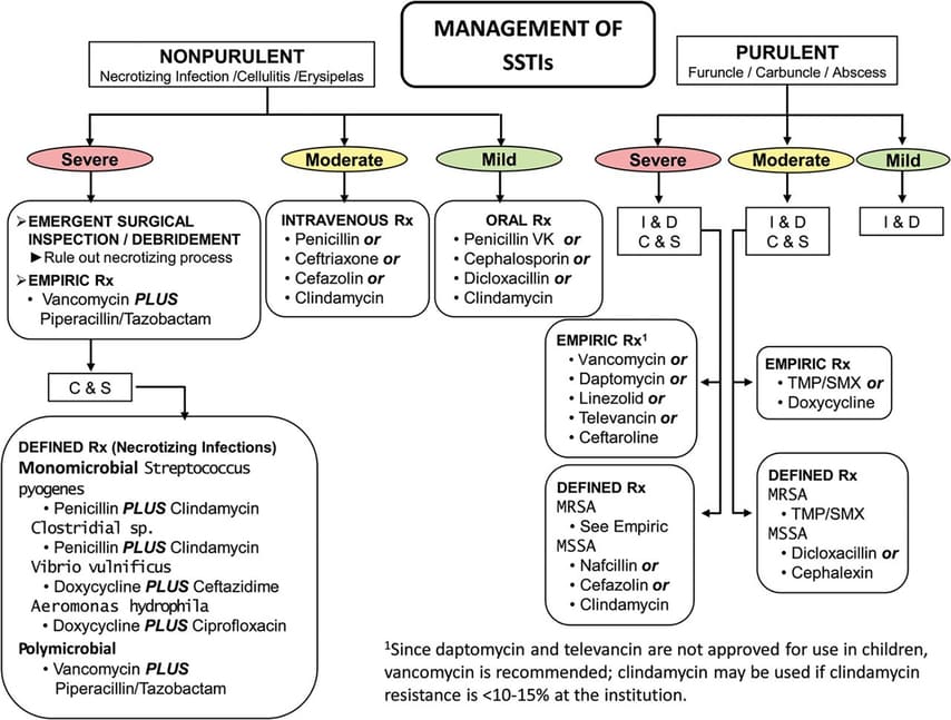
Skin and Soft Tissue Infections (SSTIs) Skin and Soft Tissue Infections (SSTIs) 1. Introduction to Skin & Soft Tissue Infections (SSTIs) General Overview: SSTIs range from minor superficial infections (like a tiny pimple) to rapidly spreading, life-threatening emergencies (like flesh-eating bacteria). The skin normally acts as an impenetrable physical and immunological barrier; infections usually require a breach (such as trauma, an insect bite, surgery, or maceration from prolonged moisture). Classic General Clinical Presentation: Local Signs: Accumulation of pus (purulence), intense redness (erythema), pain/tenderness, swelling (edema) due to increased vascular permeability. Systemic Signs: Fever, chills, malaise (as cytokines like TNF and IL-1 enter the bloodstream). Severe Complication: Bacteremia (bacteria entering the bloodstream, potentially leading to widespread sepsis and septic shock). General Diagnostic Approach: Specimen Collection and Processing Exam Trap: Never just swab a dry, intact crust or a superficial ulcer base. You must get to the active, deep infection! Swabbing dry crusts only yields dead bacteria or environmental contaminants. Collection History & Prep: The site MUST be heavily decontaminated first with soap and 70% isopropyl alcohol. Why? To avoid culturing normal, harmless skin flora (like Staphylococcus epidermidis) which will confuse the lab results and lead to the prescription of unnecessary antibiotics. The Procedure (Aspiration > Swabs): Use a sterile needle and syringe to aspirate (pull out) the loculated fluid or pus from the absolute depths of pustular/vesicle wounds or abscesses. Fluid is always vastly superior to a dry swab. Transport: Use the aspirating syringe itself as the transport container (safely capped). If there is a delay in processing, the sample MUST go into an anaerobic transport container. Clinical Reason: Deep tissues, especially in diabetics or deep bite wounds, often harbor strict anaerobes (like Bacteroides). Room air (oxygen) is toxic to them and will kill them before they reach the lab, giving you a false negative! Swabs: If a swab must be used, it should be placed in an anaerobic transport medium or inoculated directly onto culture media right at the patient’s bedside. Laboratory Processing: Gram Stain: Done first! It acts as a rapid guide for the clinician to select early empiric antibiotics (e.g., seeing Gram-positive cocci in clusters immediately suggests Staph, prompting the use of Flucloxacillin or Vancomycin) and tells the lab which specific culture media to use. Culture: The lab uses both selective and enriched non-selective media. You must know these three: 5% Sheep Blood Agar: Detects hemolysis patterns (Alpha, Beta, Gamma) crucial for identifying Streptococcus and Staphylococcus. MacConkey Agar: Selects specifically for Gram-negatives (like E. coli or Pseudomonas), inhibiting Gram-positives. Chocolate Agar: Cooked blood agar that releases internal cell nutrients, used for fastidious (picky) organisms like Haemophilus influenzae. 2. Superficial Infections (The Pyodermas) Pyoderma literally means “pus in the skin.” These are highly contagious, superficial infections predominantly affecting the epidermis. A. Impetigo (Non-Bullous) Pathophysiology & Etiology: A superficial, intraepidermal (top layer of skin), unilocular vesicopustule. It frequently occurs after minor trauma like insect bites or scratches which break the skin barrier, allowing surface bacteria to invade. Causative Agents: Group A Streptococci (GAS) (Specifically M-serotypes 2, 49, 52, 55, 57, 59, 60, 61), Group C and G Streptococci, and Staphylococcus aureus. Clinical Scenario The Honey-Crusted Child A 6-year-old boy presents to the pediatric clinic with a cluster of sores around his mouth and nose. His mother mentions he had mosquito bites there a few days ago and kept scratching them. The sores have burst, leaving a classic “honey-colored crust.” This golden crust is the absolute hallmark of non-bullous impetigo, formed by dried serum and bacterial proteins. Diagnostics & Exam Gold! The Serology Trap Gram Stain: Reveals Gram-positive cocci. Culture: Take exudate from beneath an unroofed crust. It will grow S. aureus, GAS, or a mixture of both. Serology (Exam Gold!): If caused by Streptococcus, the Anti-Streptolysin O (ASO) titer will be SCANT (negative). Why? Because the skin lipids (cholesterol in the skin) locally bind to, inhibit, and destroy Streptolysin O! Therefore, no ASO antibodies are made. Instead, you must look for an anti-DNase B response, which readily occurs and proves a recent skin Strep infection (vital if the patient later develops Post-Streptococcal Glomerulonephritis). Treatment: Topical antibiotics (like Mupirocin) for mild, localized cases. Systemic ampicillin, penicillin, erythromycin, or cephalosporins for widespread cases or immunocompromised hosts. B. Bullous Impetigo Pathophysiology & Etiology: Caused specifically by S. aureus of Phage Group II (usually type 71). This specific strain produces ETA toxin (Exfoliative Toxin A). Mechanism: The ETA toxin acts as highly specific molecular scissors. It specifically cleaves desmoglein 1 (a transmembrane glycoprotein of desmosomes that acts like velcro to hold skin cells together). This causes subcorneal separation of the epidermis, creating a pocket that fills with fluid. Clinical History & Presentation: Seen almost exclusively in newborns and young children. Lesions begin as vesicles that quickly turn into large, flaccid bullae (blisters) containing clear yellow fluid. The bullae lack a surrounding ring of redness. They quickly rupture, leaving a moist, raw red surface. C. Staphylococcal Scalded Skin Syndrome (SSSS) Pathophysiology: Similar to bullous impetigo, but instead of the toxin acting locally, the S. aureus exfoliative exotoxin enters the bloodstream and acts systemically across the entire body. Clinical History & Presentation: Begins abruptly. The patient develops a fever, intense skin tenderness, and a scarlatiniform (sandpaper-like red) rash. Large, flaccid, clear bullae form, promptly rupture, and result in the separation of massive sheets of skin. Visual Note: The child looks exactly like they have suffered a severe, widespread boiling water burn. (Unlike Toxic Epidermal Necrolysis/TEN, which involves the deeper dermal-epidermal junction and mucous membranes, SSSS is highly superficial and usually spares the mucous membranes). D. Staphylococcal Scarlet Fever & Toxic Shock Syndrome (TSS) Staphylococcal Scarlet Fever: Caused by S. aureus enterotoxins (A through D) and Toxic Shock Syndrome Toxin 1 (TSST-1). Presents with a scarlatiniform rash and skin desquamation (peeling), particularly on the palms and soles. Toxic Shock Syndrome (TSS): A severe, life-threatening acute febrile illness driven by “superantigens” that massively hyper-activate T-cells, causing a “cytokine storm.” Clinical Presentation: Generalized scarlatiniform eruption, intense desquamation, severe

History & Diagnostics in Microbiology
Microbiology

History & Diagnostics in Microbiology

History & Diagnostics in Microbiology History & Diagnostics in Microbiology PART 1: HISTORY OF MICROBIOLOGY 1. The Dark Ages of Disease Before the invention of microscopes, humans were completely blind to the microscopic world. Diseases were attributed to supernatural causes (curses, angry gods) or “miasmas” (bad, foul-smelling air from rotting organic matter). Slowly, the concept of contagion (disease spreading by touch, clothing, or proximity) began to emerge, but the actual physical agents of disease remained a complete mystery. Historical Context: During the bubonic plague (Black Death), “plague doctors” wore bird-like masks stuffed with sweet-smelling flowers. Why? Because they genuinely believed the disease was caused by inhaling the foul “miasma” smell of death, rather than being bitten by infected flea vectors! 2. The Pioneers of Microscopy (The Lens Makers) We couldn’t study bacteria until we could see them. Three men made this possible: Zacharias Janssen (1570-1638) A Dutch spectacle maker who invented the concept of compounding lenses. He placed two lenses inside a single sliding tube, creating the first rudimentary compound microscope, allowing for enlarged images of microscopic forms. Robert Hooke (1635-1703) In the 1660s, he modified the microscope (using a 6-inch tube and two convex lenses). He famously observed cork, seaweed, and sponges. He coined the term “cell” because the tiny rectangular structural boxes in cork reminded him of the bare, empty monastery rooms (cells) where monks lived. In 1665, he published his spectacular findings in his famous book, Micrographia. He was the first to describe fungi, detailing a bluish mold on leather and a white mold (which his detailed descriptions allow us to classify today as Mucor). Antony van Leeuwenhoek (1632-1723) Known forever as the Father of Microbiology. A brilliant, self-made scientist from Delft, Holland. He made 419 lenses and over 250 single-lens microscopes, achieving a staggering, crystal-clear magnification of 200-300x. He observed sperm, blood cells, and most famously, the scrapings from his own teeth (which we now know were massive bacterial biofilms!). He wrote extensively detailed letters to the British Royal Society describing tiny, moving unicellular creatures he affectionately called ‘animalcules’. In 1683, he published the very first sketches of the three principle bacterial shapes: rods (bacilli), cocci (spheres), and spirals. 3. The Great Debate: Abiogenesis vs. Biogenesis For centuries, scientists fought a bitter war over where life actually came from. Did it magically appear from non-living matter (Abiogenesis / Spontaneous Generation), or did life only come from pre-existing life (Biogenesis)? Scientist Experiment & Conclusion Stance Van Helmont (1580-1644) Placed dirty clothes and wheat/cheese in a dark stable for 21 days. Found mice. Concluded the dirt/wheat magically “created” mice. (He ignored the fact that mice simply walked in to eat the cheese!). Supported Abiogenesis Francesco Redi (1626-1697) The 3-Jar Meat Experiment. One open jar (maggots grew), one covered in parchment (no maggots), one covered in gauze (eggs laid on top of gauze, no maggots on meat). Proved flies MUST lay eggs to make maggots. Opposed Abiogenesis Louis Joblot (1645-1723) Boiled hay infusion and divided it. Covered vessel = no growth. Uncovered = microbial growth. Opening the covered one later allowed growth. Supported Biogenesis Lazzaro Spallanzani (1729-1799) Boiled meat broth for a long time to destroy heat-resistant spores and completely sealed the flask in flame. Result: No growth. Opponents stubbornly claimed he destroyed the “vital air” needed for magic generation. Opposed Abiogenesis The Final Nail in the Coffin: Louis Pasteur (1862) Louis Pasteur (1822-1895) permanently ended the spontaneous generation debate with a stroke of genius. He designed a special ‘Swan-necked’ (S-shaped) flask. He boiled nutrient broth inside it to sterilize it. Because the flask was completely open at the very end, “vital air” could easily enter, satisfying his stubborn critics. However, gravity and the S-curve of the neck physically trapped all heavy dust particles and bacteria from the air, preventing them from falling into the broth. Result: NO GROWTH. The broth remained sterile indefinitely. When he deliberately broke the neck off, allowing dust to fall directly in, microbial growth appeared immediately. Biogenesis was proven forever! 4. The Golden Age of Microbiology The late 1800s saw an explosion of life-saving discoveries, primarily led by two bitter international rivals: Pasteur (France) and Koch (Germany). Louis Pasteur (The Innovator) Discovered anaerobic bacteria (1877) during studies on butyric acid fermentation (bacteria that live without oxygen). Discovered that Yeast is the microorganism responsible for converting sugar into alcohol. Solved the massive economic crisis of souring French wine by inventing Pasteurization (mildly boiling fruit juices/milk to kill specific spoilage contaminants without ruining the taste). Vaccines & Immunology (1880): Discovered active immunization by a happy accident. While studying chicken cholera (Pasteurella spp.), he found that leaving cultures out on the bench to age made them lose their pathogenicity (virulence). Injecting these “attenuated” (weakened) older cultures didn’t kill the chickens, but amazingly protected them from future deadly doses! Created the first attenuated rabies vaccine and famously saved a young boy (Joseph Meister) who had been savagely bitten by a rabid dog. Robert Koch (1843-1910) (The Methodical Bacteriologist) A German scientist who gave us the strict laboratory techniques we still use today. Isolated the exact microorganisms causing Anthrax and Tuberculosis. Developed solid media (using agar instead of liquid broths or potatoes) for culturing bacteria and invented the streak plate technique to physically isolate pure, single colonies. Exam Trap: Koch’s Postulates Koch created 4 strict guidelines/rules to definitively prove that a specific microbe causes a specific disease. To pass the test: The microorganism must be found in abundance in ALL organisms suffering from the disease, but NOT found in healthy organisms. The microorganism must be isolated from the diseased animal and grown in pure culture in the lab. The cultured microorganism must cause the exact same disease when introduced into a healthy lab animal. The microorganism must be re-isolated from the newly diseased animal and identified as completely identical to the original specific causative agent. The Exception / Caveat (Highly Testable!): We now know there are major exceptions to Koch’s rules! – Asymptomatic carriers (like Typhoid Mary) violate

Bacterial Growth, Genetics, and Structure
Microbiology

Bacterial Growth, Genetics, and Structure

Bacterial Growth, Genetics, and Structure Microbiology Foundations: Bacterial Growth, Genetics, and Structure Module Overview Before a bacterium can divide and cause an infection, it needs the right fuel and environment. Think of bacteria as microscopic factories; they need raw materials (nutrition) and ideal factory conditions (environment). Understanding these mechanisms is the fundamental basis of targeted antibiotic therapy and infectious disease management. 1. Bacterial Growth & Nutritional Requirements A. Nutrient Requirements (The “Raw Materials”) Just like human cells, bacterial cells are highly complex and require specific building blocks to construct their membranes, DNA, and proteins. Water: Essential for all biochemical reactions. It is the universal solvent in which all intracellular metabolic processes occur. Carbon Source (C): The backbone of all living molecules (carbohydrates, lipids, proteins). Bacteria are often classified by how they get carbon (e.g., heterotrophs get it from organic compounds like glucose; autotrophs get it from CO2). Nitrogen Source (N): Crucial for building amino acids (which make up proteins) and nucleic acids (which make up DNA/RNA). Inorganic Salts, Sulfur (S), & Phosphorus (P): Phosphorus is needed to synthesize ATP (energy currency) and the phospholipid bilayer of the cell membrane. Sulfur is strictly needed for certain sulfur-containing amino acids (like cysteine and methionine) which hold proteins together via disulfide bonds. Growth Factors: Essential vitamins and amino acids that the bacteria cannot synthesize on their own. If the environment lacks these, the bacteria cannot survive. B. Environmental Factors (The “Factory Conditions”) Temperature: Most human pathogens grow best at 37°C (normal human body temperature). These are known as mesophiles. (Clinical Note: This is why the human body generates a fever—it raises the temperature above 37°C to make the environment uncomfortably hot and hostile for the invading bacteria!) Gas (Oxygen): Determines if they can breathe in air or if air is toxic to them (detailed in the next section). pH: Most bacteria prefer a neutral pH (around 7.0), though some have adapted to survive extreme acid. Example: Helicobacter pylori in the stomach survives the highly acidic gastric juice (pH ~2.0) by secreting an enzyme called urease, which creates a neutralizing “cloud” of ammonia around the bacteria. Osmotic Pressure: Salt and sugar concentrations in the environment. Clinical Example: High salt environments usually pull water out of bacteria, killing them (which is why curing meat with salt prevents rotting). However, Staphylococcus aureus is a “halophile” (salt-lover) and can easily survive on the salty surface of human skin, making it a major cause of surgical wound infections. 2. Oxygen Requirements: Aerobic vs. Anaerobic Bacteria Clinical Scenario: The Rusty Nail A patient steps on a rusty nail. The deep puncture wound closes over quickly, trapping bacteria inside with no oxygen. This is the perfect, deadly environment for an obligate anaerobe (like Clostridium tetani) to thrive and cause tetanus. Understanding oxygen requirements tells you exactly where an infection can survive in the human body! Oxygen is highly reactive. When metabolized, it creates deadly byproducts called Reactive Oxygen Species (ROS), such as superoxide radicals (O2–) and hydrogen peroxide (H2O2). To survive in oxygen, a bacteria MUST have specific enzyme “shields” (Catalase and Superoxide Dismutase – SOD) to neutralize these toxins. Type of Bacterium Effect of Oxygen Growth Pattern in a Tube Enzyme Status (The “Shields”) Classic Clinical Examples Obligate Aerobes Only aerobic growth; O2 is strictly required. Growth occurs only at the very top of the tube where O2 is highest. Have Catalase and Superoxide Dismutase (SOD) to neutralize toxic oxygen radicals. Mycobacterium tuberculosis (This is why TB infections classically target the APEX of the lungs, where oxygen concentration is highest!) Facultative Anaerobes Adaptable! Both aerobic and anaerobic growth. Greater growth with O2, but can survive without it. Growth is best at the top, but occurs throughout the entire tube. Have Catalase and SOD to neutralize toxic oxygen. Escherichia coli (E. coli) and Staphylococcus aureus. Obligate Anaerobes Only anaerobic growth. Oxygen is highly toxic/lethal. Growth occurs only at the very bottom of the tube where there is zero O2. Lacks enzymes to neutralize harmful forms of O2. Clostridium tetani, Bacteroides fragilis (Found deep in the gut). Aerotolerant Anaerobes Only anaerobic growth, but it can continue growing in the presence of O2. Growth occurs evenly throughout the entire tube; O2 has no effect. Presence of one enzyme (SOD) allows them to partially tolerate O2. Lactobacillus and Streptococcus pyogenes. Microaerophiles Only aerobic growth, but strictly requires LOW concentrations of oxygen. Growth occurs right in the middle of the tube (where O2 is low but not zero). Produce lethal amounts of toxic oxygen if exposed to normal atmospheric air. Helicobacter pylori and Campylobacter jejuni. 3. The Bacterial Growth Curve When bacteria invade a host or are put in a culture tube, they follow a predictable, 4-stage life cycle. Exam Trap: Know exactly what happens in the Log phase vs. Stationary phase! 1. Lag Phase The “Prep” Phase Bacteria are sensing their environment, gathering nutrients, and turning on specific enzymes needed to digest local food sources. There is NO increase in the number of living bacterial cells during this phase. 2. Log Phase (Exponential Phase) The “Population Boom” There is an exponential increase in the number of living cells. The bacteria are replicating at maximum speed. Clinical Pearl: This is when bacteria are rapidly building new cell walls and dividing. Therefore, this is the exact phase where antibiotics that target cell wall synthesis (like Penicillin or Cephalosporins) are most spectacularly effective! 3. Stationary Phase The “Plateau” Nutrients are running out, and toxic metabolic waste is building up. The rate of cell division exactly equals the rate of cell death. (Deep Dive: In this phase, bacteria like Clostridium and Bacillus realize they are starving and will trigger the formation of Endospores to survive the upcoming famine). Because cell wall synthesis slows down drastically here, Penicillin becomes much less effective against bacteria in an abscess (which are usually in the stationary phase). 4. Death (Decline) Phase The “Collapse” There is an exponential decrease in the number of living cells due to complete nutrient depletion and

Scroll to Top