Germ Disc, Gastrulation and Neurulation
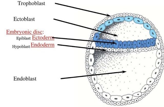
Germ Disc, Gastrulation & Neurulation: Fortion of Organs Brief Recap We concluded our last discussion with the blastocyst successfully implanted (around Day 12 post-fertilization) into the uterine endometrium. At this point: The blastocyst is fully embedded in the decidua (the transformed endometrial tissue). The trophoblast has differentiated into: Cytotrophoblast (inner layer, cellular). Syncytiotrophoblast (outer layer, invasive, multinucleated, producing hCG). The inner cell mass (embryoblast) is now clearly visible and undergoing significant changes, leading to the formation of the embryonic disc and associated cavities. Formation of the Bilaminar Embryonic/Germ Disc and Associated Cavities (Week 2 Development) This period, roughly from Day 8 to Day 14 post-fertilization, is often referred to as “the week of twos” because several structures differentiate into two layers or cavities. It’s a phase of rapid differentiation of the inner cell mass. After fertilization and cleavage, the embryo, now a blastocyst, undergoes profound organizational changes. It remodels itself from a sphere into a flattened, two-layered structure known as the Bilaminar Germ Disc. This process is crucial as it sets the stage for gastrulation, where the three primary germ layers will form. 1. From Blastocyst to Bilaminar Germ Disc This transformation begins around Day 8 post-fertilization, as the Inner Cell Mass (ICM) differentiates. A. Differentiation of the Inner Cell Mass: The inner cell mass (embryoblast) differentiates into two distinct layers that collectively form a flat, circular structure called the bilaminar embryonic disc: Epiblast (Dorsal/Upper Layer) A layer of columnar cells facing the developing amniotic cavity. Crucially, all three primary germ layers of the embryo will eventually originate from the epiblast. Location: The dorsal (upper) layer of the disc. Cell Type: Consists of tall, columnar cells. Relation to Cavity: It is directly adjacent to what will become the amniotic cavity. Significance: The epiblast is the source of all three primary germ layers during gastrulation. It is essentially the “true” embryonic component at this stage. Hypoblast (Ventral/Lower Layer) A layer of cuboidal cells facing the blastocoel. It primarily contributes to extraembryonic membranes, particularly the yolk sac. Location: The ventral (lower) layer of the disc, beneath the epiblast. Cell Type: Consists of small, cuboidal cells. Relation to Cavity: It is directly adjacent to what will become the primary yolk sac. Significance: While the hypoblast does not contribute directly to the embryo proper’s germ layers, it plays crucial roles in signaling, guiding epiblast cell movements, and forming the extraembryonic endoderm lining of the yolk sac. B. Formation of Associated Cavities: As the epiblast and hypoblast differentiate, two fluid-filled cavities form in close association with them: Amniotic Cavity Formation: A small cavity appears within the epiblast and expands. Lining: The roof of this cavity is formed by amnioblasts (cells that differentiate from the epiblast and line the amniotic cavity). The floor is the epiblast itself. Contents: It will eventually be filled with amniotic fluid, which protects the developing embryo/fetus. Primary Yolk Sac (Exocoelomic Cavity) Formation: Cells from the hypoblast migrate and spread along the inner surface of the cytotrophoblast, forming a thin membrane called the exocoelomic membrane (Heuser’s membrane). This membrane, together with the hypoblast, encloses a new cavity, the primary yolk sac. Contents: Contains fluid and plays a role in early nutrient transfer and blood cell formation. C. Development of Extraembryonic Structures: During this same period (Week 2), other crucial extraembryonic structures are forming: 1. Extraembryonic Mesoderm: Origin: A loose connective tissue layer that develops between the cytotrophoblast and the exocoelomic membrane/amnion. Cavitation: This mesoderm soon develops large cavities, forming the extraembryonic coelom (chorionic cavity). This cavity completely surrounds the amnion and the primary yolk sac, except where the embryonic disc is connected to the trophoblast by the connecting stalk (which will become the umbilical cord). Amniotic Cavity A new fluid-filled space that appears within the epiblast, enclosed by a thin membrane called the amnion. It will eventually surround the entire embryo. 2. Secondary Yolk Sac: As the extraembryonic coelom forms, the primary yolk sac shrinks, and a new, smaller secondary yolk sac forms from a second wave of hypoblast cells. This is the definitive yolk sac of the embryo. Primary Umbilical Vesicle (Yolk Sac) Forms when hypoblast cells line the blastocoel. In humans, it plays roles in early blood cell formation and nutrient transfer. 3. Chorion: The extraembryonic mesoderm, together with the two layers of the trophoblast (cytotrophoblast and syncytiotrophoblast), forms the chorion. The chorion is the outermost fetal membrane and will eventually contribute to the fetal part of the placenta. The chorionic cavity is the space within the chorion. Extraembryonic Mesoderm & Coelom A new layer of mesoderm forms between the yolk sac/amnion and the trophoblast. A large cavity, the chorionic cavity (or coelom), then forms within this mesoderm, suspending the embryo by a connecting stalk. D. Establishment of Body Axes (Preliminary): By the end of Week 2, some crucial axes begin to be established, even before gastrulation formally begins: Dorsoventral Axis: Already defined by the epiblast (dorsal) and hypoblast (ventral). Cranial-Caudal Axis: The future head end (cranial) is distinguished from the future tail end (caudal) by the appearance of a localized thickening of the hypoblast, the prechordal plate, at the future cranial region. This is an important signaling center. Left-Right Asymmetry: While not yet morphologically apparent, molecular signals are starting to be laid down that will determine left-right patterning. Summary of Bilaminar Disc Development (Week 2): Inner cell mass differentiates into Epiblast and Hypoblast. These form the Bilaminar Embryonic Disc. Amniotic Cavity forms above the epiblast. Primary Yolk Sac forms below the hypoblast, later replaced by the Secondary Yolk Sac. Extraembryonic Mesoderm and Extraembryonic Coelom develop, surrounding the amnion and yolk sac. The Chorion (trophoblast + extraembryonic mesoderm) encases everything. A Connecting Stalk links the embryonic disc to the trophoblast. Clinical Significance This highly sensitive period is critical for assessing early embryonic viability. Disruptions during germ disc formation can lead to severe birth defects, and this is when issues like ectopic pregnancies become apparent. 4. Transition to Gastrulation The formation of the bilaminar germ disc is the final preparatory