Doctors Revision

Author name: doctorsrevision@gmail.com

anatomy lecture doctors revision
Anatomy

Anatomy & Physiology 2023 Paper

Final Examination Paper Anatomy & Physiology Bachelors in Nursing • Semester 2, 2023 3 Hours Duration 100 Marks Total Marks 3 Sections A, B, C Instructions to Candidates Answer ALL questions in Section A (Objectives & Fill-ins). Answer any THREE questions from Section B. Answer any TWO questions from Section C. Write clearly and legibly. Do not write anything in the margins. SECTION A 40 Marks Part I: Objectives (20 Marks)Answer ALL questions in this part. Choose the most appropriate answer. 1. Which bone cell is responsible for resorbing (breaking down) bone matrix? A. OsteocyteB. OsteoblastC. OsteoclastD. Osteogenic cellShow AnswerAnswer: C. Osteoclast (Osteoclasts are large, multinucleated cells that break down bone tissue for remodeling). 2. The “Waiter’s Tip” position is a classic sign of injury to which part of the brachial plexus? A. Lower Trunk (C8, T1)B. Upper Trunk (C5, C6)C. Medial CordD. Posterior CordShow AnswerAnswer: B. Upper Trunk (C5, C6) (This is known as Erb-Duchenne Palsy, affecting muscles like the deltoid and biceps). 3. All muscles of facial expression are innervated by which cranial nerve? A. Trigeminal Nerve (CN V)B. Facial Nerve (CN VII)C. Accessory Nerve (CN XI)D. Hypoglossal Nerve (CN XII)Show AnswerAnswer: B. Facial Nerve (CN VII) 4. During which stage of lung maturation does surfactant production begin? A. Pseudoglandular StageB. Canalicular StageC. Saccular StageD. Alveolar StageShow AnswerAnswer: C. Saccular Stage (Type II pneumocytes differentiate and begin surfactant production). 5. Which muscle is the primary flexor of the forearm at the elbow? A. Biceps BrachiiB. BrachialisC. Triceps BrachiiD. BrachioradialisShow AnswerAnswer: B. Brachialis (It is the “workhorse” of elbow flexion, regardless of forearm position). 6. The sella turcica, which houses the pituitary gland, is a feature of which cranial bone? A. Frontal BoneB. Ethmoid BoneC. Occipital BoneD. Sphenoid BoneShow AnswerAnswer: D. Sphenoid Bone (The sphenoid is the central “keystone” bone of the cranium). 7. In oogenesis, meiosis I is completed just before ovulation, resulting in: A. One ovum and three polar bodiesB. Four functional ovaC. Two secondary oocytesD. One secondary oocyte and one polar bodyShow AnswerAnswer: D. One secondary oocyte and one polar body (The division is unequal to preserve cytoplasm). 8. Which muscle is NOT part of the rotator cuff (SITS) group? A. SupraspinatusB. Teres MajorC. InfraspinatusD. SubscapularisShow AnswerAnswer: B. Teres Major (Teres Major is an adductor and medial rotator, but not a rotator cuff muscle). 9. The primary action of the muscles in the lateral compartment of the leg (Fibularis Longus and Brevis) is: A. DorsiflexionB. InversionC. EversionD. PlantarflexionShow AnswerAnswer: C. Eversion (They are the primary everters of the foot). 10. An inability to abduct the thigh and a pelvic drop on the unsupported side (Trendelenburg sign) indicates damage to which nerve? A. Femoral NerveB. Obturator NerveC. Inferior Gluteal NerveD. Superior Gluteal NerveShow AnswerAnswer: D. Superior Gluteal Nerve (This nerve innervates the Gluteus Medius and Minimus, the main hip abductors). 11. The olecranon process is a prominent feature of which bone? A. RadiusB. HumerusC. UlnaD. ScapulaShow AnswerAnswer: C. Ulna (It forms the point of the elbow). 12. All hamstring muscles are innervated by the tibial portion of the sciatic nerve EXCEPT: A. Long head of Biceps FemorisB. Short head of Biceps FemorisC. SemitendinosusD. SemimembranosusShow AnswerAnswer: B. Short head of Biceps Femoris (It is innervated by the common fibular portion of the sciatic nerve). 13. Which of the following is NOT part of the axial skeleton? A. SternumB. RibsC. ClavicleD. VertebraeShow AnswerAnswer: C. Clavicle (The clavicle is part of the pectoral girdle, which belongs to the appendicular skeleton). 14. The “anatomical snuffbox” is formed by the tendons of all the following muscles EXCEPT: A. Abductor Pollicis LongusB. Extensor Pollicis BrevisC. Abductor Pollicis BrevisD. Extensor Pollicis LongusShow AnswerAnswer: C. Abductor Pollicis Brevis (This is a thenar muscle in the hand, not a posterior forearm muscle). 15. Referred pain to the shoulder tip is often a sign of irritation to the diaphragmatic pleura, carried by which nerve? A. Vagus NerveB. Phrenic NerveC. Intercostal NerveD. Long Thoracic NerveShow AnswerAnswer: B. Phrenic Nerve (Its root values C3-C5 correspond to the shoulder dermatome). 16. The patella is classified as which type of bone? A. Long BoneB. Irregular BoneC. Flat BoneD. Sesamoid BoneShow AnswerAnswer: D. Sesamoid Bone (It is a bone embedded within a tendon). 17. Which muscle is responsible for the first 15 degrees of arm abduction? A. DeltoidB. Pectoralis MajorC. SupraspinatusD. Latissimus DorsiShow AnswerAnswer: C. Supraspinatus (The deltoid takes over as the primary abductor after the initial 15 degrees). 18. The Adductor Pollicis muscle in the hand is innervated by the: A. Median NerveB. Radial NerveC. Musculocutaneous NerveD. Ulnar NerveShow AnswerAnswer: D. Ulnar Nerve (It is the “odd one out” of the thenar group muscles). 19. The microscopic, cylindrical unit of compact bone is called a(n): A. TrabeculaB. LamellaC. OsteonD. CanaliculusShow AnswerAnswer: C. Osteon (Also known as a Haversian system). 20. The “sit bones” are technically known as the: A. Iliac CrestsB. Pubic TuberclesC. Ischial TuberositiesD. Sacral PromontoryShow AnswerAnswer: C. Ischial Tuberosities (They bear the body’s weight when sitting). Part II: Fill in the Blanks (20 Marks)Answer ALL questions in this part. 21. The primary muscle of respiration that separates the thoracic and abdominal cavities is the [Click to reveal]. 22. The nerve that innervates the muscles of facial expression is the [Click to reveal]. 23. The final maturation stage where a round spermatid is remodeled into a spermatozoon is called [Click to reveal]. 24. The mnemonic “PAD” helps to remember that the Palmar Interossei muscles [Click to reveal] the fingers. 25. The C1 vertebra is known as the [Click to reveal], while the C2 vertebra is the [Click to reveal]. 26. The three muscles that insert at the pes anserinus on the medial side of the tibia are the Sartorius, Gracilis, and [Click to reveal]. 27. “Winging of the scapula” is caused by paralysis of the Serratus Anterior muscle due to injury to the [Click to reveal]. 28. The inorganic component that gives bone its hardness and resistance to compression is primarily [Click to reveal]. 29. In a female, a
Anatomy

Muscles of the Lower Limb

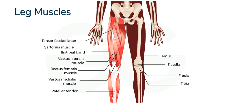
Muscles of the Lower Limb: From Pelvis to Toe. Anatomy of the Lower Extremities The Hip Joint The hip joint is one of the most important joints in the body for movement, like walking or dancing. Part 1: The Bony Pelvis & The Hip Bone The bony pelvis is a basin-shaped ring of bones connecting the vertebral column to the femurs, formed by the sacrum, coccyx, and the two hip bones (Os coxae). The Hip Bone (Os Coxa) Each large, irregularly shaped hip bone is a fusion of three primary bones that completes by the end of puberty: Ilium: The largest, most superior part, forming the prominent “wings” of the pelvis. Ischium: Forms the posteroinferior (lower-back) part of the hip bone. Pubic Bone (Pubis): Forms the anterior part of the hip bone. The Acetabulum The deep, cup-shaped socket on the lateral surface of the hip bone, formed by the union of all three bones. It articulates with the head of the femur. Key features include the crescent-shaped Lunate Surface (articular), the central Acetabular Fossa, and the fibrocartilaginous Acetabular Labrum that deepens the socket for increased stability. Detailed Anatomy of the Hip Bone Ilium: Iliac Crest: The palpable superior border, terminating anteriorly as the Anterior Superior Iliac Spine (ASIS) and posteriorly as the Posterior Superior Iliac Spine (PSIS). Other Spines: Anterior Inferior Iliac Spine (AIIS) and Posterior Inferior Iliac Spine (PIIS). Surfaces: The large, concave internal Iliac Fossa; the rough outer Gluteal Surface for gluteal muscle attachment; and the medial Auricular Surface for articulation with the sacrum. Notches: The Greater Sciatic Notch, a large indentation for passage of the sciatic nerve. Ischium: Ischial Tuberosity: The large, roughened “sitting bone” that supports body weight when seated. Ischial Spine: A pointed projection posterior to the acetabulum, separating the Greater and Lesser Sciatic Notches. Ramus of the Ischium: Projects forward to join with the pubis. Pubis: Body of Pubis: The central part that meets the other pubic bone at the Pubic Symphysis. Superior & Inferior Rami: Bars of bone that help form the acetabulum and obturator foramen. Key Markings: Includes the Pubic Tubercle and Obturator Crest for ligament and muscle attachments. Obturator Foramen The large opening created by the ischium and pubis. It is mostly closed by the obturator membrane but allows the obturator nerve and vessels to pass through the obturator canal into the thigh. The Femur (Thigh Bone) The femur is the longest, strongest, and heaviest bone in the body, transmitting weight from the hip to the tibia. Key Features of the Femur: Proximal End: Features the spherical Head (with its Fovea Capitis for the ligament of the head of the femur), the constricted Neck (a common fracture site), and the large Greater and Lesser Trochanters for muscle attachment. The Intertrochanteric Line (anterior) and Crest (posterior) connect the trochanters. Shaft: Includes the prominent posterior ridge, the Linea Aspera, for attachment of many thigh muscles. Proximally, it gives rise to the Pectineal Line and Gluteal Tuberosity. Distal End: Forms the knee joint with the large Medial and Lateral Condyles. The deep posterior notch between them is the Intercondylar Fossa. It also features the Medial and Lateral Epicondyles for ligament attachment and the anterior Patellar Surface. Key Ligaments of the Hip Joint Iliofemoral Ligament (Y-ligament of Bigelow): The strongest ligament in the body, located anteriorly. It prevents hyperextension of the hip. Pubofemoral Ligament: Located anteroinferiorly, it limits excessive abduction and extension. Ischiofemoral Ligament: Located posteriorly, it limits internal rotation and adduction. Ligament of the Head of the Femur (Ligamentum Teres): Located inside the joint, connecting the fovea capitis to the acetabulum. Transverse Acetabular Ligament: Bridges the acetabular notch, completing the socket. Muscles of the Lower Limb The powerful muscles of the lower limb are designed for stability, locomotion, and maintaining an upright posture. We will cover them regionally, starting with the hip and gluteal region. Hip Muscles: The Iliopsoas Group The Iliopsoas is the strongest hip flexor in the body. It’s a composite muscle formed by the Psoas Major and Iliacus, which merge to insert on the lesser trochanter of the femur. Psoas Major: Originates from the lumbar vertebrae. Iliacus: Originates from the iliac fossa. Main Actions: As the main flexor of the hip, it is essential for walking, running, and lifting the leg. 1. Muscles of the Gluteal Region (Buttocks) These muscles are essential for hip movement, stability, and posture, divided into superficial and deep layers. Superficial Gluteal Muscles Gluteus Maximus The largest and most superficial gluteal muscle. It is the main extensor of the thigh (crucial for climbing stairs or standing up) and a lateral rotator. Gluteus Medius Lies deep to Gluteus Maximus. It is the main abductor and a medial rotator of the thigh. It is crucial for stabilizing the pelvis during walking to prevent the hip from dropping on the unsupported side (Trendelenburg sign). Gluteus Minimus The smallest and deepest gluteal muscle. It works with the Gluteus Medius to abduct and medially rotate the thigh and stabilize the pelvis. Tensor Fasciae Latae (TFL) A small anterolateral muscle that flexes, abducts, and medially rotates the thigh. It tenses the iliotibial (IT) tract, which helps to stabilize the knee in extension. Deep Gluteal Muscles (Short External Rotators) This group of six smaller muscles lies deep to the gluteus maximus. They collectively function as powerful lateral rotators of the thigh and help stabilize the head of the femur in the acetabulum. Piriformis Origin: Anterior surface of sacrum. Insertion: Superior border of greater trochanter. Innervation: Nerve to Piriformis (S1, S2). Actions: Laterally rotates, abducts (when hip is flexed), and extends the thigh. Superior Gemellus Origin: Ischial spine. Insertion: Medial surface of greater trochanter (with Obturator Internus tendon). Innervation: Nerve to Obturator Internus (L5, S1). Actions: Laterally rotates and abducts the thigh. Obturator Internus Origin: Pelvic surface of obturator membrane. Insertion: Medial surface of greater trochanter. Innervation: Nerve to Obturator Internus (L5, S1). Actions: Laterally rotates and abducts the thigh. Inferior Gemellus Origin: Ischial tuberosity. Insertion: Medial surface of greater trochanter (with Obturator Internus

Anatomy

Muscles of the Upper Limb

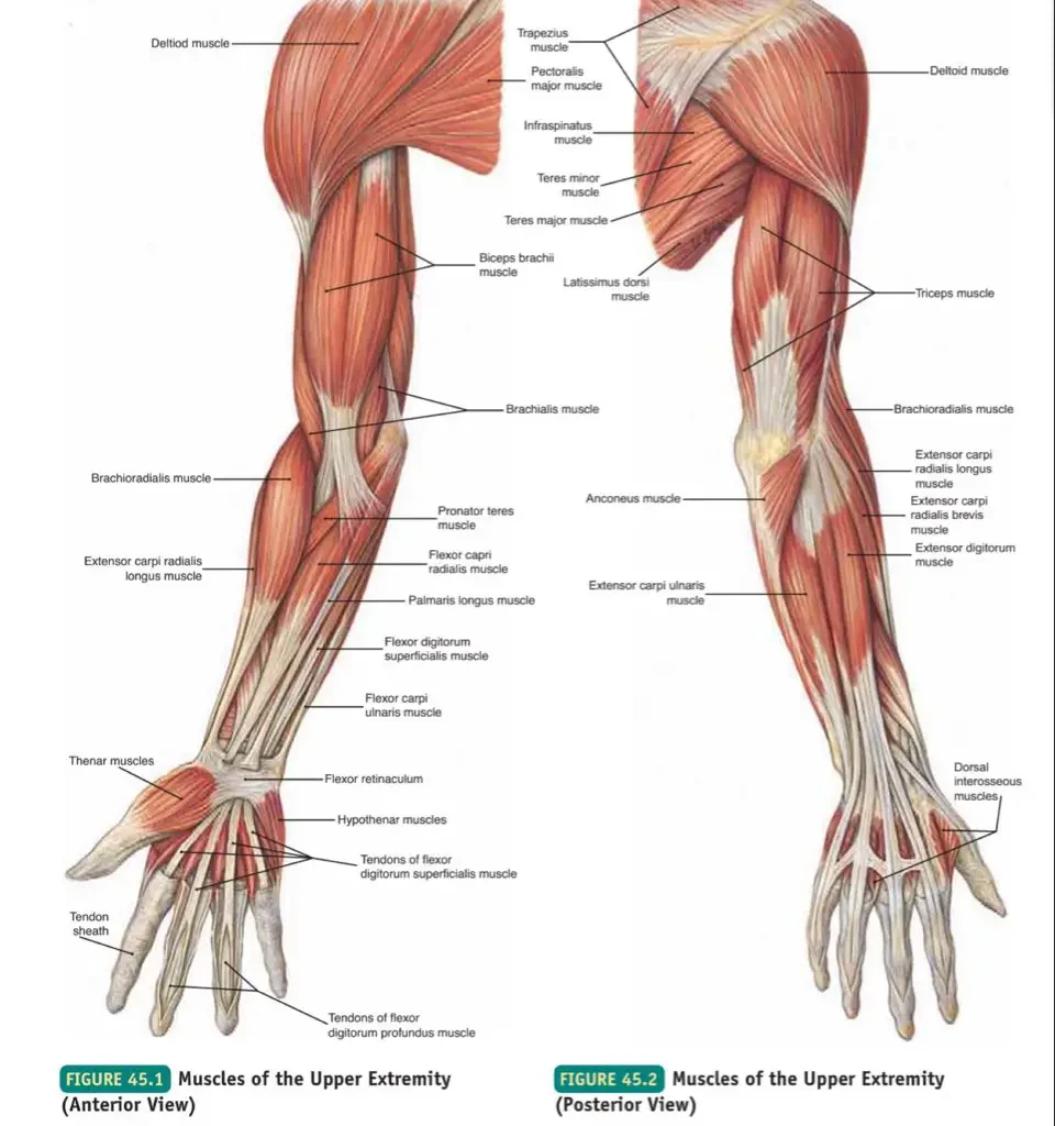
Musclesof the Upper Limbs: From Shoulder. The Brachial Plexus The brachial plexus is a complex network of nerves formed by the anterior rami of the lower four cervical nerves (C5, C6, C7, C8) and the first thoracic nerve (T1). It is responsible for the motor and sensory innervation of the entire upper limb. Understanding the plexus is best done by following its five main divisions, remembered by the mnemonic: “Real Texans Drink Cold Beer” (Roots, Trunks, Divisions, Cords, Branches). 1. Roots (C5, C6, C7, C8, T1) The five roots are the anterior primary rami of the spinal nerves, emerging between the anterior and middle scalene muscles in the neck. Key Branches from Roots: Dorsal Scapular Nerve (C5): Innervates Rhomboids and Levator Scapulae. Long Thoracic Nerve (C5, C6, C7): Innervates Serratus Anterior. 2. Trunks (Superior, Middle, Inferior) The five roots unite to form three trunks, which pass over the first rib. Upper Trunk: Formed by the union of C5 and C6 roots. Middle Trunk: A continuation of the C7 root. Lower Trunk: Formed by the union of C8 and T1 roots. Key Branches from Trunks: Suprascapular Nerve (C5, C6): From the Upper Trunk; innervates Supraspinatus and Infraspinatus. 3. Divisions (Anterior and Posterior) Each of the three trunks divides into an anterior and a posterior division, passing under the clavicle. The posterior divisions supply future extensors, while the anterior divisions supply future flexors. 4. Cords (Lateral, Posterior, Medial) The six divisions regroup to form three cords, named for their position relative to the axillary artery. Lateral Cord (C5-C7): From the anterior divisions of the upper and middle trunks. Posterior Cord (C5-T1): From the posterior divisions of all three trunks. Medial Cord (C8-T1): From the anterior division of the lower trunk. Key Branches from Cords: Lateral Pectoral Nerve: From the Lateral Cord. Upper & Lower Subscapular Nerves, Thoracodorsal Nerve: From the Posterior Cord. Medial Pectoral Nerve, Medial Cutaneous Nerves: From the Medial Cord. 5. Branches (The 5 Major Terminal Nerves) The three cords give rise to the five major terminal nerves that innervate the entire upper limb. Musculocutaneous Nerve (C5-C7) Motor: Anterior arm compartment (Biceps Brachii, Brachialis, Coracobrachialis).Sensory: Skin of the lateral forearm. Axillary Nerve (C5-C6) Motor: Deltoid and Teres Minor.Sensory: Skin over the lower deltoid (“regimental badge area”). Radial Nerve (C5-T1) Motor: All muscles of the posterior compartments of the arm and forearm (all extensors).Sensory: Posterior skin of arm and forearm, dorsal aspect of lateral 2.5 digits. Median Nerve (C5-T1) Motor: Most anterior forearm muscles (flexors/pronators), and thenar muscles of the thumb.Sensory: Skin of the lateral palm and palmar aspect of the lateral 3.5 digits. Ulnar Nerve (C8-T1) Motor: Two anterior forearm muscles (Flexor Carpi Ulnaris, medial half of FDP) and most intrinsic muscles of the hand.Sensory: Skin of the medial 1.5 digits (palmar and dorsal). Brachial Plexus Summary Table Level Components Key Nerve Branches Clinical Notes ROOTS Anterior Rami of C5, C6, C7, C8, T1 Dorsal Scapular N (C5): Rhomboids, Levator ScapulaeLong Thoracic N (C5-C7): Serratus Anterior Emerge between Scalenes. Injury to Long Thoracic N. → Winged Scapula. TRUNKS Upper: C5 + C6Middle: C7Lower: C8 + T1 Suprascapular N (C5, C6): Supraspinatus, InfraspinatusN. to Subclavius (C5, C6): Subclavius Pass over 1st rib. Erb-Duchenne palsy is an upper trunk injury. DIVISIONS Each trunk divides into an Anterior & Posterior Division No direct named branches. Posterior divisions supply extensors; Anterior supply flexors. CORDS Lateral: Ant. divisions of Upper & MiddlePosterior: Post. divisions of all 3Medial: Ant. division of Lower Lateral Pectoral N.Upper & Lower Subscapular N., Thoracodorsal N.Medial Pectoral N., Medial Cutaneous Nerves Named for position around axillary artery. BRANCHES Terminal Nerves Musculocutaneous N.Axillary N.Radial N.Median N.Ulnar N. Major nerves of the upper limb. Injuries lead to distinct motor & sensory deficits. Brachial Plexus Injuries and Clinical Correlates Upper Plexus Injury (Erb-Duchenne Palsy) Affects C5-C6 roots. Caused by an excessive angle between the neck and shoulder. Results in the classic “Waiter’s Tip” position (adducted shoulder, medially rotated arm, extended elbow). Lower Plexus Injury (Klumpke’s Palsy) Affects C8-T1 roots. Caused by excessive abduction of the arm. Affects intrinsic hand muscles, leading to a “Claw Hand” of the 4th and 5th digits. Radial Nerve Injury (Wrist Drop) Commonly caused by mid-shaft humeral fractures or compression in the axilla (“Saturday night palsy”). Results in an inability to extend the wrist and fingers. Median Nerve Injury (Carpal Tunnel Syndrome) Compression of the median nerve at the wrist. Causes numbness and tingling in the lateral 3.5 digits and weakness/atrophy of the thenar (thumb) muscles. Ulnar Nerve Injury (“Claw Hand”) Injury at the elbow (“funny bone”) or wrist. Affects intrinsic hand muscles, leading to “clawing” of the 4th and 5th digits and sensory loss over the medial hand. Muscles of the Chest (Pectoral Region) 1. Superficial Muscles of the Pectoral Region These muscles connect the upper limb to the anterior and lateral thoracic wall. a. Pectoralis Major A large, fan-shaped muscle covering the upper chest. It is a powerful adductor and medial rotator of the arm. Its clavicular head also flexes the arm, while the sternocostal head helps extend it from a flexed position. b. Pectoralis Minor A thin, triangular muscle lying deep to Pectoralis Major. It depresses the shoulder and protracts the scapula (pulls it forward and downward). c. Subclavius A small muscle located inferior to the clavicle. It anchors and depresses the clavicle, and helps protect the underlying subclavian vessels and brachial plexus. d. Serratus Anterior The “boxer’s muscle” on the lateral thoracic wall. It is the prime mover for protracting the scapula (punching/pushing) and is essential for rotating the scapula to allow for full arm elevation. Paralysis leads to “winged scapula”. 2. Deep Muscles of the Thorax (Associated with Respiration) These muscles are primarily involved in the mechanics of breathing. a. Intercostal Muscles (External, Internal, Innermost) Three layers of muscles in the intercostal spaces. The External Intercostals elevate the ribs for forced inspiration. The Internal and Innermost Intercostals depress the ribs for forced expiration. b. Transversus Thoracis A thin muscle on the inner anterior thoracic

Muscles of the Head, Neck and Trunk
Anatomy

Muscles of the Head, Neck and Trunk

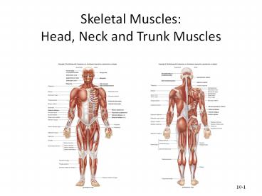
Axial Skeleton Muscles: The Footress. Muscles of the Axial Skeleton A. Muscles of the Head and Face The muscles of the head can be broadly categorized into muscles of facial expression and muscles of mastication (chewing). 1. Muscles of Facial Expression These unique muscles insert into the skin or other muscles, allowing us to show a wide range of emotions. They are all innervated by the Facial Nerve (Cranial Nerve VII). a. Occipitofrontalis (Epicranius) A broad muscle covering the top of the skull with two bellies. The Frontal belly raises the eyebrows and wrinkles the forehead, while the Occipital belly pulls the scalp posteriorly. b. Orbicularis Oculi A ring-like muscle encircling the eye. Its primary action is to close the eye (blinking, winking) and squint. c. Orbicularis Oris A complex muscle encircling the mouth. It closes and protrudes the lips, as in puckering or kissing. d. Zygomaticus Major and Minor Extend from the cheekbone to the corner of the mouth. They are the primary “smiling” muscles, raising the lateral corners of the mouth upward. e. Buccinator A thin, flat muscle of the cheek. It compresses the cheek for whistling or sucking and holds food between the teeth during chewing. f. Platysma A broad, superficial sheet of muscle in the neck. It tenses the skin of the neck, depresses the mandible, and pulls the lower lip down. 2. Muscles of Mastication (Chewing) These four pairs of muscles are responsible for moving the mandible for chewing. They are all innervated by the Mandibular division of the Trigeminal Nerve (Cranial Nerve V3). a. Masseter A powerful muscle on the side of the jaw. It is the primary elevator of the mandible (closes the jaw). b. Temporalis A fan-shaped muscle in the temporal fossa. It elevates and retracts the mandible. c. Medial Pterygoid Located deep to the mandible. It elevates the jaw and assists in side-to-side grinding movements. d. Lateral Pterygoid Located deep in the jaw. It protracts the mandible (pulls it forward), moves it side-to-side, and is the only muscle of mastication that helps open the jaw. Summary Table of Head & Face Muscles Muscle Origin Insertion Action FACIAL EXPRESSION (CN VII) Occipitofrontalis Galea aponeurotica (Frontal); Occipital bone (Occipital) Skin of eyebrows; Galea aponeurotica Raises eyebrows, wrinkles forehead, pulls scalp Orbicularis Oculi Frontal and maxillary bones Tissue of eyelid Closes eye, squints, blinks Orbicularis Oris Maxilla and mandible Skin and muscle at angles of mouth Closes and protrudes lips (puckering) Zygomaticus Major/Minor Zygomatic bone Skin and muscle at angle of mouth Raises lateral corners of mouth (smiling) Buccinator Molar region of maxilla and mandible Orbicularis oris Compresses cheek (whistling, sucking) Platysma Fascia of chest Base of mandible; skin at corner of mouth Tenses skin of neck, depresses mandible MASTICATION (CN V3) Masseter Zygomatic arch Angle and ramus of mandible Elevates mandible (closes jaw) Temporalis Temporal fossa Coronoid process of mandible Elevates and retracts mandible Medial Pterygoid Sphenoid and palatine bones Medial surface of ramus of mandible Elevates mandible, moves side-to-side Lateral Pterygoid Sphenoid bone Condylar process of mandible; TMJ capsule Protracts and depresses (opens) jaw B. Muscles of the Neck The muscles of the neck are diverse, responsible for moving the head, stabilizing the cervical spine, assisting in breathing, and facilitating swallowing and speech. They are categorized here based on location and primary actions. 1. Superficial Anterior Neck Muscles a. Sternocleidomastoid (SCM) A large, two-headed muscle on each side of the neck. When acting alone (unilaterally), it rotates the head to the opposite side and flexes it to the same side. When both act together (bilaterally), they flex the neck (chin to chest). 2. Suprahyoid Muscles (Above the Hyoid Bone) These muscles form the floor of the mouth and are primarily responsible for elevating the hyoid bone during swallowing and speaking. a. Digastric Two-bellied muscle that elevates the hyoid or depresses the mandible (opens the mouth). b. Mylohyoid Forms the floor of the mouth; elevates hyoid and floor of mouth. c. Geniohyoid Elevates and protracts the hyoid bone. d. Stylohyoid Elevates and retracts the hyoid bone. 3. Infrahyoid Muscles (Strap Muscles – Below the Hyoid) These “strap-like” muscles primarily depress the hyoid bone and larynx during swallowing and speaking. a. Sternohyoid Depresses the hyoid bone and larynx. b. Omohyoid Two-bellied muscle that depresses and retracts the hyoid. c. Sternothyroid Depresses the larynx and hyoid bone. d. Thyrohyoid Depresses the hyoid bone but elevates the larynx. 4. Deep Lateral Neck Muscles (Scalenes) The Anterior, Middle, and Posterior Scalene muscles are important for lateral flexion of the neck. They also act as accessory muscles of inspiration by elevating the first two ribs. Summary Table of Neck Muscles Muscle Origin Insertion Innervation Action Sternocleidomastoid Manubrium & Clavicle Mastoid process CN XI, C2-C3 Unilateral: Rotates head opp., flexes same side. Bilateral: Flexes neck. Digastric Mandible & Mastoid process Hyoid bone CN V3 & CN VII Elevates hyoid, depresses mandible. Mylohyoid Mandible Hyoid bone CN V3 Elevates hyoid & floor of mouth. Sternohyoid Manubrium & Clavicle Hyoid bone Ansa cervicalis Depresses hyoid and larynx. Omohyoid Scapula Hyoid bone Ansa cervicalis Depresses and retracts hyoid. Sternothyroid Manubrium Thyroid cartilage Ansa cervicalis Depresses larynx and hyoid. Thyrohyoid Thyroid cartilage Hyoid bone C1 via CN XII Depresses hyoid, elevates larynx. Scalenes (Ant, Mid, Post) Cervical vertebrae (C2-C7) First & Second ribs Cervical spinal nerves Flexes neck, elevates ribs for inspiration. C. Muscles of the Torso (Trunk) The muscles of the trunk are vital for maintaining posture, protecting internal organs, facilitating respiration, and enabling a wide range of movements. 1. Muscles of the Back These complex, layered muscles move and stabilize the vertebral column, head, and shoulders. a. Superficial Back Muscles Primarily act on the upper limbs. Includes the large Trapezius (moves scapula), Latissimus Dorsi (extends and adducts arm), and the deeper Rhomboids and Levator Scapulae (retract and elevate scapula). b. Intermediate Back Muscles Respiratory muscles. The Serratus Posterior Superior elevates ribs for inspiration, while the Serratus Posterior Inferior depresses ribs for expiration. c. Deep (Intrinsic) Back Muscles Responsible for posture

Anatomy

Axial and Appendicular Skeleton

Axial and Appendicular Skeleton The Supporters. The Axial and Appendicular Skeleton The human skeleton is divided into two major parts: the Axial Skeleton and the Appendicular Skeleton. Together, these two divisions provide the support, protection, and leverage necessary for movement. The Axial Skeleton: The Body’s Central Axis The axial skeleton forms the longitudinal axis of the body. It consists of the bones of the skull, vertebral column (spine), and thoracic cage (ribs and sternum). In brief, it comprises the head and trunk. Composition (approximately 80 bones): Skull (22 bones + 7 associated): Protects the brain and forms the face. Vertebral Column (26 bones): Protects the spinal cord and supports the head. Thoracic Cage (25 bones): Protects the heart and lungs. The Skull The skull is a bony structure that forms a protective cavity for the brain, provides the head with its shape, and is formed by 22 bones joined by fibrous joints called sutures. It consists of two main parts: the Cranium and the Face. 1. The Cranium (8 Bones) The cranium is the bony box that houses and protects the brain. Frontal Bone (1) Forms the forehead and the superior part of the orbits. Parietal Bones (2) Form the superior and lateral walls of the cranium. Temporal Bones (2) Form the inferolateral aspects of the skull and parts of the cranial base; contain the organs of hearing. Occipital Bone (1) Forms the posterior wall and most of the base of the skull. The spinal cord passes through its foramen magnum. Sphenoid Bone (1) The central “keystone” bone of the cranium; articulates with all other cranial bones. Contains the sella turcica for the pituitary gland. Ethmoid Bone (1) Forms the anterior part of the cranial floor, the medial wall of the orbits, and the roof of the nasal cavity. 2. The Face (14 Bones) These bones form the framework of the face, contain cavities for sensory organs, and provide attachment sites for facial muscles. Mandible (1) The lower jawbone; the largest and strongest bone of the face. Maxillae (2) The upper jawbones; they form the hard palate and hold the upper teeth. Zygomatic Bones (2) The cheekbones; they form the prominences of the cheeks. Nasal Bones (2) Form the bridge of the nose. Lacrimal Bones (2) Form part of the medial walls of the orbits; contain the lacrimal fossa for the tear ducts. Palatine Bones (2) Form the posterior part of the hard palate. Vomer (1) Forms the inferior part of the nasal septum. Inferior Nasal Conchae (2) Scroll-like bones forming part of the lateral walls of the nasal cavity. B. The Vertebral Column (Spine) The vertebral column serves as the main support of the body, protects the spinal cord, and provides attachment points for the ribs and muscles. It is a flexible, curved structure composed of 26 irregular bones in adults. Functions of the Vertebral Column: Support: Transmits the weight of the head and trunk to the lower limbs. Protection: Surrounds and protects the delicate spinal cord. Movement: Provides attachment points for muscles, allowing trunk and neck movement. Shock Absorption: Intervertebral discs act as shock absorbers. Regions and Curvatures The spine is divided into five regions and has four natural curves that increase its resilience. Vertebral Regions Cervical (C1-C7): 7 vertebrae in the neck.Thoracic (T1-T12): 12 vertebrae in the chest.Lumbar (L1-L5): 5 vertebrae in the lower back.Sacrum: 1 bone (5 fused vertebrae).Coccyx: 1 bone (3-5 fused vertebrae). Spinal Curvatures Cervical & Lumbar: Concave posteriorly (secondary curves).Thoracic & Sacral: Convex posteriorly (primary curves). General Structure of a Vertebra Most vertebrae share a common structural plan, consisting of a body, an arch, and various processes for muscle attachment and articulation. Vertebral Body (Centrum): The anterior, weight-bearing part. Vertebral Arch: Encloses the vertebral foramen, forming the vertebral canal for the spinal cord. Processes: Projections (spinous, transverse, articular) that serve as attachment and articulation points. Intervertebral Discs Located between adjacent vertebrae, these discs act as shock absorbers. Each is composed of an inner gelatinous nucleus pulposus and an outer collar of fibrocartilage called the anulus fibrosus. Regional Characteristics of Vertebrae Cervical Vertebrae (C1-C7) The smallest, lightest vertebrae. Their unique feature is the transverse foramina for vertebral arteries. C1 (Atlas) lacks a body and articulates with the skull (“yes” motion). C2 (Axis) has a dens that acts as a pivot for head rotation (“no” motion). Most have a bifid (split) spinous process. Thoracic Vertebrae (T1-T12) Distinguished by their articulation with the ribs via costal facets on the vertebral bodies and transverse processes. They have a heart-shaped body and a long, slender spinous process that points sharply downward. Lumbar Vertebrae (L1-L5) The largest and strongest vertebrae, designed to bear the most body weight. They have a massive, kidney-shaped body and a short, thick, blunt spinous process that projects posteriorly. Sacrum and Coccyx The Sacrum is a triangular bone formed by the fusion of 5 sacral vertebrae, forming the posterior wall of the pelvis. The Coccyx, or “tailbone,” is a small triangular bone formed by the fusion of 3-5 coccygeal vertebrae. C. The Thoracic Cage (Bony Thorax) The thoracic cage forms the protective “rib cage” around the vital organs of the chest. It includes the sternum, ribs, and the twelve thoracic vertebrae. Functions of the Thoracic Cage: Protection: Encloses and protects the heart, lungs, and major blood vessels. Support: Provides attachment points for the shoulder girdles and upper limbs. Respiration: Its ability to expand is crucial for ventilation, and it provides attachment for respiratory muscles. Bones of the Thoracic Cage The Sternum (Breastbone) A flat bone in the anterior midline of the thorax, composed of three fused parts: Manubrium: The superior part, articulating with the clavicles and the first two pairs of ribs. Features the palpable jugular (suprasternal) notch. Body (Gladiolus): The middle and largest part, articulating with ribs 2-7. Xiphoid Process: The inferior-most, small projection that serves as an attachment point for some abdominal muscles. The Ribs (12 pairs) All ribs attach posteriorly to the thoracic vertebrae and generally curve inferiorly and anteriorly. Types

Anatomy

Introduction to Musculoskeletal System Anatomy

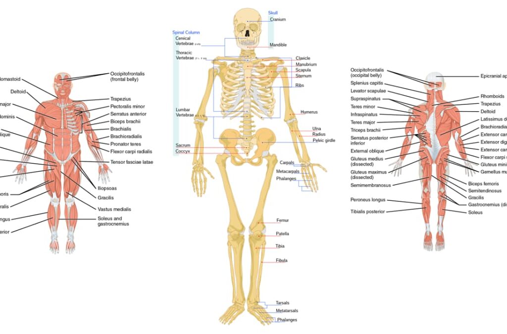
Musculoskeletal System Anatomy: The Supporters. Introduction to the Musculoskeletal System The Human Skeletal system is the body system composed of bones, cartilage, tendons, and ligaments and other tissues that perform essential functions for the human body. Altogether, the skeleton makes up about 20% of a person’s body weight. Components of the Musculoskeletal System 1. Bones The rigid organs that form the body’s structural framework. The human skeleton is composed of around 270 bones at birth, The adult human skeleton is composed of about 206 bones, which are made of specialized connective tissue with a mineralized matrix. 2. Cartilage A soft, gel-like connective tissue that protects joints, facilitates smooth movement, and provides flexible support in areas like the nose, ears, and trachea. 3. Ligaments Strong, tough bands of elastic connective tissue that connect bone to bone. They support and strengthen joints, limiting their movement to prevent injury. The body has approximately 900 ligaments. 4. Tendons Strong, fibrous bands of connective tissue that attach muscle to bone. They transmit the force generated by muscle contractions to produce movement. The body has approximately 4,000 tendons. 5. Muscles (Skeletal) Specialized contractile tissue attached to bones via tendons. Their voluntary contraction generates the force required for all conscious movement. The body has about 650 skeletal muscles. Functions of the Musculoskeletal System The coordinated action of these components provides the body with several critical functions. Support The skeleton forms the rigid internal framework that supports the body’s weight and provides its shape. Movement Bones act as levers and muscles provide the force, allowing for locomotion and manipulation. Protection The skeleton safeguards vital internal organs (e.g., skull protects the brain, rib cage protects heart and lungs). Mineral Storage Bones act as a critical reservoir for essential minerals like calcium and phosphate. Hematopoiesis Red bone marrow, found within certain bones, is responsible for producing all blood cells. Fat Storage Yellow bone marrow stores triglycerides (fat) as a source of energy. The Structure of Bone Bones are the basic unit of the human skeleton. Far from being static, they are highly vascular, living tissues that are continuously remodeled throughout life. A bone is a rigid organ that protects internal organs, produces blood cells, stores minerals, provides structural support, and enables mobility. It is composed chiefly of calcium phosphate and calcium carbonate, serving as a critical reservoir for calcium. Composition of Bone Bone tissue is a composite material, made of both organic and inorganic components that give it its unique properties. Organic Components (~35%) Composed of osteoid (unmineralized matrix), which includes Type I collagen fibers and ground substance. FUNCTION: Provides flexibility and tensile strength (resistance to twisting and pulling). Inorganic Components (~65%) Primarily hydroxyapatite (a complex of calcium phosphate) and other mineral salts like magnesium and fluoride. FUNCTION: Provides hardness and resistance to compression. Types of Bone Tissue: Compact vs. Spongy Bone has two main structural types, each with a distinct organization and function. Compact Bone (Cortical Bone) A dense, solid outer layer organized into repeating structural units called osteons (Haversian systems). Each osteon is a cylinder of concentric rings (lamellae) around a central Haversian canal, which contains blood vessels and nerves. This structure provides immense strength and protection, forming the outer layer of all bones and the shaft of long bones. Spongy Bone (Cancellous Bone) An internal, lightweight tissue that lacks osteons. It consists of an irregular latticework of thin columns of bone called trabeculae. The spaces between the trabeculae are filled with red bone marrow, the site of hematopoiesis. This structure provides strength without excessive weight and is found in the ends of long bones and in flat bones. The Four Types of Bone Cells Bone is a dynamic tissue maintained by four specialized cell types. Osteogenic Cells Function: Mesenchymal stem cells that divide and differentiate into osteoblasts. Crucial for bone growth and repair. Osteoblasts Function: Bone-building cells. They synthesize and secrete the organic osteoid matrix and initiate its calcification. Osteocytes Function: Mature, bone-maintaining cells trapped within the matrix. They act as mechanosensors, signaling for remodeling. Osteoclasts Function: Bone-resorbing cells. They break down bone matrix, which is essential for remodeling and releasing minerals into the blood. The Gross Anatomy of Bone Now that we’ve explored bone at the microscopic level, let’s examine its larger, more observable features, including its classification, overall structure, and the critical bone markings that indicate interaction points with other body structures. A. Classification of Bones by Shape Long Bones Longer than they are wide; act as levers for movement. (e.g., Femur, Humerus, Phalanges) Short Bones Cube-shaped; provide stability. (e.g., Carpals, Tarsals) Flat Bones Thin, flattened, and often curved; provide protection. (e.g., Cranial bones, Sternum, Ribs) Irregular Bones Complex and varied shapes. (e.g., Vertebrae, Hip bones) Sesamoid Bones Small bones embedded within tendons; protect tendons from stress. (e.g., Patella) B. Structure of a Long Bone Diaphysis The main, cylindrical shaft of the bone, composed of compact bone surrounding the medullary cavity. Epiphysis The expanded ends of a long bone, consisting mostly of spongy bone. Metaphysis The region where the diaphysis and epiphysis meet. Contains the epiphyseal (growth) plate. Articular Cartilage A thin layer of hyaline cartilage covering the epiphysis at a joint to reduce friction. Periosteum & Endosteum The periosteum is the tough outer membrane, while the endosteum is the thin inner lining of the medullary cavity. C. Bone Markings (Surface Features) Bone markings are characteristic projections, depressions, and openings on bone surfaces that serve as points of articulation, attachment for muscles and ligaments, or passageways for nerves and blood vessels. 1. Projections (Features that Bulge Outward) Marking Description Example Head Prominent, rounded articular surface Head of femur, Head of humerus Condyle Rounded articular projection Femoral condyles Epicondyle Raised area above a condyle Medial epicondyle of humerus Process Any bony prominence Mastoid process Spine Sharp, slender projection Ischial spine Tubercle Small, rounded projection Tubercle of humerus Tuberosity Large, rounded, roughened projection Deltoid tuberosity Trochanter Very large, blunt process (only on femur) Greater trochanter Crest Narrow, prominent ridge of bone Iliac crest Line Slight, elongated

Respiratory System
Anatomy

Respiratory System Anatomy

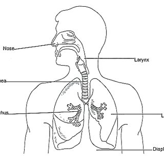
Respirator System Anatomy: Breath in, Out! Objective: To describe the macroscopic and microscopic anatomy of the respiratory system and relate structure to function in the processes of air conduction, gas exchange, and protection. Introduction to the Respiratory System The respiratory system is a complex network of organs and tissues that work together to move air into and out of the body and facilitate gas exchange. It can be broadly divided into two main parts based on function: the conducting zone (for air transport) and the respiratory zone (for gas exchange). The respiratory system is a vital biological system responsible for the exchange of gases between the body and the external environment. Its primary function is to take in oxygen (O₂) from the atmosphere and expel carbon dioxide (CO₂), a waste product of cellular metabolism. This process, known as respiration, is essential for energy production and maintaining the body’s pH balance. A. Upper Respiratory Tract (Conducting Zone) This part of the system is primarily involved in conditioning the inspired air. 1. Nose and Nasal Cavity External Nose: The visible part, supported by bone and cartilage. Nasal Cavity: Extends from the nostrils (nares) to the posterior nasal apertures (choanae). Vestibule: The anterior-most part, lined with skin and stiff hairs (vibrissae) that filter large particles. Nasal Conchae (Turbinates): Three bony projections (superior, middle, inferior) covered by mucous membranes. They dramatically increase the surface area of the nasal cavity and create turbulent airflow. Function of Turbinates & Mucosa This turbulent flow forces inhaled air to come into contact with the moist mucous membranes, which effectively: Filters: Traps dust, pollen, and other particulate matter. Warms: Heat from the underlying capillaries warms the air to body temperature. Humidifies: Water vapor from the mucus moistens the air, preventing drying of the delicate lung tissues. Mucosal Types: Olfactory Mucosa: Located in the superior nasal cavity; contains olfactory receptors for the sense of smell. Respiratory Mucosa: Lines most of the nasal cavity; composed of pseudostratified ciliated columnar epithelium with abundant goblet cells. Goblet Cells: Produce mucus. Cilia: Beat rhythmically to move mucus (and trapped particles) towards the pharynx to be swallowed. This is part of the mucociliary escalator. Paranasal Sinuses: Air-filled cavities in the frontal, sphenoid, ethmoid, and maxillary bones. They lighten the skull, warm and humidify air, and contribute to voice resonance. They drain into the nasal cavity. 2. Pharynx (Throat) A muscular tube extending from the posterior nasal cavity to the esophagus and larynx. It serves as a passageway for both air and food. Regions: Nasopharynx: Posterior to the nasal cavity. Lined with pseudostratified ciliated columnar epithelium. Contains the pharyngeal tonsils (adenoids) and the openings of the auditory (Eustachian) tubes. Oropharynx: Posterior to the oral cavity. Lined with stratified squamous epithelium (to resist abrasion from food). Contains the palatine and lingual tonsils. Laryngopharynx: Extends from the epiglottis to the esophagus. Also lined with stratified squamous epithelium. Function: Passageway for air and food; voice resonance; protective immune function (tonsils). 3. Larynx (Voice Box) Connects the pharynx to the trachea. Primarily cartilaginous structure. Main Cartilages Thyroid Cartilage: The largest, forms the “Adam’s apple.” Cricoid Cartilage: Ring-shaped, inferior to the thyroid cartilage, forms the base of the larynx. Epiglottis: Leaf-shaped elastic cartilage that guards the glottis (opening to the larynx). During swallowing, it tips posteriorly to prevent food from entering the trachea. Arytenoid, Corniculate, Cuneiform: Small cartilages involved in vocal cord movement. Vocal Folds & Function Vocal Folds (True Vocal Cords): Ligaments covered by mucous membrane, stretching across the larynx. Vibrate to produce sound as air passes over them. Tension is controlled by small intrinsic muscles. Functions: Air passageway: Keeps the airway open. Voice production (phonation). Prevention of food/liquid aspiration: Epiglottis and vocal cord closure. B. Lower Respiratory Tract (Conducting and Respiratory Zones) This part begins in the neck and extends into the thoracic cavity, leading to the lungs. 1. Trachea (Windpipe) A rigid tube extending from the larynx (C6) to the main bronchi (T4/T5, carina). Structure: Composed of 16-20 C-shaped rings of hyaline cartilage. Function of Cartilage Rings: Prevent tracheal collapse, ensuring a patent airway. The open posterior ends of the C-rings are connected by the trachealis muscle, allowing the esophagus to expand anteriorly during swallowing. Lining: Similar to the nasal cavity, it is lined with pseudostratified ciliated columnar epithelium with goblet cells, forming a robust mucociliary escalator that traps and sweeps debris upwards towards the pharynx. Carina: The point where the trachea bifurcates into the left and right main bronchi. This area is highly sensitive, and touching it triggers a strong cough reflex. 2. Bronchi The trachea divides into two main (primary) bronchi, one for each lung. Clinical Note: The right main bronchus is shorter, wider, and more vertical than the left, making it a more common site for aspirated foreign objects. Within the lungs, the branching continues: Main bronchi divide into lobar (secondary) bronchi (three on the right, two on the left, corresponding to lung lobes). Lobar bronchi then divide into segmental (tertiary) bronchi (supplying bronchopulmonary segments). Structure: Bronchi maintain cartilage (initially rings, then irregular plates) to keep them open. They are also lined with pseudostratified ciliated columnar epithelium, though it gradually becomes shorter and less abundant deeper in the system. Smooth muscle becomes more prominent as cartilage diminishes. 3. Bronchioles Bronchi continue to branch and become progressively smaller, eventually losing their cartilage support and becoming bronchioles (diameter < 1 mm). Terminal Bronchioles: The smallest airways of the conducting zone. Lined with simple cuboidal epithelium. They contain club cells (Clara cells), which secrete components of surfactant, detoxify airborne toxins, and act as stem cells. Function: These are primarily smooth muscle tubes, allowing for significant control over airway diameter and thus airflow resistance (bronchodilation and bronchoconstriction). The mucociliary escalator fades out here. 4. Respiratory Bronchioles & Alveolar Ducts Respiratory Bronchioles: The first part of the respiratory zone, where gas exchange can begin. Distinguished from terminal bronchioles by the presence of a few scattered alveoli in their walls. Lined with simple cuboidal epithelium. Alveolar Ducts: Branch off the

Cardiovascular System Anatomy
Anatomy

Cardiovascular System Anatomy

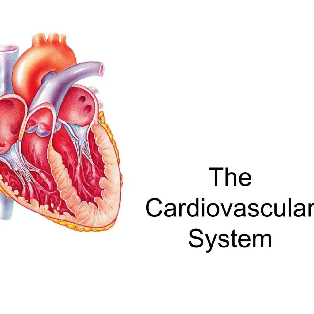
Cardiovascular System Anatomy: For the love of the Heart Cardiovascular System Anatomy Introduction to the Cardiovascular System The cardiovascular system, also known as the circulatory system, is a vast network responsible for transporting blood throughout the entire body. This system is essential for maintaining life and ensuring that every cell receives what it needs to function properly. Key Components The cardiovascular system is primarily composed of three main parts, working in perfect concert. 1. The Heart This muscular organ, roughly the size of a clenched fist, is the central pump of the system. It continuously contracts and relaxes, driving blood through the vast network of vessels. 2. Blood Vessels Arteries: Carry oxygenated blood away from the heart. Their thick, muscular walls withstand high pressure. Veins: Carry deoxygenated blood back to the heart. Their thinner walls and internal valves prevent backward blood flow. Capillaries: The smallest vessels, forming vast networks that connect arteries and veins. Their ultra-thin walls allow for the efficient exchange of gases, nutrients, and waste products with the body’s cells. 3. Blood Plasma: The liquid matrix, mostly water, that carries dissolved nutrients, hormones, and waste. Red Blood Cells (Erythrocytes): Contain hemoglobin to transport oxygen from the lungs to tissues and return carbon dioxide. White Blood Cells (Leukocytes): Key components of the immune system, defending the body against pathogens. Platelets (Thrombocytes): Small cell fragments essential for initiating the blood clotting process to stop bleeding. Primary Functions The cardiovascular system performs several indispensable functions to maintain homeostasis and sustain life. Transport of O₂ & Nutrients: Delivers oxygen and nutrients to every cell for energy and metabolic processes. Removal of Waste: Collects metabolic waste like CO₂ and urea and transports them to the lungs and kidneys for excretion. Hormone Transport: Acts as a delivery system for hormones, carrying them from glands to their target organs. Temperature Regulation: Distributes heat throughout the body and regulates its dissipation to maintain a stable internal temperature. Protection Against Disease: Circulates white blood cells and antibodies as part of the immune system to fight infections. Blood Clotting: Platelets and clotting factors in the blood prevent excessive blood loss at sites of injury. Anatomy of the Heart and Great Vessels The heart is a hollow, muscular organ located in the mediastinum, the central compartment of the thoracic cavity, nestled between the lungs. It sits slightly to the left of the midline, resting on the diaphragm. Its pointed end, the apex, points inferiorly and to the left, while the broader base points superiorly and to the right. I. The Pericardium: The Heart’s Protective Sac The heart is encased in a double-walled sac called the pericardium. It serves to anchor the heart, prevent it from overfilling, and provide a frictionless environment for its constant beating. Fibrous Pericardium: The tough, outermost layer made of dense connective tissue. It anchors the heart to the diaphragm and great vessels, preventing overfilling and providing a strong protective barrier. Serous Pericardium: A thinner, delicate inner layer, itself composed of two sub-layers: Parietal Layer: Lines the inside of the fibrous pericardium. Visceral Layer (or Epicardium): Adheres directly to the surface of the heart muscle. Pericardial Cavity: The potential space between the parietal and visceral layers, containing a thin film of serous fluid that acts as a lubricant to eliminate friction during heartbeats. II. Layers of the Heart Wall The wall of the heart itself is composed of three distinct layers, from superficial to deep. Epicardium: The outermost layer (and also the visceral layer of the serous pericardium). It is a protective layer that contains the coronary blood vessels and adipose tissue. Myocardium: The thick, muscular middle layer composed of cardiac muscle cells (cardiomyocytes). This is the contractile layer responsible for the heart’s pumping action. Its thickness is greatest in the left ventricle. Endocardium: The innermost layer, a thin, smooth membrane that lines the heart’s chambers and covers the valves. Its smooth surface minimizes friction and prevents clot formation. III. Chambers of the Heart The heart is a four-chambered organ, divided by a muscular septum into right and left sides. This separation is crucial for ensuring that oxygen-poor and oxygen-rich blood do not mix. Right Atrium (RA) Receives deoxygenated blood from the body via the Superior Vena Cava (SVC), Inferior Vena Cava (IVC), and Coronary Sinus. Pumps blood to the right ventricle. Left Atrium (LA) Receives oxygenated blood from the lungs via the four pulmonary veins. Pumps blood to the left ventricle. Right Ventricle (RV) Receives deoxygenated blood from the right atrium. Pumps deoxygenated blood to the lungs via the pulmonary artery. Left Ventricle (LV) Receives oxygenated blood from the left atrium. The strongest chamber; pumps oxygenated blood to the entire body via the aorta. IV. Heart Valves: Ensuring Unidirectional Blood Flow The heart contains four valves that act as one-way doors, preventing the backflow of blood (regurgitation). They open and close passively in response to pressure changes within the chambers. Atrioventricular (AV) Valves: Located between the atria and ventricles. Tricuspid Valve: Between the right atrium and right ventricle (has three cusps). Mitral (Bicuspid) Valve: Between the left atrium and left ventricle (has two cusps). The AV valves are anchored by fibrous chordae tendineae (“heart strings”) to papillary muscles in the ventricles. When the ventricles contract, these muscles pull on the cords, preventing the valve flaps from being pushed back up into the atria. Semilunar (SL) Valves: Located at the exit of the ventricles, preventing blood from flowing back from the great arteries. Pulmonary Valve: Between the right ventricle and the pulmonary artery. Aortic Valve: Between the left ventricle and the aorta. V. Great Vessels of the Heart These are the major blood vessels that are directly connected to the heart, responsible for carrying blood to and from its chambers. Superior & Inferior Vena Cava (SVC & IVC): Bring deoxygenated blood from the upper and lower body, respectively, to the right atrium. Pulmonary Artery: Carries deoxygenated blood from the right ventricle to the lungs. Note: It’s an artery because it carries blood AWAY from the

Common Abnormalities: Teratology and Teratogenesis
Anatomy

Common Abnormalities: Teratology and Teratogenesis

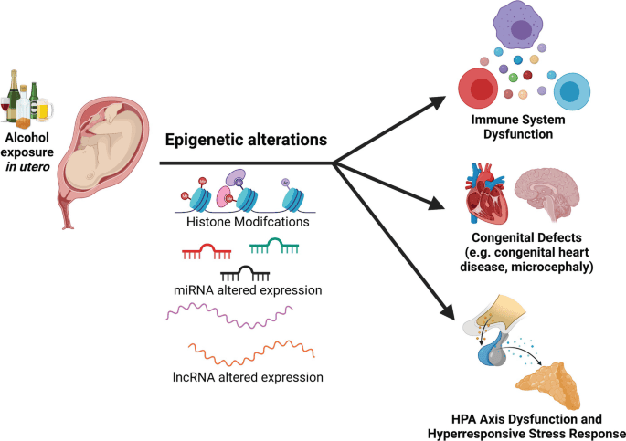
Common Abnormalities: Teratology and Teratogenesis Common Abnormalities: Teratology and Teratogenesis 1. Teratology Teratology is the scientific study of abnormal physiological development, specifically focusing on the causes, mechanisms, and patterns of birth defects, also known as congenital malformations. The term comes from the Greek “teras,” meaning monster or marvel. Key Concepts in Teratology Congenital Malformations (Birth Defects) are structural, functional, or metabolic abnormalities present at birth. These can range from minor cosmetic issues to severe, life-threatening conditions. Not all congenital conditions are visible at birth (e.g., some heart defects or metabolic disorders). They are classified into several distinct categories based on their origin. Malformation A primary structural defect resulting from an intrinsically abnormal developmental process. The blueprint itself was flawed from the beginning. Example: Polydactyly (extra fingers/toes), Spina Bifida. Disruption A defect resulting from the extrinsic breakdown of, or interference with, an originally normal developmental process. The blueprint was normal, but something damaged the structure as it was forming. Example: Limb amputation due to amniotic bands wrapping around it. Deformation An abnormal form, shape, or position of a body part caused by extrinsic mechanical forces acting on a normally developed structure. Example: Clubfoot due to intrauterine crowding, limiting space for the feet to grow properly. Dysplasia An abnormal organization of cells into tissues. The problem lies in how the cells themselves are structured and arranged. Example: Skeletal dysplasias like achondroplasia (a form of dwarfism). Syndrome A group of anomalies that occur together and have a specific, common, known cause. Example: Down syndrome (caused by Trisomy 21), Fetal Alcohol Syndrome. Association A non-random occurrence of two or more anomalies that appear together more often than by chance, but for which a common cause has not yet been identified. Example: VACTERL association (Vertebral, Anal, Cardiac, Tracheo-Esophageal, Renal, Limb defects). Factors Contributing to Birth Defects While teratogens are a major focus, it’s important to understand the broader categories of factors that can lead to congenital malformations. Causes 40-50%: Unknown Causes 20-25%: Genetic Factors (chromosomal, single gene) 20-25%: Multifactorial Inheritance (genes + environment) ~10%: Environmental Factors (Teratogens) 2. Teratogenesis Teratogenesis is the process by which a teratogen (an agent that causes birth defects) acts on an embryo or fetus to produce a congenital malformation. The study of this process is governed by a set of foundational concepts known as Wilson’s Principles. Principle 1: Susceptibility (Genotype) The genetic makeup of the embryo and mother determines their susceptibility to a teratogen. What harms one individual may have no effect on another due to genetic differences in metabolism and cellular repair. Principle 2: Dosage & Duration The amount of the teratogen and the length of exposure are critical. Generally, a higher dose or a longer duration of exposure increases the risk and severity of the resulting defect. Principle 3: Timing of Exposure (Critical Periods) This is arguably the most crucial principle. The susceptibility of an organ system to a teratogen varies dramatically with its stage of development. Pre-implantation Period (Weeks 1-2) The “all-or-nothing” period. Exposure to a teratogen usually results in either the death of the embryo or its complete recovery with no defects, as the cells are still totipotent and can be replaced. Embryonic Period (Weeks 3-8) The most sensitive period for major malformations. This is when organogenesis occurs, and each organ system has its own critical window of vulnerability (e.g., heart: weeks 3-5; CNS: weeks 3-16+). Fetal Period (Weeks 9 to Birth) Exposure during this period generally does not cause major structural defects but can lead to functional problems, growth retardation, and minor abnormalities, especially in the still-developing brain. Principle 4: Mechanisms Teratogens exert their effects through specific cellular and molecular mechanisms, such as interfering with cell proliferation or migration, inducing cell death (apoptosis), or disrupting biochemical pathways. Principle 5: Manifestations The final outcome of teratogenic exposure can be one of four manifestations: death, malformation, growth retardation, or functional deficit. Classes of Teratogens Teratogens are substances that can cause birth defects when a fetus is exposed during pregnancy. They can be broadly categorized into several classes, each with well-documented examples and associated defects. The risk and severity of abnormalities depend on the type of agent, timing, dosage, and duration of exposure. Infectious Agents (TORCH Infections) The acronym TORCH helps remember some of the most well-known infectious teratogens: Toxoplasmosis: A parasitic infection that can cause hydrocephalus and intracranial calcifications. Others (e.g., Syphilis, Varicella-Zoster, Zika, Parvovirus B19): Zika is known for causing microcephaly, while syphilis can lead to congenital deafness and other issues. Rubella (German measles): Can result in a classic triad of cataracts, cardiac malformations, and deafness. Cytomegalovirus (CMV): A common virus that can cause microcephaly, hearing loss, and intellectual disability. Herpes Simplex Virus: Can lead to skin lesions, microcephaly, and eye problems. Drugs and Chemicals Thalidomide: A classic example that caused severe limb reduction defects (phocomelia). Alcohol (Ethanol): The leading preventable cause of non-genetic birth defects, leading to Fetal Alcohol Syndrome (FAS) with distinct facial anomalies, growth retardation, and CNS dysfunction. Tobacco & Nicotine: Smoking is associated with low birth weight, premature delivery, and can affect the development of the fetal brain and lungs. Retinoids (e.g., Isotretinoin/Accutane): Highly teratogenic, causing severe CNS, facial, cardiac, and ear malformations. Anticonvulsants (e.g., Valproic Acid, Phenytoin): Associated with neural tube defects, cleft lip/palate, and cardiac defects. ACE Inhibitors: Can cause renal failure and oligohydramnios (insufficient amniotic fluid). Warfarin: An anticoagulant that can cause skeletal abnormalities, including chondrodysplasia punctata. Certain Antibiotics (e.g., Tetracycline): Can cause yellow staining of teeth and affect long bone growth. Recreational Drugs (e.g., Cocaine, Heroin): Can lead to low birth weight, withdrawal symptoms in the newborn, and learning or behavioral problems. Environmental Toxins Heavy Metals (e.g., Mercury, Lead): Can cause significant CNS damage and developmental delays. Mercury is often found in certain types of fish, and lead can be in old paint and pipes. Polychlorinated Biphenyls (PCBs): Industrial chemicals that can lead to developmental and neurological problems. Herbicides and Industrial Solvents: Exposure to certain chemicals used in agriculture and manufacturing can be harmful. Physical Agents Ionizing

Anatomy

Fetal Membranes, Placenta, Cord and Circulation

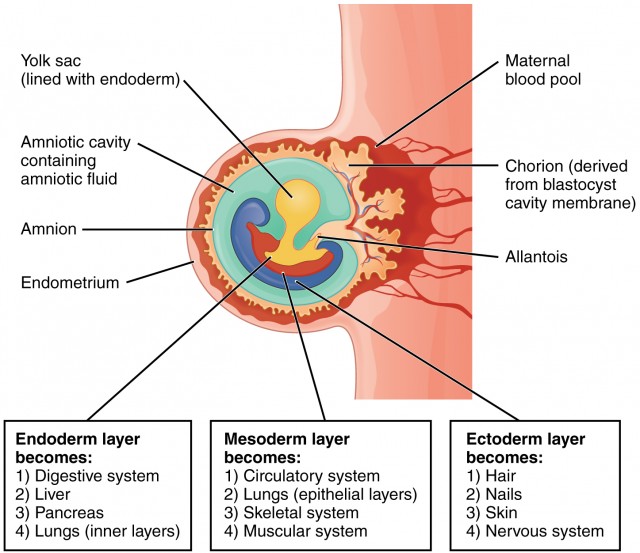
Fetal Membranes, Placenta, Cord and Circulation: Safety and Feeding Fetal Membranes, Placenta, Cord and Circulation The fetal membranes and the placenta are temporary, yet essential, organs that develop alongside the embryo and fetus. They provide a complete life-support system, handling protection, nourishment, gas exchange, waste removal, and hormonal regulation critical for successful intrauterine development. They are expelled from the body after birth. Formation of Embryonic Cavities and Membranes The period of early embryonic development (roughly Day 8 to Day 12-14 post-fertilization) is characterized by the rapid formation of several extraembryonic structures, which are vital for the embryo’s survival and subsequent development. These include the amniotic cavity, primary and secondary yolk sacs, and the chorionic cavity, along with their associated membranes. A. Formation of the Amniotic Cavity and Amnion Timeline: Begins around Day 8 post-fertilization. Process: Cavity Formation: As the blastocyst implants, a small space appears within the epiblast, which is the dorsal layer of the bilaminar germ disc (formed from the Inner Cell Mass). Enlargement: This space rapidly expands to become the amniotic cavity. Amnioblast Differentiation: Cells from the epiblast adjacent to the cytotrophoblast differentiate into thin, flattened cells called amnioblasts. Amniotic Membrane Formation: These amnioblasts, along with a layer of extraembryonic mesoderm, form the amnion, which eventually encloses the entire amniotic cavity. Roof and Floor: The roof is formed by the amnion/cytotrophoblast, while the floor is formed by the epiblast of the bilaminar germ disc. Key Features & Function of the Amnion/Amniotic Fluid: Amniotic Sac: The amnion forms the inner lining of the amniotic sac, which will eventually surround the entire embryo and then fetus. Amniotic Fluid: The cavity fills with amniotic fluid. Initially derived from maternal blood, it is later maintained largely by fetal urine excretion and fetal swallowing. It serves crucial functions: Protection: Acts as a shock absorber against mechanical trauma. Temp Regulation: Maintains a constant, optimal intrauterine temperature. Symmetry & Movement: Allows symmetrical external growth and free movement for proper musculoskeletal development (preventing contractures). Prevents Adhesion: Stops the developing embryo from physically sticking to the amnion (which would cause amniotic band syndrome). Lung/GI Development: Fetal swallowing of the fluid actively aids the maturation of the GI tract; while fetal “breathing” movements pull fluid into the lungs, providing the mechanical stretch essential for pulmonary development. Clinical Application Amniotic Fluid Volume Abnormalities Oligohydramnios (Too little fluid): Often caused by placental insufficiency or fetal renal agenesis (inability to produce urine). It can lead to Potter sequence (flattened face, clubbed feet, pulmonary hypoplasia) because the fetus is compressed without the fluid cushion. Polyhydramnios (Too much fluid): Often caused by fetal swallowing defects (e.g., esophageal atresia, anencephaly) or maternal diabetes. It can lead to premature rupture of membranes and preterm labor. B. Formation of the Yolk Sac Timeline: Primary yolk sac begins around Day 9; Secondary yolk sac around Day 12-13. 1. Primary Yolk Sac (Exocoelomic Cavity) – Day 9 Cells from the hypoblast (ventral layer) migrate and line the inner surface of the cytotrophoblast. These cells form a thin membrane called the exocoelomic membrane (Heuser’s membrane). This membrane + hypoblast encloses the primary yolk sac. Position: The bilaminar disc lies between the Amniotic Cavity (dorsal) and the Primary Yolk Sac (ventral). 2. Extraembryonic Mesoderm – Day 10-11 A new layer of loose connective tissue appears and fills the space between the exocoelomic membrane/amnion externally and the cytotrophoblast internally. 3. Secondary Yolk Sac (Definitive) – Day 12-13 The primary sac constricts due to chorionic cavity expansion. A smaller, definitive secondary yolk sac forms from a portion of the primary sac. The larger pinched-off part degenerates into exocoelomic cysts. Key Features & Function of the Yolk Sac: Nutrition Early Nutrient Transfer Plays a vital role in nutrient transfer to the embryo during weeks 2 and 3, before the uteroplacental circulation is fully functional. Blood Supply Hematopoiesis It is the primary site of early blood cell formation (Weeks 3-6). It produces primitive nucleated red blood cells expressing embryonic hemoglobin. After week 6, the fetal liver takes over this function. Reproduction Primordial Germ Cells Precursors to sperm/eggs originate in the wall of the yolk sac around week 3. They migrate along the hindgut to reach the developing gonads by week 5. Fate Vestigial Structure In humans, the yolk sac does not contain yolk. It is small, regresses rapidly by week 20, and is incorporated into the primitive gut tube and the umbilical cord. C. Formation of the Chorionic Cavity and Chorion Timeline: Begins around Day 11-12. Process: Vacuole Formation: Numerous large spaces and vacuoles appear within the extraembryonic mesoderm. Coalescence: These fuse to form a large, isolated cavity called the chorionic cavity (extraembryonic coelom). Suspension of Embryo: The embryo (with its amnion and yolk sac) is suspended in this massive cavity by the connecting stalk (which is the future umbilical cord). The Chorion (Outer Wall) The chorion forms the wall of the chorionic cavity and is strictly formed by three layers (from outside to inside): Syncytiotrophoblast (outermost) Cytotrophoblast Somatic layer of extraembryonic mesoderm (innermost) Functions: Chorionic Villi: Gives rise to the villi (the functional exchange units of the placenta). Protection: Forms an additional protective layer around the entire conceptus. Part of Placenta: The villous part (the chorion frondosum) forms the fetal component of the placenta, while the smooth part (chorion laeve) eventually fuses with the amnion. Summary of Relationships (Day 12-14): Central: Bilaminar germ disc centrally located. Dorsal: Amniotic cavity. Ventral: Secondary yolk sac. Surrounding all: Chorionic cavity (enclosed entirely by the Chorion). Bridge: Connecting stalk linking the bilaminar disc to the chorion. D. The Allantois: Development and Significance Origin: Appears around Day 16-18 as a small, sausage-shaped diverticulum (outpouching) from the caudal wall of the yolk sac (specifically the hindgut), extending directly into the connecting stalk. Vascular Development: This is its most significant role in humans. Blood vessels develop in the mesoderm wall of the allantois to become the umbilical arteries and the umbilical vein. These vessels extend through the connecting stalk to strictly link the embryonic and placental circulation. Urinary Bladder

Scroll to Top