
On this page 9 sections
The Cell Cycle

The cell cycle describes the entire lifespan of a cell, from its formation after one division until it divides again. It consists of two main stages:
- Interphase: The period of cell growth, DNA replication, and preparation for division. This is the longest phase.
- M Phase (Mitotic Phase): The period of actual cell division, including mitosis (nuclear division) and cytokinesis (cytoplasmic division).
Interphase: The Preparation Phase
Interphase is not a resting phase but a highly active period of growth and metabolic activity, crucial for preparing the cell for division. It is divided into several sub-phases.
1. G₀ Phase (Gap 0 / Quiescent Phase)
This is an optional phase where cells exit the cell cycle and stop dividing, entering a state of dormancy or terminal differentiation. While metabolically active, they are not preparing for division.
Examples of G₀ Cells:
- Terminally Differentiated: Mature muscle and nerve cells often enter G₀ permanently.
- Reversible G₀: Liver cells and lymphocytes can re-enter the cycle if stimulated.
Significance:
Prevents uncontrolled cell growth and allows cells to perform their specialized roles.
2. G₁ Phase (Gap 1 / First Growth)
This is the first growth phase after a cell division. The cell is actively growing, synthesizing proteins and RNA, and expanding its cytoplasm by creating new organelles.
G₁ Restriction Point:
At this checkpoint, the cell decides whether to commit to division and proceed to the S phase or to exit the cycle into the G₀ phase.
3. S Phase (Synthesis Phase)
The "synthesis" phase, where the most crucial event for cell division occurs: DNA replication.
Key Activities:
- Each of the 46 chromosomes is duplicated, resulting in two identical sister chromatids.
- New histone proteins are synthesized to package the newly replicated DNA.
- By the end of S phase, the cell contains double the amount of DNA.
4. G₂ Phase (Gap 2 / Second Growth)
The second growth phase and final preparatory stage before the cell enters mitosis.
G₂ Checkpoint:
The cell checks the replicated DNA for errors or damage. If damage is found, it attempts repairs. If the damage is irreparable, the cell may trigger programmed cell death (apoptosis) to prevent passing on mutations.
Cell Division
Cells reproduce through a fundamental process called cell division. This is essential for growth, repair, and reproduction in all living organisms. There are two primary types:
Mitotic Cell Division (Mitosis)
- Role: Growth and repair of tissues.
- Occurs in: Somatic cells (e.g., neurons, epithelial, muscle).
- Outcome: Two identical daughter cells.
- Chromosomes: 46 (same as parent).
Meiotic Cell Division (Meiosis)
- Role: Production of sex cells (sperm/ova).
- Occurs in: Reproductive organs only.
- Outcome: Four daughter cells.
- Chromosomes: 23 (half of parent).
Mitotic Cell Division: The Basis of Growth and Repair
Mitotic cell division is a continuous process crucial for increasing the number of cells for growth and replacing worn out, damaged, or dead cells. However, not all cells divide at the same rate—epithelial cells divide almost continuously, while mature muscle cells largely lose the ability to divide.
Key Processes in Mitotic Cell Division:
- Replication of Chromosomes: Creating exact copies of the genetic material (occurs in S phase).
- Mitosis: The division of the nucleus.
- Cytokinesis: The division of the cytoplasm.
Mechanism of Mitotic Division
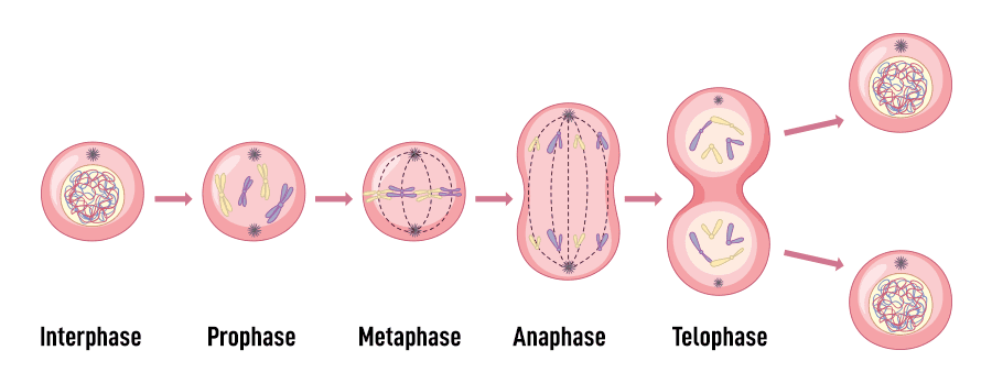
During mitosis, the cell's diffuse chromatin condenses into visible chromosomes. The centrosome duplicates, and each copy moves to opposite ends (poles) of the cell. They create spindle fibers that grab onto the chromosomes and pull them apart, ensuring that when the cell finally divides, each new daughter cell receives its own identical copy of the genetic material.
Mitotic Phases
Once interphase is complete, the cell enters mitosis. While it's a continuous process, we divide it into four sequential phases for easier understanding.
A. Prophase
- • Replicated chromosomes coil and condense, becoming visible as two identical sister chromatids joined at a centromere.
- • The nuclear envelope disappears.
- • Centrioles migrate to opposite poles, and the mitotic spindle begins to form.
B. Metaphase
- • The replicated chromosomes line up precisely at the cell's equator (the metaphase plate).
- • The centromere of each chromosome is attached to the spindle fibers.
C. Anaphase
- • Centromeres divide, and the sister chromatids separate.
- • Each separated chromatid is now considered an individual chromosome.
- • Spindle fibers pull the chromosomes towards opposite poles of the cell.
D. Telophase
- • The spindle fibers disassemble.
- • A new nuclear envelope forms around each set of chromosomes at the poles.
- • Chromosomes uncoil back into their thread-like chromatin form.
Cytokinesis: Division of the Cytoplasm
Usually occurring during late anaphase and telophase, cytokinesis is the final step. A furrow forms in the plasma membrane, deepens, and eventually pinches the parent cell into two separate, genetically identical daughter cells, each with its own nucleus and cytoplasm.
Cell Cycle Disorders: When Regulation Fails
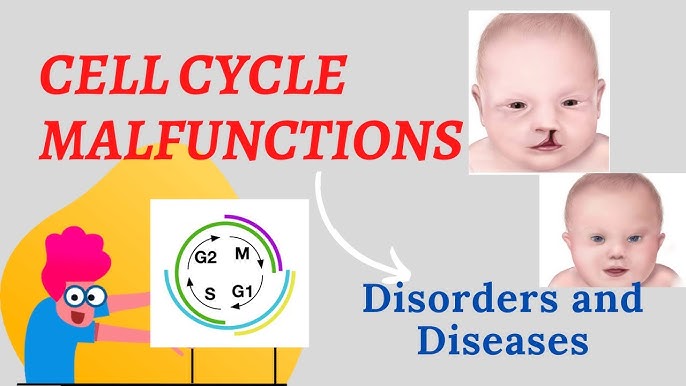
The cell cycle is a tightly regulated sequence of events with a series of checkpoints that monitor the cell's health and DNA integrity. When these regulatory mechanisms fail, the cell cycle can become dysregulated, leading to various disorders, most notably cancer.
Cells have checks and balances, and special proteins called cyclins constantly monitor the cell's health. Unhealthy cells normally self-destruct via apoptosis. Cancer cells, however, lose this ability. For many cells, the G1 checkpoint is the most important; if a cell receives a "go-ahead" signal here, it will usually complete division. If not, it enters a non-dividing state called the G₀ phase.
Key Regulators of the Cell Cycle
Before discussing disorders, it's essential to understand the main players that normally control the cell cycle:
Cyclins and CDKs
These are the "engine" of the cell cycle. Cyclin-Dependent Kinases (CDKs) are enzymes that are activated by binding to Cyclins. Different Cyclin-CDK complexes drive the cell through each phase.
Cell Cycle Checkpoints
Critical control points that monitor conditions. The main ones are the G1 Checkpoint (the "start" point), the G2 Checkpoint (checks DNA replication), and the M Checkpoint (checks spindle attachment).
Tumor Suppressor Genes
These are the "brakes." They encode proteins that inhibit cell division or repair DNA. Key examples are p53 ("Guardian of the Genome") and Rb (Retinoblastoma protein).
Proto-oncogenes & Oncogenes
Proto-oncogenes are the "accelerators" that promote normal cell growth. When mutated, they become Oncogenes, which are stuck in the "on" position, causing uncontrolled growth.
Causes of Cell Cycle Disorders
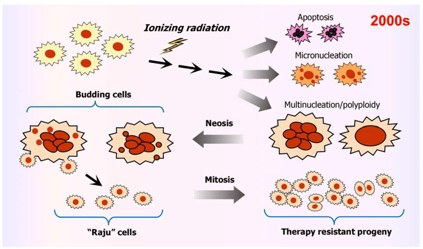
Disorders arise when the delicate balance of these activators and inhibitors is disrupted, often due to:
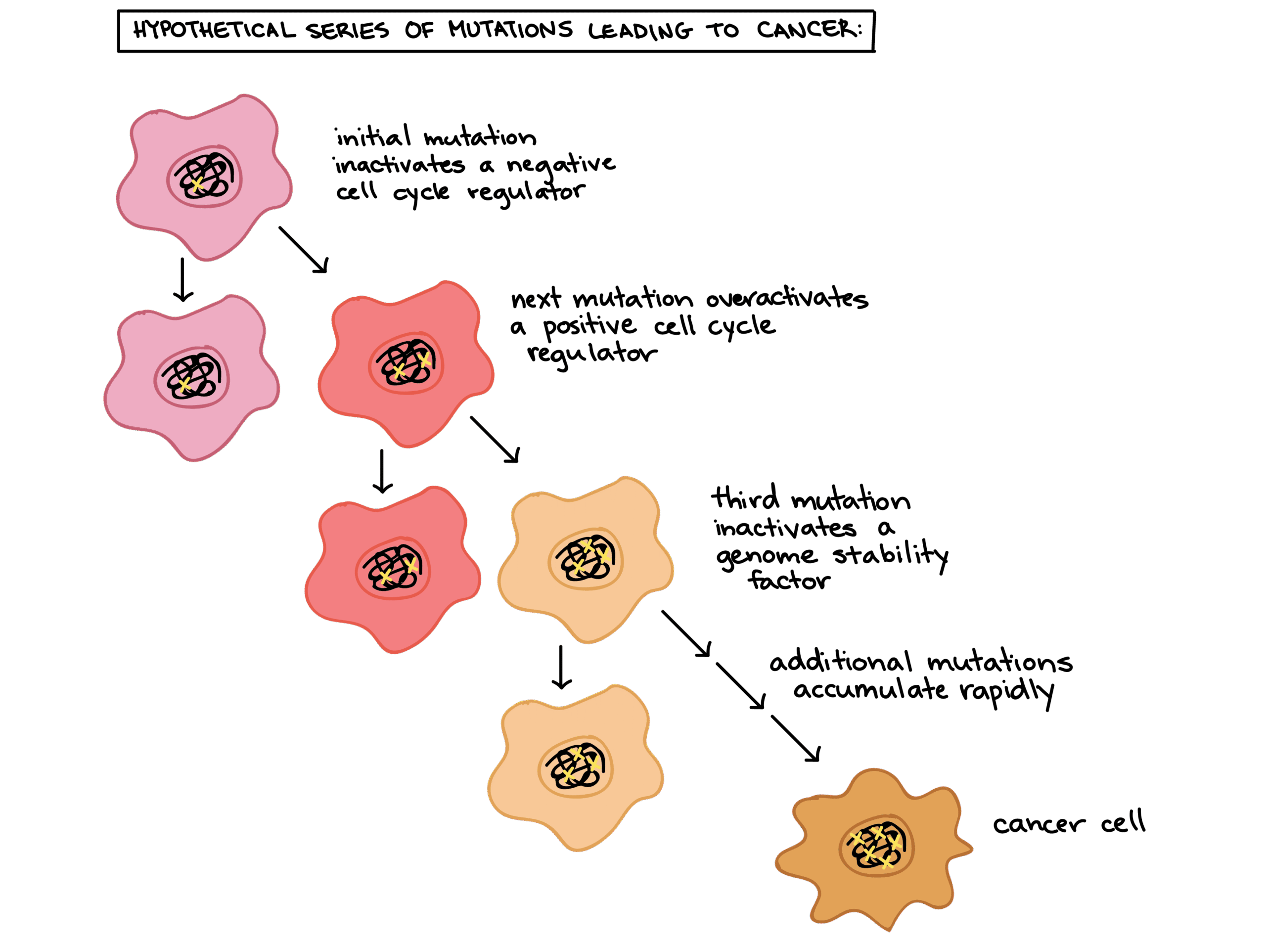
- Genetic Mutations: Inactivating "brake" genes (tumor suppressors) or activating "accelerator" genes (proto-oncogenes).
- Epigenetic Changes: Altering gene expression without changing the DNA sequence, such as silencing a tumor suppressor gene.
- Viral Infections: Some viruses, like HPV, produce proteins that disable key regulators like p53 and Rb.
- Environmental Factors: Carcinogens and radiation that cause DNA damage and lead to mutations.
Consequences & Types of Cell Cycle Disorders
1. Cancer (Malignancy)
Cancer is the primary disease of uncontrolled cell division. Cancer cells ignore the normal signals that control the cell cycle. They enter the S phase without waiting for a signal and become "immortal," escaping the normal limit on cell divisions. This is typically caused by multiple mutations that activate oncogenes and inactivate tumor suppressor genes.
Hallmarks of Cancer Cells
- Sustained proliferative signaling
- Evasion of growth suppressors
- Resistance to cell death
- Enabling replicative immortality
- Inducing angiogenesis
- Activating invasion & metastasis
2. Aneuploidy (Incorrect Chromosome Number)
A failure of the M checkpoint can lead to an unequal distribution of chromosomes during cell division. While most aneuploid cells die, some survive and can lead to genetic disorders like Down Syndrome (Trisomy 21). Aneuploidy is also a common feature of cancer cells.
3. Developmental & Premature Aging Disorders
Precise control of the cell cycle is critical during embryonic development. Errors can lead to underdevelopment (e.g., microcephaly) or overgrowth syndromes. Similarly, some premature aging syndromes are linked to defects in DNA repair mechanisms that impact cell cycle checkpoints.
Therapeutic Implications
Understanding these disorders is fundamental to developing treatments. Many cancer therapies are designed to target the cell cycle:
- Chemotherapy: Uses drugs that damage DNA or disrupt the mitotic spindle to preferentially kill rapidly dividing cancer cells.
- Targeted Therapies: Newer drugs that specifically inhibit mutated or overactive molecules, such as CDK inhibitors.
- Immunotherapy: Harnessing the immune system to recognize and destroy cancer cells that have evaded normal cell cycle controls.
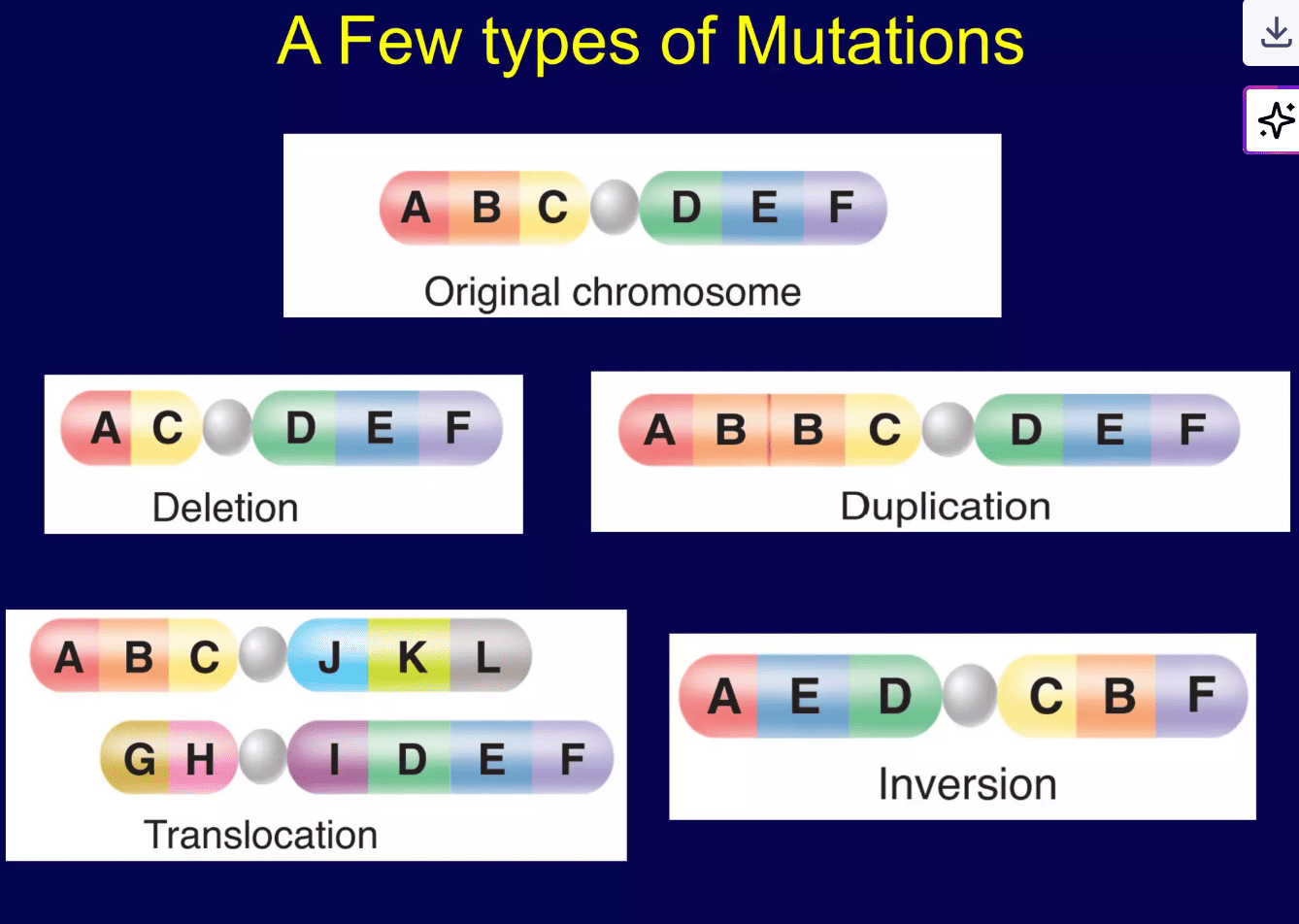
Chromosomal Mutations: Large-Scale Genetic Changes
Chromosomal mutations are significant changes affecting the structure or number of entire chromosomes. These large-scale alterations are distinct from gene (point) mutations, which involve changes to individual DNA base pairs within a gene. Such structural changes often arise from errors during meiosis or from exposure to mutagens.
Lesson map
Further reading
For deeper study, compare this lesson with current guidance and standard references from recognised medical and public-health organisations.