Doctors Revision

Author name: doctorsrevision@gmail.com

Body planes and cavities
Anatomy

Anatomical Position, Directional Terms & Planes

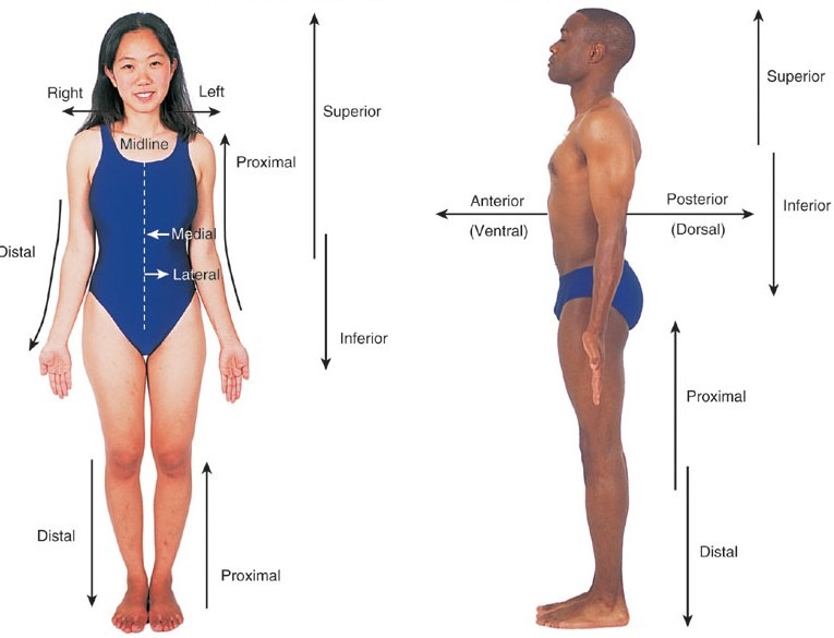
Anatomical Position, Directional Terms & Planes Anatomical Position, Directional Terms & Planes Main Questions to Answer What is the anatomical position, and why is it the universal standard? What are the specific directional terms used to navigate the human body? What are the anatomical planes and sections used in medical imaging? How do we correctly describe specific body movements and clinical patient positions? The Problem: Why Do We Need a Standard? When we describe where something is on the human body, it can quickly become confusing because the body is incredibly mobile. For example, if a person is holding their hand with the palm facing up, a mole on it is on the “front.” But if they turn their hand so the palm faces down, is that mole now on the “inside,” the “back,” or still the “front”? This ambiguity is highly dangerous in medicine (e.g., a surgeon operating on the wrong side of a limb). This confusion is exactly why anatomists and medical professionals created a single, rigid standard position to use as an absolute reference point, no matter how the body is actually positioned in real life. The Golden Rule of Anatomy No matter how a patient or a body in an image is actually positioned (sitting, lying down, upside down, or curled up), you always describe their anatomy as if they were standing in the Anatomical Position. Most Important Rule: All descriptions are from the patient’s point of view, not yours. The patient’s left is always their left, even if it is on your right side when you look at them. The Solution: The Anatomical Position The Anatomical Position is the universal starting point for describing any part of the body. It acts as the “Zero Coordinate” for the human map. The Strict Rules of Anatomical Position: Body Posture: The person is standing up straight (erect). Head and Eyes: They are facing directly forward, with eyes looking straight ahead. Lower Limbs: The legs are together or slightly apart (shoulder-width), with the feet flat on the floor and toes pointing directly forward. Upper Limbs: Their arms are hanging down at their sides. Hands (Crucial Detail): Their palms are facing forward (supinated). Because the palms face forward, their thumbs are pointing away from the body (laterally). This ensures the two bones of the forearm (radius and ulna) are parallel and not crossed over each other. Anatomical Terms of Position (Directional Terms) These terms are like a GPS for the body. They are used in pairs of opposites and help describe where one body part is strictly in relation to another. To accurately describe body parts and their positions, we use this specific set of directional terms. Front / Back Anterior (Ventral): Towards the front of the body. Example: “The sternum (breastbone) is anterior to the vertebral column (spine).” Example: “The kneecap is located on the anterior side of the leg.” Posterior (Dorsal): Towards the back of the body. Example: “The vertebral column (spine) is posterior to the sternum.” Example: “The shoulder blades are located on the posterior chest wall.” Top / Bottom (Axial Skeleton) Superior (Cranial/Cephalic): Towards the top or head. Used only for the head, neck, and trunk. Example: “The nose is superior to the mouth.” Example: “The skull is cranial to the neck.” Inferior (Caudal): Towards the bottom, feet, or tail. Used only for the head, neck, and trunk. Example: “The mouth is inferior to the nose.” Example: “The neck is caudal to the skull.” Midline / Sides Medial: Towards the imaginary midline of the body. Example: “The nose is medial to the ears.” Example: “The heart is medial to the lungs.” Lateral: Away from the midline of the body; towards the sides. Example: “The ears are lateral to the nose.” Example: “The arms are lateral to the chest.” Depth Superficial (External): Situated closer to the surface of the body. Example: “The skin is superficial to the skeletal muscles.” Deep (Internal): Situated further inward, away from the surface of the body. Example: “The bones are deep to the skin and muscles.” Limbs (Appendicular Skeleton) Proximal: Closer to the origin or attachment point of a limb to the main trunk of the body. Example: “The elbow is proximal to the wrist.” Example: “The femur (thigh) is proximal to the knee.” Distal: Farther away from the origin or attachment point of a limb. Example: “The wrist is distal to the elbow.” Example: “The toes are distal to the ankle.” Advanced / Additional Terms Ipsilateral: On the same side of the body. Example: “The right hand and right foot are ipsilateral.” Contralateral: On the opposite side of the body. Example: “A stroke on the right side of the brain causes contralateral paralysis on the left side of the body.” Rostral: Towards the nose (specifically used in neuroanatomy to describe the brain). Student Pitfall: Proximal/Distal vs. Superior/Inferior Students often make the mistake of saying “The wrist is inferior to the elbow.” While technically lower to the ground, anatomists strictly reserve Superior/Inferior for the Head and Trunk (Axial skeleton). For the arms and legs (Appendicular skeleton), you must use Proximal and Distal. Why? Because if you raise your hand above your head, your wrist is suddenly physically higher than your elbow. But anatomically, the wrist is always Distal to the elbow, no matter where your arm is reaching! Anatomical Planes and Sections To study internal anatomy, or to view the body using medical imaging (like CT scans or MRIs), the body is often sectioned (cut) along an imaginary flat 2D surface called a plane. The cut itself is called a section. 1. Sagittal Plane: A vertical line dividing the body into left and right parts. Midsagittal (Median) Plane: Cuts exactly down the absolute midline, creating equal left and right halves. Parasagittal Plane: An off-center cut, creating unequal left and right portions. 2. Coronal (Frontal) Plane: A vertical line dividing the body into anterior (front) and posterior (back) parts. Memory Aid: Think of a crown (corona) sitting across the top of your
anatomy lecture doctors revision
Anatomy

Anatomy Introduction

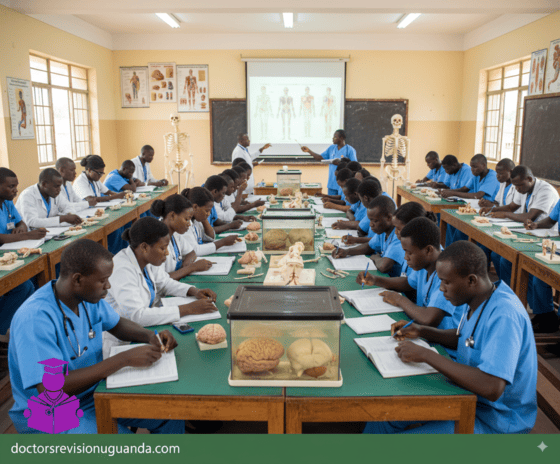
Intro to Anatomy: Terms & Concepts Introduction to Anatomy Anatomy is the scientific study of the structural organization of the human body, ranging from microscopic cells to large, visible structures like organs and bones. Derived from the Greek word for “cutting apart,” it explores how these parts are arranged to form functional systems, often in conjunction with physiology, which focuses on function. The History of Anatomy For centuries, the dissection of human bodies was taboo in many societies. The journey of anatomical study is marked by key historical milestones: Claudius Galenus: A second-century Greek physician who learned about the human form by performing vivisections on pigs. Leonardo da Vinci: Poked around in dead bodies and created beautifully detailed anatomical drawings until the Pope made him stop. 17th and 18th Centuries: Certified anatomists were allowed to perform tightly regulated human dissections. These were often popular public events attended by artists like Michelangelo and Rembrandt. The Anatomy Act (1832): The study of human anatomy became such a craze in Europe that grave robbing became a lucrative occupation until Britain passed this act, which provided students with corpses of executed murderers. Modern Day: Today, students of anatomy and physiology still use educational cadavers, which are donated by volunteers. Andreas Vesalius: Known as the ‘Father of Anatomy’. He was the first to carry out dissection to closely observe the inner structure and construction of the human body. Key Concepts in Anatomy and Physiology Function Follows Form This is the core principle of anatomy. It means that the shape of a body part (its structure or form) is perfectly designed for its job (its function). The function of a cell, organ, or whole organism always reflects its form. This is also known as the Complementarity of Structure and Function. Example: Form & Function Think of a fork. It has prongs (its form) specifically to help it pick up food (its function). Your teeth are a perfect biological example. Your sharp front teeth are for tearing food, while your flat back teeth are for grinding. Their shape is perfect for their job. Hierarchy of Organization The human body is organized in a hierarchical manner, from the smallest chemical components to the entire organism. Levels of Organization in the Body: Chemical Level: Atoms and molecules, the smallest units of matter. Cellular Level: Cells, the smallest units of living things. Tissue Level: Groups of similar cells that work together. Organ Level: Two or more tissue types performing a specific function. Organ System Level: Groups of organs working together for a common purpose. Organismal Level: The sum total of all structural levels working together to keep us alive. Homeostasis Homeostasis is the ability of all living systems to maintain stable internal conditions no matter what changes are occurring outside the body. Survival is all about maintaining this delicate balance. Example: Homeostasis Think of a thermostat. If the house gets too cold, the heat turns on. If it gets too hot, the A/C kicks in. Your body does this constantly. If you get hot, you sweat to cool down. If you get cold, you shiver to warm up. Your body is always working to keep your temperature, blood sugar, and many other factors in a perfect, stable range. Foundational Anatomical Terms Mastering the language of anatomy is the first step to understanding its complexities. This guide covers the foundational terminology you will encounter throughout your studies. These terms provide a universal standard for describing the structure and function of the human body. Human anatomy (ah-nat -o−-me−) is the study of the structure and organization of the body and the study of the relationships of body parts to one another. There are two subdivisions of anatomy: Gross anatomy involves the dissection and examination of various parts of the body without magnifying lenses. Microanatomy, also known as histology, consists of the examination of tissues and cells with various magnification techniques. Human physiology (fiz-e−-ol-o−-je−) is the study of the function of the body and its parts. Physiology involves observation and experimentation, and it usually requires the use of specialized equipment and materials. Term (Etymology) Definition Example Anatomy(ana = apart; tom = to cut) The study of the structure of living organisms. Studying the bones, muscles, and organs in a human cadaver to understand their physical arrangement. Appendicular(append = to hang) Pertaining to the upper and lower limbs. The appendicular skeleton includes the bones of the arms, legs, shoulders, and pelvis. Axial(ax = axis) Pertaining to the longitudinal axis of the body. The axial skeleton consists of the skull, vertebral column, and rib cage, forming the central support of the body. Body region(regio = boundary) A portion of the body with a special identifying name. The “cephalic region” refers to the head, while the “thoracic region” refers to the chest. Directional term(directio = act of guiding) A term that references how the position of a body part relates to the position of another body part. The nose is superior to the mouth, and the feet are inferior to the knees. The sternum (breastbone) is anterior to the spine. Effector(efet = result) A structure that functions by performing an action that is directed by an integrating center. In regulating body temperature, sweat glands are effectors that produce sweat to cool the body down when directed by the brain. Homeostasis(homeo = same; sta = make stand or stop) Maintenance of a relatively stable internal environment. The body maintaining a constant internal temperature of approximately 37°C (98.6°F) regardless of external temperature changes. Integrating center(integratus = make whole) A structure that functions to interpret information and coordinate a response. The brain acts as an integrating center when it receives signals that blood sugar is too high and then sends signals to the pancreas to release insulin. Metabolism(metabole = change) The sum of the chemical reactions in the body. The digestion of food into nutrients (catabolism) and the building of new tissues from those nutrients (anabolism) are both parts of metabolism. Parietal(paries = wall) Pertaining to the wall

histology introduction
Anatomy

Histology Introduction

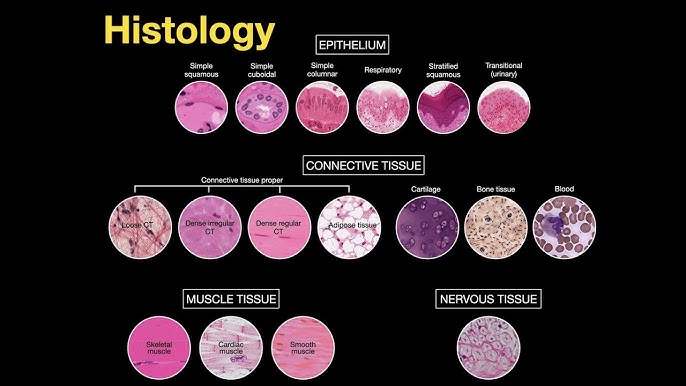
Introduction to Histology: The Study of Tissues What is Histology? Histology is the study of tissues. The word is derived from the Greek words “histo” (tissue) and “logos” (study). Therefore, histology is the science of the microscopic structure of cells, tissues, and organs. Simply put, it’s the study of tissues under a microscope. This field examines the microscopic anatomy of biological tissues and is fundamental to understanding the structure and function of the entire body. Why Health workers Need to Know Histology A strong foundation in histology is not just for doctors or researchers; it is a critical component of a professional nurse’s knowledge base. It elevates a nurse’s practice from task-oriented care to a deeper, more analytical level of patient management. Explains Form & Function Shows how tissue structure relates to its job, making treatments like oxygen therapy more meaningful. Identifies Disease Knowing normal tissue helps nurses recognize changes in disease, aiding in assessments like wound care. Enhances Practical Skills Improves participation in collecting and interpreting lab samples (e.g., biopsies). Informs Patient Education Allows nurses to better explain conditions and treatments, leading to more informed care. Medication Efficacy Helps nurses anticipate medication effects and side effects by understanding drug-cell interactions. Interdisciplinary Collaboration Facilitates clearer communication with pathologists, doctors, and other healthcare professionals. Methods of Histology Histology employs various techniques to prepare tissues for microscopic examination. These methods are crucial for preserving tissue integrity and allowing for the study of their structure and function. The main steps involve tissue preparation, staining, and microscopy. 1. Tissue Preparation Techniques This is the first and most critical step to preserve tissue and allow for thin sectioning. There are three main methods. a. Paraffin Technique This is the most common method for preparing tissues for routine histological examination. Procedures of the Paraffin Technique: Tissue Sample Collection: Obtaining the sample (biopsy, surgical excision). Fixation: Preserving the tissue, commonly with 4% formaldehyde (formalin). Dehydration: Removing water with increasing concentrations of alcohol. Clearing: Replacing alcohol with a clearing agent like xylene. Impregnation: Infiltrating the tissue with melted soft paraffin. Embedding: Transferring the tissue to hard paraffin to form a solid block. Sectioning: Cutting the block into very thin (5-8 µm) sections using a microtome. b. Celloidin Technique Provides superior support for both soft and hard tissues, such as bones, teeth, and large brain sections. Advantages: Excellent support for hard tissues Minimal shrinkage and distortion Good architectural preservation Disadvantages: Very time-consuming process Difficult to cut very thin sections Requires specialized technical skills c. Freezing Technique Rapidly prepares tissues by freezing, especially for urgent diagnoses during surgery. Advantages: Rapid diagnosis (minutes) Preserves molecules (DNA, RNA, proteins) Preserves antigens for immunostaining Disadvantages: Poor staining and cellular detail Inadequate fixation compared to paraffin Expensive and complex equipment (cryostat) 2. Staining Techniques Staining uses dyes to enhance the visibility of different tissue structures under the microscope. This is essential because most tissues are colorless. Common Stains and Their Uses: Hematoxylin and Eosin (H&E): The most common stain. Hematoxylin stains acidic structures like the nucleus blue, while Eosin stains basic structures like the cytoplasm pink. PAS (Periodic Acid-Schiff): Stains carbohydrates magenta. Useful for identifying basement membranes, mucus, glycogen, and fungal walls. Silver Stains (Reticulin): Stains reticular fibers black. Used in kidney, liver, and bone marrow biopsies. Trichrome Stains: Differentiates muscle (red), collagen (blue/green), and fibrin. Used for assessing fibrosis. Immunostains (Immunohistochemistry): Uses antibodies to detect specific molecules or cell types. Crucial for cancer diagnosis and classification. 3. Microscopy Techniques Microscopy is the use of microscopes to visualize small structures that are not visible to the naked eye. Light Microscope Uses natural or electric light to examine stained sections. This is the most commonly used microscope in routine histology. Electron Microscope Uses a beam of electrons for much higher magnification. TEM provides high-resolution internal details, while SEM provides detailed 3D surface images. Test Your Knowledge Check your understanding of the concepts covered in this post. 1. Histology is defined as the study of: Cells under a light microscope. Gross anatomy of organs. Tissues under a microscope. Chemical composition of biological structures. Rationale: The text explicitly states, “Histology therefore is the science of the microscopic structure of cells, tissues and organs OR simply put; The study of tissues under a microscope.” 2. Why is understanding histology important for nurses regarding medication efficacy? It helps them prescribe the correct dosage. It allows them to understand how drugs interact with specific cell types and tissues. It teaches them how to administer intravenous medications. It explains the cost-effectiveness of different drugs. Rationale: The text states under “Medication Efficacy,” “Understanding how drugs interact with specific cell types and tissues (e.g., receptors on cell surfaces) helps nurses anticipate medication effects and side effects.” 3. Which tissue preparation technique is most commonly used for routine histological examination due to its preservation and hardening properties? Celloidin Technique Freezing Technique Paraffin Technique Vital Staining Rationale: The text states, “The paraffin technique is the most common method for preparing tissues for routine histological examination.” 4. What is the primary disadvantage of the Celloidin Technique mentioned in the text? It causes significant tissue shrinkage and distortion. It is a very rapid process. It is time-consuming and difficult to cut very thin sections. It poorly preserves hard tissues like bone. Rationale: Under “Disadvantages of Celloidin Technique,” the text lists, “Time-Consuming: The process is lengthy,” and “Difficulty in Cutting Thin Sections: Achieving very thin sections can be challenging.” 5. In the Paraffin Technique, what is the purpose of the ‘Clearing’ step? To replace water with alcohol. To harden the tissue by coagulating proteins. To replace alcohol with a clearing agent like xylene. To embed the tissue in molten paraffin. Rationale: The text explains under “Clearing,” “Aim: To replace alcohol with xylene, which is miscible with paraffin.” 6. Which staining technique uses positively charged dyes to stain negatively charged cellular components, such as nuclei? Acidic Staining Basic Staining Neutral Staining Metachromatic Staining Rationale: The text states under “Basic Staining,” “Uses positively charged dyes to stain negatively charged

anatomy lecture doctors revision
Anatomy

Foundations of Anatomy: Understanding The Cell

Cell Theory
Alright, let’s dive into the microscopic world that makes up our bodies, starting with the fundamental concept of the Cell Theory. This theory is one of the cornerstones of biology and medicine, giving us the basic understanding of life. It essentially has three main parts, like three key rules about cells:

All living organisms are made up of one or more cells. This means whether it’s a tiny bacterium, a plant, or a human being, the basic unit of structure is the cell. Some organisms are single-celled (like amoeba), while complex organisms like us are made of trillions of cells working together.

Scroll to Top
