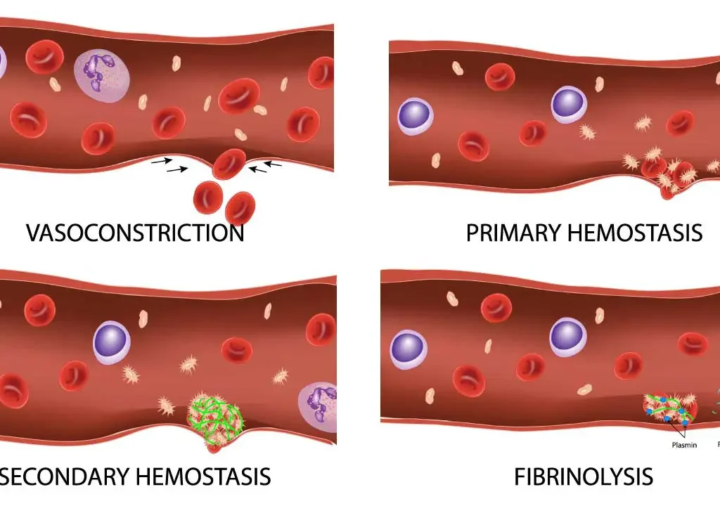
Blood Related Pathophysiology
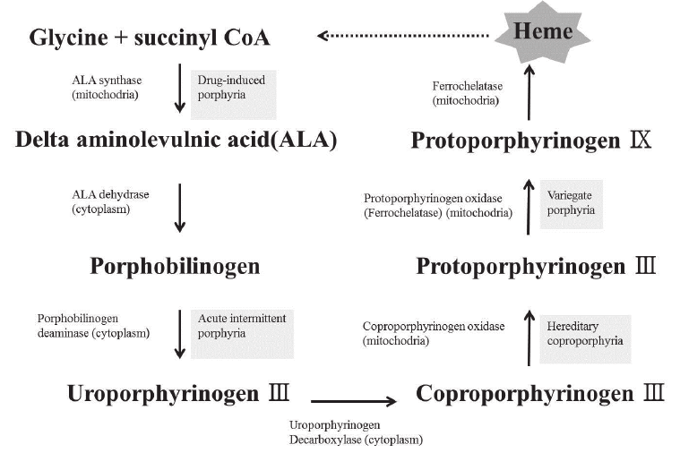
Blood : Related Physiologies Physiology of Red Blood Cells I. Erythropoiesis: The Journey of a Red Blood Cell Erythropoiesis is the highly regulated process of red blood cell (RBC) production, primarily occurring in the bone marrow in adults. It’s a continuous, dynamic process designed to maintain a stable red blood cell mass and oxygen-carrying capacity in the blood. A. Sites of Erythropoiesis Embryonic/Fetal Life Yolk Sac (0-3 mo): Initial primitive site. Liver (3-7 mo): Primary peak activity. Spleen (3-6 mo): Contributes lesser extent. Bone Marrow (5 mo+): Gradually takes over. Adult Life Red Bone Marrow: Exclusive site (Vertebrae, sternum, ribs, pelvis, proximal long bones). Extramedullary: Reversion to Liver/Spleen in severe pathology (e.g., myelofibrosis). B. Stages of Erythropoiesis Progresses from Stem Cell to Mature RBC through distinct morphological changes. 1. Pluripotent Hematopoietic Stem Cell (HSC) “Master cells” capable of self-renewal. Differentiate into Common Myeloid Progenitors (CMPs). 2. Erythroid Progenitors (BFU-E & CFU-E) BFU-E: Primitive, EPO-sensitive but not dependent. CFU-E: Mature, highly sensitive and dependent on EPO for survival. 3. Pronormoblast (Proerythroblast) First recognizable precursor. Large (20-25 µm), basophilic cytoplasm (ribosomes), prominent nucleoli. Begins globin synthesis. 4. Basophilic Normoblast Smaller, intensely basophilic cytoplasm. Active hemoglobin synthesis begins. 5. Polychromatophilic Normoblast Grayish-blue cytoplasm (mix of ribosomes + Hb). Most active stage of Hb synthesis. 6. Orthochromatophilic Normoblast Smallest nucleated precursor. Dense, pyknotic nucleus. Pink cytoplasm (massive Hb). Nucleus is extruded at this stage. 7. Reticulocyte (Polychromatophilic Erythrocyte) Anucleated, contains residual RNA network (reticulum). Released from marrow to blood. Constitutes 0.5-2.5% of circulating RBCs. Reticulocytosis: Indicates increased production (e.g., response to anemia). 8. Mature Erythrocyte Biconcave disc, anucleated, no organelles. Packed with Hemoglobin for O2 transport. Lifespan: ~120 days. C. Regulation of Erythropoiesis 1. Erythropoietin (EPO) – The Key Hormone Source: Kidneys (90%), Liver (10%). Stimulus: Renal Hypoxia (Low O2) due to anemia, altitude, lung disease. Action: Binds receptors on progenitors (CFU-E) → Promotes proliferation, survival, Hb synthesis, and early release. 2. Nutritional Requirements Iron: Essential for Heme. Deficiency = Anemia. B12 & Folate: DNA synthesis cofactors. Deficiency = Macrocytic Anemia. Protein/Vitamins: Globin synthesis, C, B6, Copper, Zinc. 3. Hormonal Influences Androgens (Testosterone): Stimulate EPO + direct marrow effect (Higher RBCs in males). Thyroid/Growth Hormone: Stimulatory effects. Hypothyroidism can cause mild anemia. II. Hemoglobin Synthesis Hemoglobin (Hb) is the primary protein within red blood cells, responsible for oxygen transport from the lungs to the tissues and carbon dioxide transport from the tissues back to the lungs. It is a complex molecule, and its synthesis is a highly coordinated process. A. Structure of Hemoglobin A mature hemoglobin molecule is a tetramer (four subunits). Each subunit has two parts: 1. Heme (Non-Protein) Porphyrin ring structure with a central Iron (Fe2+) atom. Function: Site where oxygen binds reversibly. Capacity: 4 Heme groups per Hb molecule = 4 O2 molecules. 2. Globin (Protein) Four polypeptide chains (typically 2 pairs). Adult Hb: Two alpha (α) + Two beta (β) chains. Each globin chain enfolds a heme group. Specific combination determines Hb type. B. The Synthesis Process Occurs primarily in the cytoplasm of developing RBCs (pronormoblasts through reticulocytes). 1. Globin Chain Synthesis Occurs on ribosomes in the cytoplasm. Alpha (α) chains: Encoded on Chromosome 16. Beta (β), Gamma (γ), Delta (δ), Epsilon (ε): Encoded on Chromosome 11. 2. Heme Synthesis Multi-step enzymatic pathway occurring in Mitochondria and Cytoplasm. Start: Succinyl CoA + Glycine. Rate-Limiting Step: Formation of delta-aminolevulinic acid (ALA) by ALA synthase. Intermediates: Porphobilinogen → Uroporphyrinogen → Coproporphyrinogen → Protoporphyrin. Final Step: Insertion of Ferrous Iron (Fe2+) into Protoporphyrin IX ring by Ferrochelatase (Heme synthase). Iron Delivery: Transported by Transferrin, taken up via Transferrin Receptors. 3. Assembly Heme + Globin rapidly combine. 1 Globin + 1 Heme = Globin-Heme Monomer. 4 Monomers assemble = Final Hemoglobin Tetramer. C. Types of Normal Hemoglobin & Developmental Changes Globin chain production changes to adapt to oxygen environments. 1. Embryonic Hemoglobins (First 8-10 weeks) Gower 1 (ζ2ε2): Zeta + Epsilon. Gower 2 (α2ε2): Alpha + Epsilon. Hb Portland (ζ2γ2): Zeta + Gamma. Very high O2 affinity for extraction from maternal blood. 2. Fetal Hemoglobin (HbF – α2γ2) Predominant from 10 weeks to birth. Composition: 2 Alpha (α) + 2 Gamma (γ). Function: Higher O2 affinity than adult Hb (HbA). Crucial for O2 transfer across placenta. Post-Birth: Constitutes 60-90% at birth; gradually declines and is replaced by HbA. 3. Adult Hemoglobins Hemoglobin A (HbA – α2β2): 95-97% of adult Hb. 2 Alpha (α) + 2 Beta (β). Affinity regulated by 2,3-BPG for efficient tissue release. Hemoglobin A2 (HbA2 – α2δ2): 1.5-3.5% (Minor). 2 Alpha (α) + 2 Delta (δ). Elevated in Beta-thalassemia trait. D. Regulation of Hemoglobin Synthesis Iron Availability: Most critical. Deficiency impairs heme synthesis → reduced Hb. Globin Chain Balance: Synthesis of alpha/non-alpha chains is tightly balanced. Imbalance (Thalassemias) causes unstable chains/ineffective erythropoiesis. Erythropoietin (EPO): Indirectly stimulates synthesis by promoting precursor proliferation/maturation. III. Red Blood Cell Metabolism Unlike most cells in the body, mature red blood cells (erythrocytes) are anucleated and lack mitochondria, endoplasmic reticulum, and other organelles. This means they cannot synthesize proteins or carry out oxidative phosphorylation. Their metabolism is highly specialized and focuses on two main goals: Generating energy (ATP): To maintain membrane integrity, ion gradients (Na+/K+ pump), and cell shape. Protecting hemoglobin from oxidative damage: Hemoglobin is susceptible to oxidation, which impairs function and damages the cell. A. Energy Production (ATP Generation) RBCs rely almost exclusively on Anaerobic Glycolysis (Embden-Meyerhof pathway). 1. Embden-Meyerhof Pathway Converts Glucose → Pyruvate → Lactate. Yield: Net 2 ATP per glucose. Key Functions of ATP: Ion Gradients: Powers Na+/K+ ATPase pump (prevents osmotic lysis). Cell Shape: Phosphorylation of cytoskeletal proteins maintains deformability. 2. Rapoport-Luebering Shunt Offshoot pathway producing 2,3-Bisphosphoglycerate (2,3-BPG). Significance: Binds deoxyhemoglobin, stabilizing T-state → Promotes O2 release. High BPG: Decreased affinity (Right shift) → Increased delivery. Low BPG: Increased affinity (Left shift) → Decreased delivery. Cost: Consumes 1 ATP otherwise generated by glycolysis. B. Protection Against Oxidative Damage RBCs have antioxidant systems to neutralize Reactive Oxygen Species (ROS) that cause Methemoglobin (Fe3+) or Heinz bodies (denatured Hb). 1. Hexose Monophosphate