Epithelial Tissue
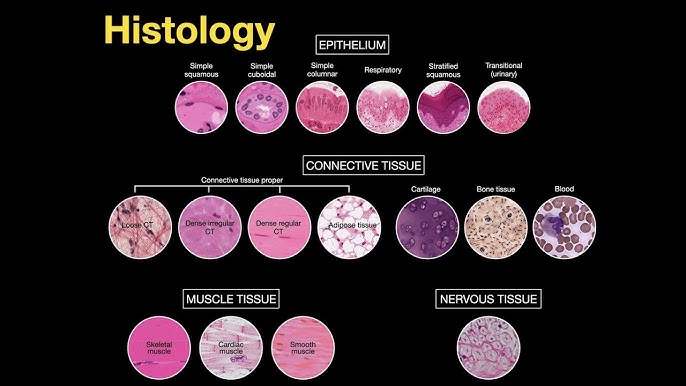
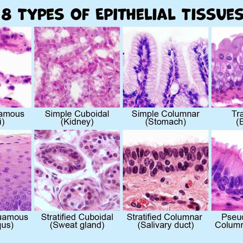
Epithelium: The Body’s Lining & Covering Tissue What is Epithelium? Epithelium forms continuous sheets of cells that line internal surfaces and cover the external surface of the body. It acts as a selective barrier that protects tissues and is often involved in absorption or secretion. A non-cellular layer called the basement membrane separates an epithelium from the underlying connective tissue. Key Characteristics: Anchorage & Polarity Cells are anchored to a basement membrane and have an apical surface (facing a free space) and a basal surface (attached to the basement membrane). Avascularity & Cellularity Epithelium contains no blood vessels (avascular) and is composed almost entirely of tightly packed cells with very little extracellular matrix (cellularity). Origin of the Epithelial Tissues Epithelial tissues are diverse in function and location, which is reflected in their origins from all three primary germ layers formed during embryonic development. These are the primary layers of cells from which all tissues and organs of the body are derived. Ectoderm (Outermost layer) Gives rise to structures that interact with the outside world and the nervous system. Epithelial Derivatives: Epidermis of the skin Lining of the oral & nasal cavities Lining of the anal canal Glands derived from skin Mesoderm (Middle layer) Forms structures related to movement, support, and circulation. Epithelial Derivatives: Endothelium (lining blood vessels) Mesothelium (lining serous cavities) Epithelium of kidney tubules Epithelium of the gonads Endoderm (Innermost layer) Forms the lining of the digestive and respiratory systems and associated glands. Epithelial Derivatives: Lining of the GI tract Lining of the respiratory tract Lining of the urinary bladder Epithelium of thyroid, pancreas, liver The Basement Membrane The basement membrane (BM) is a thin, acellular, extracellular layer that underlies all epithelial tissues, separating them from the adjacent connective tissue. It is a critical structural and functional component. Composition & Structure The BM is primarily composed of glycoproteins (like laminin), proteoglycans, and various types of collagen (especially Type IV collagen). Under an electron microscope, it is seen to have two main layers: Basal Lamina: Produced by the epithelial cells. It has a clearer layer (lamina lucida) and a denser layer (lamina densa). Reticular Lamina: Produced by the underlying connective tissue. It is composed of reticular fibers (Type III collagen) that anchor the basal lamina. Functions of the Basement Membrane Structural Support: Provides a stable base for epithelial cells. Filtration Barrier: Regulates passage of molecules (e.g., in the kidney). Cell Adhesion: Mediates strong attachment of epithelium. Maintains Polarity: Helps define the apical-basal orientation. Regulates Cell Behavior: Influences growth and differentiation. Tissue Repair: Acts as a scaffold for regeneration. Summary of Key Characteristics Anchored to basement membrane Apical and Basal surfaces (Polarity) No extracellular matrix (Cellularity) Avascular (no blood vessels) Clinical Correlation: Invasion and Cancer The basement membrane is a critical marker in cancer diagnosis. Benign tumors remain confined above the BM. A hallmark of malignant tumors (cancer) is their ability to produce enzymes that degrade the BM, allowing them to invade the underlying connective tissue and metastasize (spread). Classification of Epithelium Epithelium is classified based on two main features: the number of cell layers and the shape of the cells at the apical (free) surface. Simple Squamous Function: Ideal for diffusion and filtration. | Location: Alveoli of the lungs, lining of blood vessels (endothelium). Simple Cuboidal Function: Secretion and absorption. | Location: Kidney tubules, glands. Simple Columnar Function: Absorption and secretion. | Location: Gastrointestinal tract. Stratified Squamous Function: Protection against abrasion. Keratinized: Surface cells are dead and filled with keratin. (Location: Epidermis of the skin). Non-keratinized: Surface cells are living. (Location: Esophagus, vagina). Transitional (Urinary) Epithelium Function: Allows for distension (stretching). | Location: Urinary bladder, ureters. Pseudostratified Ciliated Columnar (Respiratory) Epithelium Function: Secretion and movement of mucus. | Location: Trachea, bronchi. Simple squamous epithelium Simple squamous epithelium is a single layer of thin, flattened cells that forms a delicate lining in areas where rapid diffusion, filtration, or smooth movement of substances is needed. The extreme thinness of the cells provides minimal protection but allows for quick transport of molecules. Classification & Defining Characteristics Simple Consists of a single layer of cells, crucial for rapid transport across the membrane. Squamous Cells are flat, thin, and scale-like (“squashed”), resembling a tiled floor from the surface. Permeable The extreme thinness of the single layer makes it highly permeable for quick exchange. Structure & Appearance Cell Shape: Irregularly shaped, flattened cells that interlock like puzzle pieces. Nucleus: Oval or flattened, often appearing as a central bulge in the thin cell. Cytoplasm: Scanty (very little), reflecting its primary role in passive transport. Basement Membrane: Rests on a thin basement membrane, separating it from underlying connective tissue. Locations and Functions Simple squamous epithelium is strategically located in areas where rapid diffusion, filtration, or a slick, friction-reducing surface is required. Lining of Blood & Lymphatic Vessels (Endothelium) Provides a smooth, clot-preventing surface for blood flow and facilitates the exchange of gases, nutrients, and waste. Lining of Serous Cavities (Mesothelium) Lines the pleura, pericardium, and peritoneum, producing a slippery serous fluid that lubricates organs and prevents friction. Alveoli of the Lungs (Type I Pneumocytes) Forms the extremely thin “air-blood barrier” essential for rapid gas exchange (oxygen in, carbon dioxide out). Glomerular Capsules (Bowman’s Capsule) in the Kidneys Forms the filtration membrane for blood, allowing water and small solutes to pass into the renal tubule while retaining large molecules. Clinical Significance Understanding the structure of simple squamous epithelium is key to diagnosing and managing several clinical conditions. Pathological Considerations Edema: Fluid accumulation (e.g., in heart failure) increases the diffusion distance across the alveolar epithelium, impairing gas exchange. Inflammation: Inflammation of serous membranes (pleuritis, peritonitis) causes fluid accumulation (effusions) and painful friction. Cancer: Malignant mesotheliomas can arise from the mesothelium, and a disrupted endothelium is a key factor in various vascular diseases. Simple cuboidal epithelium Simple cuboidal epithelium is a single layer of cube-shaped cells, often with round, central nuclei, primarily performing secretion and absorption. It is found lining surfaces like kidney tubules, ducts of glands, and the surface