Doctors Revision

Author name: doctorsrevision@gmail.com

Cardiovascular System Anatomy
Anatomy

Cardiovascular System Anatomy

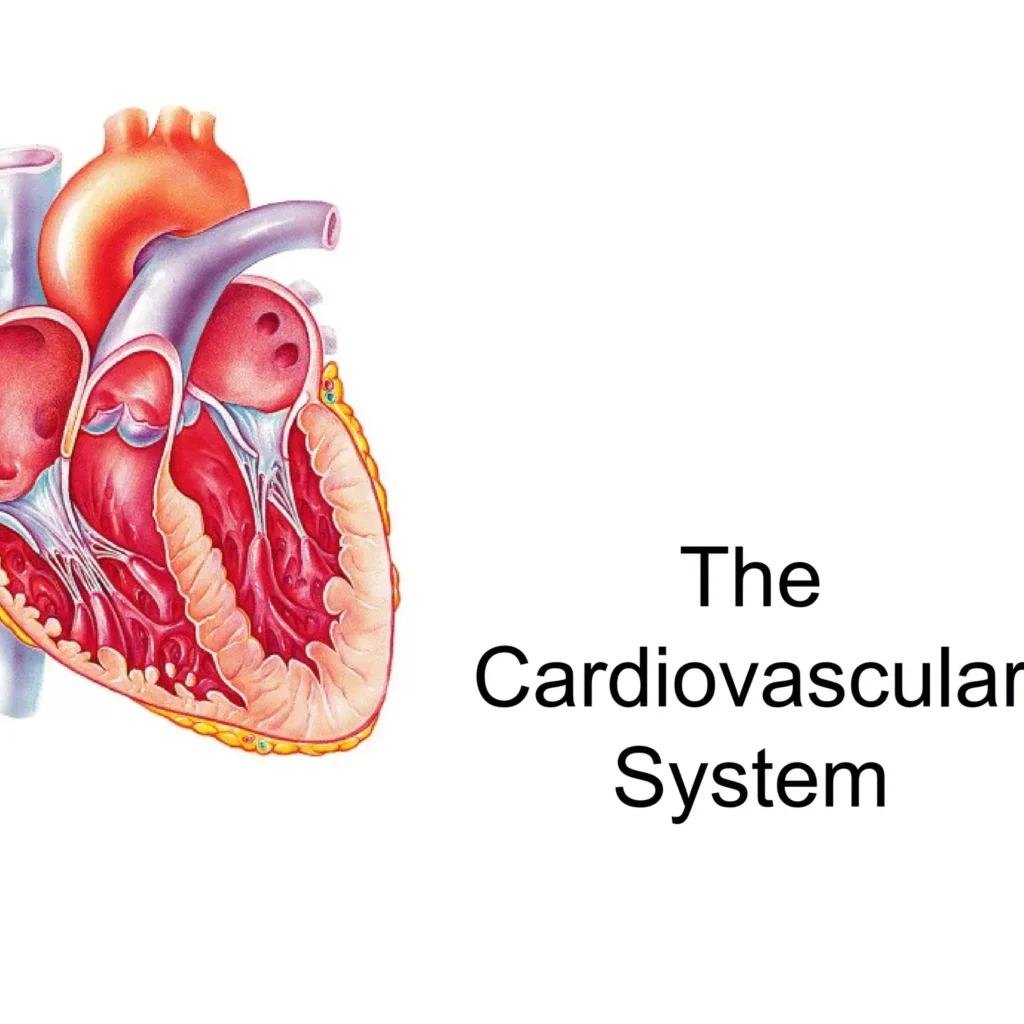
Cardiovascular System Anatomy: For the love of the Heart Cardiovascular System Anatomy Introduction to the Cardiovascular System The cardiovascular system, also known as the circulatory system, is a vast network responsible for transporting blood throughout the entire body. This system is essential for maintaining life and ensuring that every cell receives what it needs to function properly. Key Components The cardiovascular system is primarily composed of three main parts, working in perfect concert. 1. The Heart This muscular organ, roughly the size of a clenched fist, is the central pump of the system. It continuously contracts and relaxes, driving blood through the vast network of vessels. 2. Blood Vessels Arteries: Carry oxygenated blood away from the heart. Their thick, muscular walls withstand high pressure. Veins: Carry deoxygenated blood back to the heart. Their thinner walls and internal valves prevent backward blood flow. Capillaries: The smallest vessels, forming vast networks that connect arteries and veins. Their ultra-thin walls allow for the efficient exchange of gases, nutrients, and waste products with the body’s cells. 3. Blood Plasma: The liquid matrix, mostly water, that carries dissolved nutrients, hormones, and waste. Red Blood Cells (Erythrocytes): Contain hemoglobin to transport oxygen from the lungs to tissues and return carbon dioxide. White Blood Cells (Leukocytes): Key components of the immune system, defending the body against pathogens. Platelets (Thrombocytes): Small cell fragments essential for initiating the blood clotting process to stop bleeding. Primary Functions The cardiovascular system performs several indispensable functions to maintain homeostasis and sustain life. Transport of O₂ & Nutrients: Delivers oxygen and nutrients to every cell for energy and metabolic processes. Removal of Waste: Collects metabolic waste like CO₂ and urea and transports them to the lungs and kidneys for excretion. Hormone Transport: Acts as a delivery system for hormones, carrying them from glands to their target organs. Temperature Regulation: Distributes heat throughout the body and regulates its dissipation to maintain a stable internal temperature. Protection Against Disease: Circulates white blood cells and antibodies as part of the immune system to fight infections. Blood Clotting: Platelets and clotting factors in the blood prevent excessive blood loss at sites of injury. Anatomy of the Heart and Great Vessels The heart is a hollow, muscular organ located in the mediastinum, the central compartment of the thoracic cavity, nestled between the lungs. It sits slightly to the left of the midline, resting on the diaphragm. Its pointed end, the apex, points inferiorly and to the left, while the broader base points superiorly and to the right. I. The Pericardium: The Heart’s Protective Sac The heart is encased in a double-walled sac called the pericardium. It serves to anchor the heart, prevent it from overfilling, and provide a frictionless environment for its constant beating. Fibrous Pericardium: The tough, outermost layer made of dense connective tissue. It anchors the heart to the diaphragm and great vessels, preventing overfilling and providing a strong protective barrier. Serous Pericardium: A thinner, delicate inner layer, itself composed of two sub-layers: Parietal Layer: Lines the inside of the fibrous pericardium. Visceral Layer (or Epicardium): Adheres directly to the surface of the heart muscle. Pericardial Cavity: The potential space between the parietal and visceral layers, containing a thin film of serous fluid that acts as a lubricant to eliminate friction during heartbeats. II. Layers of the Heart Wall The wall of the heart itself is composed of three distinct layers, from superficial to deep. Epicardium: The outermost layer (and also the visceral layer of the serous pericardium). It is a protective layer that contains the coronary blood vessels and adipose tissue. Myocardium: The thick, muscular middle layer composed of cardiac muscle cells (cardiomyocytes). This is the contractile layer responsible for the heart’s pumping action. Its thickness is greatest in the left ventricle. Endocardium: The innermost layer, a thin, smooth membrane that lines the heart’s chambers and covers the valves. Its smooth surface minimizes friction and prevents clot formation. III. Chambers of the Heart The heart is a four-chambered organ, divided by a muscular septum into right and left sides. This separation is crucial for ensuring that oxygen-poor and oxygen-rich blood do not mix. Right Atrium (RA) Receives deoxygenated blood from the body via the Superior Vena Cava (SVC), Inferior Vena Cava (IVC), and Coronary Sinus. Pumps blood to the right ventricle. Left Atrium (LA) Receives oxygenated blood from the lungs via the four pulmonary veins. Pumps blood to the left ventricle. Right Ventricle (RV) Receives deoxygenated blood from the right atrium. Pumps deoxygenated blood to the lungs via the pulmonary artery. Left Ventricle (LV) Receives oxygenated blood from the left atrium. The strongest chamber; pumps oxygenated blood to the entire body via the aorta. IV. Heart Valves: Ensuring Unidirectional Blood Flow The heart contains four valves that act as one-way doors, preventing the backflow of blood (regurgitation). They open and close passively in response to pressure changes within the chambers. Atrioventricular (AV) Valves: Located between the atria and ventricles. Tricuspid Valve: Between the right atrium and right ventricle (has three cusps). Mitral (Bicuspid) Valve: Between the left atrium and left ventricle (has two cusps). The AV valves are anchored by fibrous chordae tendineae (“heart strings”) to papillary muscles in the ventricles. When the ventricles contract, these muscles pull on the cords, preventing the valve flaps from being pushed back up into the atria. Semilunar (SL) Valves: Located at the exit of the ventricles, preventing blood from flowing back from the great arteries. Pulmonary Valve: Between the right ventricle and the pulmonary artery. Aortic Valve: Between the left ventricle and the aorta. V. Great Vessels of the Heart These are the major blood vessels that are directly connected to the heart, responsible for carrying blood to and from its chambers. Superior & Inferior Vena Cava (SVC & IVC): Bring deoxygenated blood from the upper and lower body, respectively, to the right atrium. Pulmonary Artery: Carries deoxygenated blood from the right ventricle to the lungs. Note: It’s an artery because it carries blood AWAY from the
Common Abnormalities: Teratology and Teratogenesis
Anatomy

Common Abnormalities: Teratology and Teratogenesis

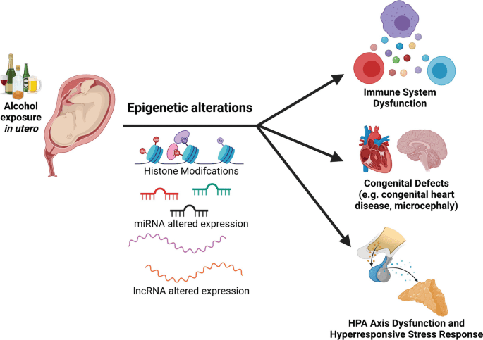
Common Abnormalities: Teratology and Teratogenesis Common Abnormalities: Teratology and Teratogenesis 1. Teratology Teratology is the scientific study of abnormal physiological development, specifically focusing on the causes, mechanisms, and patterns of birth defects, also known as congenital malformations. The term comes from the Greek “teras,” meaning monster or marvel. Key Concepts in Teratology Congenital Malformations (Birth Defects) are structural, functional, or metabolic abnormalities present at birth. These can range from minor cosmetic issues to severe, life-threatening conditions. Not all congenital conditions are visible at birth (e.g., some heart defects or metabolic disorders). They are classified into several distinct categories based on their origin. Malformation A primary structural defect resulting from an intrinsically abnormal developmental process. The blueprint itself was flawed from the beginning. Example: Polydactyly (extra fingers/toes), Spina Bifida. Disruption A defect resulting from the extrinsic breakdown of, or interference with, an originally normal developmental process. The blueprint was normal, but something damaged the structure as it was forming. Example: Limb amputation due to amniotic bands wrapping around it. Deformation An abnormal form, shape, or position of a body part caused by extrinsic mechanical forces acting on a normally developed structure. Example: Clubfoot due to intrauterine crowding, limiting space for the feet to grow properly. Dysplasia An abnormal organization of cells into tissues. The problem lies in how the cells themselves are structured and arranged. Example: Skeletal dysplasias like achondroplasia (a form of dwarfism). Syndrome A group of anomalies that occur together and have a specific, common, known cause. Example: Down syndrome (caused by Trisomy 21), Fetal Alcohol Syndrome. Association A non-random occurrence of two or more anomalies that appear together more often than by chance, but for which a common cause has not yet been identified. Example: VACTERL association (Vertebral, Anal, Cardiac, Tracheo-Esophageal, Renal, Limb defects). Factors Contributing to Birth Defects While teratogens are a major focus, it’s important to understand the broader categories of factors that can lead to congenital malformations. Causes 40-50%: Unknown Causes 20-25%: Genetic Factors (chromosomal, single gene) 20-25%: Multifactorial Inheritance (genes + environment) ~10%: Environmental Factors (Teratogens) 2. Teratogenesis Teratogenesis is the process by which a teratogen (an agent that causes birth defects) acts on an embryo or fetus to produce a congenital malformation. The study of this process is governed by a set of foundational concepts known as Wilson’s Principles. Principle 1: Susceptibility (Genotype) The genetic makeup of the embryo and mother determines their susceptibility to a teratogen. What harms one individual may have no effect on another due to genetic differences in metabolism and cellular repair. Principle 2: Dosage & Duration The amount of the teratogen and the length of exposure are critical. Generally, a higher dose or a longer duration of exposure increases the risk and severity of the resulting defect. Principle 3: Timing of Exposure (Critical Periods) This is arguably the most crucial principle. The susceptibility of an organ system to a teratogen varies dramatically with its stage of development. Pre-implantation Period (Weeks 1-2) The “all-or-nothing” period. Exposure to a teratogen usually results in either the death of the embryo or its complete recovery with no defects, as the cells are still totipotent and can be replaced. Embryonic Period (Weeks 3-8) The most sensitive period for major malformations. This is when organogenesis occurs, and each organ system has its own critical window of vulnerability (e.g., heart: weeks 3-5; CNS: weeks 3-16+). Fetal Period (Weeks 9 to Birth) Exposure during this period generally does not cause major structural defects but can lead to functional problems, growth retardation, and minor abnormalities, especially in the still-developing brain. Principle 4: Mechanisms Teratogens exert their effects through specific cellular and molecular mechanisms, such as interfering with cell proliferation or migration, inducing cell death (apoptosis), or disrupting biochemical pathways. Principle 5: Manifestations The final outcome of teratogenic exposure can be one of four manifestations: death, malformation, growth retardation, or functional deficit. Classes of Teratogens Teratogens are substances that can cause birth defects when a fetus is exposed during pregnancy. They can be broadly categorized into several classes, each with well-documented examples and associated defects. The risk and severity of abnormalities depend on the type of agent, timing, dosage, and duration of exposure. Infectious Agents (TORCH Infections) The acronym TORCH helps remember some of the most well-known infectious teratogens: Toxoplasmosis: A parasitic infection that can cause hydrocephalus and intracranial calcifications. Others (e.g., Syphilis, Varicella-Zoster, Zika, Parvovirus B19): Zika is known for causing microcephaly, while syphilis can lead to congenital deafness and other issues. Rubella (German measles): Can result in a classic triad of cataracts, cardiac malformations, and deafness. Cytomegalovirus (CMV): A common virus that can cause microcephaly, hearing loss, and intellectual disability. Herpes Simplex Virus: Can lead to skin lesions, microcephaly, and eye problems. Drugs and Chemicals Thalidomide: A classic example that caused severe limb reduction defects (phocomelia). Alcohol (Ethanol): The leading preventable cause of non-genetic birth defects, leading to Fetal Alcohol Syndrome (FAS) with distinct facial anomalies, growth retardation, and CNS dysfunction. Tobacco & Nicotine: Smoking is associated with low birth weight, premature delivery, and can affect the development of the fetal brain and lungs. Retinoids (e.g., Isotretinoin/Accutane): Highly teratogenic, causing severe CNS, facial, cardiac, and ear malformations. Anticonvulsants (e.g., Valproic Acid, Phenytoin): Associated with neural tube defects, cleft lip/palate, and cardiac defects. ACE Inhibitors: Can cause renal failure and oligohydramnios (insufficient amniotic fluid). Warfarin: An anticoagulant that can cause skeletal abnormalities, including chondrodysplasia punctata. Certain Antibiotics (e.g., Tetracycline): Can cause yellow staining of teeth and affect long bone growth. Recreational Drugs (e.g., Cocaine, Heroin): Can lead to low birth weight, withdrawal symptoms in the newborn, and learning or behavioral problems. Environmental Toxins Heavy Metals (e.g., Mercury, Lead): Can cause significant CNS damage and developmental delays. Mercury is often found in certain types of fish, and lead can be in old paint and pipes. Polychlorinated Biphenyls (PCBs): Industrial chemicals that can lead to developmental and neurological problems. Herbicides and Industrial Solvents: Exposure to certain chemicals used in agriculture and manufacturing can be harmful. Physical Agents Ionizing

Fetal Membranes and Placenta
Anatomy

Fetal Membranes, Placenta, Cord and Circulation

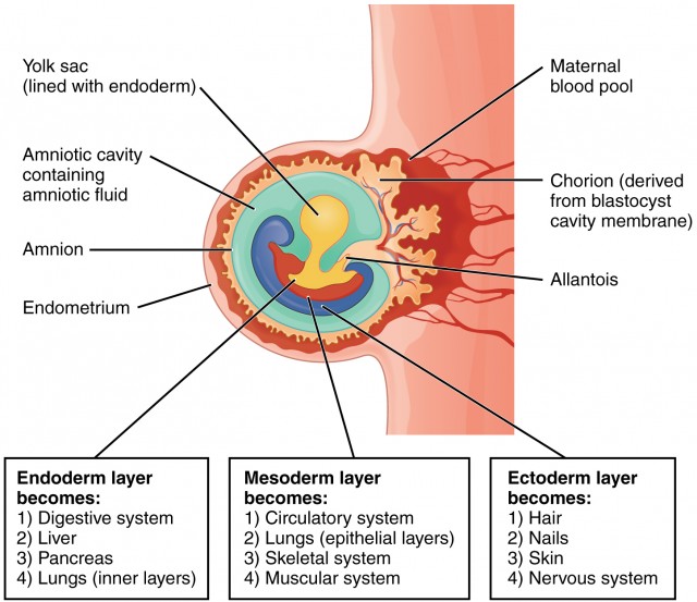
Fetal Membranes, Placenta, Cord and Circulation: Safety and Feeding Fetal Membranes, Placenta, Cord and Circulation The fetal membranes and the placenta are temporary, yet essential, organs that develop alongside the embryo and fetus. They provide a complete life-support system, handling protection, nourishment, gas exchange, waste removal, and hormonal regulation critical for successful intrauterine development. They are expelled from the body after birth. Formation of Embryonic Cavities and Membranes The period of early embryonic development (roughly Day 8 to Day 12-14 post-fertilization) is characterized by the rapid formation of several extraembryonic structures, which are vital for the embryo’s survival and subsequent development. These include the amniotic cavity, primary and secondary yolk sacs, and the chorionic cavity, along with their associated membranes. A. Formation of the Amniotic Cavity and Amnion Timeline: Begins around Day 8 post-fertilization. Process: Cavity Formation: As the blastocyst implants, a small space appears within the epiblast, which is the dorsal layer of the bilaminar germ disc (formed from the Inner Cell Mass). Enlargement: This space rapidly expands to become the amniotic cavity. Amnioblast Differentiation: Cells from the epiblast adjacent to the cytotrophoblast differentiate into thin, flattened cells called amnioblasts. Amniotic Membrane Formation: These amnioblasts, along with a layer of extraembryonic mesoderm, form the amnion, which eventually encloses the entire amniotic cavity. Roof and Floor: The roof is formed by the amnion/cytotrophoblast, while the floor is formed by the epiblast of the bilaminar germ disc. Key Features & Function of the Amnion/Amniotic Fluid: Amniotic Sac: The amnion forms the inner lining of the amniotic sac, which will eventually surround the entire embryo and then fetus. Amniotic Fluid: The cavity fills with amniotic fluid. Initially derived from maternal blood, it is later maintained largely by fetal urine excretion and fetal swallowing. It serves crucial functions: Protection: Acts as a shock absorber against mechanical trauma. Temp Regulation: Maintains a constant, optimal intrauterine temperature. Symmetry & Movement: Allows symmetrical external growth and free movement for proper musculoskeletal development (preventing contractures). Prevents Adhesion: Stops the developing embryo from physically sticking to the amnion (which would cause amniotic band syndrome). Lung/GI Development: Fetal swallowing of the fluid actively aids the maturation of the GI tract; while fetal “breathing” movements pull fluid into the lungs, providing the mechanical stretch essential for pulmonary development. Clinical Application Amniotic Fluid Volume Abnormalities Oligohydramnios (Too little fluid): Often caused by placental insufficiency or fetal renal agenesis (inability to produce urine). It can lead to Potter sequence (flattened face, clubbed feet, pulmonary hypoplasia) because the fetus is compressed without the fluid cushion. Polyhydramnios (Too much fluid): Often caused by fetal swallowing defects (e.g., esophageal atresia, anencephaly) or maternal diabetes. It can lead to premature rupture of membranes and preterm labor. B. Formation of the Yolk Sac Timeline: Primary yolk sac begins around Day 9; Secondary yolk sac around Day 12-13. 1. Primary Yolk Sac (Exocoelomic Cavity) – Day 9 Cells from the hypoblast (ventral layer) migrate and line the inner surface of the cytotrophoblast. These cells form a thin membrane called the exocoelomic membrane (Heuser’s membrane). This membrane + hypoblast encloses the primary yolk sac. Position: The bilaminar disc lies between the Amniotic Cavity (dorsal) and the Primary Yolk Sac (ventral). 2. Extraembryonic Mesoderm – Day 10-11 A new layer of loose connective tissue appears and fills the space between the exocoelomic membrane/amnion externally and the cytotrophoblast internally. 3. Secondary Yolk Sac (Definitive) – Day 12-13 The primary sac constricts due to chorionic cavity expansion. A smaller, definitive secondary yolk sac forms from a portion of the primary sac. The larger pinched-off part degenerates into exocoelomic cysts. Key Features & Function of the Yolk Sac: Nutrition Early Nutrient Transfer Plays a vital role in nutrient transfer to the embryo during weeks 2 and 3, before the uteroplacental circulation is fully functional. Blood Supply Hematopoiesis It is the primary site of early blood cell formation (Weeks 3-6). It produces primitive nucleated red blood cells expressing embryonic hemoglobin. After week 6, the fetal liver takes over this function. Reproduction Primordial Germ Cells Precursors to sperm/eggs originate in the wall of the yolk sac around week 3. They migrate along the hindgut to reach the developing gonads by week 5. Fate Vestigial Structure In humans, the yolk sac does not contain yolk. It is small, regresses rapidly by week 20, and is incorporated into the primitive gut tube and the umbilical cord. C. Formation of the Chorionic Cavity and Chorion Timeline: Begins around Day 11-12. Process: Vacuole Formation: Numerous large spaces and vacuoles appear within the extraembryonic mesoderm. Coalescence: These fuse to form a large, isolated cavity called the chorionic cavity (extraembryonic coelom). Suspension of Embryo: The embryo (with its amnion and yolk sac) is suspended in this massive cavity by the connecting stalk (which is the future umbilical cord). The Chorion (Outer Wall) The chorion forms the wall of the chorionic cavity and is strictly formed by three layers (from outside to inside): Syncytiotrophoblast (outermost) Cytotrophoblast Somatic layer of extraembryonic mesoderm (innermost) Functions: Chorionic Villi: Gives rise to the villi (the functional exchange units of the placenta). Protection: Forms an additional protective layer around the entire conceptus. Part of Placenta: The villous part (the chorion frondosum) forms the fetal component of the placenta, while the smooth part (chorion laeve) eventually fuses with the amnion. Summary of Relationships (Day 12-14): Central: Bilaminar germ disc centrally located. Dorsal: Amniotic cavity. Ventral: Secondary yolk sac. Surrounding all: Chorionic cavity (enclosed entirely by the Chorion). Bridge: Connecting stalk linking the bilaminar disc to the chorion. D. The Allantois: Development and Significance Origin: Appears around Day 16-18 as a small, sausage-shaped diverticulum (outpouching) from the caudal wall of the yolk sac (specifically the hindgut), extending directly into the connecting stalk. Vascular Development: This is its most significant role in humans. Blood vessels develop in the mesoderm wall of the allantois to become the umbilical arteries and the umbilical vein. These vessels extend through the connecting stalk to strictly link the embryonic and placental circulation. Urinary Bladder

bilaminar disc formation e1758606529816
Anatomy

Germ Disc, Gastrulation and Neurulation

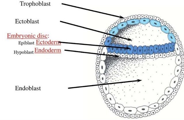
Germ Disc, Gastrulation & Neurulation: Fortion of Organs Brief Recap We concluded our last discussion with the blastocyst successfully implanted (around Day 12 post-fertilization) into the uterine endometrium. At this point: The blastocyst is fully embedded in the decidua (the transformed endometrial tissue). The trophoblast has differentiated into: Cytotrophoblast (inner layer, cellular). Syncytiotrophoblast (outer layer, invasive, multinucleated, producing hCG). The inner cell mass (embryoblast) is now clearly visible and undergoing significant changes, leading to the formation of the embryonic disc and associated cavities. Formation of the Bilaminar Embryonic/Germ Disc and Associated Cavities (Week 2 Development) This period, roughly from Day 8 to Day 14 post-fertilization, is often referred to as “the week of twos” because several structures differentiate into two layers or cavities. It’s a phase of rapid differentiation of the inner cell mass. After fertilization and cleavage, the embryo, now a blastocyst, undergoes profound organizational changes. It remodels itself from a sphere into a flattened, two-layered structure known as the Bilaminar Germ Disc. This process is crucial as it sets the stage for gastrulation, where the three primary germ layers will form. 1. From Blastocyst to Bilaminar Germ Disc This transformation begins around Day 8 post-fertilization, as the Inner Cell Mass (ICM) differentiates. A. Differentiation of the Inner Cell Mass: The inner cell mass (embryoblast) differentiates into two distinct layers that collectively form a flat, circular structure called the bilaminar embryonic disc: Epiblast (Dorsal/Upper Layer) A layer of columnar cells facing the developing amniotic cavity. Crucially, all three primary germ layers of the embryo will eventually originate from the epiblast. Location: The dorsal (upper) layer of the disc. Cell Type: Consists of tall, columnar cells. Relation to Cavity: It is directly adjacent to what will become the amniotic cavity. Significance: The epiblast is the source of all three primary germ layers during gastrulation. It is essentially the “true” embryonic component at this stage. Hypoblast (Ventral/Lower Layer) A layer of cuboidal cells facing the blastocoel. It primarily contributes to extraembryonic membranes, particularly the yolk sac. Location: The ventral (lower) layer of the disc, beneath the epiblast. Cell Type: Consists of small, cuboidal cells. Relation to Cavity: It is directly adjacent to what will become the primary yolk sac. Significance: While the hypoblast does not contribute directly to the embryo proper’s germ layers, it plays crucial roles in signaling, guiding epiblast cell movements, and forming the extraembryonic endoderm lining of the yolk sac. B. Formation of Associated Cavities: As the epiblast and hypoblast differentiate, two fluid-filled cavities form in close association with them: Amniotic Cavity Formation: A small cavity appears within the epiblast and expands. Lining: The roof of this cavity is formed by amnioblasts (cells that differentiate from the epiblast and line the amniotic cavity). The floor is the epiblast itself. Contents: It will eventually be filled with amniotic fluid, which protects the developing embryo/fetus. Primary Yolk Sac (Exocoelomic Cavity) Formation: Cells from the hypoblast migrate and spread along the inner surface of the cytotrophoblast, forming a thin membrane called the exocoelomic membrane (Heuser’s membrane). This membrane, together with the hypoblast, encloses a new cavity, the primary yolk sac. Contents: Contains fluid and plays a role in early nutrient transfer and blood cell formation. C. Development of Extraembryonic Structures: During this same period (Week 2), other crucial extraembryonic structures are forming: 1. Extraembryonic Mesoderm: Origin: A loose connective tissue layer that develops between the cytotrophoblast and the exocoelomic membrane/amnion. Cavitation: This mesoderm soon develops large cavities, forming the extraembryonic coelom (chorionic cavity). This cavity completely surrounds the amnion and the primary yolk sac, except where the embryonic disc is connected to the trophoblast by the connecting stalk (which will become the umbilical cord). Amniotic Cavity A new fluid-filled space that appears within the epiblast, enclosed by a thin membrane called the amnion. It will eventually surround the entire embryo. 2. Secondary Yolk Sac: As the extraembryonic coelom forms, the primary yolk sac shrinks, and a new, smaller secondary yolk sac forms from a second wave of hypoblast cells. This is the definitive yolk sac of the embryo. Primary Umbilical Vesicle (Yolk Sac) Forms when hypoblast cells line the blastocoel. In humans, it plays roles in early blood cell formation and nutrient transfer. 3. Chorion: The extraembryonic mesoderm, together with the two layers of the trophoblast (cytotrophoblast and syncytiotrophoblast), forms the chorion. The chorion is the outermost fetal membrane and will eventually contribute to the fetal part of the placenta. The chorionic cavity is the space within the chorion. Extraembryonic Mesoderm & Coelom A new layer of mesoderm forms between the yolk sac/amnion and the trophoblast. A large cavity, the chorionic cavity (or coelom), then forms within this mesoderm, suspending the embryo by a connecting stalk. D. Establishment of Body Axes (Preliminary): By the end of Week 2, some crucial axes begin to be established, even before gastrulation formally begins: Dorsoventral Axis: Already defined by the epiblast (dorsal) and hypoblast (ventral). Cranial-Caudal Axis: The future head end (cranial) is distinguished from the future tail end (caudal) by the appearance of a localized thickening of the hypoblast, the prechordal plate, at the future cranial region. This is an important signaling center. Left-Right Asymmetry: While not yet morphologically apparent, molecular signals are starting to be laid down that will determine left-right patterning. Summary of Bilaminar Disc Development (Week 2): Inner cell mass differentiates into Epiblast and Hypoblast. These form the Bilaminar Embryonic Disc. Amniotic Cavity forms above the epiblast. Primary Yolk Sac forms below the hypoblast, later replaced by the Secondary Yolk Sac. Extraembryonic Mesoderm and Extraembryonic Coelom develop, surrounding the amnion and yolk sac. The Chorion (trophoblast + extraembryonic mesoderm) encases everything. A Connecting Stalk links the embryonic disc to the trophoblast. Clinical Significance This highly sensitive period is critical for assessing early embryonic viability. Disruptions during germ disc formation can lead to severe birth defects, and this is when issues like ectopic pregnancies become apparent. 4. Transition to Gastrulation The formation of the bilaminar germ disc is the final preparatory

fertilization
Anatomy

Fertilization and Implantation

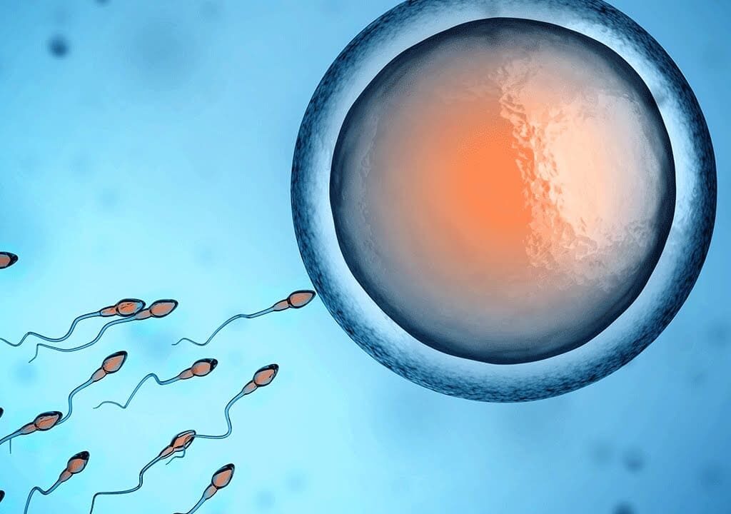
Fertilization & Implantation: The Beginning of a New Individual Fertilization Fertilization, also known as conception, is the fundamental biological process where a male gamete (sperm) and a female gamete (secondary oocyte) fuse to form a new, single-celled entity called a zygote. Fertilization is the process by which a male gamete (sperm) and a female gamete (ovum) fuse to form a new diploid cell called a zygote. This event typically occurs in the ampulla of the fallopian tube, usually within 12-24 hours after ovulation. This remarkable union restores the diploid (2n) number of chromosomes and marks the very beginning of the development of a new, genetically unique individual. Site of Fertilization In humans, fertilization typically occurs in the ampulla of the fallopian tube (oviduct). This is the wider, outer portion of the tube, close to the ovary, where the egg is captured after ovulation. The Key Players in Fertilization Successful fertilization depends on the precise interaction of four critical components. Sperm (Male Gamete) A small, motile cell designed to travel through the female reproductive tract and deliver its haploid genetic material to the egg. Egg (Secondary Oocyte) A large, non-motile cell containing the female’s haploid genetic material, cytoplasm, and all the necessary nutrients to support early embryonic development. It is arrested in Metaphase II of meiosis. Zona Pellucida A thick, glycoprotein-rich outer layer surrounding the egg. It acts as a species-specific binding site for sperm and is essential for preventing polyspermy (fertilization by more than one sperm). Corona Radiata The outermost layer of follicular (granulosa) cells that surrounds the zona pellucida, providing nourishment and protection to the ovulated egg. The Journey of the Sperm The passage of sperm through the female reproductive tract is a highly regulated and selective process, designed to ensure only sperm with normal morphology and vigorous motility reach the egg. Post-Ejaculation: Semen coagulates into a gel, protecting sperm from the vagina’s acidic environment and holding them near the cervix. This gel liquefies within an hour. The Cervix: Cervical mucus acts as a barrier, filtering out sub-motile sperm. The Uterus: Uterine myometrial contractions, aided by prostaglandins in the seminal fluid, propel the sperm towards the fallopian tubes. The first sperm enter the fallopian tubes minutes after ejaculation, but they can survive in the female reproductive tract for up to five days, awaiting ovulation. The Events of Fertilization Once an ovulated egg is present, fertilization proceeds through a highly coordinated series of events. Event 1: Capacitation A final maturation step that “arms” the sperm within the female reproductive tract. The Process: The female tract’s environment strips away cholesterol and proteins from the sperm’s head. The Result: The sperm’s tail becomes hyper-motile, and its acrosome membrane is destabilized, ready to release enzymes. Key takeaway: A sperm cannot fertilize an egg until it has been capacitated. A. Sperm Transport and Capacitation 1. The Journey Ejaculation & Vaginal Transit: Millions of sperm deposited in posterior fornix. Many lost to acidity/leukocytes. Cervical & Uterine Passage: Sperm navigate the cervix (mucus becomes permeable) and uterine cavity. Fallopian Tube: Only a few thousand reach the tubes, guided by chemotaxis and uterine contractions. 2. Capacitation (Maturation) Crucial process (2-10 hours) in female tract involving: Membrane Changes: Removal of cholesterol/glycoproteins from sperm head (acrosomal region). Increases fluidity/reactivity. Hyperactivation: Increased flagellar beating (vigorous/erratic) essential for penetrating egg layers. Result: Sperm is now capable of the acrosomal reaction. Event 2: The Acrosomal Reaction Penetrating the Corona Radiata: Hyper-motile sperm push through the outer layer of follicular cells. Binding to the Zona Pellucida: The sperm binds to species-specific ZP3 receptors on the zona pellucida, like a key in a lock. Releasing Enzymes: This binding triggers the acrosome to release digestive enzymes (like acrosin). Digesting a Path: These enzymes create a tunnel through the zona pellucida, allowing the sperm to reach the egg’s cell membrane. B. Penetration of the Egg’s Protective Layers Upon reaching the secondary oocyte, capacitated sperm must penetrate two barriers: 1. Corona Radiata Penetration Sperm use hyperactivated motility to push through. Enzymes like hyaluronidase (on sperm surface) break down hyaluronic acid in the extracellular matrix. 2. Zona Pellucida Penetration Binding: Sperm proteins bind to specific receptors (primarily ZP3 glycoprotein) on the Zona Pellucida. (Species-specific). Acrosomal Reaction: Binding to ZP3 triggers fusion of acrosomal membrane with sperm plasma membrane. Releases hydrolytic enzymes (acrosin, neuraminidase). Digestion & Motility: Enzymes digest a path; sperm tail thrusts push sperm through. C. Fusion of Sperm and Oocyte Membranes Sperm reaches the perivitelline space. Sperm head lies flat against oocyte plasma membrane. Membranes fuse. Sperm head, tail, mitochondria, and centriole enter oocyte cytoplasm. Events 3 & 4: The Blocks to Polyspermy To prevent a lethal condition where more than one sperm fertilizes the egg, the oocyte deploys a two-stage defense system. Fast Block (Immediate but Temporary) The instant fusion of the first sperm triggers a rapid influx of sodium ions (Na⁺) into the oocyte, instantly changing the membrane’s electrical charge to repel all other sperm. Slow Block (Cortical Reaction – Permanent) Sperm fusion also triggers a massive release of calcium ions (Ca²⁺) inside the oocyte. This causes cortical granules to release enzymes that destroy all ZP3 receptors and harden the zona pellucida, making it impenetrable. D. Prevention of Polyspermy (Block to Polyspermy) Mechanisms to ensure only ONE sperm fertilizes the egg (preventing lethal abnormal chromosome numbers). 1. Fast Block (Electrical) Rapid, transient depolarization of oocyte membrane prevents other sperm fusion. (Less prominent in humans). 2. Slow Block (Cortical) Primary Mechanism. Sperm fusion triggers intracellular Ca2+ surge. Cortical Reaction: Cortical granules release enzymes into perivitelline space causing: Zona Reaction: Hardens Zona Pellucida (cleaves ZP2, inactivates ZP3). Release of loosely attached sperm. E. Completion of Meiosis II The Ca2+ surge stimulates the secondary oocyte to finish division. Forms Mature Ovum (Female Pronucleus). Releases Second Polar Body. Male and Female Pronuclei swell and replicate DNA. F. Syngamy & Zygote Formation Pronuclear membranes break down. Chromosomes intermingle. Syngamy: Fusion of genetic material. Formation of diploid Zygote (46 chromosomes). Zygote immediately begins first mitotic division. The Fusion and

Menstruation Cycle
Anatomy

Menstruation Cycle

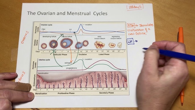
Menstruation: Preparing for pregnancy The Menstrual Cycle Learning Objectives & Overview The menstrual cycle is a monthly series of natural changes in hormone production and the structures of the uterus and ovaries. It is a complex, highly coordinated process that prepares the female body for the possibility of pregnancy. Averaging around 28 days (though a normal range is strictly defined as 21 to 35 days), the cycle is designed to produce and release an egg (ovulation) and prepare the uterus for potential implantation. If pregnancy does not occur, the uterine lining is shed, resulting in menstruation. 1. Key Organs & Hormones Involved (The HPO Axis) The entire cycle is a masterful conversation between the brain and the reproductive organs, regulated by a precise cascade of hormones known as the Hypothalamic-Pituitary-Ovarian (HPO) Axis. 1. Hypothalamus Releases Gonadotropin-Releasing Hormone (GnRH) to start the cascade. Deep Detail: GnRH must be released in a strictly pulsatile manner (every 60-90 minutes). Continuous release of GnRH actually shuts down the entire system via receptor downregulation. 2. Anterior Pituitary Gland Releases FSH (Follicle-Stimulating Hormone) & LH (Luteinizing Hormone) to stimulate the ovaries in response to GnRH. 3. Ovaries Mature the eggs and act as the primary endocrine factories, producing Estrogen (specifically Estradiol, E2), Progesterone, and Inhibins. 4. Uterus The target organ. Its inner lining (the endometrium) thickens and sheds in direct response to ovarian hormones. Hormones then feedback to the brain to regulate the cycle. Clinical Correlation: Pharmacological Menopause Because the hypothalamus must release GnRH in pulses, doctors can use continuous long-acting GnRH agonists (like Leuprolide) to intentionally shut down the pituitary. This stops FSH and LH production, halting the menstrual cycle entirely. This is used to treat severe endometriosis, uterine fibroids, and hormone-responsive cancers. The Purpose of the Cycle The menstrual cycle is elegantly designed to ensure that if fertilization occurs, the uterus is perfectly prepared to nurture the developing embryo. If fertilization doesn’t happen, the system resets itself, and the cycle begins anew, ready for the next opportunity. 2. Phases of the Menstrual Cycle The entire process is best understood by looking at two main, overlapping cycles that happen simultaneously: The Ovarian Cycle: Focuses on what happens in the ovaries (egg maturation and release). The Uterine Cycle: Focuses on what happens in the uterus (preparation and shedding of the lining). 3. The Ovarian Cycle This cycle describes the series of changes that occur within the follicles of the ovary, driven by fluctuating hormones. It is divided into three distinct phases. A. The Follicular Phase (Day 1 to ~14) This phase is highly variable in length among different women, which accounts for the difference between a 28-day and a 35-day cycle. What happens in the Ovary: Follicle Development: Under the influence of FSH, several primordial follicles begin to grow into primary, then secondary follicles. Dominant Follicle Selection: Usually, only one follicle becomes the dominant (Graafian) follicle and continues to mature, while the others undergo atresia (programmed cell death). Estrogen Production: The growing dominant follicle produces rapidly increasing amounts of estrogen. Deep Detail: The Two-Cell, Two-Gonadotropin Theory Estrogen isn’t just magically produced; it requires teamwork between two cell layers in the follicle: Theca Cells: Stimulated by LH, they take cholesterol and convert it into Androgens (like testosterone). They cannot make estrogen directly. Granulosa Cells: Stimulated by FSH, they take the androgens produced by the theca cells and use an enzyme called Aromatase to convert them into Estrogens (Estradiol). Hormonal Control: FSH (Follicle-Stimulating Hormone): Stimulates initial follicle growth. Estrogen: Initially provides negative feedback on FSH (to prevent too many follicles from growing), but as it peaks, it undergoes a unique physiological phenomenon: it switches to positive feedback, leading to the LH surge. B. Ovulation (Around Day 14) The Trigger: The sustained high surge of estrogen from the dominant follicle over 48 hours causes a sudden, dramatic release of Luteinizing Hormone (LH) from the pituitary gland (known as the “LH surge”). What happens in the Ovary: The LH surge acts on the ovary to trigger the mature dominant follicle to rupture, expelling the secondary oocyte (which is arrested in Metaphase II of meiosis) into the fallopian tube. The egg remains viable for fertilization for around 12 to 24 hours. Clinical Correlations at Ovulation Mittelschmerz: Roughly 20% of women experience mild, unilateral lower abdominal pain during ovulation, caused by the localized peritoneal irritation from the ruptured follicle bleeding slightly. Cervical Mucus Changes: The high estrogen peak just before ovulation causes cervical mucus to become thin, clear, and extremely stretchy (resembling raw egg whites). This is called Spinnbarkeit and is highly favorable for sperm penetration and survival. Ovulation Predictor Kits (OPKs): These over-the-counter urine tests specifically detect the LH Surge. Since ovulation occurs 24-36 hours after the LH surge begins, it marks the optimal window for conception. C. The Luteal Phase (~Day 14 to 28) Unlike the follicular phase, the luteal phase has a strictly fixed duration of exactly 14 days in almost all women. What happens in the Ovary: Corpus Luteum Formation: After ovulation, the ruptured follicle collapses and, driven by LH, transforms into the corpus luteum (literally “yellow body,” due to lipid accumulation). Hormone Production: The corpus luteum acts as a temporary endocrine gland, producing massive amounts of Progesterone and some estrogen. Fate of Corpus Luteum: It has an inherent lifespan. It degenerates into a white scar called the Corpus Albicans after 10-14 days if no pregnancy occurs. If pregnancy occurs, it is “rescued” by hCG to continue producing progesterone. Hormonal Control: Progesterone: Becomes the dominant hormone, preparing the uterus for implantation and raising the basal body temperature by ~0.5°C. Negative Feedback: High progesterone, estrogen, and inhibin A levels profoundly inhibit FSH and LH release from the brain, absolutely preventing new follicle development while waiting to see if a pregnancy takes hold. Clinical Correlation: Luteal Phase Defect If the corpus luteum is weak and does not produce enough progesterone, the uterine lining cannot be maintained long enough for a fertilized egg to implant. This is a known cause

GAMETOGENESIS doctors notes
Anatomy

Gametogenesis

Reproductive Cycles & Gametogenesis cells Gametogenesis Gametogenesis is the fundamental biological process where a diploid cell (2n), specifically a primordial germ cell, undergoes meiosis to form a haploid gamete (n). In simpler terms, it’s the creation of sex cells. In males, this process is called spermatogenesis and results in the production of spermatozoa (sperm). In females, it is called oogenesis, which leads to the formation of an ovum (egg). Purpose of Gametogenesis To produce genetically diverse haploid gametes (sperm and egg) that are ready for fertilization. The fusion of these cells forms a diploid zygote, initiating the development of a new, genetically unique individual. Where It Happens (The Gonads) In Males: The testes In Females: The ovaries Common Terms to Know First Understanding the following vocabulary is essential for grasping the concepts of gametogenesis. Diploid (2n) vs. Haploid (n) Diploid cells contain two complete sets of chromosomes (46 in humans), one from each parent. Most body cells are diploid. Haploid cells contain only a single set of chromosomes (23 in humans). Gametes are haploid. Primordial Germ Cells (PGCs) The earliest recognizable precursor cells for gametes. They originate outside the gonads during embryonic development and migrate into them. Mitosis Standard cell division that produces two identical diploid daughter cells. Used to multiply the number of precursor germ cells before meiosis begins. Meiosis A specialized two-stage cell division that reduces the chromosome number by half, producing four genetically unique haploid cells from one diploid cell. Meiosis I: The “reductional division” where homologous chromosome pairs are separated, making the cells haploid. Meiosis II: Similar to mitosis, where sister chromatids are separated. 1. The Fundamental Purpose of Reproduction At its core, reproduction is the biological process by which new individual organisms are produced from their parents. It is a defining characteristic of all known life, and it ensures the continuation of a species from one generation to the next. Without reproduction, a species would become extinct. A. Asexual vs. Sexual Reproduction There are two primary modes of reproduction, each with distinct characteristics and evolutionary implications: Asexual Reproduction Definition: Involves a single parent producing offspring that are genetically identical to itself. There is no fusion of gametes. Mechanisms: Binary Fission: (e.g., bacteria, amoeba) A single cell divides into two identical daughter cells. Budding: (e.g., yeast, hydra) A new organism grows out from the body of the parent. Fragmentation: (e.g., starfish, planaria) A parent organism breaks into fragments, and each fragment develops into a new individual. Vegetative Propagation: (e.g., plants) New plants grow from parts of the parent plant (stems, leaves, roots). Parthenogenesis: (e.g., some insects, reptiles) Development of an embryo from an unfertilized egg. Advantages: Rapid population growth: Can produce many offspring quickly. No need for a mate: Beneficial in sparsely populated or harsh environments. Energy efficient: Less energy investment compared to finding a mate and gamete production/fertilization. Successful in stable environments: If the parent is well-adapted, offspring will also be well-adapted. Disadvantages: Lack of genetic diversity: Offspring are clones, making the entire population vulnerable to environmental changes, diseases, or new predators. Limited adaptation: Slower evolution due to lack of variation. Sexual Reproduction Definition: Involves two parents contributing genetic material to produce offspring that are genetically unique. This typically involves the fusion of two specialized reproductive cells called gametes (sperm and egg). Mechanisms: Fertilization: The fusion of male and female gametes to form a zygote. Meiosis: A specialized type of cell division that produces haploid gametes from diploid germline cells (which we will delve into next!). Advantages: Genetic diversity: Generates new combinations of alleles through meiosis (crossing over, independent assortment) and the random fusion of gametes. This variation is the raw material for natural selection. Adaptation: Increased diversity allows populations to adapt to changing environments, resist diseases, and evolve. Removal of deleterious mutations: Sexual reproduction can help purge harmful mutations from a population more effectively over time. Disadvantages: Slower reproduction rate: Typically fewer offspring produced. Energy intensive: Requires finding a mate, courtship, and often parental care. Risk of disease transmission: Can facilitate the spread of sexually transmitted diseases. In humans and most complex animals, sexual reproduction is the primary mode, emphasizing the crucial role of genetic diversity in long-term species survival and adaptation. 2. The Role of Meiosis in Gametogenesis Sexual reproduction relies on the fusion of two gametes, each contributing a set of chromosomes. To ensure that the offspring ends up with the correct number of chromosomes (and not double the amount with each generation), a specialized cell division called Meiosis is essential. A. Overview of Chromosome Number: Diploid (2n): Cells that contain two sets of homologous chromosomes (one set inherited from each parent). Somatic (body) cells are diploid. In humans, 2n = 46 chromosomes. Haploid (n): Cells that contain only one set of chromosomes. Gametes (sperm and egg) are haploid. In humans, n = 23 chromosomes. B. What is Meiosis? Meiosis is a two-step cell division process that transforms one diploid cell into four genetically distinct haploid cells (gametes). It is unique to sexually reproducing organisms and has two main goals: Reduce the chromosome number by half: From diploid (2n) to haploid (n). Generate genetic diversity: Through processes we’ve touched upon before, and will elaborate here. C. Stages of Meiosis: Meiosis involves two consecutive cell divisions, Meiosis I and Meiosis II, each with prophase, metaphase, anaphase, and telophase stages. Meiosis I (Reductional Division) Homologous chromosomes separate. Prophase I: Chromosomes condense and become visible. Synapsis: Homologous chromosomes pair up, forming bivalents (or tetrads, as they consist of four chromatids). Crossing Over: Non-sister chromatids of homologous chromosomes exchange genetic material at points called chiasmata. This is a critical event for genetic recombination and creating new allele combinations on chromatids. Nuclear envelope breaks down; spindle fibers form. Metaphase I: Homologous chromosome pairs (bivalents) align randomly at the metaphase plate. Independent Assortment: The orientation of each homologous pair is random and independent of other pairs. This further shuffles genetic information. Anaphase I: Homologous chromosomes separate and move to opposite poles of the cell. Sister chromatids remain

nervous tissue
Anatomy

Nervous Tissue

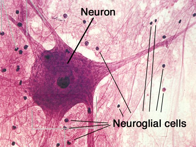
Nervous Tissue: The Body’s Communication Network Nervous Tissue Nervous tissue is the master controller and communication system of the body. It forms the brain, spinal cord, and peripheral nerves, and its primary function is to regulate and integrate all body functions by rapidly transmitting electrical signals. The Two Main Cell Types The nervous system is comprised of two principal types of cells that work in concert. Neurons (Nerve Cells) These are the primary functional cells that are specialized to transmit electrical signals (nerve impulses). They send and receive messages using chemical signals called neurotransmitters across junctions known as synapses. Neuroglia (Glial Cells) These are the non-excitable, supporting cells of the nervous system. They provide physical and metabolic support, insulation (myelin), and immune defense for the neurons. Examples include Astrocytes, Oligodendrocytes, and Schwann Cells. General Characteristics Primary Function: To receive stimuli, transmit electrical impulses, and process information to control the body’s responses. Location: Makes up the Central Nervous System (CNS)—the brain and spinal cord—and the Peripheral Nervous System (PNS)—the peripheral nerves. Key Properties of Neurons Excitability The ability to respond to a stimulus by generating an electrical change across its membrane (membrane potential). Conductivity The ability to propagate these electrical signals (nerve impulses or action potentials) rapidly along the cell membrane. The Neuron (Nerve Cell): The Signaling Unit Neurons are the excitable cells responsible for transmitting electrical signals. They are typically long-lived, amitotic (do not divide in their mature form), and have a very high metabolic rate, requiring a continuous supply of oxygen and glucose to function. 1. Structural Components of a Typical Neuron Cell Body (Soma) The neuron’s main nutritional and metabolic center. It contains the nucleus, most organelles, and prominent Nissl bodies (rough ER), reflecting its high rate of protein synthesis. Dendrites Numerous, short, highly branched processes that act as the main receptive regions. They receive incoming signals from other neurons and convey them towards the cell body. Axon A single, long process that acts as the conducting region, generating and transmitting nerve impulses (action potentials) away from the cell body. It terminates in branches called axon terminals. The Myelin Sheath Many axons are covered by a fatty, insulating layer called the myelin sheath, which is formed by glial cells (Schwann cells in the PNS and oligodendrocytes in the CNS). This sheath dramatically speeds up nerve impulse transmission. The gaps between the myelin segments are called Nodes of Ranvier, where the action potential “jumps” from node to node (saltatory conduction). 2. Functional Classification of Neurons Sensory (Afferent) Transmit impulses from sensory receptors towards the CNS. Motor (Efferent) Transmit impulses from the CNS to effector organs (muscles/glands). Interneurons Lie between sensory and motor neurons within the CNS to integrate information. Most neurons are interneurons. 3. Structural Classification of Neurons Multipolar Three or more processes (one axon, many dendrites). The most common type in the CNS. Bipolar Two processes (one axon, one dendrite). Rare; found in special sense organs like the retina. Unipolar A single, short process that divides T-like. Found in most sensory neurons in the PNS. Neuroglia (Glial Cells): The Supporting Cast Neuroglia are non-excitable cells that surround, support, insulate, and protect neurons. They are far more numerous than neurons and can divide throughout life. There are six types of neuroglia: four in the CNS and two in the PNS. A. Neuroglia of the Central Nervous System (CNS) Astrocytes (Star Cells) Most abundant and versatile. Anchor neurons to blood vessels (form the blood-brain barrier) and regulate the chemical environment. Microglial Cells The resident macrophages of the CNS. They monitor neuron health and phagocytize microorganisms and debris. Ependymal Cells Line the central cavities of the brain and spinal cord. Their cilia help circulate cerebrospinal fluid (CSF). Oligodendrocytes Form the myelin sheaths around axons in the CNS. One oligodendrocyte can myelinate several axons. B. Neuroglia of the Peripheral Nervous System (PNS) Satellite Cells Surround neuron cell bodies in PNS ganglia, providing support and regulating the chemical environment. Schwann Cells Form the myelin sheaths around thicker axons in the PNS. One Schwann cell myelinates one segment of one axon. Crucial for regeneration. Nerve Impulse (Action Potential) Generation and Transmission The ability of neurons to communicate relies on their ability to generate and transmit electrical signals, a process that involves several key stages. 1. Resting Membrane Potential A neuron at rest has a voltage difference across its membrane of about -70mV. This is maintained by the sodium-potassium pump and ion leak channels. 2. Graded Potentials Short-lived, localized changes in membrane potential. If a graded potential is strong enough to reach the threshold potential (~ -55mV) at the axon hillock, it triggers an action potential. 3. Action Potential (Nerve Impulse) A brief, rapid, all-or-none electrical impulse that travels down the axon. It involves a depolarization phase (Na⁺ rushes in) followed by a repolarization phase (K⁺ rushes out). 4. Synapses The junction where information is transferred. An arriving action potential causes the release of chemical messengers called neurotransmitters across a tiny gap (the synaptic cleft), which then bind to the next cell. Test Your Knowledge Check your understanding of the concepts covered in this post. 1. Which of the following is the primary function of nervous tissue? Support and protect organs Contract and generate force Transmit electrical signals and process information Form linings and glandular secretions Rationale: This is the overarching function of nervous tissue, allowing for rapid communication and complex thought processes. 2. The two main types of cells found in nervous tissue are: Epithelial cells and glial cells Neurons and muscle cells Neurons and neuroglia Fibroblasts and oligodendrocytes Rationale: Neurons are the excitable, signaling cells, while neuroglia (glial cells) are the supporting cells. 3. Which part of a neuron typically receives incoming signals from other neurons? Axon Axon terminal Dendrite Myelin sheath Rationale: Dendrites are the highly branched receptive regions that receive neurotransmitter signals from other neurons. 4. The ability of a neuron to respond to a stimulus by changing its membrane potential is called: Conductivity Contractility Excitability Secretion Rationale: Excitability is

muscle tissue
Anatomy

Muscle Tissue

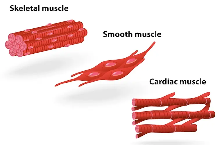
Muscle Tissue: The Body’s Engine of Movement Muscle Tissue Muscle tissue is composed of highly specialized, contractile cells that generate force to produce movement. The cells within all three types of muscle tissue are specialized for contraction (shortening), a process enabled by the interaction of specialized protein fibers. This contraction enables the movement of the whole body and many internal organs, in addition to producing heat energy. An important characteristic is that mature muscle cells have generally lost the ability to divide, so destroyed muscle cells often cannot be replaced. General Characteristics All muscle tissues share a set of key properties that allow them to function effectively. Excitability (Responsiveness) The ability to receive and respond to a stimulus (like a nerve impulse). Contractility The ability to shorten forcibly when stimulated. Extensibility The ability to be stretched or extended. Elasticity The ability to recoil and resume its resting length after stretching. The Three Types of Muscle Tissue Muscle tissue is classified into three types based on location, structure, and functional characteristics. While the cells in smooth and cardiac muscle are referred to as muscle cells, the long, cylindrical cells of skeletal muscle are often called muscle fibers. 1. Skeletal Muscle Striated (Striped appearance) Voluntary (Under conscious control) Multinucleate (Long, cylindrical cells) Location: Primarily attached to the skeleton, enabling body movement. 2. Cardiac Muscle Striated (Striped appearance) Involuntary (Not under conscious control) Branched Cells (Connected by intercalated discs) Location: Found exclusively in the wall of the heart. 3. Smooth Muscle Non-striated (Smooth appearance) Involuntary (Not under conscious control) Spindle-shaped Cells (Single central nucleus) Location: Found in the walls of hollow organs like the stomach, bladder, and blood vessels. Skeletal Muscle Named for its location, skeletal muscle tissue is usually attached to bones and skin, enabling movement of the head, trunk, and limbs. Its contractions are voluntary (under conscious control). Key Characteristics: Striated: Appears striped or banded due to the highly organized arrangement of contractile proteins (actin and myosin). Voluntary: Contraction is consciously controlled by the nervous system. Multi-nucleated Fibers: The cells (muscle fibers) are long, cylindrical, and contain multiple nuclei located at the periphery. General Organization (Macroscopic to Microscopic) A whole skeletal muscle is a complex organ containing muscle fibers, blood vessels, nerve fibers, and extensive connective tissue wrappings that hold everything together and transmit the force of contraction. 1. Entire Muscle The whole organ, surrounded by a dense irregular CT layer called the Epimysium. 2. Muscle Fascicle A bundle of muscle fibers, surrounded by a fibrous CT layer called the Perimysium. 3. Muscle Fiber (Cell) A single muscle cell, surrounded by a delicate areolar CT layer called the Endomysium. These three “mysiums” are continuous and converge to form tendons, which transmit the contractile force to the bones. Microscopic Organization of a Muscle Fiber A skeletal muscle fiber is a highly specialized, elongated cell optimized for rapid and powerful contraction. Sarcolemma & Sarcoplasm The Sarcolemma is the cell membrane, featuring deep invaginations called T-tubules. The Sarcoplasm is the cytoplasm, rich in glycogen (glycosomes) and oxygen-storing myoglobin. Sarcoplasmic Reticulum (SR) A specialized smooth ER that surrounds each myofibril. Its primary role is to store and release calcium ions (Ca²⁺), the critical trigger for muscle contraction. Myofibrils and the Sarcomere Myofibrils are the rod-like contractile elements that make up ~80% of the muscle fiber’s volume. Each myofibril is a chain of repeating sarcomeres, the smallest functional unit of muscle contraction. Sarcomere Banding Pattern (Striations) A-Band (Dark): Represents the full length of the thick (myosin) filaments. I-Band (Light): Contains only thin (actin) filaments. Z-Disc (Line): A protein sheet that anchors thin filaments and defines the boundaries of one sarcomere. Myofilaments (Contractile Proteins) Thick Filaments (Myosin): Composed of myosin protein with globular heads that bind to actin and use ATP to generate force. Thin Filaments (Actin): Composed of actin protein, which has binding sites for myosin heads. Also contains two regulatory proteins: Tropomyosin: Blocks the myosin-binding sites on actin in a relaxed muscle. Troponin: Binds to Ca²⁺, which causes it to move tropomyosin, exposing the binding sites and allowing contraction to begin. The Sliding Filament Model of Contraction This is how muscles contract: A nerve impulse triggers the release of Ca²⁺ from the Sarcoplasmic Reticulum. Ca²⁺ binds to troponin, causing it to move tropomyosin away from actin’s binding sites. Myosin heads bind to actin, forming a cross-bridge. The myosin heads pivot, pulling the thin filaments toward the center of the sarcomere (the “power stroke”). This uses ATP. The myosin head detaches, re-cocks, and is ready for another cycle as long as Ca²⁺ and ATP are present. This sliding action shortens all the sarcomeres simultaneously, causing the entire muscle fiber to contract. Satellite Cells These are quiescent (inactive) stem cells located on the surface of mature muscle fibers. When a muscle fiber is injured, satellite cells become activated. They divide and differentiate into new muscle cells to repair the damaged tissue and also contribute to muscle growth (hypertrophy) in response to exercise. Cardiac Muscle Tissue The muscle tissue located in the walls of the heart is cardiac muscle tissue. It consists of branching cells that interconnect in a netlike arrangement. The rhythmic contractions of cardiac muscle are involuntary because they cannot be consciously controlled. General Characteristics Location: Found exclusively in the myocardium, the middle and thickest layer of the heart wall. Function: Responsible for the forceful, rhythmic contractions that pump blood throughout the body. Control: Involuntary. It possesses its own intrinsic electrical conduction system (autorhythmicity). Appearance: It is striated, similar to skeletal muscle, due to the organized arrangement of contractile proteins. Energy Needs: Extremely high metabolic demand, with abundant mitochondria, and relies almost exclusively on aerobic respiration. Microscopic Organization (Cardiomyocyte) Cardiac muscle cells, or cardiomyocytes, are highly specialized cells with several unique features. The Defining Feature: Intercalated Discs These are complex, specialized junctions that connect adjacent cardiomyocytes end-to-end, appearing as dark, wavy lines. They contain two vital components: Desmosomes: Act as strong anchoring points, preventing cells from separating during powerful contractions. Gap Junctions: Channels that allow

connective tissues
Anatomy

Connective Tissues

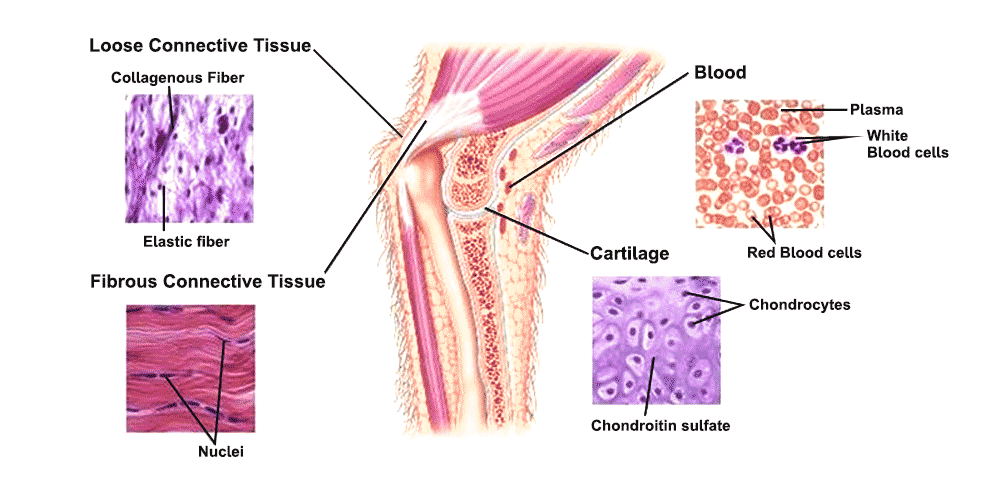
Connective Tissue: The Body’s Support System Connective Tissue Connective Tissue (CT) is a diverse group of tissues that connect, support, and bind other tissues and organs together. All connective tissues are derived from an embryonic tissue called mesenchyme. Key Distinguishing Features Origin: All connective tissues arise from mesenchyme. Vascularity: Most are well vascularized, with notable exceptions being cartilage (avascular) and dense regular CT (poorly vascularized). Extracellular Matrix (ECM): This is the defining characteristic. Cells are widely scattered within a large amount of non-living material that they produce. The ECM, consisting of ground substance and protein fibers, is responsible for the tissue’s physical properties. Components of Connective Tissue All connective tissues share three fundamental components: Ground Substance, Fibers, and Cells. 1. Ground Substance An unstructured, gel-like material that fills the space between cells and contains the fibers. It is composed of: Interstitial Fluid: Watery fluid that bathes the cells. Adhesion Proteins: (e.g., fibronectin, laminin) Act as glue, allowing cells to attach to the matrix. Proteoglycans: Large molecules that trap water, forming a gel that allows for diffusion of nutrients and waste. 2. Fibers Fibers provide support and strength to the connective tissue. There are three types: Collagen Fibers The strongest and most abundant type. Thick, rope-like bundles that provide high tensile strength (resist pulling forces). Elastic Fibers Long, thin, stretchy fibers containing elastin. Allow tissues to stretch and recoil. Found in skin, lungs, and blood vessels. Reticular Fibers Short, fine, branched collagenous fibers that form delicate networks (stroma) to support soft organs like the spleen and lymph nodes. 3. Cells of Connective Tissue Connective tissues contain a variety of resident and migrating cells with distinct roles. Primary Cell Types “Blast” Cells (Immature & Active) Fibroblasts: In CT proper. Chondroblasts: In cartilage. Osteoblasts: In bone. Hematopoietic Stem Cells: In blood. “Cyte” Cells (Mature & Maintaining) Fibrocytes: In CT proper. Chondrocytes: In cartilage. Osteocytes: In bone. Other Important Cell Types: Adipocytes (Fat Cells): Store energy (fat), provide insulation, and cushion organs. Mast Cells: Initiate local inflammatory responses by releasing histamine. Found near blood vessels. Macrophages: “Big eaters” that engulf foreign materials and dead cells as part of the immune system. Plasma Cells: Produce antibodies. Leukocytes (White Blood Cells): Migrate from the bloodstream to fight infection. Primary Functions & Main Categories The diverse composition of connective tissues allows them to perform a wide range of functions, from binding and support to transportation and immune response. They are broadly classified into four main categories. 1. Connective Tissue Proper Includes Loose CT (e.g., Areolar, Adipose) and Dense CT (e.g., tendons, dermis of the skin). 2. Cartilage Strong and flexible tissue that provides support and shock absorption. Includes Hyaline, Elastic, and Fibrocartilage. 3. Bone Tissue Hard connective tissue that forms the skeleton, with a calcified matrix. 4. Blood A fluid connective tissue where the extracellular matrix is the liquid plasma. Connective Tissue Proper This is the most diverse group of connective tissues. It is divided into two main categories: Loose Connective Tissues, which have more ground substance and fewer fibers, and Dense Connective Tissues, which have more fibers and less ground substance. A. Loose (Areolar) Connective Tissues Loose Areolar Connective Tissue Features a loose, gel-like matrix with all three fiber types (collagen, elastic, reticular) and various cells, including fibroblasts, macrophages, and mast cells. Histology Hint: Look for a sparse, web-like appearance with randomly arranged thick pink (collagen) and thin black/purple (elastic) fibers, plus many scattered black dots (cell nuclei). Function: Wraps and cushions organs, holds tissue fluid, plays a key role in inflammation. Location: Widely distributed under epithelia; forms the lamina propria of mucous membranes. Adipose Tissue (Fat) Primarily composed of large, tightly packed adipocytes (fat cells) with very little matrix. It is highly vascularized. Histology Hint: Characterized by large, empty-looking circular cells (adipocytes), as the fat droplet is typically dissolved during processing. Nuclei are flattened and pushed to the periphery. Function: Energy storage, insulation, and organ protection/cushioning. Location: Under the skin (hypodermis), around kidneys and eyeballs, in the abdomen and breasts. Reticular Connective Tissue A network of fine reticular fibers in a loose ground substance, with reticular cells (specialized fibroblasts) as the main cell type. Histology Hint: Look for a fine, branching network of dark-staining reticular fibers forming a delicate meshwork (stroma), typically filled with numerous small, round cells (like lymphocytes in a lymph node). Function: Forms a soft internal skeleton (stroma) that supports other cell types in lymphoid organs. Location: Lymphoid organs (lymph nodes, spleen, bone marrow). B. Dense (Fibrous) Connective Tissues Dense Regular Connective Tissue Densely packed, primarily parallel collagen fibers with fibroblasts as the major cell type. It is poorly vascularized. Histology Hint: Characterized by dense, wavy, parallel bundles of pink collagen fibers running in a single direction, with fibroblast nuclei squeezed and flattened between them. Function: Attaches muscles to bones (tendons) or bones to bones (ligaments). Provides great tensile strength in one direction. Location: Tendons, most ligaments, aponeuroses. Dense Irregular Connective Tissue Primarily irregularly arranged, thick collagen fibers with some elastic fibers and fibroblasts. Histology Hint: Shows thick bundles of pink collagen fibers running in many different directions, creating a chaotic appearance. Function: Withstands tension exerted in many directions, providing structural strength. Location: Dermis of the skin, fibrous capsules of organs and joints. Elastic Connective Tissue A type of dense regular connective tissue with a high proportion of elastic fibers. Histology Hint: Displays prominent, wavy, dark-staining elastic fibers arranged in parallel, often with a background of lighter pink collagen. Function: Allows tissue to recoil after stretching; maintains pulsatile blood flow and aids passive recoil of lungs. Location: Walls of large arteries, certain ligaments of the vertebral column, walls of bronchial tubes. Cartilage Cartilage is a tough, flexible connective tissue that consists of a firm, gelatinous matrix in which cartilage cells, or chondrocytes, are embedded within fluid-filled spaces called lacunae. It is avascular (lacks blood vessels) and lacks nerves, relying on diffusion for nutrients. Key Characteristics: Cells: Chondroblasts produce the matrix, which mature into chondrocytes that maintain it from within their lacunae.

Scroll to Top