
On this page 8 sections
Overview & definition
Human anatomy (ah-nat-o-me) is the scientific study of the structural organization of the human body and the physical relationships among its constituent parts. The term originates from the ancient Greek root words ana ("apart") and tome ("to cut"), reflecting its historical and ongoing reliance on dissection.
Anatomy does not exist in isolation; it forms a unified discipline alongside human physiology (fiz-e-ol-o-je), which investigates the biochemical, mechanical, and physical functions of living structures. While anatomy reveals how an organ or tissue is constructed, physiology explains how that structure works to sustain life.
History of anatomical study
For centuries, cultural, religious, and legal taboos severely restricted the dissection of human bodies. The understanding of human anatomy evolved through key historical phases:
-
2nd Century AD
Claudius Galenus (Galen)
A Greek physician practicing in Rome who served as physician to gladiators and emperors. Because human dissection was prohibited by Roman law, Galen based his extensive anatomical treatises on vivisections and dissections of animals, predominantly pigs, apes, and oxen. Although several of his mammalian extrapolations were inaccurate for humans, his teachings stood unchallenged as absolute dogma for over 1,300 years.
-
c. 1490–1515
Leonardo da Vinci
Conducted human dissections alongside physicians such as Marcantonio della Torre. Da Vinci generated hundreds of pioneering anatomical illustrations documenting muscular attachments, bones, and cardiovascular structures before ecclesiastical restrictions limited his access to cadaveric material.
-
1543
Andreas Vesalius
Universally regarded as the "Father of Modern Anatomy." A Flemish physician and professor at the University of Padua, Vesalius broke with scholastic tradition by personally dissecting human bodies rather than delegating the task to barber-surgeons. In 1543, he published De Humani Corporis Fabrica Libri Septem ("On the Fabric of the Human Body"), correcting hundreds of Galen's anatomical errors and establishing direct empirical observation as the gold standard of medicine.
-
17th & 18th Centuries
Anatomical Theatres & Public Dissections
Certified anatomists conducted tightly regulated human dissections within tiered anatomical theatres (such as those in Padua, Leiden, and Bologna). These sessions served as academic investigations and civic events attended by scholars, medical apprentices, and prominent artists.
-
1832
The British Anatomy Act
The rapid expansion of medical schools during the Industrial Revolution created a shortage of cadavers, fueling illegal grave-robbing ("resurrectionists") and homicides for dissection material (such as the Burke and Hare murders in Edinburgh). The Anatomy Act of 1832 abolished the practice of dissecting executed criminals as a legal penalty and established a licensed, inspected framework allowing unclaimed hospital bodies to be utilized for medical education.
-
Modern Era
Voluntary Cadaveric Donation
Contemporary medical, nursing, and surgical curricula rely on willed body donation programs governed by informed consent and anatomical tissue legislation. Dissection continues to provide 3D spatial orientation, tactile appreciation of tissue planes, and exposure to anatomical variation.

Core principles & organization
1. Complementarity of structure and function
A fundamental axiom of anatomical science is that function always reflects structure. What an organ, tissue, or cell is capable of performing depends directly on its physical architecture, spatial geometry, and chemical composition.
For example, the mineralized hydroxyapatite matrix of cortical bone provides compressive strength to resist gravitational load, whereas the compliant, elastin-rich walls of elastic arteries (such as the aorta) accommodate cyclic systolic pressure surges. In dentition, pointed incisors and canines shear food, while broad, cusped molars crush and grind fibrous nutrients.
2. Hierarchy of structural organization
The human body is assembled through six ascending, interdependent levels of structural organization:
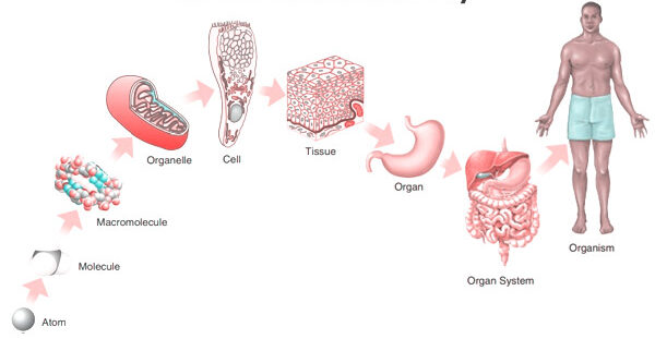
- Chemical level: Atoms (carbon, hydrogen, oxygen, nitrogen, calcium, phosphorus) bond to construct functional macromolecules, including nucleic acids, carbohydrates, lipids, and structural proteins.
- Cellular level: The fundamental structural and functional unit of life. Macromolecules assemble to construct organelles (nuclei, mitochondria, ribosomes) within specialized cells (such as erythrocytes, osteocytes, and cardiomyocytes).
- Tissue level: Aggregations of morphologically similar cells and their surrounding extracellular matrix cooperating to execute specialized tasks. The four primary tissue classifications are epithelial, connective, muscle, and nervous tissue.
- Organ level: Discrete anatomical structures composed of two or more primary tissue types organized to execute complex physiological functions (e.g., the stomach, liver, lungs, and brain).
- Organ system level: Coordinated groups of organs working synergistically to fulfill body-wide homeostatic tasks (e.g., the cardiovascular, respiratory, renal, and digestive systems).
- Organismal level: The highest echelon of integration, representing the sum total of all eleven organ systems cooperating to maintain independent biological life.
3. Homeostasis & dynamic equilibrium
Homeostasis represents the capacity of an organism to preserve a relatively stable, regulated internal physiological environment despite fluctuations in external parameters. This requires continuous physiological monitoring and corrective adjustments.
A classic homeostatic control loop consists of three primary elements:
- Receptor (sensor): Monitors the regulated variable (e.g., peripheral thermoreceptors in the skin or arterial baroreceptors in the carotid sinus) and transmits afferent data to the control center.
- Integrating (control) center: Analyzes afferent signals against a physiological set-point and dictates an efferent response (e.g., the hypothalamic preoptic nucleus for thermoregulation).
- Effector: A responsive organ, muscle, or gland that executes the corrective action (e.g., eccrine sweat glands activating diaphoresis to lower core body temperature, or peripheral vascular smooth muscle constricting to elevate systemic pressure).
Branches of anatomy
To examine anatomical structures at different dimensions and across different stages of life, anatomy is organized into distinct subdisciplines:
1. Gross (macroscopic) anatomy
The examination of large body structures readily visible to the unaided eye without the aid of optical magnification.
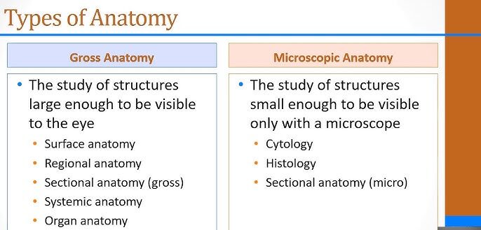
- Regional anatomy: Investigates all anatomical structures (muscles, nerves, blood vessels, bones) situated within a specific spatial territory concurrently. For example, surgical dissection of the femoral triangle or carpal tunnel.
- Systemic anatomy: Studies the human body system by system across all regions. For example, charting the vascular tree from the aorta to distal microvasculature, or examining the axial and appendicular components of the skeletal system.
- Surface anatomy: Studies internal structures as they relate to and project upon the overlying skin. It forms the baseline of physical diagnosis, including palpating the radial arterial pulse, auscultating heart valves at designated intercostal spaces, and identifying bony landmarks for spinal taps (lumbar puncture at L3/L4 or L4/L5).
2. Microscopic anatomy
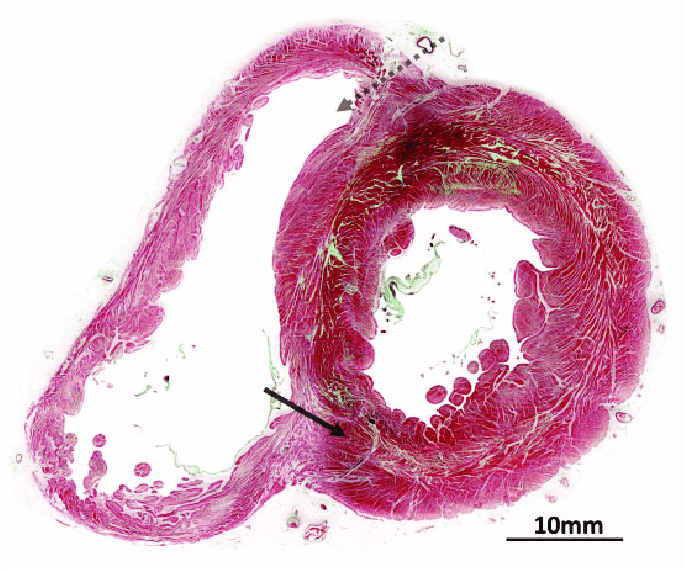
The examination of structural units that cannot be resolved without magnification. Specimens are prepared via fixation, microtome sectioning, and histochemical staining.
- Cytology: The microscopic and ultrastructural evaluation of individual cell types, their internal organelles, and inclusions (e.g., cervical cytology via Papanicolaou smears).
- Histology: The microscopic evaluation of tissue architecture and organization, distinguishing regular architectural patterns from pathological changes in biopsy specimens.
- Instrumentation: Light microscopy utilizes visible light and glass lenses to visualize cell boundaries and nuclear morphology (up to ~1,000× magnification). Transmission and scanning electron microscopy utilize accelerated electron beams to resolve ultrastructural details, such as mitochondrial cristae, nuclear pores, and the phospholipid bilayer (up to ~500,000× magnification).
3. Developmental anatomy
The study of structural changes that occur in an organism from conception throughout embryonic development, childhood, maturity, and senescence.

- Embryology: Investigates the first eight weeks of post-fertilization development in humans, covering gastrulation, neurulation, and the establishment of all primary organ systems (organogenesis).
- Ontogeny (ontogenesis): Encompasses the entire lifespan sequence of structural changes from fertilization through senescence.
Specialized clinical and applied branches
- Pathological anatomy (morbid anatomy): Examines gross and microscopic structural alterations induced by disease states (e.g., cirrhotic remodeling of liver parenchyma or atherosclerotic plaques in coronary arteries).
- Radiographic (imaging) anatomy: Utilizes non-invasive imaging technologies to study internal anatomy in living individuals. Modalities include projection radiography (X-rays), computed tomography (CT), magnetic resonance imaging (MRI), and diagnostic ultrasonography.
- Surgical (applied) anatomy: Emphasizes anatomical relationships, fascial planes, and neurovascular trajectories critical to safe surgical entry and procedural execution.
Foundational vocabulary
Anatomical terminology provides an unambiguous, internationally recognized language based primarily on Greek and Latin roots. This standardized lexicon prevents clinical errors when describing patient findings, surgical sites, and radiological images.
Scroll the table sideways on a smaller screen
| Term & etymology | Definition | Example / clinical context |
|---|---|---|
| Anatomy (ana = apart; tome = to cut) |
The scientific study of the physical structure and spatial organization of living organisms. | Dissecting the axilla to trace the cords and terminal branches of the brachial plexus. |
| Appendicular (appendere = to hang upon) |
Pertaining to the upper and lower extremities and their respective limb girdles. | The appendicular skeleton comprises the shoulder girdle, arms, forearms, hands, pelvic girdle, thighs, legs, and feet. |
| Axial (axis = central axle) |
Pertaining to the central longitudinal axis of the body. | The axial skeleton forms the vertical core, including the cranium, hyoid, vertebral column, ribs, and sternum. |
| Body region (regio = bounded boundary) |
A defined anatomical territory designated by a standardized topographical name. | The brachial region corresponds strictly to the arm (shoulder to elbow), distinct from the antebrachial region (forearm). |
| Directional term (directio = guiding towards) |
Standardized terminology used to reference the position of one body structure relative to another. | The patella is superior to the tibia and anterior to the popliteal fossa. |
| Effector (efficere = to bring about) |
An organ, muscle, or gland that carries out a homeostatic correction directed by an integrating center. | Skeletal muscle shivering acting as an effector to elevate core body temperature during hypothermia. |
| Homeostasis (homeo = similar; stasis = standing still) |
The physiological maintenance of a dynamic, stable equilibrium in the internal fluid environment. | Renal and endocrine systems regulating blood plasma osmolality within a narrow range (~285–295 mOsm/kg). |
| Integrating center (integrare = to make whole) |
A control element that processes afferent sensory data and dictates efferent corrective commands. | The respiratory center in the medulla oblongata regulating ventilation rate based on cerebrospinal fluid pH and arterial pCO₂. |
| Metabolism (metabole = change/turnover) |
The sum of all integrated biochemical pathways operating within an organism (catabolism and anabolism). | Catabolic glycolysis breaking down glucose for ATP production alongside anabolic hepatic glycogen synthesis. |
| Parietal (paries = chamber wall) |
Pertaining to or forming the outer wall of a body cavity. | The parietal peritoneum lines the inner surface of the abdominopelvic muscular wall. |
| Pericardium (peri = around; kardia = heart) |
The fibroserous sac enclosing the heart and the roots of the great vessels. | Acute inflammation of the pericardial layers results in pericarditis and may cause pericardial friction rubs. |
| Peritoneum (peri = around; teinein = to stretch) |
The extensive serous membrane lining the abdominal cavity and investing abdominal viscera. | Peritonitis represents a life-threatening inflammation of this membrane following gastrointestinal perforation. |
| Physiology (physis = nature; logos = study) |
The scientific study of the mechanical, physical, and biochemical functions of living structures. | Investigating cardiac excitation-contraction coupling through calcium-induced calcium release in myocytes. |
| Plane (planum = flat surface) |
An imaginary two-dimensional geometrical surface along which a structure or body is sectioned. | A sagittal plane oriented parallel to the longitudinal midline of the human body. |
| Pleura (pleura = rib/side) |
The double-layered serous membrane lining the thoracic cavity and enveloping each lung. | Pleural effusion refers to the pathological accumulation of fluid in the pleural space between the membranes. |
| Receptor (recipere = to receive) |
A specialized sensory structure or macromolecule designed to monitor changes in physiological variables. | Aortic body chemoreceptors detecting severe arterial hypoxemia and transmitting signals via the vagus nerve. |
| Section (sectio = a cutting) |
A flat physical surface of a structure exposed by a cut along a designated anatomical plane. | An axial cross-section of the thigh revealing compartments enclosed by fascia lata. |
| Serous membrane (serum = watery fluid; membrana = film) |
A thin, smooth, two-layered mesothelial membrane that lines closed body cavities and secretes lubricating serous fluid. | The pleura, pericardium, and peritoneum are the three primary serous membrane envelopes in the human body. |
| Visceral (viscus = internal organ) |
Pertaining to or directly investing an internal organ situated within a body cavity. | The visceral pleura adheres firmly to the outer parenchymal surface of the lung, dipping into its interlobar fissures. |
Planes, directions & cavities
The standard anatomical position
All anatomical descriptions are calibrated to an invariant universal reference posture: the anatomical position. In this posture:
- The individual stands erect, facing forward toward the observer.
- The head, gaze, and eyes are directed straight ahead horizontally.
- The upper limbs are suspended along the sides of the trunk.
- The palms of the hands face anteriorly (supinated), with thumbs directed away from the body.
- The lower extremities are parallel, with feet flat on the floor and toes directed anteriorly.
Anatomical planes and sections
Three cardinal orthogonal planes section the human body in the anatomical position:
- Sagittal plane: A vertical plane extending from anterior to posterior, dividing the body into right and left segments. The midsagittal (median) plane passes directly through the midline, creating equal right and left halves. Planes parallel to the median are termed parasagittal.
- Coronal (frontal) plane: A vertical plane oriented at a right angle to the sagittal plane, dividing the body into anterior (front) and posterior (back) segments.
- Transverse (horizontal / axial) plane: A horizontal plane oriented perpendicular to both sagittal and frontal planes, dividing the body into superior (upper) and inferior (lower) segments. Standard clinical CT and MRI scans are generated largely in this plane.
Relative directional terms
Directional terms operate in opposing conceptual pairs:
- Superior (cranial) vs. Inferior (caudal): Closer to the head vs. closer to the feet. Example: The thyroid gland is superior to the thymus.
- Anterior (ventral) vs. Posterior (dorsal): Toward the front surface of the body vs. toward the back surface. Example: The sternum is anterior to the pericardium.
- Medial vs. Lateral: Closer to the midsagittal plane vs. further away from the midline. Example: The ulna is medial to the radius in the anatomical position.
- Proximal vs. Distal: Employed primarily for limbs; closer to the point of origin (or limb attachment to the trunk) vs. farther from the point of attachment. Example: The elbow is proximal to the wrist, while the phalanges are distal to the metacarpals.
- Superficial (external) vs. Deep (internal): Closer to the external surface of the body vs. positioned deeper within internal tissue layers. Example: The epidermis is superficial to the dermis; the pectoralis major muscle is superficial to the pectoralis minor.
Body cavities and membranes
The human body organizes internal organs within two major fluid-cushioned cavities:
Dorsal (posterior) body cavity
Enclosed by bone and invested by protective fibrous meningeal membranes:
- Cranial cavity: Formed by neurocranial bones, housing the brain and bathed in cerebrospinal fluid.
- Vertebral (spinal) cavity: Formed by the stacked vertebral foramina, protecting the spinal cord and spinal nerve roots.
Ventral (anterior) body cavity
Subdivided by the muscular diaphragm into superior thoracic and inferior abdominopelvic compartments:
- Thoracic cavity: Contains the right and left pleural cavities (each enveloping a lung) and the central mediastinum (containing the pericardial cavity enclosing the heart, along with the trachea, esophagus, and thoracic aorta).
- Abdominopelvic cavity: Extends from the inferior surface of the diaphragm to the pelvic floor. It houses digestive organs (stomach, intestines, liver, pancreas, spleen), renal structures, and pelvic viscera (urinary bladder, internal reproductive organs, and rectum).
Methods of examination
Mastering anatomy requires synthesizing spatial relationships, visual recognition, and physical diagnostic skills. Clinical and educational examination relies on several core methodologies:
- Cadaveric dissection: The foundational method of physical isolation and exposure of anatomical layers, preserving true tactile feedback and spatial depth that cannot be fully replicated in synthetic or digital media.
- Inspection & observation: Rigorous visual appraisal of surface contours, symmetry, color changes, and obvious structural deviations (e.g., assessing thoracic expansion or pupillary symmetry).
- Palpation: Feeling surface and deep anatomical structures with the fingertips or palms to assess consistency, mobility, tenderness, temperature, and arterial pulsations (e.g., palpating lymph nodes, abdominal viscera, or peripheral pulses).
- Auscultation: Listening to acoustic signals produced within active body chambers, primarily through a stethoscope (e.g., valvular heart sounds, bronchial breath sounds, and intestinal borborygmi).
- Percussion: Tapping directly on the body surface to emit resonant acoustic waves, allowing clinicians to delineate underlying tissue densities (e.g., differentiating resonant air-filled lung parenchyma from dull fluid effusions or solid hepatic borders).
- Diagnostic medical imaging: Correlating cross-sectional radiological slices (CT, MRI) and acoustic sonography with known anatomical planes to detect pathological tissue changes in living patients.