Doctors Revision

Author name: doctorsrevision@gmail.com

nervous tissue
Anatomy

Nervous Tissue

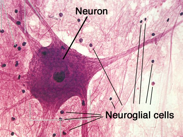
Nervous Tissue: The Body’s Communication Network Nervous Tissue Nervous tissue is the master controller and communication system of the body. It forms the brain, spinal cord, and peripheral nerves, and its primary function is to regulate and integrate all body functions by rapidly transmitting electrical signals. The Two Main Cell Types The nervous system is comprised of two principal types of cells that work in concert. Neurons (Nerve Cells) These are the primary functional cells that are specialized to transmit electrical signals (nerve impulses). They send and receive messages using chemical signals called neurotransmitters across junctions known as synapses. Neuroglia (Glial Cells) These are the non-excitable, supporting cells of the nervous system. They provide physical and metabolic support, insulation (myelin), and immune defense for the neurons. Examples include Astrocytes, Oligodendrocytes, and Schwann Cells. General Characteristics Primary Function: To receive stimuli, transmit electrical impulses, and process information to control the body’s responses. Location: Makes up the Central Nervous System (CNS)—the brain and spinal cord—and the Peripheral Nervous System (PNS)—the peripheral nerves. Key Properties of Neurons Excitability The ability to respond to a stimulus by generating an electrical change across its membrane (membrane potential). Conductivity The ability to propagate these electrical signals (nerve impulses or action potentials) rapidly along the cell membrane. The Neuron (Nerve Cell): The Signaling Unit Neurons are the excitable cells responsible for transmitting electrical signals. They are typically long-lived, amitotic (do not divide in their mature form), and have a very high metabolic rate, requiring a continuous supply of oxygen and glucose to function. 1. Structural Components of a Typical Neuron Cell Body (Soma) The neuron’s main nutritional and metabolic center. It contains the nucleus, most organelles, and prominent Nissl bodies (rough ER), reflecting its high rate of protein synthesis. Dendrites Numerous, short, highly branched processes that act as the main receptive regions. They receive incoming signals from other neurons and convey them towards the cell body. Axon A single, long process that acts as the conducting region, generating and transmitting nerve impulses (action potentials) away from the cell body. It terminates in branches called axon terminals. The Myelin Sheath Many axons are covered by a fatty, insulating layer called the myelin sheath, which is formed by glial cells (Schwann cells in the PNS and oligodendrocytes in the CNS). This sheath dramatically speeds up nerve impulse transmission. The gaps between the myelin segments are called Nodes of Ranvier, where the action potential “jumps” from node to node (saltatory conduction). 2. Functional Classification of Neurons Sensory (Afferent) Transmit impulses from sensory receptors towards the CNS. Motor (Efferent) Transmit impulses from the CNS to effector organs (muscles/glands). Interneurons Lie between sensory and motor neurons within the CNS to integrate information. Most neurons are interneurons. 3. Structural Classification of Neurons Multipolar Three or more processes (one axon, many dendrites). The most common type in the CNS. Bipolar Two processes (one axon, one dendrite). Rare; found in special sense organs like the retina. Unipolar A single, short process that divides T-like. Found in most sensory neurons in the PNS. Neuroglia (Glial Cells): The Supporting Cast Neuroglia are non-excitable cells that surround, support, insulate, and protect neurons. They are far more numerous than neurons and can divide throughout life. There are six types of neuroglia: four in the CNS and two in the PNS. A. Neuroglia of the Central Nervous System (CNS) Astrocytes (Star Cells) Most abundant and versatile. Anchor neurons to blood vessels (form the blood-brain barrier) and regulate the chemical environment. Microglial Cells The resident macrophages of the CNS. They monitor neuron health and phagocytize microorganisms and debris. Ependymal Cells Line the central cavities of the brain and spinal cord. Their cilia help circulate cerebrospinal fluid (CSF). Oligodendrocytes Form the myelin sheaths around axons in the CNS. One oligodendrocyte can myelinate several axons. B. Neuroglia of the Peripheral Nervous System (PNS) Satellite Cells Surround neuron cell bodies in PNS ganglia, providing support and regulating the chemical environment. Schwann Cells Form the myelin sheaths around thicker axons in the PNS. One Schwann cell myelinates one segment of one axon. Crucial for regeneration. Nerve Impulse (Action Potential) Generation and Transmission The ability of neurons to communicate relies on their ability to generate and transmit electrical signals, a process that involves several key stages. 1. Resting Membrane Potential A neuron at rest has a voltage difference across its membrane of about -70mV. This is maintained by the sodium-potassium pump and ion leak channels. 2. Graded Potentials Short-lived, localized changes in membrane potential. If a graded potential is strong enough to reach the threshold potential (~ -55mV) at the axon hillock, it triggers an action potential. 3. Action Potential (Nerve Impulse) A brief, rapid, all-or-none electrical impulse that travels down the axon. It involves a depolarization phase (Na⁺ rushes in) followed by a repolarization phase (K⁺ rushes out). 4. Synapses The junction where information is transferred. An arriving action potential causes the release of chemical messengers called neurotransmitters across a tiny gap (the synaptic cleft), which then bind to the next cell. Test Your Knowledge Check your understanding of the concepts covered in this post. 1. Which of the following is the primary function of nervous tissue? Support and protect organs Contract and generate force Transmit electrical signals and process information Form linings and glandular secretions Rationale: This is the overarching function of nervous tissue, allowing for rapid communication and complex thought processes. 2. The two main types of cells found in nervous tissue are: Epithelial cells and glial cells Neurons and muscle cells Neurons and neuroglia Fibroblasts and oligodendrocytes Rationale: Neurons are the excitable, signaling cells, while neuroglia (glial cells) are the supporting cells. 3. Which part of a neuron typically receives incoming signals from other neurons? Axon Axon terminal Dendrite Myelin sheath Rationale: Dendrites are the highly branched receptive regions that receive neurotransmitter signals from other neurons. 4. The ability of a neuron to respond to a stimulus by changing its membrane potential is called: Conductivity Contractility Excitability Secretion Rationale: Excitability is
muscle tissue
Anatomy

Muscle Tissue

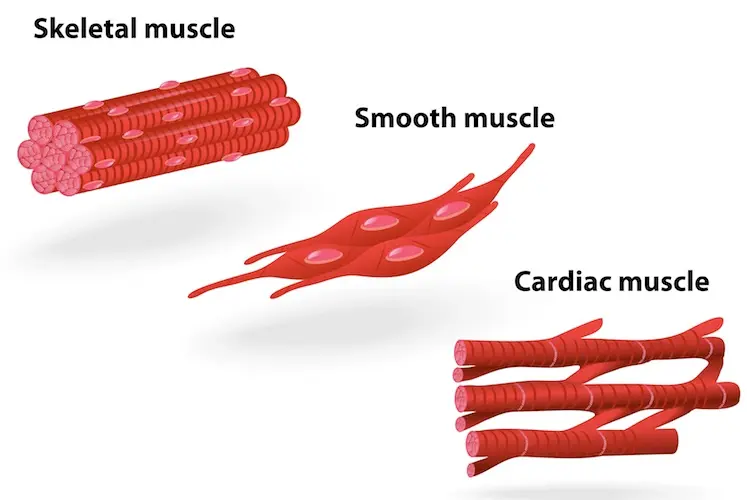
Muscle Tissue: The Body’s Engine of Movement Muscle Tissue Muscle tissue is composed of highly specialized, contractile cells that generate force to produce movement. The cells within all three types of muscle tissue are specialized for contraction (shortening), a process enabled by the interaction of specialized protein fibers. This contraction enables the movement of the whole body and many internal organs, in addition to producing heat energy. An important characteristic is that mature muscle cells have generally lost the ability to divide, so destroyed muscle cells often cannot be replaced. General Characteristics All muscle tissues share a set of key properties that allow them to function effectively. Excitability (Responsiveness) The ability to receive and respond to a stimulus (like a nerve impulse). Contractility The ability to shorten forcibly when stimulated. Extensibility The ability to be stretched or extended. Elasticity The ability to recoil and resume its resting length after stretching. The Three Types of Muscle Tissue Muscle tissue is classified into three types based on location, structure, and functional characteristics. While the cells in smooth and cardiac muscle are referred to as muscle cells, the long, cylindrical cells of skeletal muscle are often called muscle fibers. 1. Skeletal Muscle Striated (Striped appearance) Voluntary (Under conscious control) Multinucleate (Long, cylindrical cells) Location: Primarily attached to the skeleton, enabling body movement. 2. Cardiac Muscle Striated (Striped appearance) Involuntary (Not under conscious control) Branched Cells (Connected by intercalated discs) Location: Found exclusively in the wall of the heart. 3. Smooth Muscle Non-striated (Smooth appearance) Involuntary (Not under conscious control) Spindle-shaped Cells (Single central nucleus) Location: Found in the walls of hollow organs like the stomach, bladder, and blood vessels. Skeletal Muscle Named for its location, skeletal muscle tissue is usually attached to bones and skin, enabling movement of the head, trunk, and limbs. Its contractions are voluntary (under conscious control). Key Characteristics: Striated: Appears striped or banded due to the highly organized arrangement of contractile proteins (actin and myosin). Voluntary: Contraction is consciously controlled by the nervous system. Multi-nucleated Fibers: The cells (muscle fibers) are long, cylindrical, and contain multiple nuclei located at the periphery. General Organization (Macroscopic to Microscopic) A whole skeletal muscle is a complex organ containing muscle fibers, blood vessels, nerve fibers, and extensive connective tissue wrappings that hold everything together and transmit the force of contraction. 1. Entire Muscle The whole organ, surrounded by a dense irregular CT layer called the Epimysium. 2. Muscle Fascicle A bundle of muscle fibers, surrounded by a fibrous CT layer called the Perimysium. 3. Muscle Fiber (Cell) A single muscle cell, surrounded by a delicate areolar CT layer called the Endomysium. These three “mysiums” are continuous and converge to form tendons, which transmit the contractile force to the bones. Microscopic Organization of a Muscle Fiber A skeletal muscle fiber is a highly specialized, elongated cell optimized for rapid and powerful contraction. Sarcolemma & Sarcoplasm The Sarcolemma is the cell membrane, featuring deep invaginations called T-tubules. The Sarcoplasm is the cytoplasm, rich in glycogen (glycosomes) and oxygen-storing myoglobin. Sarcoplasmic Reticulum (SR) A specialized smooth ER that surrounds each myofibril. Its primary role is to store and release calcium ions (Ca²⁺), the critical trigger for muscle contraction. Myofibrils and the Sarcomere Myofibrils are the rod-like contractile elements that make up ~80% of the muscle fiber’s volume. Each myofibril is a chain of repeating sarcomeres, the smallest functional unit of muscle contraction. Sarcomere Banding Pattern (Striations) A-Band (Dark): Represents the full length of the thick (myosin) filaments. I-Band (Light): Contains only thin (actin) filaments. Z-Disc (Line): A protein sheet that anchors thin filaments and defines the boundaries of one sarcomere. Myofilaments (Contractile Proteins) Thick Filaments (Myosin): Composed of myosin protein with globular heads that bind to actin and use ATP to generate force. Thin Filaments (Actin): Composed of actin protein, which has binding sites for myosin heads. Also contains two regulatory proteins: Tropomyosin: Blocks the myosin-binding sites on actin in a relaxed muscle. Troponin: Binds to Ca²⁺, which causes it to move tropomyosin, exposing the binding sites and allowing contraction to begin. The Sliding Filament Model of Contraction This is how muscles contract: A nerve impulse triggers the release of Ca²⁺ from the Sarcoplasmic Reticulum. Ca²⁺ binds to troponin, causing it to move tropomyosin away from actin’s binding sites. Myosin heads bind to actin, forming a cross-bridge. The myosin heads pivot, pulling the thin filaments toward the center of the sarcomere (the “power stroke”). This uses ATP. The myosin head detaches, re-cocks, and is ready for another cycle as long as Ca²⁺ and ATP are present. This sliding action shortens all the sarcomeres simultaneously, causing the entire muscle fiber to contract. Satellite Cells These are quiescent (inactive) stem cells located on the surface of mature muscle fibers. When a muscle fiber is injured, satellite cells become activated. They divide and differentiate into new muscle cells to repair the damaged tissue and also contribute to muscle growth (hypertrophy) in response to exercise. Cardiac Muscle Tissue The muscle tissue located in the walls of the heart is cardiac muscle tissue. It consists of branching cells that interconnect in a netlike arrangement. The rhythmic contractions of cardiac muscle are involuntary because they cannot be consciously controlled. General Characteristics Location: Found exclusively in the myocardium, the middle and thickest layer of the heart wall. Function: Responsible for the forceful, rhythmic contractions that pump blood throughout the body. Control: Involuntary. It possesses its own intrinsic electrical conduction system (autorhythmicity). Appearance: It is striated, similar to skeletal muscle, due to the organized arrangement of contractile proteins. Energy Needs: Extremely high metabolic demand, with abundant mitochondria, and relies almost exclusively on aerobic respiration. Microscopic Organization (Cardiomyocyte) Cardiac muscle cells, or cardiomyocytes, are highly specialized cells with several unique features. The Defining Feature: Intercalated Discs These are complex, specialized junctions that connect adjacent cardiomyocytes end-to-end, appearing as dark, wavy lines. They contain two vital components: Desmosomes: Act as strong anchoring points, preventing cells from separating during powerful contractions. Gap Junctions: Channels that allow

connective tissues
Anatomy

Connective Tissues

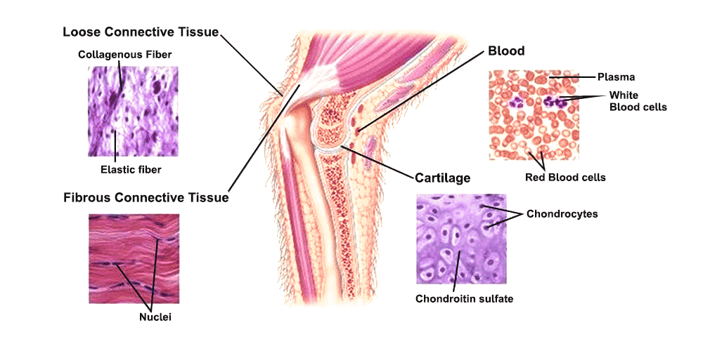
Connective Tissue: The Body’s Support System Connective Tissue Connective Tissue (CT) is a diverse group of tissues that connect, support, and bind other tissues and organs together. All connective tissues are derived from an embryonic tissue called mesenchyme. Key Distinguishing Features Origin: All connective tissues arise from mesenchyme. Vascularity: Most are well vascularized, with notable exceptions being cartilage (avascular) and dense regular CT (poorly vascularized). Extracellular Matrix (ECM): This is the defining characteristic. Cells are widely scattered within a large amount of non-living material that they produce. The ECM, consisting of ground substance and protein fibers, is responsible for the tissue’s physical properties. Components of Connective Tissue All connective tissues share three fundamental components: Ground Substance, Fibers, and Cells. 1. Ground Substance An unstructured, gel-like material that fills the space between cells and contains the fibers. It is composed of: Interstitial Fluid: Watery fluid that bathes the cells. Adhesion Proteins: (e.g., fibronectin, laminin) Act as glue, allowing cells to attach to the matrix. Proteoglycans: Large molecules that trap water, forming a gel that allows for diffusion of nutrients and waste. 2. Fibers Fibers provide support and strength to the connective tissue. There are three types: Collagen Fibers The strongest and most abundant type. Thick, rope-like bundles that provide high tensile strength (resist pulling forces). Elastic Fibers Long, thin, stretchy fibers containing elastin. Allow tissues to stretch and recoil. Found in skin, lungs, and blood vessels. Reticular Fibers Short, fine, branched collagenous fibers that form delicate networks (stroma) to support soft organs like the spleen and lymph nodes. 3. Cells of Connective Tissue Connective tissues contain a variety of resident and migrating cells with distinct roles. Primary Cell Types “Blast” Cells (Immature & Active) Fibroblasts: In CT proper. Chondroblasts: In cartilage. Osteoblasts: In bone. Hematopoietic Stem Cells: In blood. “Cyte” Cells (Mature & Maintaining) Fibrocytes: In CT proper. Chondrocytes: In cartilage. Osteocytes: In bone. Other Important Cell Types: Adipocytes (Fat Cells): Store energy (fat), provide insulation, and cushion organs. Mast Cells: Initiate local inflammatory responses by releasing histamine. Found near blood vessels. Macrophages: “Big eaters” that engulf foreign materials and dead cells as part of the immune system. Plasma Cells: Produce antibodies. Leukocytes (White Blood Cells): Migrate from the bloodstream to fight infection. Primary Functions & Main Categories The diverse composition of connective tissues allows them to perform a wide range of functions, from binding and support to transportation and immune response. They are broadly classified into four main categories. 1. Connective Tissue Proper Includes Loose CT (e.g., Areolar, Adipose) and Dense CT (e.g., tendons, dermis of the skin). 2. Cartilage Strong and flexible tissue that provides support and shock absorption. Includes Hyaline, Elastic, and Fibrocartilage. 3. Bone Tissue Hard connective tissue that forms the skeleton, with a calcified matrix. 4. Blood A fluid connective tissue where the extracellular matrix is the liquid plasma. Connective Tissue Proper This is the most diverse group of connective tissues. It is divided into two main categories: Loose Connective Tissues, which have more ground substance and fewer fibers, and Dense Connective Tissues, which have more fibers and less ground substance. A. Loose (Areolar) Connective Tissues Loose Areolar Connective Tissue Features a loose, gel-like matrix with all three fiber types (collagen, elastic, reticular) and various cells, including fibroblasts, macrophages, and mast cells. Histology Hint: Look for a sparse, web-like appearance with randomly arranged thick pink (collagen) and thin black/purple (elastic) fibers, plus many scattered black dots (cell nuclei). Function: Wraps and cushions organs, holds tissue fluid, plays a key role in inflammation. Location: Widely distributed under epithelia; forms the lamina propria of mucous membranes. Adipose Tissue (Fat) Primarily composed of large, tightly packed adipocytes (fat cells) with very little matrix. It is highly vascularized. Histology Hint: Characterized by large, empty-looking circular cells (adipocytes), as the fat droplet is typically dissolved during processing. Nuclei are flattened and pushed to the periphery. Function: Energy storage, insulation, and organ protection/cushioning. Location: Under the skin (hypodermis), around kidneys and eyeballs, in the abdomen and breasts. Reticular Connective Tissue A network of fine reticular fibers in a loose ground substance, with reticular cells (specialized fibroblasts) as the main cell type. Histology Hint: Look for a fine, branching network of dark-staining reticular fibers forming a delicate meshwork (stroma), typically filled with numerous small, round cells (like lymphocytes in a lymph node). Function: Forms a soft internal skeleton (stroma) that supports other cell types in lymphoid organs. Location: Lymphoid organs (lymph nodes, spleen, bone marrow). B. Dense (Fibrous) Connective Tissues Dense Regular Connective Tissue Densely packed, primarily parallel collagen fibers with fibroblasts as the major cell type. It is poorly vascularized. Histology Hint: Characterized by dense, wavy, parallel bundles of pink collagen fibers running in a single direction, with fibroblast nuclei squeezed and flattened between them. Function: Attaches muscles to bones (tendons) or bones to bones (ligaments). Provides great tensile strength in one direction. Location: Tendons, most ligaments, aponeuroses. Dense Irregular Connective Tissue Primarily irregularly arranged, thick collagen fibers with some elastic fibers and fibroblasts. Histology Hint: Shows thick bundles of pink collagen fibers running in many different directions, creating a chaotic appearance. Function: Withstands tension exerted in many directions, providing structural strength. Location: Dermis of the skin, fibrous capsules of organs and joints. Elastic Connective Tissue A type of dense regular connective tissue with a high proportion of elastic fibers. Histology Hint: Displays prominent, wavy, dark-staining elastic fibers arranged in parallel, often with a background of lighter pink collagen. Function: Allows tissue to recoil after stretching; maintains pulsatile blood flow and aids passive recoil of lungs. Location: Walls of large arteries, certain ligaments of the vertebral column, walls of bronchial tubes. Cartilage Cartilage is a tough, flexible connective tissue that consists of a firm, gelatinous matrix in which cartilage cells, or chondrocytes, are embedded within fluid-filled spaces called lacunae. It is avascular (lacks blood vessels) and lacks nerves, relying on diffusion for nutrients. Key Characteristics: Cells: Chondroblasts produce the matrix, which mature into chondrocytes that maintain it from within their lacunae.

epithelium-classification-types
Anatomy

Epithelial Tissue

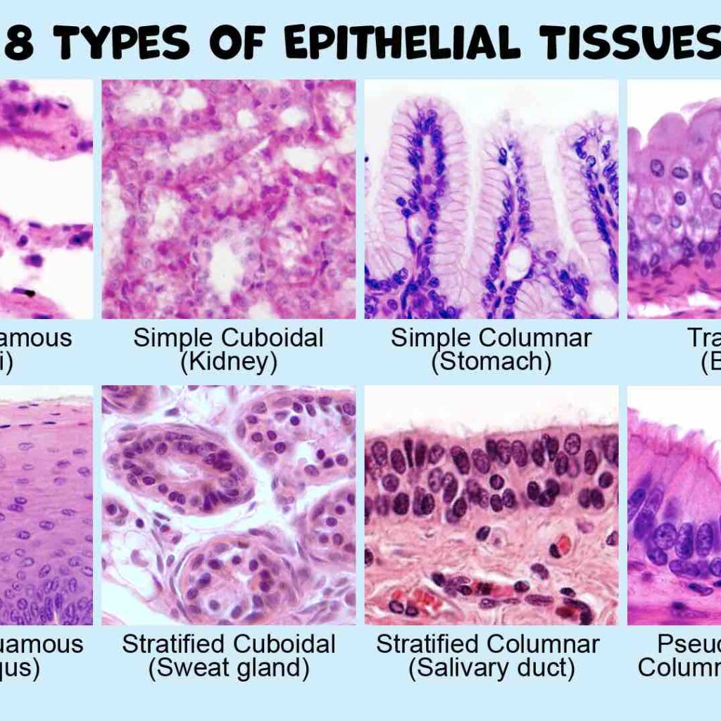
Epithelium: The Body’s Lining & Covering Tissue What is Epithelium? Epithelium forms continuous sheets of cells that line internal surfaces and cover the external surface of the body. It acts as a selective barrier that protects tissues and is often involved in absorption or secretion. A non-cellular layer called the basement membrane separates an epithelium from the underlying connective tissue. Key Characteristics: Anchorage & Polarity Cells are anchored to a basement membrane and have an apical surface (facing a free space) and a basal surface (attached to the basement membrane). Avascularity & Cellularity Epithelium contains no blood vessels (avascular) and is composed almost entirely of tightly packed cells with very little extracellular matrix (cellularity). Origin of the Epithelial Tissues Epithelial tissues are diverse in function and location, which is reflected in their origins from all three primary germ layers formed during embryonic development. These are the primary layers of cells from which all tissues and organs of the body are derived. Ectoderm (Outermost layer) Gives rise to structures that interact with the outside world and the nervous system. Epithelial Derivatives: Epidermis of the skin Lining of the oral & nasal cavities Lining of the anal canal Glands derived from skin Mesoderm (Middle layer) Forms structures related to movement, support, and circulation. Epithelial Derivatives: Endothelium (lining blood vessels) Mesothelium (lining serous cavities) Epithelium of kidney tubules Epithelium of the gonads Endoderm (Innermost layer) Forms the lining of the digestive and respiratory systems and associated glands. Epithelial Derivatives: Lining of the GI tract Lining of the respiratory tract Lining of the urinary bladder Epithelium of thyroid, pancreas, liver The Basement Membrane The basement membrane (BM) is a thin, acellular, extracellular layer that underlies all epithelial tissues, separating them from the adjacent connective tissue. It is a critical structural and functional component. Composition & Structure The BM is primarily composed of glycoproteins (like laminin), proteoglycans, and various types of collagen (especially Type IV collagen). Under an electron microscope, it is seen to have two main layers: Basal Lamina: Produced by the epithelial cells. It has a clearer layer (lamina lucida) and a denser layer (lamina densa). Reticular Lamina: Produced by the underlying connective tissue. It is composed of reticular fibers (Type III collagen) that anchor the basal lamina. Functions of the Basement Membrane Structural Support: Provides a stable base for epithelial cells. Filtration Barrier: Regulates passage of molecules (e.g., in the kidney). Cell Adhesion: Mediates strong attachment of epithelium. Maintains Polarity: Helps define the apical-basal orientation. Regulates Cell Behavior: Influences growth and differentiation. Tissue Repair: Acts as a scaffold for regeneration. Summary of Key Characteristics Anchored to basement membrane Apical and Basal surfaces (Polarity) No extracellular matrix (Cellularity) Avascular (no blood vessels) Clinical Correlation: Invasion and Cancer The basement membrane is a critical marker in cancer diagnosis. Benign tumors remain confined above the BM. A hallmark of malignant tumors (cancer) is their ability to produce enzymes that degrade the BM, allowing them to invade the underlying connective tissue and metastasize (spread). Classification of Epithelium Epithelium is classified based on two main features: the number of cell layers and the shape of the cells at the apical (free) surface. Simple Squamous Function: Ideal for diffusion and filtration. | Location: Alveoli of the lungs, lining of blood vessels (endothelium). Simple Cuboidal Function: Secretion and absorption. | Location: Kidney tubules, glands. Simple Columnar Function: Absorption and secretion. | Location: Gastrointestinal tract. Stratified Squamous Function: Protection against abrasion. Keratinized: Surface cells are dead and filled with keratin. (Location: Epidermis of the skin). Non-keratinized: Surface cells are living. (Location: Esophagus, vagina). Transitional (Urinary) Epithelium Function: Allows for distension (stretching). | Location: Urinary bladder, ureters. Pseudostratified Ciliated Columnar (Respiratory) Epithelium Function: Secretion and movement of mucus. | Location: Trachea, bronchi. Simple squamous epithelium Simple squamous epithelium is a single layer of thin, flattened cells that forms a delicate lining in areas where rapid diffusion, filtration, or smooth movement of substances is needed. The extreme thinness of the cells provides minimal protection but allows for quick transport of molecules. Classification & Defining Characteristics Simple Consists of a single layer of cells, crucial for rapid transport across the membrane. Squamous Cells are flat, thin, and scale-like (“squashed”), resembling a tiled floor from the surface. Permeable The extreme thinness of the single layer makes it highly permeable for quick exchange. Structure & Appearance Cell Shape: Irregularly shaped, flattened cells that interlock like puzzle pieces. Nucleus: Oval or flattened, often appearing as a central bulge in the thin cell. Cytoplasm: Scanty (very little), reflecting its primary role in passive transport. Basement Membrane: Rests on a thin basement membrane, separating it from underlying connective tissue. Locations and Functions Simple squamous epithelium is strategically located in areas where rapid diffusion, filtration, or a slick, friction-reducing surface is required. Lining of Blood & Lymphatic Vessels (Endothelium) Provides a smooth, clot-preventing surface for blood flow and facilitates the exchange of gases, nutrients, and waste. Lining of Serous Cavities (Mesothelium) Lines the pleura, pericardium, and peritoneum, producing a slippery serous fluid that lubricates organs and prevents friction. Alveoli of the Lungs (Type I Pneumocytes) Forms the extremely thin “air-blood barrier” essential for rapid gas exchange (oxygen in, carbon dioxide out). Glomerular Capsules (Bowman’s Capsule) in the Kidneys Forms the filtration membrane for blood, allowing water and small solutes to pass into the renal tubule while retaining large molecules. Clinical Significance Understanding the structure of simple squamous epithelium is key to diagnosing and managing several clinical conditions. Pathological Considerations Edema: Fluid accumulation (e.g., in heart failure) increases the diffusion distance across the alveolar epithelium, impairing gas exchange. Inflammation: Inflammation of serous membranes (pleuritis, peritonitis) causes fluid accumulation (effusions) and painful friction. Cancer: Malignant mesotheliomas can arise from the mesothelium, and a disrupted endothelium is a key factor in various vascular diseases. Simple cuboidal epithelium Simple cuboidal epithelium is a single layer of cube-shaped cells, often with round, central nuclei, primarily performing secretion and absorption. It is found lining surfaces like kidney tubules, ducts of glands, and the surface

cell cycle
Anatomy

Cell Cycle and Disorders

The Cell Cycle: A Cell’s Life Journey The Cell Cycle The cell cycle describes the entire lifespan of a cell, from its formation after one division until it divides again. It consists of two main stages: Interphase: The period of cell growth, DNA replication, and preparation for division. This is the longest phase. M Phase (Mitotic Phase): The period of actual cell division, including mitosis (nuclear division) and cytokinesis (cytoplasmic division). Interphase: The Preparation Phase Interphase is not a resting phase but a highly active period of growth and metabolic activity, crucial for preparing the cell for division. It is divided into several sub-phases. 1. G₀ Phase (Gap 0 / Quiescent Phase) This is an optional phase where cells exit the cell cycle and stop dividing, entering a state of dormancy or terminal differentiation. While metabolically active, they are not preparing for division. Examples of G₀ Cells: Terminally Differentiated: Mature muscle and nerve cells often enter G₀ permanently. Reversible G₀: Liver cells and lymphocytes can re-enter the cycle if stimulated. Significance: Prevents uncontrolled cell growth and allows cells to perform their specialized roles. 2. G₁ Phase (Gap 1 / First Growth) This is the first growth phase after a cell division. The cell is actively growing, synthesizing proteins and RNA, and expanding its cytoplasm by creating new organelles. Critical “Decision Point”: At this checkpoint, the cell decides whether to commit to division and proceed to the S phase or to exit the cycle into the G₀ phase. 3. S Phase (Synthesis Phase) The “synthesis” phase, where the most crucial event for cell division occurs: DNA replication. Key Activities: Each of the 46 chromosomes is duplicated, resulting in two identical sister chromatids. New histone proteins are synthesized to package the newly replicated DNA. By the end of S phase, the cell contains double the amount of DNA. 4. G₂ Phase (Gap 2 / Second Growth) The second growth phase and final preparatory stage before the cell enters mitosis. “Quality Control” Checkpoint: The cell checks the replicated DNA for errors or damage. If damage is found, it attempts repairs. If the damage is irreparable, the cell may trigger programmed cell death (apoptosis) to prevent passing on mutations. Cell Division Cells reproduce through a fundamental process called cell division. This is essential for growth, repair, and reproduction in all living organisms. There are two primary types: Mitotic Cell Division (Mitosis) Role: Growth and repair of tissues. Occurs in: Somatic cells (e.g., neurons, epithelial, muscle). Outcome: Two identical daughter cells. Chromosomes: 46 (same as parent). Meiotic Cell Division (Meiosis) Role: Production of sex cells (sperm/ova). Occurs in: Reproductive organs only. Outcome: Four daughter cells. Chromosomes: 23 (half of parent). Mitotic Cell Division: The Basis of Growth and Repair Mitotic cell division is a continuous process crucial for increasing the number of cells for growth and replacing worn out, damaged, or dead cells. However, not all cells divide at the same rate—epithelial cells divide almost continuously, while mature muscle cells largely lose the ability to divide. Key Processes in Mitotic Cell Division: Replication of Chromosomes: Creating exact copies of the genetic material (occurs in S phase). Mitosis: The division of the nucleus. Cytokinesis: The division of the cytoplasm. The Core Mechanism During mitosis, the cell’s diffuse chromatin condenses into visible chromosomes. The centrosome duplicates, and each copy moves to opposite ends (poles) of the cell. They create spindle fibers that grab onto the chromosomes and pull them apart, ensuring that when the cell finally divides, each new daughter cell receives its own identical copy of the genetic material. Mitotic Phases Once interphase is complete, the cell enters mitosis. While it’s a continuous process, we divide it into four sequential phases for easier understanding. A. Prophase • Replicated chromosomes coil and condense, becoming visible as two identical sister chromatids joined at a centromere. • The nuclear envelope disappears. • Centrioles migrate to opposite poles, and the mitotic spindle begins to form. B. Metaphase • The replicated chromosomes line up precisely at the cell’s equator (the metaphase plate). • The centromere of each chromosome is attached to the spindle fibers. C. Anaphase • Centromeres divide, and the sister chromatids separate. • Each separated chromatid is now considered an individual chromosome. • Spindle fibers pull the chromosomes towards opposite poles of the cell. D. Telophase • The spindle fibers disassemble. • A new nuclear envelope forms around each set of chromosomes at the poles. • Chromosomes uncoil back into their thread-like chromatin form. Cytokinesis: Division of the Cytoplasm Usually occurring during late anaphase and telophase, cytokinesis is the final step. A furrow forms in the plasma membrane, deepens, and eventually pinches the parent cell into two separate, genetically identical daughter cells, each with its own nucleus and cytoplasm. Cell Cycle Disorders: When Regulation Fails The cell cycle is a tightly regulated sequence of events with a series of checkpoints that monitor the cell’s health and DNA integrity. When these regulatory mechanisms fail, the cell cycle can become dysregulated, leading to various disorders, most notably cancer. Cells have checks and balances, and special proteins called cyclins constantly monitor the cell’s health. Unhealthy cells normally self-destruct via apoptosis. Cancer cells, however, lose this ability. For many cells, the G1 checkpoint is the most important; if a cell receives a “go-ahead” signal here, it will usually complete division. If not, it enters a non-dividing state called the G₀ phase. Key Regulators of the Cell Cycle Before discussing disorders, it’s essential to understand the main players that normally control the cell cycle: Cyclins and CDKs These are the “engine” of the cell cycle. Cyclin-Dependent Kinases (CDKs) are enzymes that are activated by binding to Cyclins. Different Cyclin-CDK complexes drive the cell through each phase. Cell Cycle Checkpoints Critical control points that monitor conditions. The main ones are the G1 Checkpoint (the “start” point), the G2 Checkpoint (checks DNA replication), and the M Checkpoint (checks spindle attachment). Tumor Suppressor Genes These are the “brakes.” They encode proteins that inhibit cell division or

Body planes and cavities
Anatomy

Anatomical Position, Directional Terms & Planes

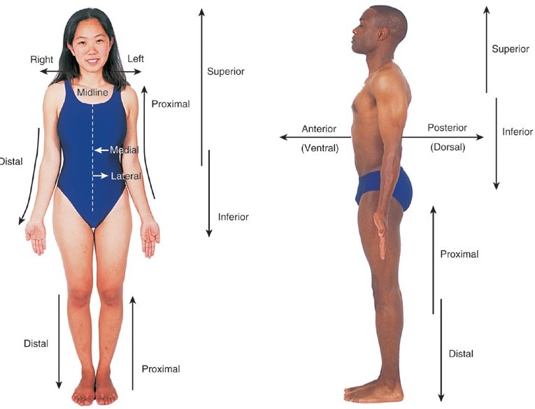
Anatomical Position, Directional Terms & Planes Anatomical Position, Directional Terms & Planes Main Questions to Answer What is the anatomical position, and why is it the universal standard? What are the specific directional terms used to navigate the human body? What are the anatomical planes and sections used in medical imaging? How do we correctly describe specific body movements and clinical patient positions? The Problem: Why Do We Need a Standard? When we describe where something is on the human body, it can quickly become confusing because the body is incredibly mobile. For example, if a person is holding their hand with the palm facing up, a mole on it is on the “front.” But if they turn their hand so the palm faces down, is that mole now on the “inside,” the “back,” or still the “front”? This ambiguity is highly dangerous in medicine (e.g., a surgeon operating on the wrong side of a limb). This confusion is exactly why anatomists and medical professionals created a single, rigid standard position to use as an absolute reference point, no matter how the body is actually positioned in real life. The Golden Rule of Anatomy No matter how a patient or a body in an image is actually positioned (sitting, lying down, upside down, or curled up), you always describe their anatomy as if they were standing in the Anatomical Position. Most Important Rule: All descriptions are from the patient’s point of view, not yours. The patient’s left is always their left, even if it is on your right side when you look at them. The Solution: The Anatomical Position The Anatomical Position is the universal starting point for describing any part of the body. It acts as the “Zero Coordinate” for the human map. The Strict Rules of Anatomical Position: Body Posture: The person is standing up straight (erect). Head and Eyes: They are facing directly forward, with eyes looking straight ahead. Lower Limbs: The legs are together or slightly apart (shoulder-width), with the feet flat on the floor and toes pointing directly forward. Upper Limbs: Their arms are hanging down at their sides. Hands (Crucial Detail): Their palms are facing forward (supinated). Because the palms face forward, their thumbs are pointing away from the body (laterally). This ensures the two bones of the forearm (radius and ulna) are parallel and not crossed over each other. Anatomical Terms of Position (Directional Terms) These terms are like a GPS for the body. They are used in pairs of opposites and help describe where one body part is strictly in relation to another. To accurately describe body parts and their positions, we use this specific set of directional terms. Front / Back Anterior (Ventral): Towards the front of the body. Example: “The sternum (breastbone) is anterior to the vertebral column (spine).” Example: “The kneecap is located on the anterior side of the leg.” Posterior (Dorsal): Towards the back of the body. Example: “The vertebral column (spine) is posterior to the sternum.” Example: “The shoulder blades are located on the posterior chest wall.” Top / Bottom (Axial Skeleton) Superior (Cranial/Cephalic): Towards the top or head. Used only for the head, neck, and trunk. Example: “The nose is superior to the mouth.” Example: “The skull is cranial to the neck.” Inferior (Caudal): Towards the bottom, feet, or tail. Used only for the head, neck, and trunk. Example: “The mouth is inferior to the nose.” Example: “The neck is caudal to the skull.” Midline / Sides Medial: Towards the imaginary midline of the body. Example: “The nose is medial to the ears.” Example: “The heart is medial to the lungs.” Lateral: Away from the midline of the body; towards the sides. Example: “The ears are lateral to the nose.” Example: “The arms are lateral to the chest.” Depth Superficial (External): Situated closer to the surface of the body. Example: “The skin is superficial to the skeletal muscles.” Deep (Internal): Situated further inward, away from the surface of the body. Example: “The bones are deep to the skin and muscles.” Limbs (Appendicular Skeleton) Proximal: Closer to the origin or attachment point of a limb to the main trunk of the body. Example: “The elbow is proximal to the wrist.” Example: “The femur (thigh) is proximal to the knee.” Distal: Farther away from the origin or attachment point of a limb. Example: “The wrist is distal to the elbow.” Example: “The toes are distal to the ankle.” Advanced / Additional Terms Ipsilateral: On the same side of the body. Example: “The right hand and right foot are ipsilateral.” Contralateral: On the opposite side of the body. Example: “A stroke on the right side of the brain causes contralateral paralysis on the left side of the body.” Rostral: Towards the nose (specifically used in neuroanatomy to describe the brain). Student Pitfall: Proximal/Distal vs. Superior/Inferior Students often make the mistake of saying “The wrist is inferior to the elbow.” While technically lower to the ground, anatomists strictly reserve Superior/Inferior for the Head and Trunk (Axial skeleton). For the arms and legs (Appendicular skeleton), you must use Proximal and Distal. Why? Because if you raise your hand above your head, your wrist is suddenly physically higher than your elbow. But anatomically, the wrist is always Distal to the elbow, no matter where your arm is reaching! Anatomical Planes and Sections To study internal anatomy, or to view the body using medical imaging (like CT scans or MRIs), the body is often sectioned (cut) along an imaginary flat 2D surface called a plane. The cut itself is called a section. 1. Sagittal Plane: A vertical line dividing the body into left and right parts. Midsagittal (Median) Plane: Cuts exactly down the absolute midline, creating equal left and right halves. Parasagittal Plane: An off-center cut, creating unequal left and right portions. 2. Coronal (Frontal) Plane: A vertical line dividing the body into anterior (front) and posterior (back) parts. Memory Aid: Think of a crown (corona) sitting across the top of your

anatomy lecture doctors revision
Anatomy

Anatomy Introduction

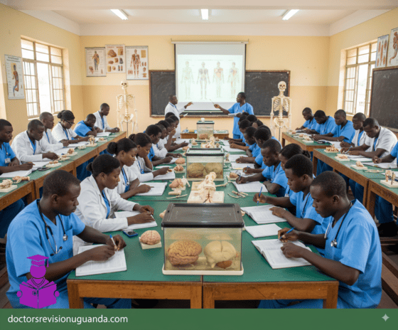
Intro to Anatomy: Terms & Concepts Introduction to Anatomy Anatomy is the scientific study of the structural organization of the human body, ranging from microscopic cells to large, visible structures like organs and bones. Derived from the Greek word for “cutting apart,” it explores how these parts are arranged to form functional systems, often in conjunction with physiology, which focuses on function. The History of Anatomy For centuries, the dissection of human bodies was taboo in many societies. The journey of anatomical study is marked by key historical milestones: Claudius Galenus: A second-century Greek physician who learned about the human form by performing vivisections on pigs. Leonardo da Vinci: Poked around in dead bodies and created beautifully detailed anatomical drawings until the Pope made him stop. 17th and 18th Centuries: Certified anatomists were allowed to perform tightly regulated human dissections. These were often popular public events attended by artists like Michelangelo and Rembrandt. The Anatomy Act (1832): The study of human anatomy became such a craze in Europe that grave robbing became a lucrative occupation until Britain passed this act, which provided students with corpses of executed murderers. Modern Day: Today, students of anatomy and physiology still use educational cadavers, which are donated by volunteers. Andreas Vesalius: Known as the ‘Father of Anatomy’. He was the first to carry out dissection to closely observe the inner structure and construction of the human body. Key Concepts in Anatomy and Physiology Function Follows Form This is the core principle of anatomy. It means that the shape of a body part (its structure or form) is perfectly designed for its job (its function). The function of a cell, organ, or whole organism always reflects its form. This is also known as the Complementarity of Structure and Function. Example: Form & Function Think of a fork. It has prongs (its form) specifically to help it pick up food (its function). Your teeth are a perfect biological example. Your sharp front teeth are for tearing food, while your flat back teeth are for grinding. Their shape is perfect for their job. Hierarchy of Organization The human body is organized in a hierarchical manner, from the smallest chemical components to the entire organism. Levels of Organization in the Body: Chemical Level: Atoms and molecules, the smallest units of matter. Cellular Level: Cells, the smallest units of living things. Tissue Level: Groups of similar cells that work together. Organ Level: Two or more tissue types performing a specific function. Organ System Level: Groups of organs working together for a common purpose. Organismal Level: The sum total of all structural levels working together to keep us alive. Homeostasis Homeostasis is the ability of all living systems to maintain stable internal conditions no matter what changes are occurring outside the body. Survival is all about maintaining this delicate balance. Example: Homeostasis Think of a thermostat. If the house gets too cold, the heat turns on. If it gets too hot, the A/C kicks in. Your body does this constantly. If you get hot, you sweat to cool down. If you get cold, you shiver to warm up. Your body is always working to keep your temperature, blood sugar, and many other factors in a perfect, stable range. Foundational Anatomical Terms Mastering the language of anatomy is the first step to understanding its complexities. This guide covers the foundational terminology you will encounter throughout your studies. These terms provide a universal standard for describing the structure and function of the human body. Human anatomy (ah-nat -o−-me−) is the study of the structure and organization of the body and the study of the relationships of body parts to one another. There are two subdivisions of anatomy: Gross anatomy involves the dissection and examination of various parts of the body without magnifying lenses. Microanatomy, also known as histology, consists of the examination of tissues and cells with various magnification techniques. Human physiology (fiz-e−-ol-o−-je−) is the study of the function of the body and its parts. Physiology involves observation and experimentation, and it usually requires the use of specialized equipment and materials. Term (Etymology) Definition Example Anatomy(ana = apart; tom = to cut) The study of the structure of living organisms. Studying the bones, muscles, and organs in a human cadaver to understand their physical arrangement. Appendicular(append = to hang) Pertaining to the upper and lower limbs. The appendicular skeleton includes the bones of the arms, legs, shoulders, and pelvis. Axial(ax = axis) Pertaining to the longitudinal axis of the body. The axial skeleton consists of the skull, vertebral column, and rib cage, forming the central support of the body. Body region(regio = boundary) A portion of the body with a special identifying name. The “cephalic region” refers to the head, while the “thoracic region” refers to the chest. Directional term(directio = act of guiding) A term that references how the position of a body part relates to the position of another body part. The nose is superior to the mouth, and the feet are inferior to the knees. The sternum (breastbone) is anterior to the spine. Effector(efet = result) A structure that functions by performing an action that is directed by an integrating center. In regulating body temperature, sweat glands are effectors that produce sweat to cool the body down when directed by the brain. Homeostasis(homeo = same; sta = make stand or stop) Maintenance of a relatively stable internal environment. The body maintaining a constant internal temperature of approximately 37°C (98.6°F) regardless of external temperature changes. Integrating center(integratus = make whole) A structure that functions to interpret information and coordinate a response. The brain acts as an integrating center when it receives signals that blood sugar is too high and then sends signals to the pancreas to release insulin. Metabolism(metabole = change) The sum of the chemical reactions in the body. The digestion of food into nutrients (catabolism) and the building of new tissues from those nutrients (anabolism) are both parts of metabolism. Parietal(paries = wall) Pertaining to the wall

histology introduction
Anatomy

Histology Introduction

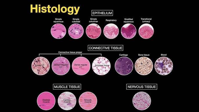
Introduction to Histology: The Study of Tissues What is Histology? Histology is the study of tissues. The word is derived from the Greek words “histo” (tissue) and “logos” (study). Therefore, histology is the science of the microscopic structure of cells, tissues, and organs. Simply put, it’s the study of tissues under a microscope. This field examines the microscopic anatomy of biological tissues and is fundamental to understanding the structure and function of the entire body. Why Health workers Need to Know Histology A strong foundation in histology is not just for doctors or researchers; it is a critical component of a professional nurse’s knowledge base. It elevates a nurse’s practice from task-oriented care to a deeper, more analytical level of patient management. Explains Form & Function Shows how tissue structure relates to its job, making treatments like oxygen therapy more meaningful. Identifies Disease Knowing normal tissue helps nurses recognize changes in disease, aiding in assessments like wound care. Enhances Practical Skills Improves participation in collecting and interpreting lab samples (e.g., biopsies). Informs Patient Education Allows nurses to better explain conditions and treatments, leading to more informed care. Medication Efficacy Helps nurses anticipate medication effects and side effects by understanding drug-cell interactions. Interdisciplinary Collaboration Facilitates clearer communication with pathologists, doctors, and other healthcare professionals. Methods of Histology Histology employs various techniques to prepare tissues for microscopic examination. These methods are crucial for preserving tissue integrity and allowing for the study of their structure and function. The main steps involve tissue preparation, staining, and microscopy. 1. Tissue Preparation Techniques This is the first and most critical step to preserve tissue and allow for thin sectioning. There are three main methods. a. Paraffin Technique This is the most common method for preparing tissues for routine histological examination. Procedures of the Paraffin Technique: Tissue Sample Collection: Obtaining the sample (biopsy, surgical excision). Fixation: Preserving the tissue, commonly with 4% formaldehyde (formalin). Dehydration: Removing water with increasing concentrations of alcohol. Clearing: Replacing alcohol with a clearing agent like xylene. Impregnation: Infiltrating the tissue with melted soft paraffin. Embedding: Transferring the tissue to hard paraffin to form a solid block. Sectioning: Cutting the block into very thin (5-8 µm) sections using a microtome. b. Celloidin Technique Provides superior support for both soft and hard tissues, such as bones, teeth, and large brain sections. Advantages: Excellent support for hard tissues Minimal shrinkage and distortion Good architectural preservation Disadvantages: Very time-consuming process Difficult to cut very thin sections Requires specialized technical skills c. Freezing Technique Rapidly prepares tissues by freezing, especially for urgent diagnoses during surgery. Advantages: Rapid diagnosis (minutes) Preserves molecules (DNA, RNA, proteins) Preserves antigens for immunostaining Disadvantages: Poor staining and cellular detail Inadequate fixation compared to paraffin Expensive and complex equipment (cryostat) 2. Staining Techniques Staining uses dyes to enhance the visibility of different tissue structures under the microscope. This is essential because most tissues are colorless. Common Stains and Their Uses: Hematoxylin and Eosin (H&E): The most common stain. Hematoxylin stains acidic structures like the nucleus blue, while Eosin stains basic structures like the cytoplasm pink. PAS (Periodic Acid-Schiff): Stains carbohydrates magenta. Useful for identifying basement membranes, mucus, glycogen, and fungal walls. Silver Stains (Reticulin): Stains reticular fibers black. Used in kidney, liver, and bone marrow biopsies. Trichrome Stains: Differentiates muscle (red), collagen (blue/green), and fibrin. Used for assessing fibrosis. Immunostains (Immunohistochemistry): Uses antibodies to detect specific molecules or cell types. Crucial for cancer diagnosis and classification. 3. Microscopy Techniques Microscopy is the use of microscopes to visualize small structures that are not visible to the naked eye. Light Microscope Uses natural or electric light to examine stained sections. This is the most commonly used microscope in routine histology. Electron Microscope Uses a beam of electrons for much higher magnification. TEM provides high-resolution internal details, while SEM provides detailed 3D surface images. Test Your Knowledge Check your understanding of the concepts covered in this post. 1. Histology is defined as the study of: Cells under a light microscope. Gross anatomy of organs. Tissues under a microscope. Chemical composition of biological structures. Rationale: The text explicitly states, “Histology therefore is the science of the microscopic structure of cells, tissues and organs OR simply put; The study of tissues under a microscope.” 2. Why is understanding histology important for nurses regarding medication efficacy? It helps them prescribe the correct dosage. It allows them to understand how drugs interact with specific cell types and tissues. It teaches them how to administer intravenous medications. It explains the cost-effectiveness of different drugs. Rationale: The text states under “Medication Efficacy,” “Understanding how drugs interact with specific cell types and tissues (e.g., receptors on cell surfaces) helps nurses anticipate medication effects and side effects.” 3. Which tissue preparation technique is most commonly used for routine histological examination due to its preservation and hardening properties? Celloidin Technique Freezing Technique Paraffin Technique Vital Staining Rationale: The text states, “The paraffin technique is the most common method for preparing tissues for routine histological examination.” 4. What is the primary disadvantage of the Celloidin Technique mentioned in the text? It causes significant tissue shrinkage and distortion. It is a very rapid process. It is time-consuming and difficult to cut very thin sections. It poorly preserves hard tissues like bone. Rationale: Under “Disadvantages of Celloidin Technique,” the text lists, “Time-Consuming: The process is lengthy,” and “Difficulty in Cutting Thin Sections: Achieving very thin sections can be challenging.” 5. In the Paraffin Technique, what is the purpose of the ‘Clearing’ step? To replace water with alcohol. To harden the tissue by coagulating proteins. To replace alcohol with a clearing agent like xylene. To embed the tissue in molten paraffin. Rationale: The text explains under “Clearing,” “Aim: To replace alcohol with xylene, which is miscible with paraffin.” 6. Which staining technique uses positively charged dyes to stain negatively charged cellular components, such as nuclei? Acidic Staining Basic Staining Neutral Staining Metachromatic Staining Rationale: The text states under “Basic Staining,” “Uses positively charged dyes to stain negatively charged

anatomy lecture doctors revision
Anatomy

Foundations of Anatomy: Understanding The Cell

Cell Theory
Alright, let’s dive into the microscopic world that makes up our bodies, starting with the fundamental concept of the Cell Theory. This theory is one of the cornerstones of biology and medicine, giving us the basic understanding of life. It essentially has three main parts, like three key rules about cells:

All living organisms are made up of one or more cells. This means whether it’s a tiny bacterium, a plant, or a human being, the basic unit of structure is the cell. Some organisms are single-celled (like amoeba), while complex organisms like us are made of trillions of cells working together.

Scroll to Top