Axial and Appendicular Skeleton
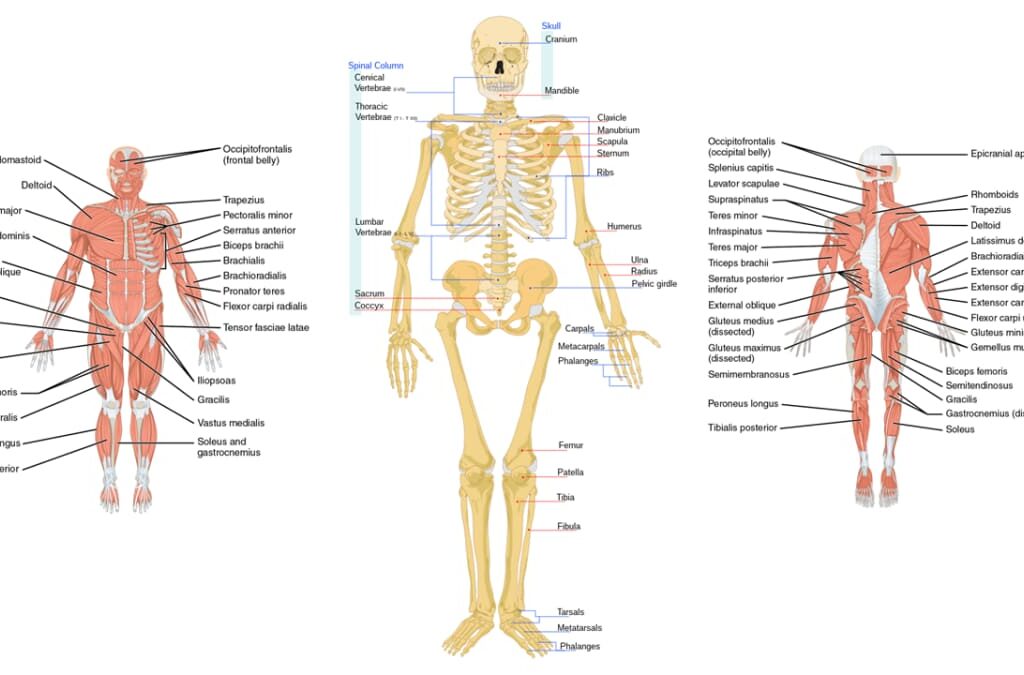
Axial and Appendicular Skeleton The Supporters. The Axial and Appendicular Skeleton The human skeleton is divided into two major parts: the Axial Skeleton and the Appendicular Skeleton. Together, these two divisions provide the support, protection, and leverage necessary for movement. The Axial Skeleton: The Body’s Central Axis The axial skeleton forms the longitudinal axis of the body. It consists of the bones of the skull, vertebral column (spine), and thoracic cage (ribs and sternum). In brief, it comprises the head and trunk. Composition (approximately 80 bones): Skull (22 bones + 7 associated): Protects the brain and forms the face. Vertebral Column (26 bones): Protects the spinal cord and supports the head. Thoracic Cage (25 bones): Protects the heart and lungs. The Skull The skull is a bony structure that forms a protective cavity for the brain, provides the head with its shape, and is formed by 22 bones joined by fibrous joints called sutures. It consists of two main parts: the Cranium and the Face. 1. The Cranium (8 Bones) The cranium is the bony box that houses and protects the brain. Frontal Bone (1) Forms the forehead and the superior part of the orbits. Parietal Bones (2) Form the superior and lateral walls of the cranium. Temporal Bones (2) Form the inferolateral aspects of the skull and parts of the cranial base; contain the organs of hearing. Occipital Bone (1) Forms the posterior wall and most of the base of the skull. The spinal cord passes through its foramen magnum. Sphenoid Bone (1) The central “keystone” bone of the cranium; articulates with all other cranial bones. Contains the sella turcica for the pituitary gland. Ethmoid Bone (1) Forms the anterior part of the cranial floor, the medial wall of the orbits, and the roof of the nasal cavity. 2. The Face (14 Bones) These bones form the framework of the face, contain cavities for sensory organs, and provide attachment sites for facial muscles. Mandible (1) The lower jawbone; the largest and strongest bone of the face. Maxillae (2) The upper jawbones; they form the hard palate and hold the upper teeth. Zygomatic Bones (2) The cheekbones; they form the prominences of the cheeks. Nasal Bones (2) Form the bridge of the nose. Lacrimal Bones (2) Form part of the medial walls of the orbits; contain the lacrimal fossa for the tear ducts. Palatine Bones (2) Form the posterior part of the hard palate. Vomer (1) Forms the inferior part of the nasal septum. Inferior Nasal Conchae (2) Scroll-like bones forming part of the lateral walls of the nasal cavity. B. The Vertebral Column (Spine) The vertebral column serves as the main support of the body, protects the spinal cord, and provides attachment points for the ribs and muscles. It is a flexible, curved structure composed of 26 irregular bones in adults. Functions of the Vertebral Column: Support: Transmits the weight of the head and trunk to the lower limbs. Protection: Surrounds and protects the delicate spinal cord. Movement: Provides attachment points for muscles, allowing trunk and neck movement. Shock Absorption: Intervertebral discs act as shock absorbers. Regions and Curvatures The spine is divided into five regions and has four natural curves that increase its resilience. Vertebral Regions Cervical (C1-C7): 7 vertebrae in the neck.Thoracic (T1-T12): 12 vertebrae in the chest.Lumbar (L1-L5): 5 vertebrae in the lower back.Sacrum: 1 bone (5 fused vertebrae).Coccyx: 1 bone (3-5 fused vertebrae). Spinal Curvatures Cervical & Lumbar: Concave posteriorly (secondary curves).Thoracic & Sacral: Convex posteriorly (primary curves). General Structure of a Vertebra Most vertebrae share a common structural plan, consisting of a body, an arch, and various processes for muscle attachment and articulation. Vertebral Body (Centrum): The anterior, weight-bearing part. Vertebral Arch: Encloses the vertebral foramen, forming the vertebral canal for the spinal cord. Processes: Projections (spinous, transverse, articular) that serve as attachment and articulation points. Intervertebral Discs Located between adjacent vertebrae, these discs act as shock absorbers. Each is composed of an inner gelatinous nucleus pulposus and an outer collar of fibrocartilage called the anulus fibrosus. Regional Characteristics of Vertebrae Cervical Vertebrae (C1-C7) The smallest, lightest vertebrae. Their unique feature is the transverse foramina for vertebral arteries. C1 (Atlas) lacks a body and articulates with the skull (“yes” motion). C2 (Axis) has a dens that acts as a pivot for head rotation (“no” motion). Most have a bifid (split) spinous process. Thoracic Vertebrae (T1-T12) Distinguished by their articulation with the ribs via costal facets on the vertebral bodies and transverse processes. They have a heart-shaped body and a long, slender spinous process that points sharply downward. Lumbar Vertebrae (L1-L5) The largest and strongest vertebrae, designed to bear the most body weight. They have a massive, kidney-shaped body and a short, thick, blunt spinous process that projects posteriorly. Sacrum and Coccyx The Sacrum is a triangular bone formed by the fusion of 5 sacral vertebrae, forming the posterior wall of the pelvis. The Coccyx, or “tailbone,” is a small triangular bone formed by the fusion of 3-5 coccygeal vertebrae. C. The Thoracic Cage (Bony Thorax) The thoracic cage forms the protective “rib cage” around the vital organs of the chest. It includes the sternum, ribs, and the twelve thoracic vertebrae. Functions of the Thoracic Cage: Protection: Encloses and protects the heart, lungs, and major blood vessels. Support: Provides attachment points for the shoulder girdles and upper limbs. Respiration: Its ability to expand is crucial for ventilation, and it provides attachment for respiratory muscles. Bones of the Thoracic Cage The Sternum (Breastbone) A flat bone in the anterior midline of the thorax, composed of three fused parts: Manubrium: The superior part, articulating with the clavicles and the first two pairs of ribs. Features the palpable jugular (suprasternal) notch. Body (Gladiolus): The middle and largest part, articulating with ribs 2-7. Xiphoid Process: The inferior-most, small projection that serves as an attachment point for some abdominal muscles. The Ribs (12 pairs) All ribs attach posteriorly to the thoracic vertebrae and generally curve inferiorly and anteriorly. Types