cell cycle

Cell Cycle and Disorders

The Cell Cycle: A Cell's Life Journey

The Cell Cycle

The cell cycle describes the entire lifespan of a cell, from its formation after one division until it divides again. It consists of two main stages:

  • Interphase: The period of cell growth, DNA replication, and preparation for division. This is the longest phase.
  • M Phase (Mitotic Phase): The period of actual cell division, including mitosis (nuclear division) and cytokinesis (cytoplasmic division).

Interphase: The Preparation Phase

Interphase is not a resting phase but a highly active period of growth and metabolic activity, crucial for preparing the cell for division. It is divided into several sub-phases.

1. G₀ Phase (Gap 0 / Quiescent Phase)

This is an optional phase where cells exit the cell cycle and stop dividing, entering a state of dormancy or terminal differentiation. While metabolically active, they are not preparing for division.

Examples of G₀ Cells:

  • Terminally Differentiated: Mature muscle and nerve cells often enter G₀ permanently.
  • Reversible G₀: Liver cells and lymphocytes can re-enter the cycle if stimulated.

Significance:

Prevents uncontrolled cell growth and allows cells to perform their specialized roles.

2. G₁ Phase (Gap 1 / First Growth)

This is the first growth phase after a cell division. The cell is actively growing, synthesizing proteins and RNA, and expanding its cytoplasm by creating new organelles.

Critical "Decision Point":

At this checkpoint, the cell decides whether to commit to division and proceed to the S phase or to exit the cycle into the G₀ phase.

3. S Phase (Synthesis Phase)

The "synthesis" phase, where the most crucial event for cell division occurs: DNA replication.

Key Activities:

  • Each of the 46 chromosomes is duplicated, resulting in two identical sister chromatids.
  • New histone proteins are synthesized to package the newly replicated DNA.
  • By the end of S phase, the cell contains double the amount of DNA.

4. G₂ Phase (Gap 2 / Second Growth)

The second growth phase and final preparatory stage before the cell enters mitosis.

"Quality Control" Checkpoint:

The cell checks the replicated DNA for errors or damage. If damage is found, it attempts repairs. If the damage is irreparable, the cell may trigger programmed cell death (apoptosis) to prevent passing on mutations.

Cell Division

Cells reproduce through a fundamental process called cell division. This is essential for growth, repair, and reproduction in all living organisms. There are two primary types:

Mitotic Cell Division (Mitosis)

  • Role: Growth and repair of tissues.
  • Occurs in: Somatic cells (e.g., neurons, epithelial, muscle).
  • Outcome: Two identical daughter cells.
  • Chromosomes: 46 (same as parent).

Meiotic Cell Division (Meiosis)

  • Role: Production of sex cells (sperm/ova).
  • Occurs in: Reproductive organs only.
  • Outcome: Four daughter cells.
  • Chromosomes: 23 (half of parent).

Mitotic Cell Division: The Basis of Growth and Repair

Mitotic cell division is a continuous process crucial for increasing the number of cells for growth and replacing worn out, damaged, or dead cells. However, not all cells divide at the same rate—epithelial cells divide almost continuously, while mature muscle cells largely lose the ability to divide.

Key Processes in Mitotic Cell Division:

  • Replication of Chromosomes: Creating exact copies of the genetic material (occurs in S phase).
  • Mitosis: The division of the nucleus.
  • Cytokinesis: The division of the cytoplasm.

The Core Mechanism

During mitosis, the cell's diffuse chromatin condenses into visible chromosomes. The centrosome duplicates, and each copy moves to opposite ends (poles) of the cell. They create spindle fibers that grab onto the chromosomes and pull them apart, ensuring that when the cell finally divides, each new daughter cell receives its own identical copy of the genetic material.
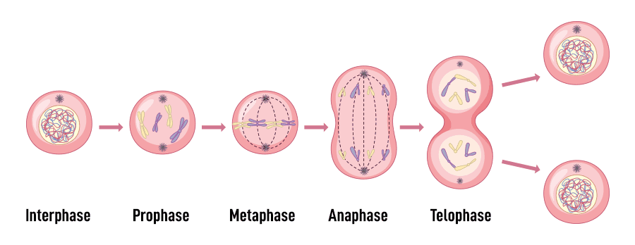
Mitotic Phases

Once interphase is complete, the cell enters mitosis. While it's a continuous process, we divide it into four sequential phases for easier understanding.

A. Prophase

  • • Replicated chromosomes coil and condense, becoming visible as two identical sister chromatids joined at a centromere.
  • • The nuclear envelope disappears.
  • • Centrioles migrate to opposite poles, and the mitotic spindle begins to form.

B. Metaphase

  • • The replicated chromosomes line up precisely at the cell's equator (the metaphase plate).
  • • The centromere of each chromosome is attached to the spindle fibers.

C. Anaphase

  • • Centromeres divide, and the sister chromatids separate.
  • • Each separated chromatid is now considered an individual chromosome.
  • • Spindle fibers pull the chromosomes towards opposite poles of the cell.

D. Telophase

  • • The spindle fibers disassemble.
  • • A new nuclear envelope forms around each set of chromosomes at the poles.
  • • Chromosomes uncoil back into their thread-like chromatin form.

Cytokinesis: Division of the Cytoplasm

Usually occurring during late anaphase and telophase, cytokinesis is the final step. A furrow forms in the plasma membrane, deepens, and eventually pinches the parent cell into two separate, genetically identical daughter cells, each with its own nucleus and cytoplasm.

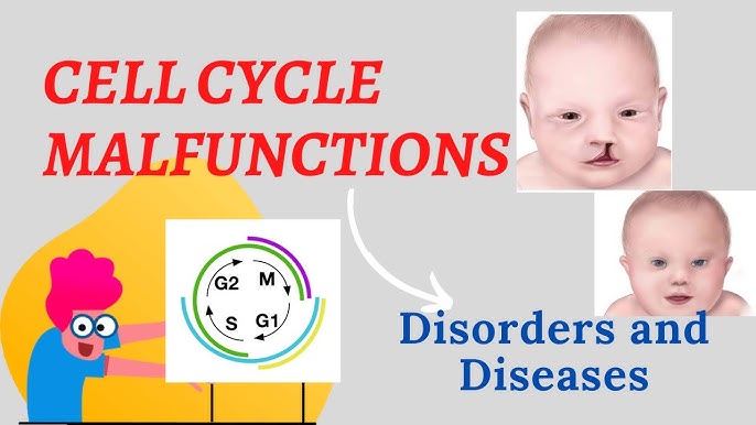
Cell Cycle Disorders: When Regulation Fails

The cell cycle is a tightly regulated sequence of events with a series of checkpoints that monitor the cell's health and DNA integrity. When these regulatory mechanisms fail, the cell cycle can become dysregulated, leading to various disorders, most notably cancer.

Cells have checks and balances, and special proteins called cyclins constantly monitor the cell's health. Unhealthy cells normally self-destruct via apoptosis. Cancer cells, however, lose this ability. For many cells, the G1 checkpoint is the most important; if a cell receives a "go-ahead" signal here, it will usually complete division. If not, it enters a non-dividing state called the G₀ phase.

Key Regulators of the Cell Cycle

Before discussing disorders, it's essential to understand the main players that normally control the cell cycle:

Cyclins and CDKs

These are the "engine" of the cell cycle. Cyclin-Dependent Kinases (CDKs) are enzymes that are activated by binding to Cyclins. Different Cyclin-CDK complexes drive the cell through each phase.

Cell Cycle Checkpoints

Critical control points that monitor conditions. The main ones are the G1 Checkpoint (the "start" point), the G2 Checkpoint (checks DNA replication), and the M Checkpoint (checks spindle attachment).

Tumor Suppressor Genes

These are the "brakes." They encode proteins that inhibit cell division or repair DNA. Key examples are p53 ("Guardian of the Genome") and Rb (Retinoblastoma protein).

Proto-oncogenes & Oncogenes

Proto-oncogenes are the "accelerators" that promote normal cell growth. When mutated, they become Oncogenes, which are stuck in the "on" position, causing uncontrolled growth.

Causes of Cell Cycle Disorders

Disorders arise when the delicate balance of these activators and inhibitors is disrupted, often due to:

  • Genetic Mutations: Inactivating "brake" genes (tumor suppressors) or activating "accelerator" genes (proto-oncogenes).
  • Epigenetic Changes: Altering gene expression without changing the DNA sequence, such as silencing a tumor suppressor gene.
  • Viral Infections: Some viruses, like HPV, produce proteins that disable key regulators like p53 and Rb.
  • Environmental Factors: Carcinogens and radiation that cause DNA damage and lead to mutations.

Consequences & Types of Cell Cycle Disorders

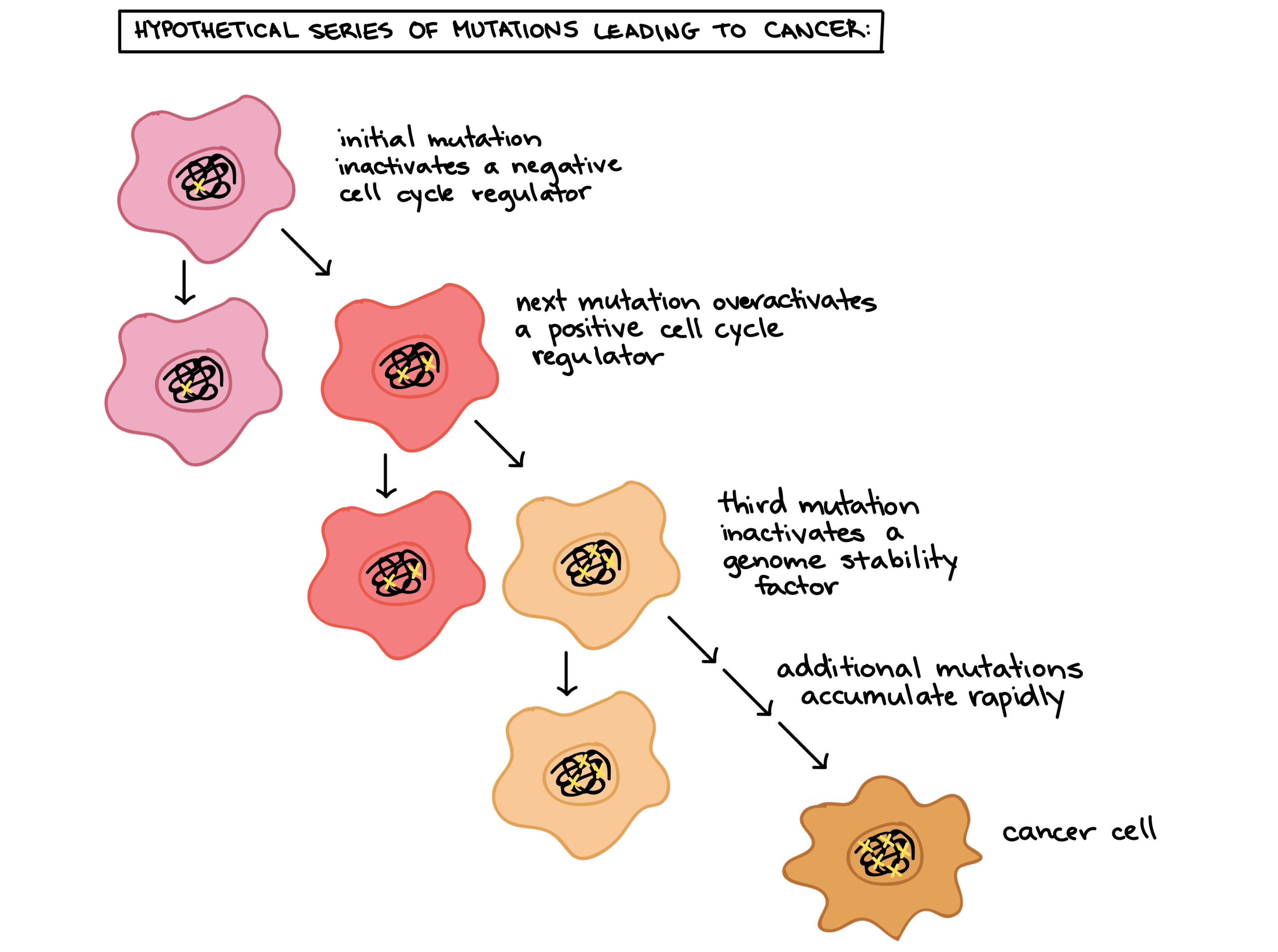
1. Cancer (Malignancy)

Cancer is the primary disease of uncontrolled cell division. Cancer cells ignore the normal signals that control the cell cycle. They enter the S phase without waiting for a signal and become "immortal," escaping the normal limit on cell divisions. This is typically caused by multiple mutations that activate oncogenes and inactivate tumor suppressor genes.

Hallmarks of Cancer Cells

  • Sustained proliferative signaling
  • Evasion of growth suppressors
  • Resistance to cell death
  • Enabling replicative immortality
  • Inducing angiogenesis
  • Activating invasion & metastasis

2. Aneuploidy (Incorrect Chromosome Number)

A failure of the M checkpoint can lead to an unequal distribution of chromosomes during cell division. While most aneuploid cells die, some survive and can lead to genetic disorders like Down Syndrome (Trisomy 21). Aneuploidy is also a common feature of cancer cells.

3. Developmental & Premature Aging Disorders

Precise control of the cell cycle is critical during embryonic development. Errors can lead to underdevelopment (e.g., microcephaly) or overgrowth syndromes. Similarly, some premature aging syndromes are linked to defects in DNA repair mechanisms that impact cell cycle checkpoints.

Therapeutic Implications

Understanding these disorders is fundamental to developing treatments. Many cancer therapies are designed to target the cell cycle:

  • Chemotherapy: Uses drugs that damage DNA or disrupt the mitotic spindle to preferentially kill rapidly dividing cancer cells.
  • Targeted Therapies: Newer drugs that specifically inhibit mutated or overactive molecules, such as CDK inhibitors.
  • Immunotherapy: Harnessing the immune system to recognize and destroy cancer cells that have evaded normal cell cycle controls.

Chromosomal Mutations: Large-Scale Genetic Changes

Chromosomal mutations are significant changes affecting the structure or number of entire chromosomes. These large-scale alterations are distinct from gene (point) mutations, which involve changes to individual DNA base pairs within a gene. Such structural changes often arise from errors during meiosis or from exposure to mutagens.
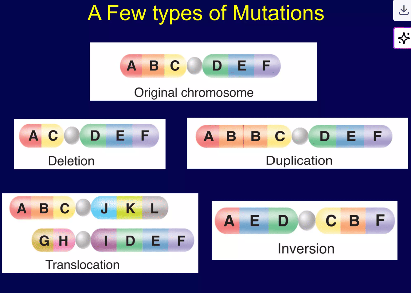
Diagram illustrating different types of chromosomal mutations

Deletion

A segment of the chromosome, containing one or more genes, is lost or excised.

Example: A chromosome originally containing gene segments [A-B-C-D-E-F] loses the [C] segment, resulting in [A-B-D-E-F].

Impact: Results in a loss of genetic information. The consequences can range from mild to severe, depending on the size and function of the deleted genes. (e.g., Cri-du-chat syndrome).

Duplication

A segment of the chromosome is repeated, resulting in extra copies of genes.

Example: The [B-C] segment is repeated, resulting in [A-B-C-B-C-D-E-F].

Impact: While sometimes benign, duplications can disrupt normal gene dosage and cellular processes, leading to developmental problems.

Inversion

A segment of a chromosome breaks off, flips 180 degrees, and reattaches to the same chromosome.

Example: The [B-C-D] segment is inverted, resulting in [A-D-C-B-E-F].

Impact: The genetic material is still present but in a reversed order. While the individual may be normal, inversions can cause issues during meiosis, potentially leading to nonviable gametes or offspring with unbalanced chromosomes.

Translocation

A segment of one chromosome breaks off and attaches to a different, non-homologous chromosome.

Example: A segment from chromosome 8 breaks off and attaches to chromosome 14. This is an exchange of genetic material between two different chromosomes.

Impact: Balanced translocations (no net loss/gain of DNA) may not affect the individual but can lead to fertility issues. Unbalanced translocations in offspring, where there is extra or missing genetic material, typically cause significant health problems.

Test Your Knowledge

Check your understanding of the concepts covered in this post.

1. Which of the following best describes the primary function of the G1 checkpoint in the cell cycle?

  • To ensure that DNA replication has been completed accurately.
  • To check for proper attachment of spindle fibers to chromatids.
  • To assess cell size, nutrient availability, and DNA integrity before DNA synthesis.
  • To synthesize new histone proteins for DNA packaging.
Rationale: The G1 checkpoint (or restriction point) is a critical decision point where the cell determines if conditions are favorable for division, checking for sufficient resources, appropriate size, and absence of DNA damage before committing to DNA replication (S phase).

2. A cell that has sustained significant DNA damage would most likely be arrested at which cell cycle checkpoint, primarily by the action of p53?

  • G0 phase
  • S phase
  • G2 checkpoint
  • M (Spindle Assembly) checkpoint
Rationale: The G2 checkpoint is where the cell primarily checks for complete DNA replication and any DNA damage before entering mitosis (M phase). p53 plays a crucial role here, halting the cycle to allow for repair or triggering apoptosis if damage is too extensive. While p53 can also act at G1, G2 is a key point for assessing the integrity of the replicated genome.

3. During which phase of the cell cycle does DNA replication occur, ensuring that each daughter cell receives a complete set of genetic material?

  • G1 phase
  • S phase
  • G2 phase
  • M phase
Rationale: S phase stands for "Synthesis" phase, referring to the synthesis (replication) of DNA. During this phase, each chromosome is duplicated to form two identical sister chromatids.

4. The formation of the mitotic spindle and the breakdown of the nuclear envelope are characteristic events of which stage of mitosis?

  • Prophase
  • Metaphase
  • Anaphase
  • Telophase
Rationale: Prophase is the initial stage of mitosis characterized by chromosome condensation, the beginning of mitotic spindle formation, and the breakdown of the nuclear envelope (or prometaphase, which is often considered part of prophase).

5. What is the primary role of tumor suppressor genes like p53 and Rb?

  • To accelerate cell division by producing growth factors.
  • To repair damaged DNA during the S phase.
  • To act as "brakes" on the cell cycle, inhibiting uncontrolled proliferation.
  • To form the mitotic spindle during cell division.
Rationale: Tumor suppressor genes encode proteins that regulate cell growth and division, often by halting the cell cycle or initiating apoptosis in response to damage or abnormal conditions. They prevent uncontrolled cell proliferation.

6. Which type of chromosomal mutation results from a segment of a chromosome breaking off, flipping 180 degrees, and reattaching to the same chromosome?

  • Deletion
  • Duplication
  • Translocation
  • Inversion
Rationale: An inversion involves a segment of a chromosome being reversed end to end. While the genetic material is still present, its linear order is altered.

7. Cancer cells often exhibit which of the following characteristics regarding cell cycle control?

  • Strict adherence to the G1 checkpoint.
  • Enhanced apoptosis in response to DNA damage.
  • Loss of density-dependent inhibition (contact inhibition).
  • Reduced growth factor requirements.
Rationale: Normal cells stop dividing when they come into contact with other cells (density-dependent inhibition). Cancer cells lose this control, continuing to divide and pile up on each other. They typically also have reduced apoptosis and often ignore G1 checkpoints.

8. Aneuploidy, a condition of an abnormal number of chromosomes, is most directly caused by errors during which process?

  • DNA replication in S phase.
  • Cytokinesis.
  • Spindle fiber attachment and chromosome segregation in M phase.
  • G1 phase cell growth.
Rationale: Aneuploidy results from nondisjunction, which is the failure of homologous chromosomes or sister chromatids to separate properly during anaphase of meiosis or mitosis. This is directly related to errors in spindle fiber attachment and chromosome segregation.

9. The activity of Cyclin-Dependent Kinases (CDKs) is dependent on their association with which other class of proteins?

  • Growth factors
  • Tumor suppressors
  • Cyclins
  • Oncogenes
Rationale: CDKs are inactive on their own. They require binding to specific cyclin proteins to become active and phosphorylate target proteins, thereby driving the cell cycle forward.

10. Which of the following is an example of a chromosomal duplication?

  • Loss of a segment of chromosome 5, leading to Cri-du-chat syndrome.
  • An extra copy of chromosome 21, causing Down Syndrome.
  • A repeated segment on chromosome 15, associated with some forms of autism.
  • Exchange of genetic material between chromosome 9 and chromosome 22, linked to chronic myeloid leukemia.
Rationale: This option directly describes a repeated segment, which is the definition of a duplication. Cri-du-chat is a deletion. Down Syndrome (Trisomy 21) is a numerical chromosomal abnormality (aneuploidy), not a structural duplication of a segment within a chromosome. The exchange between chromosome 9 and 22 (Philadelphia chromosome) is a translocation.

11. The phase of the cell cycle where a cell exits the cycle and enters a non-dividing state, often temporarily or permanently, is known as the ________________ phase.

Rationale: The G0 phase is a quiescent state where cells are metabolically active but are not dividing or preparing for division.

12. Programmed cell death, a crucial mechanism for removing damaged or unwanted cells, is called ________________.

Rationale: Apoptosis is the process of controlled cellular suicide, essential for development and maintaining tissue homeostasis.

13. The part of the cell cycle where the cell grows and prepares for division, but isn't actually dividing yet, is called ________________.

Rationale: Interphase is the longest part of the cell cycle, consisting of G1, S, and G2 phases, where the cell grows, replicates its DNA, and prepares for mitosis.

14. When a piece of a chromosome breaks off and is lost, this type of mutation is called a ________________.

Rationale: A deletion is a chromosomal mutation where a segment of a chromosome is removed or lost.

15. The special proteins that activate CDKs and regulate the cell cycle are called ________________.

Rationale: Cyclins are proteins that bind to and activate cyclin-dependent kinases (CDKs), which then drive the progression of the cell cycle.
Body planes and cavities

Anatomical Position, Directional Terms & Planes

Anatomical Position, Directional Terms & Planes

Main Questions to Answer:

  • What is the anatomical position?
  • What are the directional terms used in anatomy?
  • What are the anatomical planes and sections?

The Problem: Why Do We Need a Standard?

When we describe where something is on the body, it can be confusing. If a person is holding their hand with the palm facing up, a circle on it is on the "front." If they turn their hand so the palm faces down, is that circle now on the "inside" or still the "front"? This confusion is why anatomists and medical professionals created a single standard position to use as a reference point, no matter how the body is actually positioned.

The Golden Rule

No matter how a patient or a body in an image is actually positioned (sitting, lying down, etc.), you always describe it as if it were in the anatomical position.

The Solution: The Anatomical Position

The Anatomical Position is the universal starting point for describing any part of the body. It is an initial point of reference to accurately describe location and direction.

The Rules of Anatomical Position:

  • The person is standing up straight (erect).
  • They are facing forward.
  • Their arms are down at their sides.
  • Their palms are facing forward.
  • Their thumbs are pointing away from the body (to the side).

Most Important Rule

All descriptions are from the patient's point of view, not yours. The patient's left is always their left, even if it's on your right.

Anatomical Terms of Position (Directional Terms)

These terms are like a GPS for the body. They are used in pairs of opposites and help describe where one body part is in relation to another. To accurately describe body parts and their positions, we use a set of directional terms.

Paired Terms:

Anterior (Ventral)

Towards the front of the body.

Example: "The sternum (breastbone) is anterior to the vertebral column (spine)."

Posterior (Dorsal)

Towards the back of the body.

Example: "The vertebral column (spine) is posterior to the sternum."

Superior

(Used for Axial Skeleton - Trunk/Head)

Towards the top or head.

Example: "The nose is superior to the mouth."

Inferior

(Used for Axial Skeleton - Trunk/Head)

Towards the bottom or feet.

Example: "The mouth is inferior to the nose."

Cranial (or Cephalic)

Towards the head.

Example: "The skull is cranial to the neck."

Caudal

Towards the tail or bottom.

Example: "The neck is caudal to the skull."

Medial

Towards the midline of the body.

Example: "The nose is medial to the ears."

Lateral

Away from the midline of the body.

Example: "The ears are lateral to the nose."

Superficial (External)

Situated on the surface of the body.

Example: "The skin is superficial to the bones."

Deep (Internal)

Situated towards the inside of the body.

Example: "The bones are deep to the skin."

Proximal

(Used for Appendicular Skeleton - Limbs)

Closer to the origin or attachment point of a limb.

Example: "The elbow is proximal to the wrist."

Distal

(Used for Appendicular Skeleton - Limbs)

Farther from the origin or attachment point of a limb.

Example: "The wrist is distal to the elbow."

Planes and Sections

To study the internal anatomy, the body is often sectioned (cut) along an imaginary flat surface called a plane. The cut itself is called a section.

The Three Main Body Planes:

1. Sagittal Plane

A vertical line dividing the body into left and right parts.

2. Coronal (Frontal) Plane

A vertical line dividing the body into anterior and posterior parts.

3. Axial (Transverse) Plane

A horizontal line dividing the body into superior and inferior parts.

Special Note: Viewing Axial Sections

The standard in medicine (for CT/MRI scans) is the Radiology View: you are looking up from the patient's feet toward their head. This is why the Right/Left markers on a scan seem reversed.

Regional Terminology

This is like learning the names of countries on a map, but for the human body. We divide the body into two main areas: Axial (head, neck, trunk) and Appendicular (limbs).

A) Axial Skeleton Regions (Head, Neck, and Trunk)

Frontal: Forehead

Orbital: Eye area

Nasal: Nose area

Oral: Mouth area

Mental: Chin

Occipital: Back of head

Otic: Ear area

Cervical: Neck

Sternal: Breastbone area

Axillary: Armpit

Mammary: Breast area

Scapular: Shoulder blade

Vertebral: Spine area

Abdominal: Belly

Umbilical: Belly button

Inguinal: Groin

Pubic: Genital region

Lumbar: Lower back

Sacral: Near tailbone

B) Appendicular Skeleton Regions (The Limbs)

1. Upper Limb (The Arm)

Acromial: Tip of shoulder

Brachial: Upper arm

Antecubital: Front of elbow

Olecranal: Back of elbow

Antebrachial: Forearm

Carpal: Wrist

Palmar: Palm of hand

Pollex: Thumb

Digital: Fingers

2. Lower Limb (The Leg)

Coxal: Hip area

Femoral: Thigh

Patellar: Kneecap

Popliteal: Back of knee

Crural: Shin area

Sural: Calf area

Fibular: Side of lower leg

Tarsal: Ankle

Calcaneal: Heel

Hallux: Big toe

Digital: Toes

Body Movements

Describing how our bodies move seems simple, but terms like "up," "down," or "sideways" can be confusing because their meaning changes depending on our position. To create a clear and universal language for healthcare professionals, trainers, and scientists, anatomy uses a specific set of terms for every possible motion.

All of these movements are described from a single, consistent starting point: the Anatomical Position. These notes break down the essential anatomical movement terms, providing simple definitions and memory aids to help you understand and describe motion accurately.

Core Movement Pairs

Flexion

Bending a joint or decreasing the angle between two body parts.

Example: Bending your elbow.

Memory Aid: Think of curling into the "Fetal" position—everything is in Flexion.

Extension

Straightening a joint or increasing the angle between two body parts.

Example: Straightening your knee.

Abduction

Moving a limb away from the body's midline.

Example: Lifting your arm out to the side.

Memory Aid: An alien abduction takes you away.

Adduction

Moving a limb toward the body's midline.

Example: Bringing your arm back to your side.

Memory Aid: You are "adding" the limb back to your body.

Rotational Movements

Medial (Internal) Rotation

Rotating a limb inward, toward the body's midline.

Example: Turning your foot inward to be "pigeon-toed."

Lateral (External) Rotation

Rotating a limb outward, away from the body's midline.

Example: Turning your foot outward.

Specialized Movements

1. Forearm: Supination & Pronation

Supination: Rotating the forearm so the palm faces up.

Memory Aid: You can hold a bowl of "soup" in your palm.

Pronation: Rotating the forearm so the palm faces down.

Memory Aid: You are "prone" to dropping things.

2. Ankle: Dorsiflexion, Plantarflexion, Inversion & Eversion

Dorsiflexion: Pointing your toes up toward your shin.

Plantarflexion: Pointing your toes down, away from the shin.

Inversion: Turning the sole of the foot inward (medially).

Eversion: Turning the sole of the foot outward (laterally).

3. Scapula (Shoulder Blade)

Elevation: Moving the shoulder blades up (shrugging).

Depression: Moving the shoulder blades down.

Protraction: Moving the shoulder blades forward and apart.

Retraction: Pulling the shoulder blades back and together.

Complex Movement

Circumduction

A circular, cone-like movement of a limb that combines flexion, extension, abduction, and adduction.

Example: Making large circles with your arm or leg.

Body Positions

These are standardized postures or orientations of the human body used in anatomy, nursing, surgery, and critical care to ensure consistency in patient care, examination, and procedural execution.

1. Supine Position

The patient lies flat on their back, facing upward, with arms typically at their sides and legs extended.

Clinical Uses & Advantages

  • Physical Examination (anterior)
  • CPR & Surgeries
  • Comfort & Accessibility
  • Stable Hemodynamics

Disadvantages & Risks

  • Risk of Aspiration
  • Respiratory Distress
  • Pressure Injuries (sacrum, heels)
  • Urinary Stasis

2. Prone Position

The patient lies flat on their stomach, facing downward, with the head turned to one side.

Clinical Uses & Advantages

  • Posterior Body Procedures (e.g., spine surgery)
  • Improving Oxygenation in ARDS
  • Secretion Drainage
  • Pressure Relief (anterior)

Disadvantages & Risks

  • Difficult Airway Management
  • Access Challenges (IVs, drains)
  • Pressure Injuries (face, eyes, chest)
  • Cardiovascular Compromise

3. Lateral (Side-Lying) Position

The patient lies on either their left or right side, typically with a pillow between the knees.

Clinical Uses & Advantages

  • Aspiration Risk Reduction
  • Rectal Procedures & Enemas
  • Hip or Kidney Surgery
  • Pressure Ulcer Prevention

Disadvantages & Risks

  • Nerve Compression (brachial plexus)
  • Pressure on Dependent Shoulder/Hip
  • Requires Careful Spinal Alignment
  • Limited Access to Opposite Side

4. Fowler’s Position

Patient lies on their back with the head and trunk elevated (Semi-Fowler's: 30-45°, High Fowler's: 60-90°).

Clinical Uses & Advantages

  • Facilitates Breathing (respiratory distress)
  • Reduces Aspiration Risk During Feeding
  • Increases Comfort
  • Reduces Intracranial Pressure (ICP)

Disadvantages & Risks

  • Shearing Forces (sliding down bed)
  • Pressure Ulcers (sacrum, heels)
  • Risk of Foot Drop
  • Can Cause Hypotension

5. Trendelenburg Position

The patient lies supine with the entire bed tilted so the head is lower than the feet.

Clinical Uses & Advantages

  • Pelvic/Lower Abdominal Surgeries
  • Central Venous Catheter Insertion
  • Management of Air Embolism
  • Temporarily Improves Venous Return

Disadvantages & Risks

  • Increases Intracranial Pressure (ICP)
  • Worsens Respiratory Distress
  • Cardiovascular Strain
  • High Risk of Gastric Reflux/Aspiration

6. Reverse Trendelenburg Position

Patient lies supine with the entire bed tilted so the head is elevated above the feet.

Clinical Uses & Advantages

  • Reduces GERD Symptoms
  • Decreases Intracranial Pressure
  • Improves Visualization in Upper Abdominal Surgery
  • Reduces Head/Neck Swelling Post-Op

Disadvantages & Risks

  • Can Cause Hypotension
  • Increased Pressure on Feet
  • Risk of Patient Sliding Down

7. Lithotomy Position

Patient lies on their back with hips and knees flexed, and feet often placed in stirrups.

Clinical Uses & Advantages

  • Childbirth & Gynecological Procedures
  • Urological & Rectal Surgeries
  • Excellent Perineal Access

Disadvantages & Risks

  • High Risk of Nerve Injury (peroneal)
  • Musculoskeletal Strain on Hips/Knees
  • Risk of Compartment Syndrome
  • Cardiovascular Effects

8. Sims' (Semi-Prone) Position

Patient lies on their left side with the right leg sharply flexed towards the chest; the left arm is behind the body.

Clinical Uses & Advantages

  • Rectal Examinations & Enemas
  • Prevents Aspiration in Unconscious Patients
  • Comfortable Resting Position
  • Reduces Pressure on Sacrum

Disadvantages & Risks

  • Limited Access to Body
  • Pressure on Dependent Shoulder/Hip
  • Difficult to Maintain Position

9. Dorsal Recumbent Position

Patient lies supine with knees bent and feet flat on the bed.

Clinical Uses & Advantages

  • Female Catheterization
  • Perineal Care & Vaginal Exams
  • Reduces Pressure on Heels

Disadvantages & Risks

  • Pressure on Sacrum
  • Can Cause Back Strain
  • Respiratory Compromise

10. Genu-Pectoral (Knee-Chest) Position

Patient kneels on the bed with their chest resting on a pillow, thighs perpendicular to the bed.

Clinical Uses & Advantages

  • Umbilical Cord Prolapse (Emergency)
  • Rectal/Sigmoidoscopy Procedures
  • Maximal Rectal Exposure

Disadvantages & Risks

  • Extremely Uncomfortable
  • Compromises Respiration
  • Cardiovascular Strain
  • High Risk of Pressure Injuries

Test Your Knowledge

Check your understanding of the concepts covered in this post.

1. What is the main reason anatomists and medical professionals use the Anatomical Position as a standard reference?

  • To ensure all patients are examined in the same physical stance.
  • To simplify the naming of body organs.
  • To create a consistent and unambiguous reference point for describing body parts, regardless of actual body orientation.
  • To perform surgery more efficiently.
Rationale: The text explicitly states, "This confusion is why anatomists and medical professionals created a single standard position to use as a reference point, no matter how the body is actually positioned."

2. The sternum (breastbone) is _________ to the vertebral column (spine).

  • Posterior
  • Lateral
  • Anterior
  • Superior
Rationale: The definition of Anterior is "Towards the front of the body," and the example given is "The sternum (breastbone) is anterior to the vertebral column (spine)."

3. Which body plane divides the body into anterior (front) and posterior (back) parts?

  • Sagittal plane
  • Axial plane
  • Coronal plane
  • Transverse plane
Rationale: The Coronal Plane is defined as "A vertical line that divides a structure into anterior (front) and posterior (back) parts."

4. Moving a limb away from the body's midline is described as:

  • Adduction
  • Flexion
  • Abduction
  • Extension
Rationale: Abduction is defined as "Moving a limb away from the body's midline," with the memory aid "An alien abduction takes you away."

5. The anatomical term for the back of the knee is:

  • Patellar
  • Crural
  • Popliteal
  • Femoral
Rationale: Under Lower Limb regions, Popliteal is listed as "The back of the knee."

6. When describing the left and right sides of the body in an anatomical context, whose perspective should always be used?

  • The observer's (your) perspective.
  • The patient's perspective.
  • The perspective of the person taking the image.
  • It depends on the specific body part being described.
Rationale: The "Most Important Rule" under Anatomical Position states, "All descriptions are from the patient's point of view, not yours. The patient's left is always their left, even if it's on your right."

7. Rotating the forearm so the palm faces up, as if holding a bowl of soup, is called:

  • Pronation
  • Eversion
  • Supination
  • Dorsiflexion
Rationale: Supination is defined as "Rotating the forearm so the palm faces up," with the memory aid "You can hold a bowl of 'soup' in your palm."

8. In a standard radiology view (for CT scans & MRIs) of an axial section, if you are looking at the image, your right hand will correspond to which side of the patient?

  • The patient's right side.
  • The patient's left side.
  • The patient's superior side.
  • The patient's inferior side.
Rationale: The text explains, "When you look at the image, your right hand will line up with the patient's left side, and your left hand will line up with the patient's right side. This is why the 'Right' and 'Left' markers on a scan seem reversed."

9. The wrist is _________ to the elbow.

  • Proximal
  • Superior
  • Medial
  • Distal
Rationale: Distal is defined as "Situated farther from the origin or attachment point of a limb," and the example given is "The wrist is distal to the elbow."

10. Circumduction is a complex movement that involves a combination of which of the following?

  • Only flexion and extension.
  • Only abduction and adduction.
  • Flexion, extension, abduction, and adduction.
  • Rotation and pronation.
Rationale: Circumduction is described as "A circular, cone-like movement of a limb. It is a combination of flexion, extension, abduction, and adduction."

11. A vertical line that divides a structure into left and right parts is called a ____________ plane.

Rationale: The Sagittal Plane is defined as "A vertical line that divides a structure into left and right parts."

12. The movement that involves moving a limb toward the body's midline is called ____________.

Rationale: Adduction is defined as "Moving a limb toward the body's midline," with the memory aid "You are 'adding' the limb back to your body."

13. The anatomical term for the forearm (from the elbow to the wrist) is ____________.

Rationale: Under Upper Limb regions, Antebrachial is listed as "The forearm (from the elbow to the wrist)."

14. Moving the shoulder blades up, such as in shrugging, is known as ____________.

Rationale: Elevation (of the scapula) is defined as "Moving the shoulder blades up (shrugging)."

15. When describing body parts, the term ____________ means situated towards the inside of the body.

Rationale: Deep (Internal) is defined as "Situated towards the inside of the body."
anatomy lecture doctors revision

Anatomy Introduction

Introduction to Anatomy: Key Terms & Concepts

The History of Anatomy

  • For centuries, the dissection of human bodies was taboo in many societies. Claudius Galenus, a second-century Greek physician, learned about the human form by performing vivisections on pigs.
  • Leonardo da Vinci poked around in dead bodies and created beautifully detailed anatomical drawings until the Pope made him stop.
  • By the 17th and 18th centuries, certified anatomists were allowed to perform tightly regulated human dissections. These were often popular public events attended by artists like Michelangelo and Rembrandt.
  • The study of human anatomy became such a craze in Europe that grave robbing became a lucrative occupation until 1832, when Britain passed the Anatomy Act, which provided students with corpses of executed murderers.
  • Today, students of anatomy and physiology still use educational cadavers, which are donated by volunteers.
  • Andreas Vesalius is known as the 'Father of Anatomy'. He was the first to carry out dissection to closely observe the inner structure and construction of the human body

Key Concepts in Anatomy and Physiology

Function Follows Form

This is the core principle of anatomy. It means that the shape of a body part (its structure or form) is perfectly designed for its job (its function). The function of a cell, organ, or whole organism always reflects its form. This is also known as the Complementarity of Structure and Function.

Example: Form & Function

Think of a fork. It has prongs (its form) specifically to help it pick up food (its function). Your teeth are a perfect biological example. Your sharp front teeth are for tearing food, while your flat back teeth are for grinding. Their shape is perfect for their job.

Hierarchy of Organization

The human body is organized in a hierarchical manner, from the smallest chemical components to the entire organism.

Levels of Organization in the Body:

  • Chemical Level: Atoms and molecules, the smallest units of matter.
  • Cellular Level: Cells, the smallest units of living things.
  • Tissue Level: Groups of similar cells that work together.
  • Organ Level: Two or more tissue types performing a specific function.
  • Organ System Level: Groups of organs working together for a common purpose.
  • Organismal Level: The sum total of all structural levels working together to keep us alive.

Homeostasis

Homeostasis is the ability of all living systems to maintain stable internal conditions no matter what changes are occurring outside the body. Survival is all about maintaining this delicate balance.

Example: Homeostasis

Think of a thermostat. If the house gets too cold, the heat turns on. If it gets too hot, the A/C kicks in. Your body does this constantly. If you get hot, you sweat to cool down. If you get cold, you shiver to warm up. Your body is always working to keep your temperature, blood sugar, and many other factors in a perfect, stable range.

Mastering the language of anatomy is the first step to understanding its complexities. This guide covers the foundational terminology you will encounter throughout your studies. These terms provide a universal standard for describing the structure and function of the human body.
Human anatomy (ah-nat -o−-me−) is the study of the structure and organization of the body and the study of the relationships of body parts to one another.
There are two subdivisions of anatomy.

  • Gross anatomy involves the dissection and examination of various parts of the body without magnifying lenses.

  • Microanatomy, also known as histology, consists of the examination of tissues and cells with various magnification techniques.
  • Human physiology (fiz-e−-ol-o−-je−) is the study of the function of the body and its parts. Physiology involves observation and experimentation, and it usually requires the use of specialized equipment and materials.

    Foundational Anatomical Terms

    Anatomy

    (ana = apart; tom = to cut)

    The study of the structure of living organisms.

    Example: Studying the bones, muscles, and organs in a human cadaver to understand their physical arrangement.

    Appendicular

    (append = to hang)

    Pertaining to the upper and lower limbs.

    Example: The appendicular skeleton includes the bones of the arms, legs, shoulders, and pelvis.

    Axial

    (ax = axis)

    Pertaining to the longitudinal axis of the body.

    Example: The axial skeleton consists of the skull, vertebral column, and rib cage, forming the central support of the body.

    Body region

    (regio = boundary)

    A portion of the body with a special identifying name.

    Example: The "cephalic region" refers to the head, while the "thoracic region" refers to the chest.

    Directional term

    (directio = act of guiding)

    A term that references how the position of a body part relates to the position of another body part.

    Example: The nose is superior to the mouth, and the feet are inferior to the knees. The sternum (breastbone) is anterior to the spine.

    Effector

    (efet = result)

    A structure that functions by performing an action that is directed by an integrating center.

    Example: In regulating body temperature, sweat glands are effectors that produce sweat to cool the body down when directed by the brain.

    Homeostasis

    (homeo = same; sta = make stand or stop)

    Maintenance of a relatively stable internal environment.

    Example: The body maintaining a constant internal temperature of approximately 37°C (98.6°F) regardless of external temperature changes.

    Integrating center

    (integratus = make whole)

    A structure that functions to interpret information and coordinate a response.

    Example: The brain acts as an integrating center when it receives signals that blood sugar is too high and then sends signals to the pancreas to release insulin.

    Metabolism

    (metabole = change)

    The sum of the chemical reactions in the body.

    Example: The digestion of food into nutrients (catabolism) and the building of new tissues from those nutrients (anabolism) are both parts of metabolism.

    Parietal

    (paries = wall)

    Pertaining to the wall of a body cavity.

    Example: The parietal pleura is the outer membrane lining the wall of the thoracic (chest) cavity.

    Pericardium

    (peri = around; cardi = heart)

    The membrane surrounding the heart.

    Example: The pericardium provides protection and lubrication for the heart as it beats within the chest cavity.

    Peritoneum

    (peri = around; ton = to stretch)

    The membrane lining the abdominal cavity and covering the abdominal organs.

    Example: The peritoneum allows organs like the intestines to slide past each other without friction during digestion.

    Physiology

    (physio = nature; logy = study of)

    The study of the functioning of living organisms.

    Example: Studying how the heart pumps blood through the circulatory system or how the kidneys filter waste from the blood.

    Plane

    (planum = flat surface)

    Imaginary two-dimensional flat surface that marks the direction of a cut through a structure.

    Example: A sagittal plane divides the body vertically into right and left parts.

    Pleura

    (pleura = rib)

    The membrane lining the thoracic cavity and covering the lungs.

    Example: The pleura secretes a fluid that allows the lungs to expand and contract smoothly within the rib cage during breathing.

    Receptor

    (recipere = receive)

    A structure that functions to collect information.

    Example: Temperature receptors in the skin detect changes in environmental temperature and send signals to the brain.

    Section

    (sectio = cutting)

    A flat surface of the body produced by a cut through a plane of the body.

    Example: A cross-section (or transverse section) of the small intestine would show its internal layers, like the mucosa and muscle layers.

    Serous membrane

    (serum = watery fluid; membrana = thin layer)

    A two-layered membrane that lines body cavities and covers the internal organs.

    Example: The pleura, pericardium, and peritoneum are all examples of serous membranes.

    Visceral

    (viscus = internal organ)

    Pertaining to organs in a body cavity.

    Example: The visceral pleura is the inner membrane that directly covers the surface of the lungs.


    Understanding Anatomy: Structure, Branches, and How to Study

    You're embarking on a fascinating and challenging journey—the study of the human body. As you progress, you will begin to understand the complex structures and functions of the human organism.

    What is Anatomy?

    Imagine you're taking apart a complex toy to see how it's built. Anatomy is very similar – it's the study of the body's structure, like looking at all the pieces of that toy.

    • Body Parts: This includes everything from the smallest cells to the largest organs and how they all fit together.
    • Relationships: It's not just about what the parts are, but also how they interact. Think of how a gear connects to another gear in that toy.

    Analogy: If you're building a house, anatomy is like looking at the blueprint and understanding where all the walls, pipes, and wires go.

    Branches of Anatomy: Different Ways to Look at the Body

    Anatomy is a huge field, so scientists have divided it into different ways to study the body, kind of like having different magnifying glasses to look at the same object.

    1. Gross (Macroscopic) Anatomy: What You Can See

    This is about the big stuff, the parts of the body you can see with your naked eye without a microscope.

    • "Gross" here means large, not disgusting!
    • Example: When you see a doctor examining a bruise on your arm, or when a surgeon operates and sees organs like the heart or lungs directly, that's gross anatomy in action.
    • Origin of the word "Anatomy": It comes from Greek words meaning "to cut apart." This makes sense for gross anatomy, as doctors and scientists often dissect (cut up) bodies or organs to study them.
    Subdivisions of Gross Anatomy:
    • Regional Anatomy: Studying everything in one specific area.
      • Imagine: You're studying the "head region." You'd look at the bones of the skull, the muscles of the face, the nerves, and blood vessels all within that one area at the same time.
      • Another example: If you're studying the "leg," you'd look at the femur bone, the quadriceps muscle, the femoral artery, and the sciatic nerve, all as they exist in the leg.
    • Systemic Anatomy: Studying one body system throughout the entire body.
      • Imagine: You're studying the "circulatory system." You'd follow the heart, arteries, veins, and capillaries all over the body, from your head to your toes.
      • Another example: When you study the "skeletal system," you learn about all the bones in the body, their names, and how they connect, regardless of where they are located.
    • Surface Anatomy: Looking at what's under the skin by observing the surface.
      • Imagine: A bodybuilder flexing their biceps. You can see the shape of the muscle just by looking at their arm, even though the muscle is under the skin.
      • Another example: A nurse feeling for a pulse in your wrist is using surface anatomy to locate the radial artery, even though they can't see it directly.

    2. Microscopic Anatomy: What You Need a Microscope For

    This branch deals with the tiny structures you can't see without magnification.

    • Example: Think about how you need a magnifying glass to see the details of a tiny insect. For microscopic anatomy, we use powerful microscopes.
    • How it's done: Scientists take very thin slices of body tissue, stain them (to make different parts visible), and then look at them under a microscope.
    Subdivisions of Microscopic Anatomy:
    • Cytology: The study of individual cells.
      • Imagine: Looking at a single brick of a house. Cytology is studying that individual brick – its shape, what's inside it, how it functions.
      • Example: Examining a red blood cell to see its biconcave shape and lack of a nucleus.
    • Histology: The study of tissues (groups of similar cells working together).
      • Imagine: Looking at a whole wall of a house, which is made up of many bricks. Histology is studying how those cells (bricks) are organized into tissues (walls).
      • Example: Looking at a piece of muscle tissue and seeing how the muscle cells are arranged to allow for contraction.
    Microscopes Used:
    • Light Microscope (for Histology): Uses light to magnify. It's good for seeing tissues and larger cells, but has limitations.
    • Electron Microscope (for Cytology/Ultrastructure): Uses a beam of electrons for much higher magnification. This allows us to see the tiny structures inside cells (like organelles).
      • Analogy: A light microscope is like seeing a blurry photo, while an electron microscope is like a super high-definition photo, letting you see every tiny detail.

    3. Developmental Anatomy: How the Body Changes Over Time

    This branch focuses on how the body grows and changes throughout an individual's entire life.

    • Example: How does a single fertilized egg develop into a baby, then a child, an adult, and eventually an elderly person? Developmental anatomy studies all these transformations.
    Subdivisions of Developmental Anatomy:
    • Embryology: The study of development before birth.
      • Imagine: Watching a tiny seed sprout and grow into a small plant before it even breaks the surface of the soil. Embryology is studying the development of a baby inside the mother's womb.
      • Example: Understanding how the heart forms from simple tubes into a four-chambered organ during the first few weeks of pregnancy.
    • Ontogeny (Ontogenesis/Morphogenesis): The study of development from conception (fertilized egg) all the way through old age.
      • Imagine: Following that plant from the seed, through its growth into a mature plant, producing flowers and fruits, and eventually withering and dying. Ontogeny covers the entire lifespan.
      • Example: Studying how bones grow and change density from childhood to adulthood and how they might weaken in old age.

    Other Specialized Branches (for Medical and Research Purposes)

    These are like specific tools used for particular jobs in medicine and science.

    • Pathological Anatomy: Studies how diseases change the body's structures.
      • Example: Examining a cancerous tumor to understand how the cells have changed and what kind of cancer it is.
    • Radiographic Anatomy: Studies internal structures using imaging techniques.
      • Example: An X-ray to look at a broken bone, an ultrasound to see a baby in the womb, or a CT scan to create detailed images of organs. These help doctors see inside without cutting the body open.
    • Molecular Biology: Investigates the structure of tiny biological molecules (like DNA or proteins).
      • Example: Studying the shape of a specific protein to understand how it functions in the body or how a drug might interact with it.

    How to Study Anatomy

    It's not just about memorizing names!

    • Anatomical Terminology: Learning the specific language used to describe body parts and directions (e.g., "anterior" for front, "posterior" for back). This is like learning the vocabulary for a new language.
    • Observation: Looking closely at models, diagrams, or actual specimens.
    • Manipulation: Handling models or specimens to understand their 3D relationships.
    • Palpation: Feeling organs or structures with your hands (e.g., a doctor feeling your lymph nodes).
    • Auscultation: Listening to body sounds with a stethoscope (e.g., a doctor listening to your heart or lungs).

    Test Your Knowledge

    Check your understanding of the concepts covered in this post.

    1. Which of the following terms describes the study of the functioning of living organisms?

    • Anatomy
    • Histology
    • Physiology
    • Embryology
    Rationale: Physiology is defined as "The study of the functioning of living organisms." Anatomy is the study of structure, while Histology and Embryology are specific branches of anatomy.

    2. What does the term "Axial" pertain to in anatomy?

    • The upper and lower limbs
    • The longitudinal axis of the body
    • The wall of a body cavity
    • Organs in a body cavity
    Rationale: "Axial" is defined as "Pertaining to the longitudinal axis of the body," which includes the head, neck, and trunk.

    3. Which level of organization is defined as "groups of similar cells that work together"?

    • Organ Level
    • Chemical Level
    • Organismal Level
    • Tissue Level
    Rationale: According to the hierarchy of organization, the Tissue Level is where "Tissues are groups of similar cells that work together."

    4. The ability of all living systems to maintain stable internal conditions regardless of external changes is known as:

    • Metabolism
    • Homeostasis
    • Complementarity
    • Ontogeny
    Rationale: Homeostasis is explicitly defined as "The ability of all living systems to maintain stable internal conditions no matter what changes are occurring outside the body."

    5. Which branch of anatomy focuses on structures that can be seen with the naked eye?

    • Cytology
    • Microscopic Anatomy
    • Gross Anatomy
    • Embryology
    Rationale: Gross (Macroscopic) Anatomy is described as studying "the big stuff, the parts of the body you can see with your naked eye without a microscope."

    6. If you are studying the development of a human from conception through old age, which branch of developmental anatomy are you primarily focused on?

    • Embryology
    • Histology
    • Ontogeny
    • Regional Anatomy
    Rationale: Ontogeny is described as "The study of development from conception (fertilized egg) all the way through old age." Embryology specifically covers development before birth.

    7. A doctor uses an X-ray to examine a patient's broken bone. This is an application of which specialized branch of anatomy?

    • Pathological Anatomy
    • Molecular Biology
    • Radiographic Anatomy
    • Surface Anatomy
    Rationale: Radiographic Anatomy "Studies internal structures using imaging techniques," such as X-rays, CT scans, and ultrasounds.

    8. What is the term for a structure that functions to collect information, like temperature receptors in the skin?

    • Effector
    • Integrating center
    • Receptor
    • Visceral
    Rationale: A receptor is defined as "A structure that functions to collect information."

    9. The principle that states the shape of a body part is designed for its job is known as:

    • Hierarchy of Organization
    • Homeostasis
    • Metabolism
    • Function Follows Form / Complementarity of Structure and Function
    Rationale: The content explicitly states, "Function Follows Form: This means that the shape of a body part is designed for its job. The function of a cell, organ, or whole organism always reflects its form."

    10. Leonardo da Vinci's anatomical drawings were notable for their detail. Which of the following statements about his work is true according to the provided text?

    • He exclusively performed vivisections on pigs.
    • The Pope encouraged his dissection work.
    • He created detailed anatomical drawings by "poking around in dead bodies."
    • He was one of the certified anatomists allowed tightly regulated human dissections in the 17th century.
    Rationale: The text states, "Leonardo da Vinci poked around in dead bodies and created beautifully detailed anatomical drawings until the Pope made him stop." The other options are either attributed to others or contradict the text.

    11. The study of the structure of living organisms is called ____________.

    Rationale: Anatomy is introduced as "The study of the structure of living organisms."

    12. The membrane lining the abdominal cavity and covering the abdominal organs is the ____________.

    Rationale: The definition provided for Peritoneum is exactly "The membrane lining the abdominal cavity and covering the abdominal organs."

    13. In the hierarchy of organization, the smallest units of living things are at the ____________ Level.

    Rationale: The hierarchy states, "Cellular Level: Cells are the smallest units of living things."

    14. A term that references how the position of a body part relates to the position of another body part is a ____________ term.

    Rationale: Directional term is defined as "A term that references how the position of a body part relates to the position of another body part."

    15. The study of individual cells is known as ____________.

    Rationale: Under Microscopic Anatomy, Cytology is defined as "The study of individual cells."
    ```
    anatomy lecture doctors revision

    Foundations of Anatomy: Understanding The Cell

    Cell Theory
    Alright, let’s dive into the microscopic world that makes up our bodies, starting with the fundamental concept of the Cell Theory. This theory is one of the cornerstones of biology and medicine, giving us the basic understanding of life. It essentially has three main parts, like three key rules about cells:

    All living organisms are made up of one or more cells. This means whether it’s a tiny bacterium, a plant, or a human being, the basic unit of structure is the cell. Some organisms are single-celled (like amoeba), while complex organisms like us are made of trillions of cells working together.

    histology introduction

    Histology Introduction

    Introduction to Histology: The Study of Tissues

    What is Histology?

    Histology is the study of tissues. The word is derived from the Greek words “histo” (tissue) and “logos” (study). Therefore, histology is the science of the microscopic structure of cells, tissues, and organs. Simply put, it's the study of tissues under a microscope.

    This field examines the microscopic anatomy of biological tissues and is fundamental to understanding the structure and function of the entire body.

    Why Health workers Need to Know Histology

    A strong foundation in histology is not just for doctors or researchers; it is a critical component of a professional nurse's knowledge base. It elevates a nurse's practice from task-oriented care to a deeper, more analytical level of patient management.

    Explains Form & Function

    Shows how tissue structure relates to its job, making treatments like oxygen therapy more meaningful.

    Identifies Disease

    Knowing normal tissue helps nurses recognize changes in disease, aiding in assessments like wound care.

    Enhances Practical Skills

    Improves participation in collecting and interpreting lab samples (e.g., biopsies).

    Informs Patient Education

    Allows nurses to better explain conditions and treatments, leading to more informed care.

    Medication Efficacy

    Helps nurses anticipate medication effects and side effects by understanding drug-cell interactions.

    Interdisciplinary Collaboration

    Facilitates clearer communication with pathologists, doctors, and other healthcare professionals.

    Methods of Histology

    Histology employs various techniques to prepare tissues for microscopic examination. These methods are crucial for preserving tissue integrity and allowing for the study of their structure and function. The main steps involve tissue preparation, staining, and microscopy.

    1. Tissue Preparation Techniques

    This is the first and most critical step to preserve tissue and allow for thin sectioning. There are three main methods.

    a. Paraffin Technique

    This is the most common method for preparing tissues for routine histological examination.

    Procedures of the Paraffin Technique:
    1. Tissue Sample Collection: Obtaining the sample (biopsy, surgical excision).
    2. Fixation: Preserving the tissue, commonly with 4% formaldehyde (formalin).
    3. Dehydration: Removing water with increasing concentrations of alcohol.
    4. Clearing: Replacing alcohol with a clearing agent like xylene.
    5. Impregnation: Infiltrating the tissue with melted soft paraffin.
    6. Embedding: Transferring the tissue to hard paraffin to form a solid block.
    7. Sectioning: Cutting the block into very thin (5-8 µm) sections using a microtome.

    b. Celloidin Technique

    Provides superior support for both soft and hard tissues, such as bones, teeth, and large brain sections.

    Advantages:
    • Excellent support for hard tissues
    • Minimal shrinkage and distortion
    • Good architectural preservation
    Disadvantages:
    • Very time-consuming process
    • Difficult to cut very thin sections
    • Requires specialized technical skills

    c. Freezing Technique

    Rapidly prepares tissues by freezing, especially for urgent diagnoses during surgery.

    Advantages:
    • Rapid diagnosis (minutes)
    • Preserves molecules (DNA, RNA, proteins)
    • Preserves antigens for immunostaining
    Disadvantages:
    • Poor staining and cellular detail
    • Inadequate fixation compared to paraffin
    • Expensive and complex equipment (cryostat)

    2. Staining Techniques

    Staining uses dyes to enhance the visibility of different tissue structures under the microscope. This is essential because most tissues are colorless.

    Common Stains and Their Uses:

    Hematoxylin and Eosin (H&E): The most common stain. Hematoxylin stains acidic structures like the nucleus blue, while Eosin stains basic structures like the cytoplasm pink.
    PAS (Periodic Acid-Schiff): Stains carbohydrates magenta. Useful for identifying basement membranes, mucus, glycogen, and fungal walls.
    Silver Stains (Reticulin): Stains reticular fibers black. Used in kidney, liver, and bone marrow biopsies.
    Trichrome Stains: Differentiates muscle (red), collagen (blue/green), and fibrin. Used for assessing fibrosis.
    Immunostains (Immunohistochemistry): Uses antibodies to detect specific molecules or cell types. Crucial for cancer diagnosis and classification.

    3. Microscopy Techniques

    Microscopy is the use of microscopes to visualize small structures that are not visible to the naked eye.

    Light Microscope

    Uses natural or electric light to examine stained sections. This is the most commonly used microscope in routine histology.

    Electron Microscope

    Uses a beam of electrons for much higher magnification. TEM provides high-resolution internal details, while SEM provides detailed 3D surface images.

    Test Your Knowledge

    Check your understanding of the concepts covered in this post.

    1. Histology is defined as the study of:

    • Cells under a light microscope.
    • Gross anatomy of organs.
    • Tissues under a microscope.
    • Chemical composition of biological structures.
    Rationale: The text explicitly states, "Histology therefore is the science of the microscopic structure of cells, tissues and organs OR simply put; The study of tissues under a microscope."

    2. Why is understanding histology important for nurses regarding medication efficacy?

    • It helps them prescribe the correct dosage.
    • It allows them to understand how drugs interact with specific cell types and tissues.
    • It teaches them how to administer intravenous medications.
    • It explains the cost-effectiveness of different drugs.
    Rationale: The text states under "Medication Efficacy," "Understanding how drugs interact with specific cell types and tissues (e.g., receptors on cell surfaces) helps nurses anticipate medication effects and side effects."

    3. Which tissue preparation technique is most commonly used for routine histological examination due to its preservation and hardening properties?

    • Celloidin Technique
    • Freezing Technique
    • Paraffin Technique
    • Vital Staining
    Rationale: The text states, "The paraffin technique is the most common method for preparing tissues for routine histological examination."

    4. What is the primary disadvantage of the Celloidin Technique mentioned in the text?

    • It causes significant tissue shrinkage and distortion.
    • It is a very rapid process.
    • It is time-consuming and difficult to cut very thin sections.
    • It poorly preserves hard tissues like bone.
    Rationale: Under "Disadvantages of Celloidin Technique," the text lists, "Time-Consuming: The process is lengthy," and "Difficulty in Cutting Thin Sections: Achieving very thin sections can be challenging."

    5. In the Paraffin Technique, what is the purpose of the 'Clearing' step?

    • To replace water with alcohol.
    • To harden the tissue by coagulating proteins.
    • To replace alcohol with a clearing agent like xylene.
    • To embed the tissue in molten paraffin.
    Rationale: The text explains under "Clearing," "Aim: To replace alcohol with xylene, which is miscible with paraffin."

    6. Which staining technique uses positively charged dyes to stain negatively charged cellular components, such as nuclei?

    • Acidic Staining
    • Basic Staining
    • Neutral Staining
    • Metachromatic Staining
    Rationale: The text states under "Basic Staining," "Uses positively charged dyes to stain negatively charged cellular components (e.g., nuclei with hematoxylin, methylene blue)."

    7. Which stain is described as the "most routinely used" and provides a basic architectural overview of tissues, staining nuclei blue and cytoplasm pink?

    • PAS (Periodic Acid-Schiff)
    • Silver Stains
    • Trichrome Stains
    • Hematoxylin and Eosin (H&E)
    Rationale: The text states under "Common Stains - Hematoxylin and Eosin (H&E)," "Most routinely used stain. Hematoxylin stains nuclei blue... Eosin stains cytoplasm pink. Provides the basic architectural overview of tissues."

    8. The Freezing Technique is particularly useful for:

    • Ensuring minimal shrinkage over several days.
    • Providing rapid diagnosis during surgical procedures.
    • Creating very thin sections for routine examination.
    • Hardening very delicate tissues like brain.
    Rationale: The text highlights, "Rapid Diagnosis: Frozen sections can be prepared and examined within minutes, crucial for intraoperative consultations to guide immediate surgical decisions."

    9. What is a key advantage of the Freezing Technique for molecular studies?

    • It causes significant protein denaturation.
    • It allows for rapid decomposition of cellular enzymes.
    • It preserves biomolecules like DNA, RNA, and enzymes.
    • It requires extensive prior chemical fixation.
    Rationale: Under "Advantages of Freezing Technique," it notes, "Molecular Preservation: Freezing preserves biomolecules (DNA, RNA, proteins, enzymes), ideal for molecular detection and enzyme activity assessment."

    10. Which type of electron microscope provides high-resolution images of the internal details of a specimen by passing electrons through it?

    • Scanning Electron Microscope (SEM)
    • Transmission Electron Microscope (TEM)
    • Light Microscope
    • Cryostat
    Rationale: The text specifies, "Transmission Electron Microscope (TEM): A beam of electrons passes through the specimen, providing high-resolution internal details."

    11. The Greek word "histo" in histology means ________________.

    Rationale: The definition states, "The word histology is derived from Greek words “histo” meaning tissue..."

    12. In the Paraffin Technique, ________________ is used to remove water from the tissue by immersing it in increasing concentrations of alcohol.

    Rationale: The text explains under "Dehydration," "Tissue is immersed in increasing concentrations of alcohol... Aim: To remove water from tissue spaces..."

    13. The primary fixative commonly used in the Paraffin Technique is ________________.

    Rationale: The text states under "Fixation," "Commonly uses 4% formaldehyde (formalin)."

    14. The technique that uses antibodies to show specific molecules or cell types, crucial for cancer diagnosis, is called ________________.

    Rationale: The text describes under "Immunostains (Immunohistochemistry)," "Uses antibodies to show specific molecules or cell types. Crucial for cancer diagnosis..."

    15. A cryostat is used to perform sectioning for the ________________ technique.

    Rationale: The text states under "Freezing Technique," "Sectioning is performed using a cryostat (a freezing microtome)."