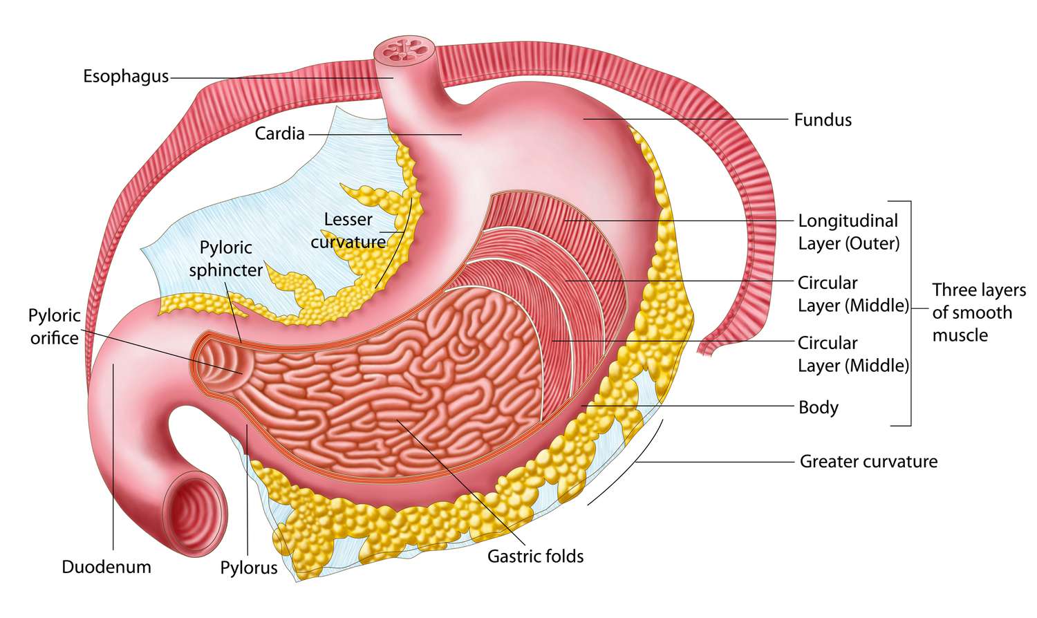
Stomach and Intestines Anatomy

Stomach and Intestines Anatomy

Stomach & Intestines

Digestive System Anatomy: Stomach & Intestines
GASTROINTESTINAL ANATOMY

Stomach

The stomach is a dilated, J-shaped organ of the alimentary canal, situated between the esophagus and the duodenum.

Functions

  • Storage of food: It acts as a temporary reservoir for ingested food.
  • Mixing and mechanical digestion: It churns food with gastric juices to form chyme.
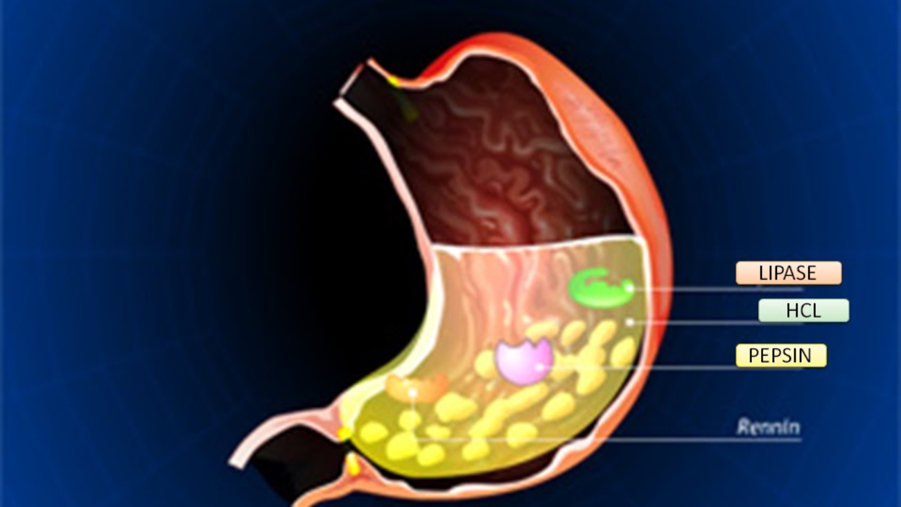
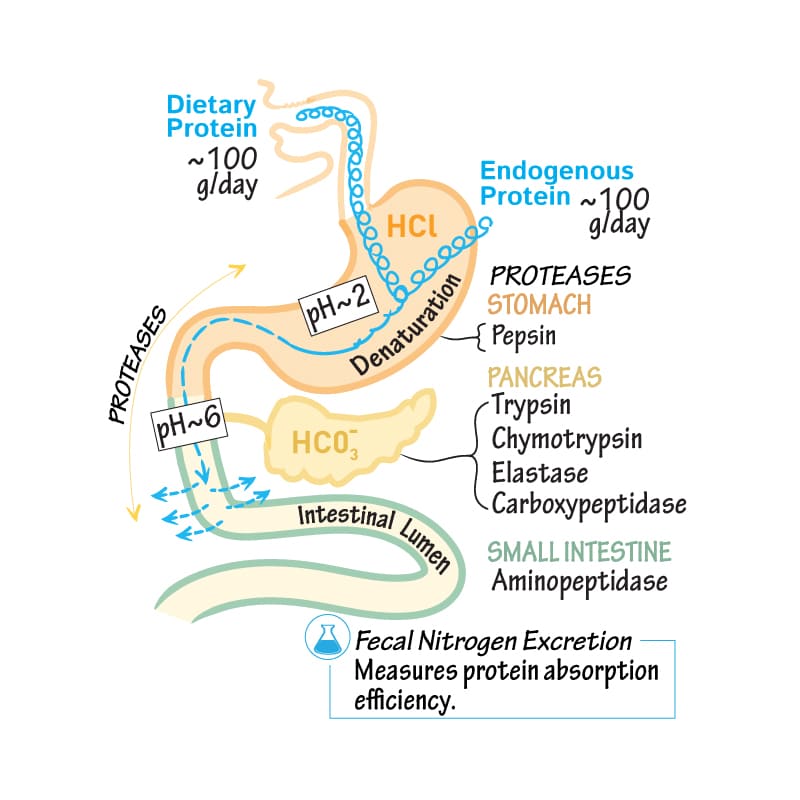
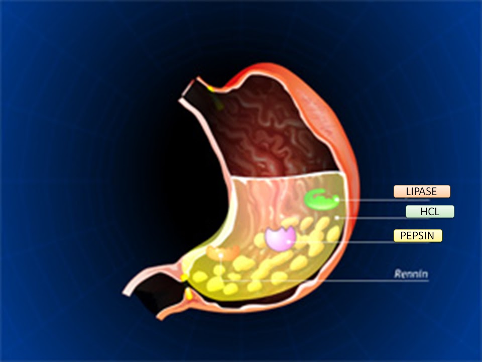
  • Chemical digestion: Gastric juices (containing hydrochloric acid and enzymes like pepsin) initiate protein digestion.
  • Controlled release: Regulates the slow release of chyme into the duodenum for further digestion and absorption.

General Characteristics

  • Capacity: The total capacity of the stomach is approximately 1500 ml.
  • Shape and Position:
    • Its position and shape vary significantly among individuals and with posture and respiration. In short and obese individuals, it tends to be high and more transverse. In tall and thin individuals, it is often elongated and vertical.
    • It typically occupies the epigastric and umbilical regions of the abdomen and is partly covered by the costal diaphragm and the lower ribs.

General Structure

The stomach has two surfaces, two apertures (orifices), and two curvatures.

  • Surfaces: Anterior surface, Posterior surface.
  • Orifices (Apertures):
    • Cardia: The opening from the esophagus into the stomach.
    • Pylorus: The opening from the stomach into the duodenum.
  • Curvatures:
    • Lesser curvature: The shorter, concave, right border of the stomach.
    • Greater curvature: The longer, convex, left and inferior border of the stomach.

Specific Features:

Cardia (Cardiac Orifice)

  • Located at the esophagogastric junction.
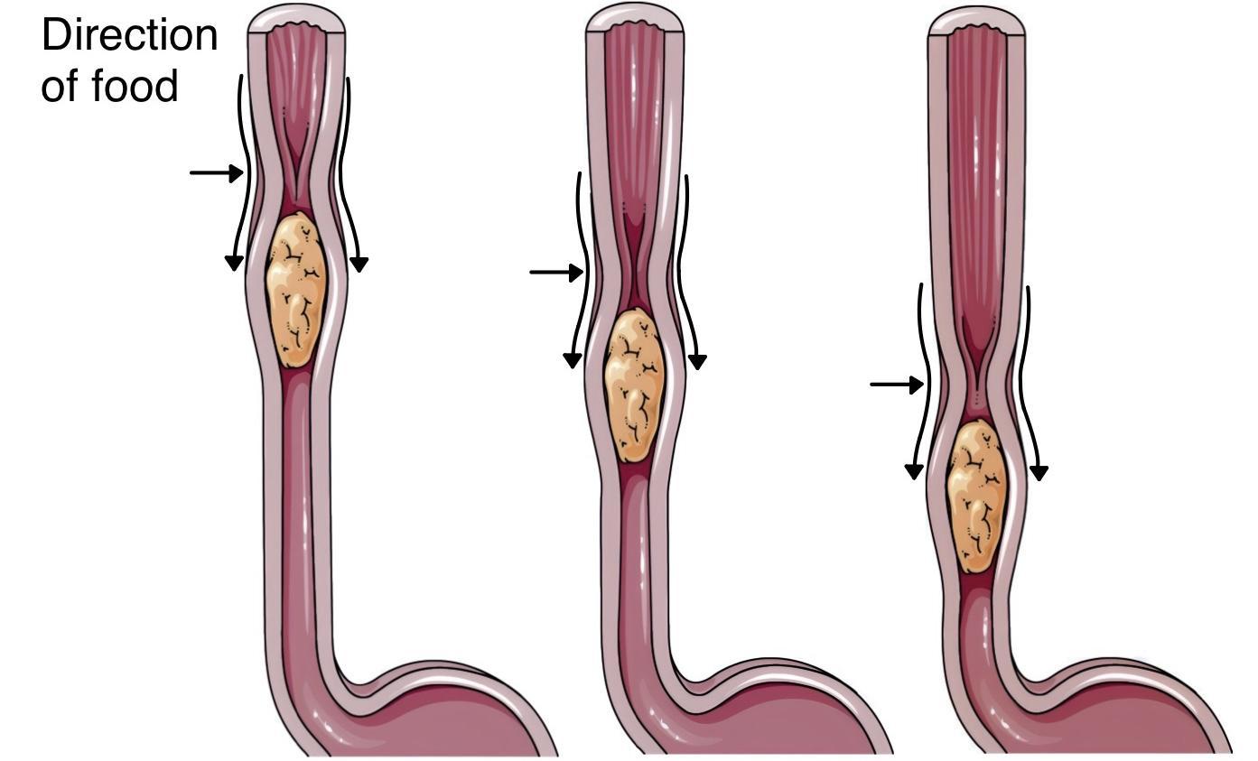
  • There is no anatomical sphincter in the traditional sense. Instead, a physiological sphincter mechanism is formed by the circular muscle layer of the distal esophagus and stomach, the acute angle of His (where the esophagus joins the stomach), and the diaphragmatic crus.
  • This physiological sphincter relaxes during swallowing to allow food entry and closes afterward to prevent gastroesophageal reflux (regurgitation) of gastric contents back into the esophagus.

Pylorus

  • The distal opening of the stomach into the duodenum, located at the gastroduodenal junction.

Lesser Curvature

  • Located on the right margin of the stomach.
  • Serves as the attachment site for the lesser omentum.
  • The angular incisure (incisura angularis) is a constant notch on the lesser curvature, marking the junction between the body and the pyloric part of the stomach.

Greater Curvature

  • Located on the left margin and inferior border of the stomach.
  • Provides attachment for the gastrosplenic ligament (connecting to the spleen) and the greater omentum (which extends inferiorly, folds back, and attaches to the transverse colon).
  • Anterior Surface: Covered by the peritoneum. The left vagus nerve forms the anterior vagal trunk and predominantly supplies the anterior surface of the stomach.
  • Posterior Surface: Also covered by the peritoneum. The right vagus nerve forms the posterior vagal trunk and predominantly supplies the posterior surface of the stomach.

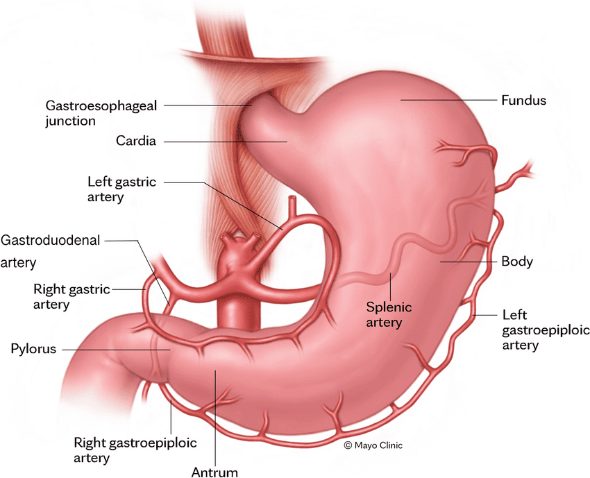
Parts of the Stomach

The stomach is typically divided into four main parts:

  1. Cardia: The region immediately surrounding the cardiac orifice.
  2. Fundus: The dome-shaped part that projects superiorly and to the left of the cardia. It often contains gas.
  3. Body: The main part of the stomach, extending from the cardia/fundus to the angular incisure on the lesser curvature.
  4. Pylorus (Pyloric Part): The tubular distal part of the stomach connecting it to the duodenum. It is further divided into:
    • Pyloric Antrum: Wider, more proximal part.
    • Pyloric Canal: Narrower, distal part.
    • Pyloric Sphincter: A thickened ring of circular smooth muscle at the gastroduodenal junction. It controls the discharge of chyme from the stomach into the duodenum. The pylorus typically lies on the transpyloric plane (L1).

Peritoneal Attachments (Omenta):

  • Lesser Omentum: A two-layered fold of peritoneum that extends from the porta hepatis (on the liver) to the lesser curvature of the stomach and the superior part of the duodenum. It is further divided into:
    • Hepatogastric ligament: From the liver to the lesser curvature of the stomach.
    • Hepatoduodenal ligament: From the liver to the superior part of the duodenum. This is clinically very important as it contains the portal triad: the common bile duct, the proper hepatic artery, and the hepatic portal vein.
  • Greater Omentum: A large, apron-like fold of peritoneum that hangs down from the greater curvature of the stomach and covers the intestines. It contains varying amounts of fat.
  • Gastrosplenic Omentum (Ligament): Connects the greater curvature of the stomach to the hilum of the spleen.

Mucous Membrane of the Stomach:

The internal lining of the stomach is thrown into numerous folds called rugae. These folds allow the stomach to expand significantly when filled with food and flatten out as it distends.

Muscle Layer of the Stomach:

The muscularis externa of the stomach is unique among the alimentary canal because it has three layers of smooth muscle, which contribute to its powerful churning action:

  1. Outer Longitudinal Layer: Primarily present along the curvatures (lesser and greater).
  2. Middle Circular Layer: Surrounds the entire stomach but is particularly prominent at the pylorus (forming the pyloric sphincter) and cardia.
  3. Innermost Oblique Layer: Found mainly in the body and fundus, allowing for a unique churning motion.

Peritoneum of the Stomach: The stomach is almost entirely intraperitoneal, meaning it is nearly completely covered by visceral peritoneum. The peritoneum leaves the stomach to form the various omenta and ligaments (lesser omentum, greater omentum, gastrosplenic omentum).


Relations of the Stomach

Anteriorly

  • Anterior abdominal wall
  • Costal margin
  • Left lobe of the liver
  • Left pleura and lung (superiorly)
  • Diaphragm

Posteriorly (Stomach Bed)

Separated by the lesser sac (omental bursa):

  • Diaphragm (posteriorly, superiorly)
  • Spleen (laterally)
  • Splenic artery (superior border of the pancreas)
  • Left kidney
  • Left suprarenal gland
  • Pancreas
  • Transverse mesocolon and transverse colon

Blood Supply of the Stomach

The stomach has a rich arterial supply from branches of the celiac trunk, ensuring robust collateral circulation.

Arterial Supply

  • Lesser Curvature:
    • Left Gastric Artery: Direct branch of the celiac trunk. Supplies the upper part of the lesser curvature and the abdominal esophagus.
    • Right Gastric Artery: A branch of the proper hepatic artery (which comes from the common hepatic artery, a celiac trunk branch). Supplies the lower part of the lesser curvature.
  • Greater Curvature:
    • Left Gastro-omental (Gastroepiploic) Artery: A branch of the splenic artery. Supplies the upper part of the greater curvature.
    • Right Gastro-omental (Gastroepiploic) Artery: A branch of the gastroduodenal artery (which comes from the common hepatic artery). Supplies the lower part of the greater curvature.
  • Fundus:
    • Short Gastric Arteries (5-7 branches): Direct branches of the splenic artery. Supply the fundus of the stomach.

Venous Drainage

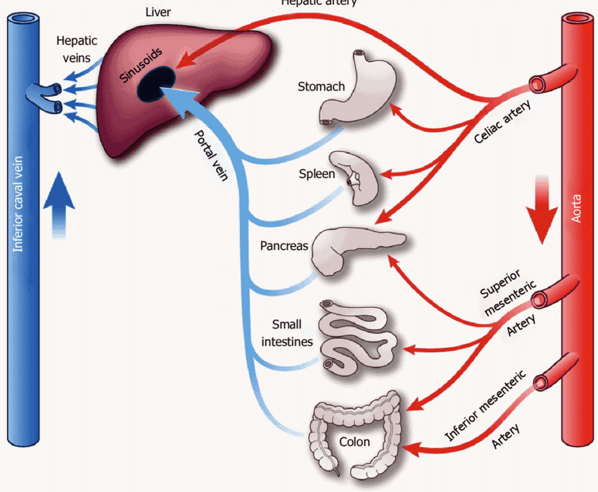
The veins of the stomach generally follow the arteries and drain into the portal venous system.

  • Left and Right Gastric Veins: Drain the lesser curvature directly into the hepatic portal vein.
  • Left Gastro-omental Vein: Drains the greater curvature into the splenic vein.
  • Right Gastro-omental Vein: Drains the greater curvature into the superior mesenteric vein.
  • Short Gastric Veins: Drain the fundus into the splenic vein.

Lymphatic Drainage: Lymphatic vessels generally follow the arterial supply and drain into regional lymph nodes, eventually leading to the celiac lymph nodes around the celiac trunk.
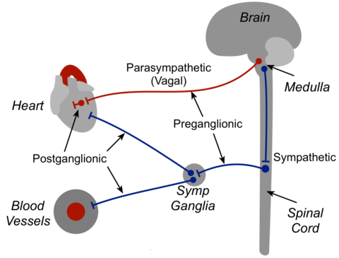
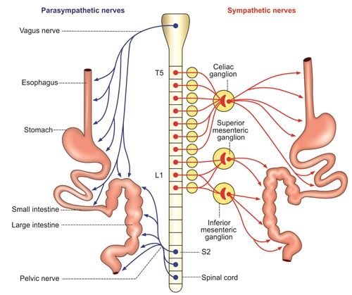
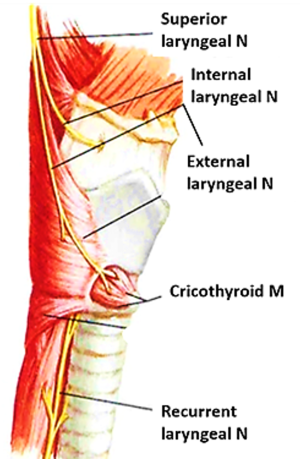
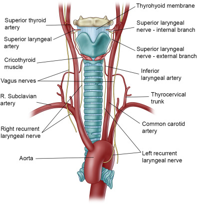
Nerve Supply:

  • Parasympathetic Innervation: Primarily from the vagus nerves. The left vagus forms the anterior vagal trunk, and the right vagus forms the posterior vagal trunk. They increase gastric motility and glandular secretion.
  • Sympathetic Innervation: From the celiac plexus, originating from spinal cord segments T6-T9. They generally inhibit gastric motility and secretion, and mediate pain.

Clinical Notes:

  • Trauma to the Stomach: The stomach is relatively mobile and protected by the rib cage, making blunt trauma less likely to cause injury unless severe. However, penetrating injuries (e.g., stab wounds, gunshot wounds) can lead to perforation and leakage of gastric contents into the peritoneal cavity, causing peritonitis, a serious inflammatory condition.
  • Gastric Ulcers: These are open sores that develop on the gastric mucosa. They are common at the pylorus and lesser curvature, areas where the mucosa is exposed to acidic gastric contents (despite the note stating "alkaline producing mucosa," these areas are indeed exposed to acid and are common ulcer sites; the pyloric region, however, also has some bicarbonate secretion). Ulcers can perforate the stomach wall, leading to peritonitis.
  • Gastric Pain: Pain originating from the stomach (e.g., from ulcers, gastritis) is typically referred to the epigastrium (upper central abdomen) via the sympathetic nerves.
  • Cancer of the Stomach: Gastric cancer can spread to regional lymph nodes. Surgical treatment often involves removing the stomach (gastrectomy) and associated regional lymph nodes, and sometimes neighboring structures, depending on the extent of spread.
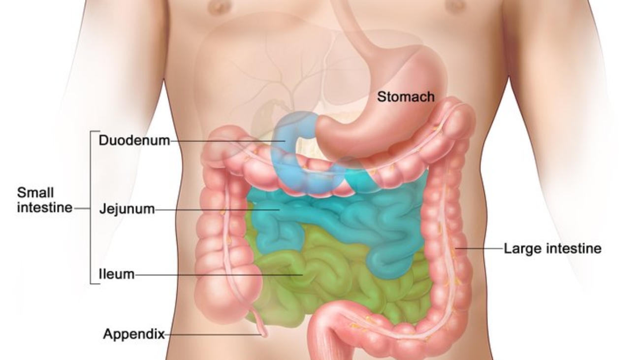
Small Intestines

The small intestine is the longest part of the alimentary canal, extending from the pylorus of the stomach to the ileocecal junction. Its primary function is the absorption of nutrients.

  • Length: Approximately 6 meters (20 feet) long in a living person, but can be much longer post-mortem due to loss of muscle tone.
  • Location: Occupies mainly the epigastric, umbilical, and hypogastric regions of the abdomen.
  • Divisions: It is divided into three main parts:
    1. Duodenum
    2. Jejunum
    3. Ileum

Duodenum

The duodenum is the first and shortest part of the small intestine.

  • Length: Approximately 25 cm (10 inches) long.
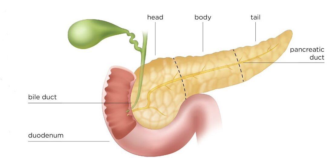
  • Course: It is C-shaped, wrapping around the head of the pancreas.
  • Peritoneal Covering:
    • The first 2.5 cm (1 inch) of the first part is intraperitoneal, resembling the stomach in structure and mobility. It is covered by peritoneum on its anterior and posterior surfaces, has the lesser sac behind it, and receives attachments from the lesser and greater omentum.
    • The remainder of the duodenum (the vast majority) is retroperitoneal, meaning it is fixed to the posterior abdominal wall and covered by peritoneum only on its anterior surface.
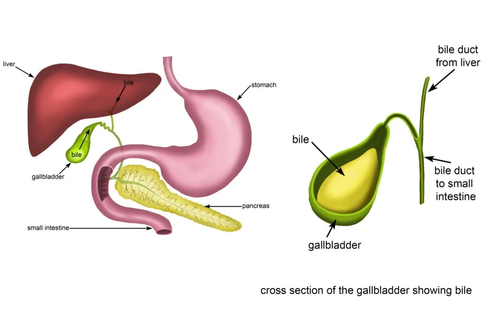
  • Key Feature: Receives the openings of the bile duct (carrying bile from the liver and gallbladder) and the pancreatic ducts (carrying digestive enzymes from the pancreas).

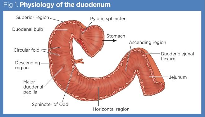
Parts of the Duodenum:

The duodenum is traditionally divided into four parts:

1. First Part (Superior Part)

  • Length: Approximately 5 cm (2 inches) long.
  • Location: Lies at the transpyloric plane (L1). The initial 2.5 cm is the most mobile, forming the "duodenal cap" or "ampulla." It runs upwards and backwards to the right of L1.
  • Relations:
    • Posteriorly: Lesser sac (initially), bile duct, portal vein, gastroduodenal artery, inferior vena cava (IVC).
    • Anteriorly: Liver (quadrate lobe) and gallbladder.
    • Superiorly: The opening into the lesser sac (epiploic foramen of Winslow).
    • Inferiorly: Head of the pancreas.

2. Second Part (Descending Part)

  • Length: Approximately 7.5 cm (3 inches) long.
  • Location: Descends on the right side of the vertebral bodies L2 and L3, within the concavity of the head of the pancreas.
  • Key Feature: Contains the major duodenal papilla (of Vater), where the bile duct and main pancreatic duct typically unite and open, and sometimes a minor duodenal papilla (for the accessory pancreatic duct).
  • Relations:
    • Anteriorly: Gallbladder (occasionally), right lobe of liver, coils of small intestines, transverse colon.
    • Medially: Head of the pancreas, and the openings of the bile and pancreatic ducts.
    • Laterally: Ascending colon, right colic flexure, right lobe of the liver.
    • Posteriorly: Right kidney, right ureter, right psoas major muscle, IVC, aorta (more medially).

3. Third Part (Horizontal or Inferior Part)

  • Length: Approximately 7.5 cm (3 inches) long.
  • Location: Runs horizontally to the left, typically at the level of L3, inferior to the head of the pancreas.
  • Key Feature: The superior mesenteric artery and vein cross anterior to this part.
  • Relations:
    • Superiorly: Head of the pancreas.
    • Inferiorly: Coils of jejunum.
    • Posteriorly: Aorta, IVC, right ureter, right psoas major muscle.
    • Anteriorly: The root of the mesentery of the small intestines (containing the superior mesenteric artery and vein), and coils of jejunum.

4. Fourth Part (Ascending Part)

  • Length: Approximately 5 cm (2 inches) long.
  • Location: Ascends superiorly and to the left, reaching the level of L2, to join the jejunum at the duodenojejunal flexure.
  • Key Feature: The duodenojejunal flexure is suspended by the ligament of Treitz (suspensory muscle of the duodenum), which attaches to the diaphragm.
  • Relations:
    • Anteriorly: Root of the mesentery and coils of jejunum.
    • Posteriorly: Aorta and left psoas major muscle.

Histology of the Duodenum:

  • Mucosa: The inner lining of the duodenum (and much of the small intestine) is thrown into numerous circular folds called plicae circulares (valves of Kerckring), which increase the surface area for absorption. The epithelium is simple columnar with abundant goblet cells and intestinal glands (crypts of Lieberkühn).
  • Brunner's Glands: Unique to the duodenum, these are submucosal glands that produce alkaline mucus to neutralize acidic chyme from the stomach.

Blood Supply of the Duodenum:

The duodenum has a dual blood supply, forming an important anastomotic arcade.

  • Superior Pancreaticoduodenal Artery: A branch of the gastroduodenal artery (from the common hepatic artery). Supplies the superior part of the duodenum.
  • Inferior Pancreaticoduodenal Artery: A branch of the superior mesenteric artery. Supplies the inferior part of the duodenum.
  • Veins and Lymphatics: Generally follow the arteries. Venous drainage is to the hepatic portal vein system.
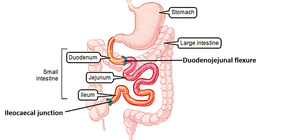
Jejunum and Ileum

These two parts constitute the mobile, coiled portion of the small intestine, primarily responsible for nutrient absorption.

  • Total Length: Approximately 6 meters (20 feet). The jejunum makes up the proximal 2/5ths, and the ileum makes up the distal 3/5ths.
  • Mesentery: Both the jejunum and ileum are suspended from the posterior abdominal wall by a double layer of peritoneum called the mesentery of the small intestine. The root of this mesentery extends obliquely from the left of L2 to the right sacroiliac joint.

Differences Between Jejunum and Ileum:

While there is a gradual transition, some characteristic differences exist:

Feature Jejunum Ileum
Location Occupies the upper part of the peritoneal cavity, mostly to the left of the midline. Occupies the lower part of the peritoneal cavity, mostly to the right of the midline, and within the pelvis.
Diameter Wider (about 2-4 cm). Narrower (about 1.5-3 cm).
Wall Thickness Thicker-walled. Thinner-walled.
Vascularity/Color Redder, more vascular. Paler, less vascular.
Plicae Circulares More numerous, taller, and more closely packed. Fewer, smaller, and more widely spaced; absent in the distal ileum.
Mesentery Fat Fat deposition in the mesentery is near the root, with long windows (clear areas) in the mesentery between blood vessels. Fat deposition extends from the root of the mesentery almost to the intestinal wall, with short windows or no windows in the mesentery.
Vascular Arcades Forms one or two large, long arterial arcades. Forms numerous short, smaller arterial arcades.
Vasa Recta Longer and less branched. Shorter and more branched.
Lymphoid Tissue Solitary lymphoid follicles are present, but Peyer's patches are absent. Large aggregations of lymphoid follicles called Peyer's patches are characteristic, especially in the distal ileum, along the antimesenteric border.

Blood Supply of Jejunum and Ileum:

  • Primarily supplied by jejunal and ileal branches that arise from the superior mesenteric artery (SMA). These arteries form arcades within the mesentery, from which straight vessels (vasa recta) arise to supply the intestinal wall.
  • Veins and Lymphatics: Follow the arteries and drain into the superior mesenteric vein (eventually to the portal vein) and superior mesenteric lymph nodes, respectively.
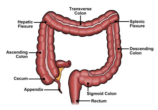
Large Intestines

The large intestine extends from the ileocecal junction to the anus.

  • Primary Function: Absorption of water and electrolytes from undigested food material, and the storage and compaction of fecal matter prior to defecation.
  • Divisions:
    1. Cecum
    2. Appendix
    3. Ascending Colon
    4. Transverse Colon
    5. Descending Colon
    6. Sigmoid Colon
    7. Rectum
    8. Anal Canal

Characteristic Features (except rectum and anal canal)

  • Teniae Coli: Three distinct longitudinal bands of smooth muscle (converging at the appendix).
  • Haustra: Sacculations or pouches of the colon, formed by the contraction of the teniae coli.
  • Omental (Epiploic) Appendages: Small, fat-filled peritoneal pouches projecting from the serosal surface.

Cecum

The cecum is the blind-ended pouch that forms the beginning of the large intestine.

  • Location: Lies in the right iliac fossa, below the level of the ileocecal junction.
  • Mobility: It is relatively mobile despite lacking a mesentery in most individuals (it has peritoneal folds).
  • Teniae Coli: The three teniae coli of the colon converge at the base of the cecum, providing a landmark for locating the appendix.
  • Relations:
    • Anteriorly: Anterior abdominal wall, coils of small intestines, greater omentum.
    • Posteriorly: Psoas major muscle, iliacus muscle, femoral nerve, lateral cutaneous nerve of the thigh, and the appendix.
  • Blood Supply: From the anterior and posterior cecal arteries, which are branches of the ileocolic artery (a branch of the superior mesenteric artery).
  • Veins and Lymphatics: Follow the arteries and drain into the superior mesenteric system.

Vermiform Appendix

The appendix is a narrow, tubular diverticulum extending from the cecum.

  • Structure: Contains a large amount of lymphoid tissue.
  • Length: Varies from 8 to 13 cm.
  • Attachment: Attached to the posteromedial surface of the base of the cecum, approximately 2.5 cm below the ileocecal junction.
  • Mesoappendix: It has its own peritoneal fold, the mesoappendix, which contains the appendicular artery.
  • Location: Often lies in the right iliac fossa. Its base can be roughly located at McBurney's point, which is 1/3 of the way along a line joining the anterior superior iliac spine (ASIS) to the umbilicus.
  • Common Positions: The appendix can lie in various positions relative to the cecum:
    • Pelvic (Descending): Most common, descending into the pelvis.
    • Retrocecal: Behind the cecum (second most common).
    • Paracecal: Beside the cecum.
    • Preileal: In front of the terminal ileum.
    • Postileal: Behind the terminal ileum.
  • Blood Supply: The appendicular artery, a branch of the posterior cecal artery (from the ileocolic artery).
  • Veins and Lymphatics: Follow the artery.
  • Nerve Supply: Superior mesenteric plexus (sympathetic for pain, vagal for parasympathetic).

Ascending Colon

The ascending colon is the part of the large intestine that travels upwards on the right side of the abdominal cavity.

  • Length: Approximately 13 cm long.
  • Course: Extends from the cecum, superior to the ileocolic junction, to the right colic flexure (hepatic flexure), where it turns left to become the transverse colon.
  • Peritoneal Covering: It is typically retroperitoneal, fixed to the posterior abdominal wall.
  • Relations:
    • Anteriorly: Anterior abdominal wall, coils of small intestines, greater omentum.
    • Posteriorly: Iliopsoas muscle, quadratus lumborum muscle, iliac crest, origin of transversus abdominis muscle, iliohypogastric nerve, ilioinguinal nerve, right kidney.
  • Blood Supply: From the ileocolic artery and right colic artery (both branches of the superior mesenteric artery).
  • Veins and Lymphatics: Follow the arteries and drain into the superior mesenteric system.

Transverse Colon

The transverse colon spans across the upper abdomen.

  • Length: Approximately 38 cm long.
  • Course: Extends from the right colic flexure to the left colic flexure (splenic flexure), where it turns inferiorly to become the descending colon.
  • Mesentery: It is uniquely suspended by the transverse mesocolon, making it the most mobile part of the large intestine. The greater omentum is attached to its superior border, and the transverse mesocolon is attached to its inferior border.
  • Relations:
    • Anteriorly: Greater omentum and anterior abdominal wall.
    • Posteriorly: Second part of the duodenum, head of the pancreas, coils of ileum and jejunum.
  • Blood Supply: Has a dual blood supply due to its developmental origin (part from midgut, part from hindgut).
    • Proximal 2/3 (right side): Supplied by the middle colic artery (a branch of the superior mesenteric artery).
    • Distal 1/3 (left side): Supplied by the left colic artery (a branch of the inferior mesenteric artery).
  • Veins and Lymphatics: Follow the arteries; veins drain into the superior and inferior mesenteric veins.

Descending Colon

The descending colon travels downwards on the left side of the abdominal cavity.

  • Length: Approximately 25 cm long.
  • Course: Extends from the left colic flexure to the sigmoid colon at the pelvic inlet.
  • Peritoneal Covering: It is typically retroperitoneal, fixed to the posterior abdominal wall.
  • Relations:
    • Anteriorly: Coils of small intestines, greater omentum, and anterior abdominal wall.
    • Posteriorly: Left kidney, left psoas major muscle, spleen (more superiorly), quadratus lumborum muscle, ilioinguinal and iliohypogastric nerves, femoral nerve, lateral cutaneous nerve of the thigh, iliac crest.
  • Blood Supply: Primarily from the left colic artery (a branch of the inferior mesenteric artery).
  • Veins and Lymphatics: Follow the arteries and drain into the inferior mesenteric system.

Sigmoid Colon

The sigmoid colon is the S-shaped terminal portion of the colon, connecting the descending colon to the rectum.

  • Length: 25 to 38 cm long.
  • Course: Extends from the pelvic brim (as a continuation of the descending colon) and ends at the level of S3 (the third sacral vertebra), where it transitions into the rectum.
  • Mesentery: It has a distinct sigmoid mesocolon, making it very mobile.
  • Relations:
    • Anteriorly: In females, the uterus and upper part of the vagina. In males, the upper part of the urinary bladder.
    • Posteriorly: Sacrum, rectum, coils of ileum.
  • Blood Supply: From the sigmoid arteries (usually 2-4 branches) of the inferior mesenteric artery.
  • Veins and Lymphatics: Follow the arteries and drain into the inferior mesenteric system.

Rectum

The rectum is the final section of the large intestine, connecting the sigmoid colon to the anal canal.

  • Length: Approximately 13 cm long.
  • Course: Extends from the level of S3 (as a continuation of the sigmoid colon) and ends in front of the coccyx, where it pierces the pelvic diaphragm to become the anal canal. The puborectalis muscle forms a sling around the rectosigmoid junction, contributing to fecal continence.
  • Peritoneal Covering:
    • Upper 1/3: Covered by peritoneum on its anterior and lateral surfaces.
    • Middle 1/3: Covered by peritoneum only on its anterior surface.
    • Lower 1/3: Devoid of peritoneal covering (subperitoneal).
  • Shape: It follows the concavity of the sacrum.
  • Internal Features: The mucosa and circular muscle layer form three permanent transverse folds called transverse folds of the rectum (valves of Houston). The longitudinal muscle layer unites from the teniae coli to form a single continuous layer.
  • Relations:
    • Anteriorly:
      • In females: Sigmoid colon (superiorly), uterus, and vagina.
      • In males: Sigmoid colon (superiorly), urinary bladder, prostate, seminal vesicles, and vas deferens.
    • Posteriorly: Sacrum, coccyx, piriformis muscle, coccygeus muscle, levator ani muscles.
  • Blood Supply: Has a rich, anastomosing blood supply from three main sources:
    • Upper 1/3: Superior rectal artery (the terminal branch of the inferior mesenteric artery).
    • Middle 1/3: Middle rectal arteries (branches of the internal iliac arteries).
    • Lower 1/3: Inferior rectal arteries (branches of the internal pudendal arteries, which come from the internal iliac).
  • Veins and Lymphatics: Follow the arteries. Venous drainage is crucial for portosystemic anastomoses. The superior rectal vein drains into the portal system, while the middle and inferior rectal veins drain into the systemic system. Lymphatics drain to internal iliac nodes.
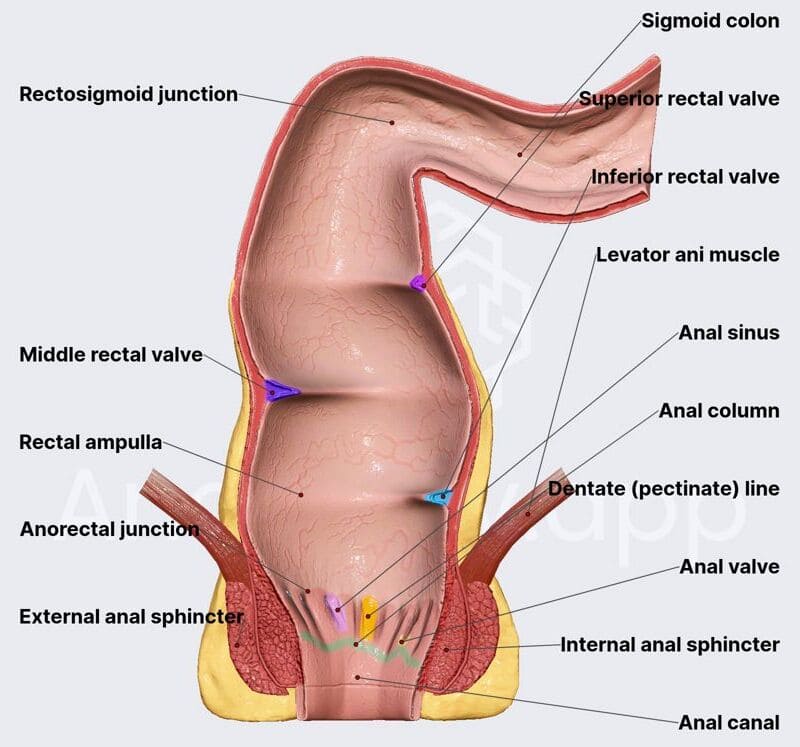
Anal Canal

The anal canal is the terminal part of the large intestine and alimentary canal.

  • Length: Approximately 4 cm long.
  • Course: Begins at the level of the levator ani muscles and ends at the anus.
  • Relations:
    • Posteriorly: Anococcygeal body (a fibromuscular structure).
    • Laterally: Ischiorectal fossae (fat-filled spaces).
    • Anteriorly:
      • In males: Perineal body, urogenital diaphragm, membranous urethra, and bulb of the penis.
      • In females: Perineal body, urogenital diaphragm, and lower half of the vagina.

Mucosa of the Anal Canal:

The anal canal has a distinct mucosal lining that reflects its embryological development and innervation.

Upper Half (above the pectinate/dentate line)

  • Epithelium: Simple columnar epithelium (similar to the rectum).
  • Features: Has longitudinal folds called anal columns (of Morgagni), which contain terminal branches of the superior rectal artery and vein.
  • Innervation: Visceral afferent (pain is dull, poorly localized). Supplied by superior rectal nerves.
  • Blood Supply: Superior rectal artery and vein.
  • Lymphatics: Drain to the inferior mesenteric lymph nodes.

Lower Half (below the pectinate/dentate line)

  • Epithelium: Stratified squamous epithelium (non-keratinized initially, becoming keratinized at the anus).
  • Features: No anal columns.
  • Innervation: Somatic afferent (highly sensitive to pain, touch, temperature). Supplied by inferior rectal nerves (branches of the pudendal nerve).
  • Blood Supply: Inferior rectal arteries and veins.
  • Lymphatics: Drain to the superficial inguinal lymph nodes.

Anal Sphincters:

Two main sphincters control defecation:

  1. Internal Anal Sphincter:
    • Composition: A thickened, involuntary (smooth) muscle layer formed by the circular muscle of the anal canal.
    • Control: Under autonomic (involuntary) control. It maintains tonic contraction to prevent leakage of fecal material.
  2. External Anal Sphincter:
    • Composition: Composed of voluntary (striated) muscle fibers. It consists of three parts (subcutaneous, superficial, and deep).
    • Control: Under somatic (voluntary) control, allowing conscious control over defecation.
    • Innervation: Pudendal nerve.

These sphincters work in coordination to control the expulsion of fecal material from the gut, maintaining continence.

Stomach & Intestines Quiz

Systems Anatomy

Enter your details to begin the examination.

🛡️ Privacy Note: Results are for tracking and certification purposes only.

heart anatomy (1)

Heart & Great Vessels

Heart & Great Vessels

Anatomy: Heart, Pericardium, Great Vessels & Esophagus
GROSS ANATOMY

The Heart, Pericardium, and Great Vessels


Pericardium

The pericardium is a tough, double-layered fibroserous sac that encloses the heart and the roots of the great vessels (aorta, pulmonary trunk, venae cavae, pulmonary veins).

Main Functions

  • Lubrication: Contains a small amount of fluid that reduces friction between the moving heart and the surrounding structures.
  • Restriction of Movement: Anchors the heart in the mediastinum, preventing excessive movement and overdistention during sudden increases in blood volume.
  • Protection: Acts as a physical barrier against infection and external trauma.

Components

The pericardium is composed of two main layers:

  1. Fibrous Pericardium (Outer layer)
  2. Serous Pericardium (Inner layer)

1. Fibrous Pericardium

The fibrous pericardium is the robust, outermost layer of the pericardial sac.

  • Description: It is a thick, tough, inelastic, and conical-shaped fibrous bag that surrounds the heart.
  • Attachments:
    • Inferiorly (Base): It is firmly and broadly fused to the central tendon of the diaphragm. This attachment is crucial for the diaphragm's role in cardiac stability.
    • Anteriorly: It is loosely attached to the posterior surface of the sternum by the sternopericardial ligaments.
    • Superiorly (Apex): It blends and is fused with the outer connective tissue coats (adventitia) of the great vessels as they enter and leave the heart (aorta, pulmonary trunk, superior vena cava, inferior vena cava, pulmonary veins).
    • Posteriorly: It is attached to the structures in the posterior mediastinum, like the esophagus and aorta.
  • Embryological Origin: Its primary origin is debated, but contributions are thought to come from the septum transversum (which also gives rise to the central tendon of the diaphragm) and pleuropericardial membranes.

2. Serous Pericardium

The serous pericardium is a thin, delicate, two-layered membrane that lines the inner surface of the fibrous pericardium and covers the external surface of the heart.

  • Layers: It is divided into two continuous layers:
    • Parietal Layer of Serous Pericardium: This layer lines the inner surface of the fibrous pericardium.
    • Visceral Layer of Serous Pericardium (Epicardium): This layer tightly adheres to the outer surface of the heart itself. It is considered the outermost layer of the heart wall.
  • Pericardial Cavity: The potential space located between the parietal and visceral layers of the serous pericardium. This space normally contains a small amount of serous fluid.

Pericardial Cavity and Sinuses


A. Pericardial Cavity

  1. Description: A potential space located between the parietal and visceral layers of the serous pericardium.
  2. Contents: Normally contains a small amount (typically 15-50 mL) of thin, straw-colored serous fluid.
  3. Function of Fluid: The pericardial fluid acts as a lubricant, allowing the heart to beat smoothly and with minimal friction within the pericardial sac.
  4. Clinical Significance:

    a. Pericardial Effusion: An abnormal accumulation of fluid in the pericardial cavity. This can be caused by various conditions, including infections (e.g., tuberculosis), inflammation (e.g., pericarditis), autoimmune diseases, trauma, and malignancies (tumors). The term "water in the heart" is a colloquial and somewhat misleading description; it's fluid around the heart.

    b. Cardiac Tamponade: A life-threatening condition where a large or rapidly accumulating pericardial effusion compresses the heart, restricting its ability to fill adequately with blood during diastole. This leads to reduced cardiac output and can be fatal if not treated promptly.

    c. Pericardiocentesis: A medical procedure to aspirate (remove) excess fluid from the pericardial cavity to relieve pressure in cases of pericardial effusion or cardiac tamponade.

B. Pericardial Sinuses

These are reflections of the serous pericardium that create cul-de-sacs or recesses.

  1. Oblique Pericardial Sinus:
    • Location: A blind-ended, inverted U-shaped cul-de-sac located posterior to the heart's left atrium.
    • Boundaries: It is situated between the reflections of the serous pericardium around the four pulmonary veins and the inferior vena cava (IVC). The posterior wall of the left atrium forms its anterior boundary.
    • Clinical Significance: Allows for surgical access to the posterior surface of the heart.
  2. Transverse Pericardial Sinus:
    • Location: A short, transverse tunnel or passage that runs between the great arteries (aorta and pulmonary trunk) anteriorly and the great veins (superior vena cava, inferior vena cava, and pulmonary veins) posteriorly.
    • Clinical Significance: This sinus is strategically important for cardiac surgeons. A surgical clamp can be passed through the transverse sinus to temporarily occlude the aorta and pulmonary trunk during cardiac surgery (e.g., for coronary artery bypass grafting or valve replacement), isolating the heart from the systemic circulation.

Innervation and Blood Supply of Pericardium

A. Blood Supply of the Pericardium

The pericardium receives its arterial supply from several sources:

  • Pericardiacophrenic Artery: The main arterial supply, a branch of the internal thoracic artery. It accompanies the phrenic nerve.
  • Musculophrenic Artery: Another branch of the internal thoracic artery.
  • Branches of the Thoracic Aorta: Small branches directly from the aorta (e.g., bronchial and esophageal arteries).
  • Coronary Arteries: The visceral layer (epicardium) receives some small branches from the coronary arteries.
  • Venous Drainage: Follows the arterial supply, draining into corresponding veins (e.g., pericardiacophrenic veins, internal thoracic veins, azygos system).

B. Innervation of the Pericardium

The innervation differs for the fibrous/parietal serous layers and the visceral serous layer.

  • Fibrous Pericardium and Parietal Layer of Serous Pericardium:
    • Innervation: Primarily supplied by the phrenic nerves (C3, C4, C5).
    • Sensitivity: These layers are richly innervated and are sensitive to pain, temperature, pressure (touch), and stretch.
    • Referred Pain: Because the phrenic nerves also supply sensory innervation to the C3-C5 dermatomes, pain originating from these pericardial layers is often referred to the ipsilateral (same side) shoulder, neck, and supraclavicular region. (The original mention of "left jaw" is less common for pericardial pain referral than the shoulder and neck).
  • Visceral Layer of Serous Pericardium (Epicardium):
    • Innervation: Supplied by the autonomic nervous system (sympathetic and parasympathetic fibers) from the cardiac plexuses.
    • Sensitivity: This layer is generally considered insensitive to pain, temperature, and touch. It is primarily sensitive to stretch.
    • Function: Autonomic innervation primarily modulates cardiac function rather than providing somatic sensation from the epicardium itself.

The Heart

The heart is a hollow, muscular organ that acts as a pump, circulating blood throughout the entire body to deliver oxygen and nutrients and remove waste products.

  • Shape: It is often described as conical or roughly pyramidal in shape, with an apex (pointing infero-anteriorly) and a base (directed postero-superiorly).
  • Location: It is situated in the middle mediastinum, slightly to the left of the midline, resting on the diaphragm.
  • Mobility:
    • The base of the heart (where the great vessels enter and leave) is relatively fixed by its attachment to the great vessels.
    • The ventricles (and apex), however, are more mobile within the pericardial sac, allowing for the pumping action.
    • The position of the heart changes subtly with respiration and with the cardiac cycle (systole and diastole).

Surfaces of the Heart

The heart has several anatomical surfaces that are important for understanding its relations to surrounding structures and for clinical examination.

  1. Anterior (Sternocostal) Surface:
    • Description: This surface faces anteriorly towards the sternum and costal cartilages.
    • Components: Primarily formed by the right ventricle (largest part), a portion of the right atrium, and a small strip of the left ventricle (along the left border).
    • Grooves: The anterior interventricular groove (containing the anterior interventricular artery/LAD) and the right atrioventricular (coronary) groove (containing the right coronary artery, often embedded in fat) are visible on this surface.
  2. Posterior Surface (Base):
    • Description: This surface is directed posteriorly, superiorly, and slightly to the right. It is generally the fixed part of the heart.
    • Components: Primarily formed by the left atrium, with a smaller contribution from the right atrium.
    • Vessels: The four pulmonary veins enter the left atrium on this surface. The superior vena cava enters the right atrium on this surface.
    • Important Note: The base of the heart is where the great vessels attach and is relatively fixed, but it does not rest on the diaphragm. The diaphragmatic surface rests on the diaphragm.
  3. Inferior (Diaphragmatic) Surface:
    • Description: This surface rests directly on the central tendon of the diaphragm.
    • Components: Primarily formed by the left ventricle (approximately 2/3) and the right ventricle (approximately 1/3). A small portion of the right atrium where the inferior vena cava (IVC) enters is also part of this surface.
    • Grooves: The posterior interventricular groove and parts of the coronary groove are found here.
  4. Apex:
    • Description: The blunt, inferolateral tip of the heart.
    • Component: Formed entirely by the left ventricle.
    • Location (Clinical): Typically located in the left 5th intercostal space, approximately 9 cm (3.5 inches) from the midsternal line (just medial to the midclavicular line). This is where the apex beat (point of maximal impulse) can be palpated.

Borders of the Heart

The heart has distinct borders when viewed from an anterior perspective, which are useful for radiographic interpretation and understanding its anatomical relationships.

  1. Right Border:
    • Components: Formed exclusively by the right atrium.
    • Course: Extends from the superior vena cava to the inferior vena cava.
  2. Left Border:
    • Components: Formed primarily by the left ventricle, with a small contribution superiorly from the left auricle (a part of the left atrium).
    • Course: Extends from the left auricle to the apex.
  3. Inferior Border:
    • Components: Formed mainly by the right ventricle, a small part of the left ventricle near the apex, and a small part of the right atrium near the IVC entrance.
    • Course: Extends from the inferior vena cava to the apex.
  4. Superior Border:
    • Components: Formed by the great vessels entering and leaving the heart (aorta, pulmonary trunk, superior vena cava). It is somewhat obscured by these vessels.

Skeleton of the Heart

The fibrous skeleton of the heart is a crucial structural and functional component, despite being largely fibrous (not bony, except in some animals).

  1. Description: It is a complex framework of dense connective tissue (fibrous rings) that surrounds the atrioventricular and arterial orifices. It's often described as being in the shape of a figure-8 or two interlocking rings.
  2. Components: Primarily composed of:
    • Annuli Fibrosi: Four fibrous rings that encircle the:
      • Right atrioventricular orifice (tricuspid valve).
      • Left atrioventricular orifice (mitral valve).
      • Aortic orifice.
      • Pulmonary orifice.
    • Trigonum Fibrosum: Two fibrous trigones (right and left) that connect the fibrous rings.
    • Membranous Part of the Interventricular and Interatrial Septa: The fibrous skeleton contributes to these septa.
  3. Functions:
    • Structural Support: Provides a rigid framework for the attachment of the heart valves, maintaining their shape and preventing their overstretching and becoming incompetent (leaky).
    • Muscle Attachment: Serves as the origin and insertion for the cardiac muscle fibers of the atria and ventricles.
    • Electrical Isolation: Crucially, it forms an electrical barrier between the atria and the ventricles. This electrical discontinuity ensures that the electrical impulses from the atria are only conducted to the ventricles via the atrioventricular (AV) bundle, allowing the atria to contract first, followed by the ventricles in a coordinated sequence.
  4. Os Cordis: In some animals (e.g., cattle), a small bone called the "os cordis" can be found within the fibrous skeleton, serving similar functions. It is not present in humans.

Walls of the Heart

The wall of the heart is composed of three distinct layers, from superficial to deep:

  1. Epicardium:
    • Description: This is the outermost layer of the heart wall.
    • Composition: It is synonymous with the visceral layer of the serous pericardium. It consists of a mesothelium and underlying connective tissue, often containing fat, coronary arteries, and veins.
  2. Myocardium:
    • Description: This is the middle and thickest layer, forming the bulk of the heart wall.
    • Composition: It is composed of specialized cardiac muscle tissue. The thickness of the myocardium varies between the different chambers, being thickest in the left ventricle due to its high-pressure pumping demands.
  3. Endocardium:
    • Description: This is the innermost layer that lines the heart chambers and covers the heart valves.
    • Composition: It consists of a single layer of flattened epithelial cells called endothelium, supported by a thin layer of connective tissue. It is continuous with the endothelium of the blood vessels entering and leaving the heart.

Chambers of the Heart

The human heart is a four-chambered organ, divided into two atria and two ventricles, which work in a coordinated fashion to pump blood.

  1. Right Atrium (RA): Receives deoxygenated blood from the body.
  2. Right Ventricle (RV): Pumps deoxygenated blood to the lungs.
  3. Left Atrium (LA): Receives oxygenated blood from the lungs.
  4. Left Ventricle (LV): Pumps oxygenated blood to the rest of the body.

1. Right Atrium

The right atrium (RA) is the right upper chamber of the heart, forming its right border.

  • Receives Blood From: It collects deoxygenated blood from three main sources:
    • Superior Vena Cava (SVC): Drains blood from the head, neck, and upper limbs.
    • Inferior Vena Cava (IVC): Drains blood from the trunk, lower limbs, and abdominal viscera.
    • Coronary Sinus: The main vein that collects deoxygenated blood from the walls of the heart itself.
  • Pumps Blood To: From the right atrium, blood passes through the right atrioventricular (tricuspid) orifice into the right ventricle.
  • Structure:
    • Main Cavity: The main, larger portion of the atrium.
    • Right Auricle: A small, ear-shaped muscular pouch that projects anteriorly from the main atrial cavity.
  • Internal Features (Embryological Significance):
    • Sulcus Terminalis (External): A shallow groove on the external surface of the right atrium, running between the SVC and IVC.
    • Crista Terminalis (Internal): An internal muscular ridge that corresponds to the sulcus terminalis. It divides the right atrium into two embryologically distinct parts:
      • Smooth-Walled Part (Sinus Venarum): The posterior, smooth part of the right atrium (posterior to the crista terminalis) is derived from the embryonic sinus venosus (specifically, its right horn). This is where the SVC, IVC, and coronary sinus open.
      • Rough-Walled Part: The anterior part (anterior to the crista terminalis), including the right auricle, has prominent muscular ridges called musculi pectinati (pectinate muscles). This part is derived from the embryonic primitive atrium.

Openings and Remnants in the Right Atrium

A. Openings in the Right Atrium:

  1. Opening of Superior Vena Cava (SVC):
    • Location: In the upper, posterior part of the right atrium.
    • Valve: No valve guards the SVC opening.
  2. Opening of Inferior Vena Cava (IVC):
    • Location: In the lower, posterior part of the right atrium.
    • Valve: Possesses a rudimentary (non-functional in adults) valve, the valve of the IVC (Eustachian valve). In fetal life, this valve directed oxygenated blood from the IVC through the foramen ovale into the left atrium.
  3. Opening of Coronary Sinus:
    • Location: Situated between the opening of the IVC and the right atrioventricular orifice, near the septal cusp of the tricuspid valve.
    • Function: Returns most of the deoxygenated blood from the heart wall (myocardium) to the right atrium.
    • Valve: Also guarded by a rudimentary (non-functional in adults) valve, the valve of the coronary sinus (Thebesian valve).
  4. Right Atrioventricular (Tricuspid) Orifice:
    • Location: The large opening between the right atrium and the right ventricle.
    • Valve: Guarded by the tricuspid valve, a functional valve with three cusps (leaflets):
      • i. Anterior cusp.
      • ii. Posterior (or inferior) cusp.
      • iii. Septal cusp.
    • Note: The posterior/inferior cusp is often the smallest.

B. Remnants in the Right Atrium (on the Interatrial Septum):

These structures are important remnants of fetal circulation.

  1. Fossa Ovalis:
    • Description: A shallow, oval depression located on the interatrial septum (the wall separating the right and left atria), on the posterior wall of the right atrium.
    • Represents: It is the remnant of the foramen ovale, an opening in the fetal heart that allowed oxygenated blood to bypass the lungs and flow directly from the right atrium to the left atrium.
  2. Annulus Ovalis (Limbus Fossa Ovalis):
    • Description: A prominent, crescent-shaped ridge that forms the superior and anterior margin of the fossa ovalis.
    • Formation: It is formed from the lower edge of the embryonic septum secundum, which covered the foramen ovale during fetal development.

2. Right Ventricle

The right ventricle (RV) is the right lower chamber of the heart, situated anteriorly and forming most of the anterior (sternocostal) surface of the heart.

  1. Receives Blood From: Receives deoxygenated blood from the right atrium through the right atrioventricular (tricuspid) orifice.
  2. Pumps Blood To: Pumps this deoxygenated blood to the lungs via the pulmonary trunk (pulmonary artery).
  3. Internal Features:
    • Inflow Part: The main part of the right ventricle, receiving blood from the right atrium. Its internal surface is characterized by prominent muscular ridges.
    • Outflow Part (Infundibulum / Conus Arteriosus): As the cavity of the right ventricle approaches the pulmonary trunk, it becomes a smooth-walled, funnel-shaped outflow tract called the infundibulum or conus arteriosus. This smooth walls allow for efficient blood ejection into the pulmonary trunk.
    • Trabeculae Carneae: Unlike the right atrium which has both smooth and rough parts, the internal surface of the right ventricle (except for the infundibulum) is lined by irregular muscular ridges called trabeculae carneae. These are generally described as three types:
      • i. Papillary Muscles: Conical muscular projections that arise from the ventricular wall. Their apices are connected to the free margins and ventricular surfaces of the tricuspid valve cusps by thin, fibrous cords called chordae tendineae. There are typically three papillary muscles: anterior, posterior, and septal. They contract just before ventricular systole to prevent the valve cusps from prolapsing into the right atrium.
      • ii. Moderator Band (Septomarginal Trabecula): A distinct, often prominent, muscular band that extends from the inferior part of the interventricular septum to the base of the anterior papillary muscle. It is important because it transmits a part of the right bundle branch of the cardiac conducting system, facilitating efficient conduction to the anterior papillary muscle and the ventricular wall.
      • iii. Prominent Ridges: Irregular, raised muscular ridges that crisscross the ventricular wall.

Pulmonary Valve

The pulmonary valve is one of the two semilunar valves of the heart, regulating blood flow from the right ventricle into the pulmonary trunk.

  • Location: Guards the pulmonary orifice, the opening between the right ventricle and the pulmonary trunk.
  • Composition: It is composed of three semilunar cusps (leaflets), which are delicate, pocket-like structures without chordae tendineae or papillary muscles.
  • Attachment: The curved, lower (proximal) margins of the cusps are attached to the fibrous ring surrounding the pulmonary orifice and to the arterial wall of the pulmonary trunk.
  • Direction of Opening: The cusps are concave towards the pulmonary trunk. During ventricular systole, they are pushed open, and their "upper mouths" (free edges) are directed into the pulmonary trunk, allowing blood to flow out. During ventricular diastole, blood in the pulmonary trunk tries to flow back into the ventricle, filling the cusps and forcing them closed.
  • Arrangement of Cusps: The three cusps are typically named based on their embryonic position (though slight variations exist in anatomical texts):
    • Anterior cusp.
    • Right cusp.
    • Left cusp. (Note: The original "one posterior (left cusp) and two anterior (anterior and right)" is a common descriptive but slightly conflicting categorization. Standard anatomical texts usually list anterior, right, and left for the pulmonary valve.)
  • Relative Position: The pulmonary orifice is indeed located slightly superior and to the left of the aortic orifice, although the aortic valve is generally considered to be positioned more centrally in the fibrous skeleton.

3. Left Atrium

The left atrium (LA) is the left upper chamber of the heart.

  • Location: It is located posterior to the right atrium, and forms the bulk of the base of the heart.
  • Relations:
    • Posteriorly: It is closely related to the oblique pericardial sinus and the esophagus. This close relationship means that enlargement of the left atrium (e.g., in mitral valve disease) can compress the esophagus, and can sometimes be visualized in diagnostic imaging like a barium swallow (where barium outlines the esophagus).
  • Structure:
    • Main Cavity: The main, larger portion of the atrium.
    • Left Auricle: A small, ear-shaped muscular pouch that projects anteriorly and superiorly.
  • Receives Blood From: It receives oxygenated blood from the lungs via the four pulmonary veins (typically two from the right lung and two from the left lung). These veins enter the posterior wall of the left atrium.
    • (Correction: The original text incorrectly states "enclosed in a common sleeve of serous pericardium together with the IVC and the SVC." While the pulmonary veins are covered by a sleeve of serous pericardium, the IVC and SVC are associated with the right atrium, not directly with the left atrium's pulmonary veins in a common sleeve).
  • Internal Features (Embryological Significance):
    • Smooth Walled: Unlike the right atrium, the majority of the internal surface of the left atrium is smooth-walled. This smooth part is embryologically derived from the incorporation of the pulmonary veins into the primitive left atrium.
    • Rough-Walled Auricle: Only the left auricle typically has prominent muscular ridges, the musculi pectinati, derived from the primitive atrium.

Openings in the Left Atrium

The left atrium has two main types of openings:

  1. Openings of the Four Pulmonary Veins:
    • Number: Typically four openings (two superior and two inferior) on the posterior wall of the left atrium.
    • Valves: These openings are not guarded by valves. Instead, the oblique course of these veins through the atrial wall and the contraction of the left atrium create a functional sphincter-like action that helps prevent significant backflow of blood during atrial systole.
  2. Left Atrioventricular (Mitral) Orifice:
    • Location: The opening between the left atrium and the left ventricle.
    • Valve: Guarded by the bicuspid valve, more commonly known as the mitral valve. It is a functional valve with two cusps (leaflets):
      • i. Anterior cusp.
      • ii. Posterior cusp.
    • Cusp Characteristics: The cusps of the mitral valve are generally thicker and more robust than those of the tricuspid valve, reflecting the higher pressure in the left side of the heart. The anterior cusp is typically larger, thicker, and more rigid than the posterior cusp, and is sometimes referred to as the septal cusp due to its proximity to the interventricular septum.

4. Left Ventricle

The left ventricle (LV) is the left lower chamber of the heart, forming the apex of the heart and a significant portion of its left and diaphragmatic surfaces.

  • Receives Blood From: Receives oxygenated blood from the left atrium through the left atrioventricular (mitral) orifice.
  • Pumps Blood To: Pumps this oxygenated blood to the entire body via the aorta (specifically, the ascending aorta).
  • Wall Thickness and Pressure: The left ventricular wall is significantly thicker (approximately 3 times thicker) than the right ventricular wall. This reflects its role in pumping blood against much higher systemic resistance, resulting in systolic pressures that are typically 5-6 times higher than in the right ventricle.
  • Internal Features:
    • Trabeculae Carneae: Similar to the right ventricle, the internal surface of the left ventricle is characterized by prominent trabeculae carneae (muscular ridges).
    • Papillary Muscles: It possesses two large papillary muscles (anterior and posterior) that connect to the mitral valve cusps via chordae tendineae.
    • No Moderator Band: The left ventricle does not have a moderator band; the conduction system (left bundle branch) has a different branching pattern.
    • Aortic Vestibule: The smooth-walled outflow tract leading from the main ventricular cavity to the aortic orifice is called the aortic vestibule. This smooth wall ensures efficient blood ejection into the aorta.
  • Cross-sectional Shape: In cross-section, the left ventricle is typically described as circular or oval-shaped, while the right ventricle is more crescentic, wrapping around the left ventricle. (The original "triangular" for LV cross-section is not standard; it's typically circular/oval due to the high pressure).

Openings in the Left Ventricle

The left ventricle has two crucial openings:

  1. Left Atrioventricular (Mitral) Orifice:
    • This opening, as discussed earlier, leads from the left atrium into the left ventricle and is guarded by the mitral valve.
  2. Aortic Opening (Aortic Orifice):
    • Location: Leads from the left ventricle into the ascending aorta.
    • Relative Position: It is located posterior and to the right of the pulmonary orifice, and slightly inferior to it.
    • Valve: Guarded by the aortic valve, which is composed of three semilunar cusps:
      • i. Right coronary cusp.
      • ii. Left coronary cusp.
      • iii. Posterior (non-coronary) cusp.
    • Arrangement and Function: These cusps have a similar semilunar arrangement and function to the pulmonary valve, preventing backflow of blood into the left ventricle during diastole.
    • Aortic Sinuses (Sinuses of Valsalva): Just above the aortic cusps are three dilatations in the wall of the ascending aorta called the aortic sinuses. These are crucial:
      • i. The right coronary artery originates from the right aortic sinus (corresponding to the right coronary cusp).
      • ii. The left coronary artery originates from the left aortic sinus (corresponding to the left coronary cusp).
      • iii. The posterior (non-coronary) sinus does not give rise to a coronary artery.

Conducting System of the Heart

The heart's ability to pump blood relies on an intrinsic electrical system that generates and conducts impulses, ensuring a coordinated and rhythmic contraction. This system consists of specialized cardiac muscle cells.

1. Sinoatrial (SA) Node

  • Location: Situated in the upper part of the right atrium, near the junction with the superior vena cava (SVC). It typically occupies an area to the left of the sulcus terminalis.
  • Description: It extends approximately 1 cm along the superior border of the right auricle and then tapers downwards along the crista terminalis for about 2 cm.
  • Pacemaker Function: It is often referred to as the natural pacemaker of the heart because it normally initiates the electrical impulses that trigger cardiac muscle contraction. It has the highest inherent rhythmicity (typically 60-100 beats per minute).
  • Innervation: Has a rich supply of both sympathetic (accelerator) and parasympathetic (vagal) (depressor) nerve fibers. These autonomic influences modulate its intrinsic rate, speeding up or slowing down the heart rate in response to the body's needs.
  • Conduction: From the SA node, impulses spread through the atrial muscle (via internodal pathways) to the AV node.

2. Atrioventricular (AV) Node

  • Location: A small nodule located in the inferior part of the interatrial septum, just above the attachment of the septal cusp of the tricuspid valve, near the opening of the coronary sinus.
  • Function: Its primary role is to delay the electrical impulse from the atria to the ventricles. This delay (approximately 0.1 second) is crucial, allowing the atria to contract and fully empty their blood into the ventricles before ventricular contraction begins.
  • Output: Gives rise to the atrioventricular (AV) bundle (Bundle of His).

3. Atrioventricular (AV) Bundle (Bundle of His)

  • Origin: Arises from the AV node.
  • Course: Descends along the inferior border of the membranous part of the interventricular septum.
  • Branching: Upon reaching the upper part of the muscular interventricular septum, it divides into the right and left bundle branches.

4. Right Bundle Branch

  • Course: Passes down the right side of the interventricular septum.
  • Distribution: It is then carried across the lumen of the right ventricle, often within the moderator band (septomarginal trabecula), to the anterior wall of the right ventricle. It subsequently divides into Purkinje fibers that rapidly spread throughout the right ventricular myocardium.

5. Left Bundle Branch

  • Course: Descends on the left side of the interventricular septum.
  • Distribution: It quickly divides into anterior and posterior fascicles, which then fan out and spread over the entire left ventricular wall, merging with the Purkinje fiber network.

6. Purkinje Fibers

  • These are specialized, large-diameter cardiac muscle fibers that rapidly conduct electrical impulses throughout the ventricular myocardium, ensuring synchronized ventricular contraction.

Blood Supply of the Heart (Coronary Circulation)

The heart muscle (myocardium) receives its own rich blood supply through the coronary arteries, which arise directly from the aorta.

A. Arterial Supply:

The heart is primarily supplied by two main arteries: the right coronary artery (RCA) and the left coronary artery (LCA).

Right Coronary Artery (RCA)

  • Origin: Arises from the anterior (right) aortic sinus of the ascending aorta.
  • Course: Passes between the right auricle and the pulmonary infundibulum, then descends vertically in the right atrioventricular groove (coronary sulcus). It reaches the inferior border of the heart, turns posteriorly, and continues in the coronary sulcus on the diaphragmatic surface.
  • Major Branches and Distribution:
    • Right Conus Artery: Supplies the anterior surface and upper part of the anterior wall of the right ventricle.
    • Anterior Ventricular Branches (2-3): Supply the anterior part of the right ventricle.
    • Marginal Branch (Acute Marginal Artery): A prominent branch that runs along the inferior margin of the right ventricle, supplying it.
    • Posterior Ventricular Branches (2): Supply the diaphragmatic (inferior) part of the right ventricle.
    • Posterior Interventricular (Descending) Artery (PDA): A crucial branch that descends in the posterior interventricular groove. It supplies the diaphragmatic surfaces of both the right and left ventricles, and the posterior one-third of the interventricular septum, and the AV node (in 90% of cases).
    • Atrial Branches: Supply the right atrium and, in about 60% of cases, the SA node artery.

Left Coronary Artery (LCA)

  • Origin: Arises from the left posterior (left) aortic sinus of the ascending aorta.
  • Course: Typically shorter than the RCA, it passes between the left auricle and the pulmonary trunk, then quickly bifurcates (or trifurcates) into its main branches.
  • Major Branches and Distribution:
    • Anterior Interventricular (Descending) Artery (LAD): The largest branch of the LCA, it descends in the anterior interventricular groove towards the apex. It supplies the anterior two-thirds of the interventricular septum, most of the anterior wall of both ventricles (including the pulmonary conus), and the papillary muscles of the left ventricle. It often anastomoses with the posterior interventricular artery near the apex.
    • Circumflex Artery (LCx): Continues in the left atrioventricular groove around the left border of the heart. It supplies the left atrium and the posterior wall of the left ventricle. It often gives off a left marginal artery (obtuse marginal) that runs along the left border. In about 40% of individuals, it gives off a sizable branch that runs on the posterior surface of the left atrium, between the pulmonary veins and the right auricle, supplying the SA node.

General Distribution Summary:

  • Right Coronary Artery: Primarily supplies the right atrium, most of the right ventricle, the SA node (60%), and the AV node (90%), and the posterior one-third of the interventricular septum.
  • Left Coronary Artery: Primarily supplies the left atrium, most of the left ventricle, the anterior two-thirds of the interventricular septum, and the SA node (40%).

B. Coronary Anastomosis:

  • Description: While small anastomoses (connections) exist between the distal branches of the major coronary arteries (e.g., between LAD and PDA), these are generally not adequate to provide sufficient blood supply to an area of the myocardium if a major coronary artery is suddenly blocked.
  • Functional End Arteries: Due to this inadequacy, the coronary arteries are often considered "functional end arteries"—meaning that while some connections exist, they are not usually sufficient to prevent tissue death (ischemia and infarction) in the event of acute occlusion of a main branch.
Clinical Significance:
  • Slow Blockage: If a coronary artery gradually narrows (e.g., due to atherosclerosis), the small anastomotic channels can sometimes enlarge over time, providing some collateral circulation.
  • Rapid Blockage: A sudden and rapid blockage of a major coronary artery leads to ischemic necrosis (death) of the heart muscle, resulting in a myocardial infarction (heart attack).
  • Angina Pectoris: Ischemia (reduced blood flow) to the heart muscle, often due to coronary artery disease, can cause angina pectoris—a severe, crushing retrosternal chest pain that can radiate to the left jaw, left shoulder, and left side of the neck.

Potential Extracardiac Anastomosis: Potential anastomoses also exist between the coronary arteries and smaller arteries outside the heart (e.g., pericardial-phrenic, bronchial, internal thoracic arteries) around the roots of the great vessels. In rare cases, these can open up to provide some blood supply to the heart if a main coronary artery is blocked.

C. Venous Drainage:

Most of the deoxygenated blood from the heart muscle drains into the right atrium, primarily via the coronary sinus and anterior cardiac veins.

  1. Coronary Sinus:
    • The largest vein of the heart, located in the posterior part of the left atrioventricular groove. It empties directly into the right atrium.
    • Tributaries of the Coronary Sinus:
      • Great Cardiac Vein: Accompanies the anterior interventricular artery (LAD) and then the circumflex artery. It drains blood from the anterior part of both ventricles and the left atrium.
      • Middle Cardiac Vein: Accompanies the posterior interventricular artery (PDA) and drains blood from the diaphragmatic surfaces of both ventricles.
      • Small Cardiac Vein: Accompanies the right marginal artery and then the right coronary artery in the right atrioventricular groove. Drains the right ventricle and part of the right atrium.
      • Posterior Vein of the Left Ventricle: Drains the posterior aspect of the left ventricle.
      • Oblique Vein of the Left Atrium (Vein of Marshall): A small vein that runs on the posterior surface of the left atrium and often empties into the great cardiac vein near the coronary sinus. It is a remnant of the left superior vena cava.
  2. Anterior Cardiac Veins: A group of 2-4 small veins that drain directly from the anterior surface of the right ventricle into the right atrium, bypassing the coronary sinus.
  3. Venae Cordis Minimae (Thebesian Veins): Numerous very small veins that drain directly from the myocardial capillaries into all four chambers of the heart.

D. Lymphatic Drainage:

  • Lymphatic vessels from the heart drain into several groups of lymph nodes, primarily the tracheobronchial and mediastinal lymph nodes.

Nerve Supply of the Heart

The heart receives both sympathetic and parasympathetic innervation, which modulate its rate and force of contraction.

  • Parasympathetic Innervation:
    • Supplied by the vagus nerves (cranial nerve X).
    • Action: Primarily causes a decrease in heart rate (bradycardia) and a reduction in the force of atrial contraction. It has less effect on ventricular contractility.
  • Sympathetic Innervation:
    • Supplied by fibers originating from the sympathetic trunk (specifically, upper thoracic spinal cord segments via cervical and upper thoracic ganglia).
    • Action: Primarily causes an increase in heart rate (tachycardia) and an increase in the force of both atrial and ventricular contraction.

Surface Markings of Heart Valves

When listening to heart sounds (auscultation) or visualizing valve positions, it's important to understand where the valves project onto the anterior chest wall. These are not the optimal places for auscultation, but their anatomical projections.

  1. Tricuspid Valve: Projection: Medial end of the sternum, usually opposite the right 4th intercostal space (ICS).
  2. Mitral Valve: Projection: Behind the left half of the sternum, usually opposite the left 4th ICS.
  3. Pulmonary Valve: Projection: Medial end of the sternum, usually opposite the left 3rd costal cartilage.
  4. Aortic Valve: Projection: Behind the left half of the sternum, usually opposite the left 3rd ICS.

Congenital Anomalies of the Heart

These are structural defects in the heart that are present at birth.

1. Atrial Septal Defects (ASD)

  • Description: A hole in the interatrial septum, allowing oxygenated blood from the left atrium to shunt to the right atrium (left-to-right shunt).
  • Prevalence: Accounts for approximately 25% of all congenital heart defects (though this percentage varies depending on classification).
  • Types: Several types exist, with secundum ASD being the most common.

2. Ventricular Septal Defects (VSD)

  • Description: A hole in the interventricular septum, allowing oxygenated blood from the left ventricle to shunt to the right ventricle (left-to-right shunt).
  • Prevalence: The most common congenital heart defect.

3. Tetralogy of Fallot

Description: A complex cyanotic (blue baby) heart defect characterized by four distinct abnormalities:

  1. Ventricular Septal Defect (VSD): A large hole in the interventricular septum.
  2. Overriding Aorta: The aorta is displaced to the right, sitting directly over the VSD, receiving blood from both ventricles.
  3. Pulmonary Stenosis: Narrowing of the pulmonary outflow tract (subvalvular, valvular, or supravalvular).
  4. Right Ventricular Hypertrophy: Thickening of the right ventricular muscle due to increased workload from pumping against the stenotic pulmonary artery and systemic pressure via the VSD.

Prevalence: Responsible for a significant proportion (around 9%) of all congenital heart defects.


Great Vessels

The great vessels are the large arteries and veins connected to the heart. They are "great" due to their size and critical role in the systemic and pulmonary circulation.

A. Great Arteries


Aorta

The largest artery in the body, originating from the left ventricle.

Parts:

  1. Ascending Aorta: Rises from the left ventricle.
    • Branches: Gives off the right and left coronary arteries (supplying the heart itself).
  2. Aortic Arch: Curves over the right pulmonary artery and the left bronchus.
    • Branches: Gives off three major arteries that supply the head, neck, and upper limbs:
      • i. Brachiocephalic Artery (innominate artery): Divides into the right subclavian artery and the right common carotid artery.
      • ii. Left Common Carotid Artery.
      • iii. Left Subclavian Artery.
  3. Descending Aorta: Extends from the aortic arch downwards.
    • a. Thoracic Aorta: The part in the thorax.
      • i. Branches: Gives off various branches, including esophageal, bronchial, pericardial, and posterior intercostal arteries.
    • b. Abdominal Aorta: Continues from the thoracic aorta after piercing the diaphragm at the level of T12 (not T10, as the original states, T10 is for the IVC, T8 for esophagus).
      • i. Branches: Gives off numerous branches to the abdominal organs and walls:
        1. Three Anterior Visceral Branches: Celiac artery, superior mesenteric artery, inferior mesenteric artery (unpaired).
        2. Three Lateral Visceral Branches: Renal arteries, suprarenal arteries, gonadal arteries (paired, ovarian/testicular).
        3. Five Lateral Abdominal Wall Branches: Inferior phrenic arteries (1 pair), lumbar arteries (4 pairs).
        4. Three Terminal Branches: Divides at the level of L4 into the right and left common iliac arteries and the small, unpaired median sacral artery.

Pulmonary Artery (Pulmonary Trunk)

  • Origin: Originates from the right ventricle.
  • Function: Carries deoxygenated blood to the lungs for oxygenation.
  • Fetal Life: In fetal life, because the lungs are non-functional, most of the blood in the pulmonary trunk bypasses the lungs and flows directly into the aorta via the ductus arteriosus.
  • Post-birth: After birth, the ductus arteriosus closes to become the ligamentum arteriosum.

B. Great Veins


Superior Vena Cava (SVC)

  • Formation: Formed by the union of the right and left brachiocephalic veins.
  • Brachiocephalic Vein Formation: Each brachiocephalic vein is formed by the union of the subclavian vein (drains blood from the upper limbs) and the internal jugular vein (drains blood from the brain and parts of the head/neck). The original text incorrectly states the subclavian vein returns blood from the "scalp via external jugular vein"—the external jugular vein itself drains into the subclavian, but the subclavian's primary role is upper limb.
  • Drainage: Drains deoxygenated blood from the head, neck, and upper limbs into the right atrium.

Inferior Vena Cava (IVC)

  • Formation: Starts at the level of L5 (not pelvic inlet) as a union of the right and left common iliac veins and receives the median sacral vein.
  • Course: Ascends through the abdomen, pierces the diaphragm at the level of T8 to drain into the right atrium.
  • Tributaries: Corresponds to the abdominal aorta in its tributary pattern:
    • Tributaries of Origin: Median sacral vein, right and left common iliac veins.
    • Anterior Visceral: Hepatic veins (right, middle, left), draining the liver.
    • Lateral Visceral: Renal veins, suprarenal veins, gonadal veins.
    • Posterior Abdominal Wall: Lumbar veins, inferior phrenic veins.

Azygos Venous System

  • Function: Drains blood from the chest wall (intercostal veins) and thoracic viscera (e.g., esophagus, bronchi) into the superior vena cava.
  • Components:
    • Azygos Vein: Located on the right side of the vertebral column. It arches over the root of the right lung and drains into the SVC at the level of T4-T5 (sternal angle).
    • Hemiazygos Vein: Located on the left side of the vertebral column in the lower thorax.
    • Accessory Hemiazygos Vein: Located on the left side in the upper thorax.
  • Connections: The hemiazygos and accessory hemiazygos veins typically drain into the azygos vein (crossing over at about T8-T9 and T7 respectively), which then drains into the SVC. (The original text had an error stating "The main azygos is found on the left" and the hemiazygos veins on the right).

Pulmonary Veins

  • Number: Typically four in number (two from each lung).
  • Function: Carry oxygenated blood from the lungs back to the heart.
  • Drainage: End by draining into the posterior part of the left atrium.

Esophagus

The esophagus is a muscular tube that transports food from the pharynx to the stomach.

  • Length: Approximately 25 cm long.
  • Course: Extends from the level of the 6th cervical vertebra (C6) to the cardia of the stomach. It passes through the diaphragm at the level of the 10th thoracic vertebra (T10).

Relations

The esophagus has numerous important relations with surrounding structures:

  • Anteriorly:
    • Trachea (in the neck and upper thorax)
    • Recurrent laryngeal nerves (traveling in the tracheoesophageal groove)
    • Arch of the aorta (crosses over the left bronchus and esophagus)
    • Pericardium (lining the heart)
    • Left atrium (posterior to the heart)
    • Diaphragm
    • Left lobe of the liver
    • Left vagus nerve
    • Left bronchus
  • Posteriorly:
    • Vertebral column
    • Aorta (thoracic aorta)
    • Thoracic duct
    • Azygos vein
    • Right vagus nerve
  • Right Side:
    • Azygos vein
    • Pleura (lining the right lung)
  • Left Side:
    • Thoracic duct
    • Subclavian artery
    • Pleura (lining the left lung)

Narrowings

The esophagus has three physiological constrictions, which are important clinically as sites where foreign bodies may lodge, or where strictures and cancers are more likely to develop.

  1. At the beginning: At the level of the cricopharyngeus muscle (C6).
  2. At the level of the sternal angle (T4/T5): Caused by the arch of the aorta and the left main bronchus crossing it.
  3. At T10: Where it pierces the diaphragm to enter the abdominal cavity.

Blood Supply

The esophagus receives a segmental blood supply from various arteries along its course:

  • Upper 1/3: Primarily from branches of the inferior thyroid arteries.
  • Mid 1/3: From direct esophageal branches of the thoracic aorta.
  • Lower 1/3: From esophageal branches of the left gastric artery (a branch of the celiac trunk).

Venous Drainage

The venous drainage generally parallels the arterial supply, but with a critical distinction in the lower third.

  • Upper 2/3: Drains into systemic veins, primarily the azygos vein and other smaller veins that eventually lead to the superior vena cava.
  • Lower 1/3: Drains into the left gastric vein, which is a tributary of the portal venous system.
Clinical Significance: The junction between the systemic and portal venous drainage in the lower esophagus is a key site for portosystemic anastomoses. In conditions like portal hypertension (e.g., due to liver cirrhosis), these veins can become engorged and form esophageal varices, which are prone to rupture and can cause life-threatening hemorrhage.
```

Heart & Great Vessels Quiz

Systems Anatomy

Enter your details to begin the examination.

🛡️ Privacy Note: Results are for tracking and certification purposes only.

rib cage and diaphragm anatomy

Rib Cage & Diaphragm Anatomy

Rib Cage & Diaphragm Anatomy

Anatomy: The Rib Cage and Diaphragm
THORACIC ANATOMY

The Rib Cage: An Overview

The thoracic cage, commonly known as the rib cage, is a robust bony-cartilaginous framework that forms the skeletal wall of the chest. It serves several critical functions:

  • Protection: Encapsulates and safeguards vital thoracic organs, including the heart, lungs, and great vessels, as well as parts of the upper abdominal organs (liver, spleen).
  • Muscle Attachment: Provides numerous points of attachment for muscles of the neck, back, chest, and upper limbs, playing a role in posture and movement.
  • Respiration: Its flexible, expandable structure is fundamental to the mechanics of breathing, allowing for changes in thoracic volume during inspiration and expiration.
  • Support: Forms the central axial skeleton to which the pectoral girdle and upper limbs are attached.
  • Shape and Location:
    • The rib cage is situated in the thorax, the region of the trunk between the neck and the abdomen.
    • Its overall shape is that of a truncated cone, wider inferiorly than superiorly.
    • It is characteristically flattened anteriorly and posteriorly (dorsoventrally) but rounded laterally, creating a spacious cavity within.

Boundaries of the Thoracic Cage

The thoracic cage forms a well-defined compartment with distinct boundaries:

  • Anterior Boundary: Formed by the sternum (breastbone) and the articulating costal cartilages.
  • Posterior Boundary: Formed by the twelve thoracic vertebrae and their associated intervertebral discs.
  • Lateral Boundaries: Consist of the twelve pairs of ribs, which extend from the thoracic vertebrae posteriorly to the sternum or costal cartilages anteriorly.
  • Superior Boundary: Thoracic Inlet (Superior Thoracic Aperture):
    • A relatively small, kidney-shaped opening.
    • Formed by: The superior aspect of the first thoracic vertebra (T1) posteriorly, the medial border of the first ribs laterally, and the superior border of the manubrium anteriorly.
    • This aperture provides passage for structures (e.g., trachea, esophagus, major vessels, nerves) between the neck and the thoracic cavity.
  • Inferior Boundary: Thoracic Outlet (Inferior Thoracic Aperture):
    • A much larger, irregular opening.
    • Formed by: The 12th thoracic vertebra (T12) posteriorly, the 11th and 12th pairs of ribs laterally, and the costal margin (formed by the cartilages of ribs 7-10) and the xiphoid process anteriorly.
    • This aperture is almost completely sealed by the diaphragm, which separates the thoracic cavity from the abdominal cavity.

The Sternum (Breastbone)

The sternum, or breastbone, is a flat, elongated bone positioned in the central anterior aspect of the thoracic cage. It forms the anterior articulation for the ribs via their costal cartilages.

Composition: The sternum is typically divided into three fused parts:

Manubrium Sterni (Handle)

  • Location: The broadest, most superior part of the sternum.
  • Vertebral Level: Roughly lies opposite the T3 and T4 vertebral bodies.
  • Articulations:
    • Superiorly: The suprasternal (jugular) notch is at its superior border. Lateral to the notch are the clavicular notches for articulation with the clavicles (forming the sternoclavicular joints).
    • Laterally: Possesses facets for articulation with the costal cartilages of the first pair of ribs and the upper half of the second pair of ribs. These are synovial joints, permitting slight movement.
    • Inferiorly: Articulates with the body of the sternum at the manubriosternal joint.

Body of the Sternum (Gladiolus)

  • Location: The longest, central part of the sternum.
  • Articulations:
    • Superiorly: Articulates with the manubrium at the manubriosternal joint.
    • Laterally: Contains facets for articulation with the costal cartilages of the lower half of the second ribs through the seventh ribs.
    • Inferiorly: Articulates with the xiphoid process at the xiphisternal joint.

Xiphoid Process

  • Location: The smallest, most inferior part of the sternum.
  • Vertebral Level: The xiphisternal joint is typically at the level of the T9 vertebral body.
  • Characteristics:
    • It is highly variable in shape and size.
    • It generally does not articulate with any ribs.
    • It remains cartilaginous in young individuals and gradually undergoes ossification (hardens into bone) from its proximal end, a process that completes in adulthood and continues into old age.

Joints of the Sternum:

  • Manubriosternal Joint (Sternal Angle / Angle of Louis): A secondary cartilaginous joint (symphysis) between the manubrium and the body. It forms a palpable transverse ridge.
  • Xiphisternal Joint: A secondary cartilaginous joint between the body and the xiphoid process. It typically fuses completely in older adults.

Clinical Uses of the Sternum


A. Diagnostic and Therapeutic Procedures

  1. Median Sternotomy (Median Thoracotomy):
    • Procedure: A surgical incision made longitudinally down the center of the sternum, which is then divided using a saw.
    • Purpose: Provides wide surgical access to the mediastinum (heart, great vessels, trachea, thymus) for procedures such as cardiac surgery (e.g., coronary artery bypass grafting, valve replacement) and lung transplantation. The sternum is typically rejoined with wires post-surgery.
  2. Bone Marrow Biopsy/Aspiration:
    • Procedure: Due to its broad, flat, and relatively superficial nature, the sternum (particularly the manubrium) is a common site for obtaining bone marrow samples. A needle is inserted into the sternum to extract marrow for diagnostic purposes (e.g., in cases of leukemia, anemia, or other hematological disorders). This site is chosen to avoid vital organs directly below and due to its accessibility.

B. Congenital Anomalies of the Sternum

Congenital malformations of the sternum primarily involve abnormalities in its shape, which can affect respiratory and cardiac function in severe cases.

  1. Pectus Carinatum (Pigeon Chest):
    • Description: A chest wall deformity characterized by an outward protrusion of the sternum and costal cartilages.
    • Appearance: Gives the chest a prominent, "pigeon-breasted" appearance.
    • Clinical Significance: Usually cosmetic, but severe cases can restrict lung expansion and cardiac function, especially during strenuous exercise.
  2. Pectus Excavatum (Funnel Chest):
    • Description: The most common sternal deformity, characterized by an inward depression of the sternum and costal cartilages.
    • Appearance: Creates a "funnel-shaped" indentation in the chest.
    • Clinical Significance: Ranges from mild cosmetic concerns to severe cases where the sternum compresses the heart and lungs, potentially leading to respiratory and cardiac compromise (e.g., reduced exercise tolerance).
    • (The "Cup-shaped deformity" and "Saucer-shaped" descriptions in the original text likely refer to variations or degrees of Pectus Excavatum.)
  3. Horns-of-Steer Deformity:
    • (This term is less common in standard medical literature but may refer to a specific variant of chest wall deformity where the costal cartilages project laterally or superiorly, resembling horns, often associated with other sternal defects or genetic syndromes.) It suggests unusual or distorted projections of the ribs or costal cartilages in relation to the sternum.

Anatomical Happenings at the Sternal Angle (Angle of Louis)

The sternal angle (Angle of Louis) is a crucial anatomical landmark due to the significant structures and events that occur at its level. It is a palpable transverse ridge formed by the junction of the manubrium and the body of the sternum.
  1. Skeletal Level: It marks the level of the intervertebral disc between the 4th and 5th thoracic vertebrae (T4/T5) posteriorly.
  2. Rib Articulation: It is at the level where the costal cartilage of the second rib articulates with the sternum. This makes the second rib the easiest to identify, and from it, other ribs can be counted.
  3. Tracheal Bifurcation: It is the level at which the trachea bifurcates into the right and left main bronchi (at the carina).
  4. Aortic Arch: It marks the beginning and end of the aortic arch:
    • The ascending aorta ends (or becomes the aortic arch).
    • The aortic arch begins and ends, giving rise to its three major branches.
    • The descending aorta begins (at the inferior aspect of the T4 vertebra).
  5. Great Vessels:
    • The ligamentum arteriosum, a remnant of the fetal ductus arteriosus, connects the arch of the aorta to the left pulmonary artery at this level.
    • The azygos vein drains into the superior vena cava (SVC) at or just above this level.
    • The SVC itself enters the right atrium at this level.
  6. Nerve Relation: The left recurrent laryngeal nerve loops around the inferior aspect of the arch of the aorta, just posterior to the ligamentum arteriosum, ascending to the larynx.
  7. Mediastinal Division: It serves as the arbitrary anatomical plane that divides the superior mediastinum from the inferior mediastinum.
  8. Lymphatic Drainage: The main lymphatic drainage ducts (thoracic duct and right lymphatic duct) may cross or terminate in the vicinity of this level.

Ribs: Structure and Function

The ribs are curved, flat bones that form the greater part of the thoracic cage. In humans, there are 12 pairs of ribs.

Functions in Humans:

  • Respiration: The primary function of the ribs, along with their associated muscles and cartilage, is to facilitate respiration. Their mobility allows for expansion and contraction of the thoracic cavity, essential for ventilation.
  • Protection: Contrary to the original statement, the ribs provide significant protection for the delicate vital organs within the thoracic cavity (heart, lungs, great vessels) and superior abdominal organs (liver, spleen, kidneys). While severe trauma can still damage these organs even with an intact rib cage, the ribs undoubtedly reduce their vulnerability.
  • Muscle Attachment: Serve as crucial attachment points for numerous muscles of the chest, back, neck, and upper limbs, playing roles in movement, posture, and respiration.

Comparative Anatomy (Other Animals - for interest, but not primary human anatomy):

  • Snakes: Ribs extend almost the entire length of the trunk and are highly mobile, aiding in locomotion. (The phrase "inside feet" is incorrect for snakes, as they are limbless; their ribs are part of their axial skeleton).
  • Fish: Ribs primarily provide attachment for swimming muscles and offer some protection against external pressure (including hydrostatic pressure in deep-water species).

Classification of Ribs

There are 12 pairs of ribs in humans, and they are classified in two main ways:

A. Classification by Sternum Articulation:

  • True Ribs (Vertebrosternal Ribs): Ribs 1-7
    • Each true rib articulates directly with the sternum via its own dedicated costal cartilage.
  • False Ribs (Vertebrochondral Ribs): Ribs 8-10
    • These ribs do not directly articulate with the sternum. Instead, their costal cartilages attach to the costal cartilage of the rib immediately above them (typically the 7th costal cartilage), thereby indirectly articulating with the sternum.
  • Floating Ribs (Vertebral/Free Ribs): Ribs 11-12
    • These ribs have no anterior attachment to the sternum or to the cartilages of other ribs. Their costal cartilages end freely in the abdominal musculature.

B. Classification by Structural Features:

  • Typical Ribs (Ribs 3-9): Share a common set of features (described in detail below).
  • Atypical Ribs (Ribs 1, 2, 10, 11, 12): Possess unique features that distinguish them from typical ribs.
  • Specific Atypical Features mentioned (and expanded):
    • Ribs 1, 10, 11, and 12: Have only one articular facet on their head. This single facet articulates with the body of its numerically corresponding vertebra. (Typical ribs have two facets, articulating with their own vertebra and the one superior to it).
    • Ribs 11 and 12:
      • Possess no neck (the segment between the head and the tubercle).
      • Possess no tubercle (the prominence for articulation with the transverse process). Therefore, they do not articulate with the transverse processes of their corresponding vertebrae, only being attached by ligaments.

Features of a Typical Rib (Ribs 3-9)

A typical rib (ribs 3-9) is characterized by the following anatomical features:

  1. Head: The posterior, expanded end of the rib.
    • It has two articular facets (demifacets), separated by a crest.
    • The inferior facet articulates with the superior costal facet on the body of its numerically corresponding vertebra.
    • The superior facet articulates with the inferior costal facet on the body of the vertebra superior to it.
  2. Neck: The flattened, constricted portion extending laterally from the head.
  3. Tubercle: A prominence located at the junction of the neck and shaft.
    • It has two parts:
      • Articular part: A smooth facet that articulates with the transverse process of its numerically corresponding vertebra (forming a costotransverse joint).
      • Non-articular part: A roughened elevation for the attachment of the costotransverse ligament.
  4. Angle: The point of greatest curvature of the rib, located just lateral to the tubercle. It also serves as an attachment point for certain muscles.
  5. Shaft (Body): The main, elongated, curved part of the rib.
    • It is generally smooth on its superior border and sharp on its inferior border.
    • Costal Groove: Along the inferior inner surface of the shaft, there is a prominent costal groove. This groove provides a protected pathway for the intercostal vein, artery, and nerve (VAN – running from superior to inferior within the groove).
    • Anterior End: The anterior end of the shaft is roughened and articulates with the costal cartilage.

Atypical Ribs (Ribs 1, 2, 10, 11, 12)

The atypical ribs possess distinct features that differentiate them from the typical ribs:

First Rib (Rib 1):

  • Unique Characteristics: It is the shortest, broadest, and most sharply curved of all ribs.
  • Flattening: It is horizontally flattened, with superior and inferior surfaces (unlike other ribs which have medial and lateral surfaces).
  • Head: Has only one articular facet for articulation with the body of the T1 vertebra.
  • Neck & Tubercle: Has a distinct neck and tubercle, with the tubercle articulating with the transverse process of T1.
  • Superior Surface Features:
    • Scalene Tubercle: A prominent roughened projection on its superior surface, midway along the medial border, for the insertion of the scalenus anterior muscle.
    • Grooves:
      • Anterior to the scalene tubercle: A shallow groove for the subclavian vein.
      • Posterior to the scalene tubercle: A deeper groove for the subclavian artery and the lower trunk of the brachial plexus.
  • Clinical Significance: Due to its close relation to the subclavian artery and brachial plexus, abnormalities or trauma to the first rib can lead to Thoracic Outlet Syndrome.

Second Rib (Rib 2):

  • Length & Curvature: Longer and less curved than the first rib, but still more curved than typical ribs.
  • Head: Has two articular facets (for T1 and T2 vertebrae), but its angle of curvature is sharp.
  • Tuberosity for Serratus Anterior: A prominent roughened area on its outer (lateral) surface, near its angle, for the origin of the serratus anterior muscle.

Tenth Rib (Rib 10):

  • Head: Similar to the first rib, its head usually has only one articular facet, articulating solely with the body of the T10 vertebra. (Sometimes it may have two, making it typical).

Eleventh Rib (Rib 11):

  • Head: Has only one articular facet for articulation with the body of the T11 vertebra.
  • No Neck: The neck is virtually absent.
  • No Tubercle: Lacks a prominent tubercle, and therefore does not articulate with the transverse process of T11.
  • No Costal Groove: The costal groove is very shallow or absent.

Twelfth Rib (Rib 12):

  • Head: Similar to the 11th rib, its head has only one articular facet for articulation with the body of the T12 vertebra.
  • No Neck or Tubercle: It also lacks a neck and a tubercle, and thus does not articulate with the transverse process of T12.
  • No Costal Groove: Costal groove is absent.
  • Length: Often shorter than the 11th rib.

Joints of the Ribs

The ribs form several important articulations within the thoracic cage, allowing for the necessary flexibility for respiration.

Costovertebral Joints (Posterior Articulations):


A. Joints of the Heads of the Ribs:

  • Type: Synovial plane joints.
  • Articulations:
    • Typical Ribs (2-9): The head of each rib articulates with two vertebral bodies (its own number and the one above) and the intervertebral disc between them.
    • Atypical Ribs (1, 10, 11, 12): The head of each articulates with only one vertebral body (its own number).
  • Movement: Limited gliding movements, contributing to the "pump-handle" and "bucket-handle" movements of the rib cage during respiration.

B. Costotransverse Joints:

  • Type: Synovial plane joints.
  • Articulations: Formed between the tubercle of a rib and the transverse process of its numerically corresponding vertebra.
  • Presence: Present for ribs 1-10. Ribs 11 and 12 lack tubercles and transverse process articulations.
  • Movement: Allow slight gliding and rotational movements.

Sternocostal Joints (Anterior Articulations):


A. Costochondral Joints:

  • Type: Primary cartilaginous joints (synchondroses).
  • Articulations: Between the anterior end of the rib and the lateral end of its costal cartilage.
  • Movement: No movement is possible at these joints; the cartilage is firmly united to the bone.

B. Chondrosternal (Sternocostal) Joints:

  • Articulations: Between the medial ends of the costal cartilages and the sternum.
  • Type:
    • 1st Rib: Forms a primary cartilaginous joint (synchondrosis) with the manubrium. No movement is possible.
    • Ribs 2-7: Form synovial plane joints with the sternum (body or manubrium). These joints allow for slight gliding movements, crucial for respiratory mechanics.

C. Interchondral Joints:

  • Articulations: Formed between the costal cartilages of ribs 8, 9, and 10, where they attach to the cartilage immediately above.
  • Type: Mostly synovial plane joints, but the 9th and 10th may be fibrous.

Floating Ribs (Ribs 11 and 12): Their anterior ends and costal cartilages do not articulate with the sternum or other costal cartilages; they terminate freely in the abdominal wall musculature.


Clinical Notes on Ribs

The ribs are frequently involved in trauma and various medical conditions due to their superficial location and integral role in respiration.

1. Flail Chest:

  • Cause: A life-threatening condition resulting from multiple rib fractures in two or more places on the same side, or fracture of the sternum combined with fractures of multiple ribs. This creates a segment of the thoracic wall that is no longer rigidly attached to the rest of the rib cage.
  • Paradoxical Movement: During inspiration, the flail segment is sucked inward by the negative intrathoracic pressure. During expiration, it is pushed outward by the positive intrathoracic pressure. This "paradoxical movement" impairs effective ventilation and gas exchange.
  • Complications: Often associated with underlying lung contusion, leading to severe respiratory distress.

2. Rib Grafts:

  • Usage: Ribs are a common source of autologous bone grafts (bone harvested from the patient's own body). Their curved shape and cancellous (spongy) bone content make them suitable for reconstructing various bony defects.
  • Example: As mentioned, they can be used to replace the mandible (lower jawbone) following a mandibulectomy (surgical removal of part or all of the mandible), for instance, due to cancer. They can also be used in facial reconstruction, orthopedic procedures, and spinal fusion.

3. Rib Contusion:

  • Cause: A bruise to the rib or surrounding tissues, typically resulting from direct trauma to the chest.
  • Symptoms: Localized pain, tenderness, and swelling. Unlike a fracture, there is no break in the bone.
  • Misconception in Original: The statement "Small hemorrhage below peritoneum" seems to be an error or misplacement. A rib contusion itself involves soft tissue and bone, but bleeding below the peritoneum (which lines the abdominal cavity) would indicate intra-abdominal injury, potentially from a fractured rib piercing the diaphragm and abdominal organs, not just a simple contusion. A rib contusion would cause hemorrhage within the chest wall musculature or periosteum.

Vertebrae: General Features and Thoracic Vertebrae

The vertebrae are the irregular bones that form the vertebral column (spine), providing support, protection for the spinal cord, and points of attachment for muscles.

A. General Features of a Typical Vertebra:

  • Main Parts:
    • Vertebral Body (Anterior): The large, cylindrical anterior portion that bears weight.
    • Vertebral Arch (Posterior): Formed by two pedicles and two laminae, which enclose the vertebral foramen. (The original text's "anterior arch and posterior body" is generally reversed; the body is anterior, the arch is posterior).
  • Processes (7 in total): Arising from the vertebral arch, these serve as attachment points for muscles and ligaments, and for articulation with adjacent vertebrae:
    • Spinous Process (1): Projects posteriorly (the "spine" you can feel).
    • Transverse Processes (2): Project laterally from the junction of the pedicle and lamina.
    • Superior Articular Processes (2): Project superiorly, with smooth superior articular facets for articulation with the inferior articular facets of the vertebra above.
    • Inferior Articular Processes (2): Project inferiorly, with smooth inferior articular facets for articulation with the superior articular facets of the vertebra below.
  • Vertebral Foramen: The opening enclosed by the vertebral body and arch, which collectively form the vertebral canal that houses the spinal cord.

B. Regions of the Vertebral Column and Number of Vertebrae:

  • Cervical (C1-C7): 7 vertebrae in the neck.
  • Thoracic (T1-T12): 12 vertebrae in the chest region.
  • Lumbar (L1-L5): 5 vertebrae in the lower back.
  • Sacral (S1-S5): 5 fused vertebrae forming the sacrum.
  • Coccygeal (Co1-Co4): Typically 4 small fused vertebrae forming the coccyx (tailbone).

C. Distinctive Features of Thoracic Vertebrae (T1-T12):

  • Number: There are 12 thoracic vertebrae.
  • Vertebral Body: They have a medium-sized, heart-shaped body (when viewed superiorly).
  • Vertebral Foramen: Generally small and circular.
  • Spinous Process: Characteristically long and slender, sloping sharply downwards (inferiorly), often overlapping the vertebra below. This downward slope limits hyperextension.
  • Costal Facets (Demifacets) on Bodies: All thoracic vertebrae have articular facets (or demifacets) on their lateral sides of the bodies for articulation with the heads of the ribs.
    • Typical thoracic vertebrae (T2-T9) have two demifacets on each side: a superior one and an inferior one.
    • Atypical thoracic vertebrae (T1, T10-T12) have variations, often a single full facet.
  • Costal Facets on Transverse Processes: Thoracic vertebrae (T1-T10) possess articular facets on their transverse processes for articulation with the tubercles of the ribs (forming costotransverse joints).
    • T11 and T12 lack these facets on their transverse processes, as ribs 11 and 12 do not have tubercles or articulate with transverse processes.

Anatomy of a Typical Intercostal Space

An intercostal space is the anatomical region between two adjacent ribs. Each space contains a neurovascular bundle that runs along the inferior margin of the rib superior to it, protected within the costal groove. The primary components of this bundle are arranged from superior to inferior as Vein, Artery, Nerve (VAN).

Contents of a Typical Intercostal Space:

  1. Intercostal Nerve (1 per space): A ventral ramus of a thoracic spinal nerve.
  2. Intercostal Arteries (typically 3 per space):
    • a. One posterior intercostal artery.
    • b. Two anterior intercostal arteries.
  3. Intercostal Veins (typically 3 per space):
    • a. One posterior intercostal vein.
    • b. Two anterior intercostal veins.

Muscles: The intercostal spaces are primarily filled with three layers of intercostal muscles: external, internal, and innermost intercostals.

Intercostal Nerves

The intercostal nerves are the ventral rami of the first eleven thoracic spinal nerves (T1-T11). The ventral ramus of T12 is called the subcostal nerve.

  1. Type: They are mixed nerves, containing both motor and sensory fibers.
  2. Course:
    • Each nerve emerges from the intervertebral foramen and immediately enters its respective intercostal space.
    • Initially, they run between the parietal pleura and the innermost intercostal muscle.
    • For the majority of their course, they lie in the costal groove on the inferior border of the rib, positioned between the internal intercostal muscle and the innermost intercostal muscle (or transversus thoracis group), along with the intercostal artery and vein (VAN bundle).
  3. Branches and Distribution:
    • Motor Branches: Supply the intercostal, subcostal, transversus thoracis, levatores costarum, and serratus posterior muscles, aiding in respiration.
    • Collateral Branch: Given off near the angle of the rib, it runs along the superior border of the rib below, supplying the intercostal muscles, parietal pleura, and periosteum of the ribs.
    • Lateral Cutaneous Branch: Pierces the intercostal muscles and fascia, emerging laterally to supply the overlying skin of the lateral thoracic and abdominal walls. It divides into anterior and posterior branches.
    • Anterior Cutaneous Branch (Terminal Branch): The continuation of the main nerve, it pierces the intercostal muscles, fascia, and pectoralis major/abdominal muscles anteriorly to supply the skin over the anterior aspect of the thorax and abdomen.
  4. Lower Intercostal and Subcostal Nerves (T7-T12):
    • These nerves pass from their intercostal spaces inferiorly and anteriorly, crossing the costal margin.
    • They continue to run between the muscle layers of the anterior abdominal wall, supplying the abdominal muscles (external oblique, internal oblique, transversus abdominis) and the skin of the anterior abdominal wall. The subcostal nerve (T12) also plays a significant role in supplying the abdominal muscles and skin below the umbilicus.

Intercostal Arteries

The intercostal spaces receive a rich arterial supply from both posterior and anterior sources, forming an anastomotic network.

A. Posterior Intercostal Arteries (11 pairs):

  • These arteries supply the posterior and lateral aspects of the intercostal spaces.
  • Upper Two Spaces (1st and 2nd):
    • Supplied by branches of the superior intercostal artery.
    • The superior intercostal artery is a branch of the costocervical trunk, which in turn arises from the second part of the subclavian artery.
  • Lower Nine Spaces (3rd-11th):
    • Supplied directly by branches of the thoracic aorta.
  • Subcostal Artery: The artery in the 12th space, running inferior to the 12th rib, is called the subcostal artery and is also a branch of the thoracic aorta.

B. Anterior Intercostal Arteries (9 pairs, not 11-12 pairs as per number of spaces for anterior supply):

  • These arteries supply the anterior aspects of the intercostal spaces.
  • Upper Six Spaces (1st-6th):
    • Arise as direct branches from the internal thoracic artery (also known as the internal mammary artery).
  • Lower Three Spaces (7th-9th):
    • Arise from the musculophrenic artery, which is one of the two terminal branches of the internal thoracic artery (the other being the superior epigastric artery).
  • (Note: The 10th and 11th intercostal spaces generally do not receive anterior intercostal arterial supply, as their cartilages are short or absent; the 12th space has no anterior intercostal artery due to the nature of the floating rib).
  • Anastomoses: The posterior and anterior intercostal arteries anastomose (connect) within each intercostal space, ensuring collateral blood supply to the region.

Intercostal Veins

  • The drainage pattern of the intercostal veins generally mirrors the arterial supply, although with some asymmetry, particularly on the left side.
    • General Pattern: Each intercostal space typically contains:
      • One posterior intercostal vein.
      • Two anterior intercostal veins.

A. Anterior Intercostal Veins:

  • These veins drain the anterior part of the intercostal spaces.
  • They drain into the internal thoracic veins (for spaces 1-6) and the musculophrenic veins (for spaces 7-9), corresponding to their arterial supply.
  • The internal thoracic veins eventually drain into the brachiocephalic veins.

B. Posterior Intercostal Veins:

  • These veins drain the posterior and lateral parts of the intercostal spaces. Their drainage is more complex and asymmetrical:
    • 1st Posterior Intercostal Vein:
      • Usually drains directly into the vertebral vein or the brachiocephalic vein (left or right).
    • 2nd, 3rd, and sometimes 4th Posterior Intercostal Veins:
      • On the right side, they typically unite to form the right superior intercostal vein, which then drains into the azygos vein.
      • On the left side, they usually unite to form the left superior intercostal vein, which drains into the left brachiocephalic vein.
    • Remaining Posterior Intercostal Veins (typically 5th-11th):
      • On the right side, they drain directly into the azygos vein.
      • On the left side, the 5th-8th (or 9th) drain into the accessory hemiazygos vein, and the 9th-11th drain into the hemiazygos vein. Both the accessory hemiazygos and hemiazygos veins typically drain into the azygos vein.
  • Subcostal Vein (12th space): Drains into the azygos vein on the right and the hemiazygos vein on the left.

Thoracic Inlet (Superior Thoracic Aperture)

The thoracic inlet, also known as the superior thoracic aperture, is the opening at the top of the thoracic cage that serves as a passageway for structures moving between the neck and the thorax. It is an important anatomical bottleneck.

  • Location: An obliquely oriented opening situated between the neck superiorly and the thoracic cavity inferiorly.
  • Boundaries:
    • Anteriorly: The superior border of the manubrium sterni (the "sternal notch" is a palpable depression in its midline).
    • Laterally: The medial borders of the first pair of ribs and their costal cartilages.
    • Posteriorly: The superior border of the body of the first thoracic vertebra (T1).
  • Contents: Numerous vital structures pass through this relatively narrow opening, including the trachea, esophagus, common carotid and subclavian arteries, internal jugular and subclavian veins, vagus and phrenic nerves, brachial plexus, and apex of the lungs covered by pleura.
  • Roof: The thoracic inlet is effectively roofed by the suprapleural membrane (Sibson's fascia), which reinforces the cervical dome of the pleura.

Thoracic Inlet Syndrome (Thoracic Outlet Syndrome)

The term "Thoracic Inlet Syndrome" is less commonly used than Thoracic Outlet Syndrome (TOS), which describes a group of disorders caused by compression of nerves (brachial plexus) or blood vessels (subclavian artery and vein) in the space between the clavicle and the first rib.

Location of Compression: The "thoracic outlet" refers to the space (not an opening) through which the neurovascular bundle passes from the neck into the arm. Key areas of compression include:
  • Scalene Triangle: Formed by the anterior and middle scalene muscles and the first rib.
  • Costoclavicular Space: Between the clavicle and the first rib.
  • Pectoralis Minor Space: Beneath the pectoralis minor muscle.
Causes of Compression:
  • Cervical Rib: A congenital anomaly where an extra rib develops from the C7 vertebra. This extra rib can significantly narrow the thoracic outlet, compressing the subclavian artery and the lower trunk of the brachial plexus.
  • Anomalous Fibrous Bands: Connective tissue bands that are present from birth.
  • Muscle Anomalies: Hypertrophy or abnormal insertion of scalene muscles.
  • Trauma: Fractures of the clavicle or first rib, whiplash injuries.
  • Repetitive Arm and Shoulder Movements: Can contribute to muscle hypertrophy and compression.
Clinical Presentation: Symptoms depend on which structures are compressed:
  • Neurogenic TOS (most common): Compression of the brachial plexus leads to pain, numbness, tingling (paresthesia), and weakness in the arm, hand, and fingers.
  • Arterial TOS: Compression of the subclavian artery can cause:
    • Ischemic pain (pain due to reduced blood flow, especially during exertion).
    • Coldness, pallor, and fatigue in the arm and hand.
    • Reduced or absent pulses in the affected limb.
  • Venous TOS: Compression of the subclavian vein can lead to swelling, discoloration (cyanosis), and a feeling of heaviness in the arm, often referred to as Paget-Schroetter syndrome.

Suprapleural Membrane (Sibson's Fascia)

The suprapleural membrane, also known as Sibson's fascia, is a strong, dense fascial layer that reinforces the cervical pleura at the thoracic inlet.

  • Location: It forms a fibrous dome or cap over the apex of the lung and the cervical pleura, stretching across the thoracic inlet.
  • Attachments:
    • Laterally: Attached to the inner border (medial border) of the first rib and its costal cartilage. (The original note "Not attached to neck of 1st rib" is accurate as its attachment is more medial to the rib's neck).
    • Posteriorly: Attached to the transverse process of the seventh cervical vertebra (C7).
    • Anteriorly: Merges with the inner aspects of the manubrium.
    • Medially: It becomes thinner and blends with the mediastinal pleura.
  • Orientation: It lies in the same oblique plane as the thoracic inlet itself.
  • Relationship to Cervical Pleura: The cervical dome (cupula) of the parietal pleura is directly attached to and supported by the undersurface of the suprapleural membrane.
  • Relationship to Neurovascular Structures: Crucially, the subclavian vessels (artery and vein) and the brachial plexus (and other structures passing into the upper limb) pass superior to (on its outer surface) the suprapleural membrane as they cross the first rib.
  • Function: Its primary function is to provide rigidity and structural support to the thoracic inlet. By doing so, it prevents the cervical pleura and the apex of the lung from being sucked up into the neck during the significant pressure changes that occur within the thoracic cavity during deep inspiration (negative intrathoracic pressure).

Thoracic Outlet (Inferior Thoracic Aperture)

The thoracic outlet, also known as the inferior thoracic aperture, is the large, irregular opening at the bottom of the thoracic cage. It forms the boundary between the thoracic and abdominal cavities.

  • Location: Forms the broad, inferior anatomical exit of the thoracic cavity.
  • Boundaries:
    • Anteriorly: The xiphoid process of the sternum.
    • Anterolaterally: The costal arch (or subcostal margin), which is formed by the conjoined costal cartilages of ribs 7-10.
    • Posterolaterally: The tips of the 11th and 12th ribs.
    • Posteriorly: The body of the twelfth thoracic vertebra (T12).
  • Covering: Unlike the thoracic inlet, the thoracic outlet is almost entirely closed off by the large, dome-shaped diaphragm, a musculofibrous septum.
  • Passages through the Diaphragm: The diaphragm, while forming a barrier, contains several essential openings (hiatuses) that allow for the passage of vital structures between the thorax and the abdomen. These include:
    • Vena Caval Foramen: For the inferior vena cava.
    • Esophageal Hiatus: For the esophagus and vagus nerves.
    • Aortic Hiatus: For the aorta, thoracic duct, and azygos vein.
    • Other smaller openings for nerves and vessels.

Diaphragm

The diaphragm is a large, dome-shaped musculofibrous septum that separates the thoracic cavity from the abdominal cavity. It is the primary muscle of respiration.

  • Location: Situated at the base of the thoracic cavity, inferior to the lungs and heart, and superior to the abdominal organs. (The term "distal to the lungs" is anatomically imprecise; "inferior to the lungs" is more accurate).
  • Presence: The diaphragm is a characteristic feature of mammals (including placental mammals, but also monotremes and marsupials), not exclusively "placentalia."
  • Structure: It is composed of two main parts:
    • Peripheral Muscular Part: Consists of skeletal muscle fibers that originate from the circumference of the thoracic outlet (sternum, lower six costal cartilages and ribs, and lumbar vertebrae) and ascend to insert into the central tendon.
    • Central Tendon: A strong, aponeurotic (tendinous) structure located in the center of the diaphragm. It is trilobate (trefoil shaped) and is the highest point of the diaphragm when relaxed.
  • Essential Function: Its most critical function is respiration, specifically inspiration. Contraction of the diaphragm flattens its domes, increasing the vertical dimension of the thoracic cavity and drawing air into the lungs.
  • Domes: The diaphragm consists of a right dome and a left dome.
    • The right dome is typically higher than the left dome.
    • This difference in height is primarily attributed to the presence of the large liver occupying space beneath the right dome, pushing it superiorly.

Diaphragmatic Apertures (Openings)

The diaphragm, despite being a muscular barrier, contains several essential openings or "apertures" that allow structures to pass between the thoracic and abdominal cavities.

A. Major Diaphragmatic Apertures:

Aperture Vertebral Level Location Structures Passing Through
1. Caval Opening (Foramen for Vena Cava) Approximately T8 Within the central tendon of the diaphragm. i. Inferior Vena Cava (IVC): The largest vein, returning deoxygenated blood from the lower body. It is often intimately fused with the margins of the opening.
ii. Terminal branches of the Right Phrenic Nerve: Primarily sensory fibers to the diaphragm and surrounding pleura/pericardium.
iii. Lymphatic vessels from the liver.
2. Esophageal Hiatus Approximately T10 Within the muscular part of the diaphragm, formed mainly by the right crus. i. Esophagus: The muscular tube connecting the pharynx to the stomach.
ii. Vagus Nerves (Anterior and Posterior Trunks): Innervating abdominal organs.
iii. Esophageal branches of the Left Gastric Artery and Vein: Supplying the distal esophagus.
iv. Lymphatic vessels.
3. Aortic Hiatus Approximately T12 Posterior to the diaphragm, formed by the right and left crura and the vertebral column. It is technically behind the diaphragm and not an opening through the diaphragm itself. i. Aorta: The main artery carrying oxygenated blood from the heart.
ii. Azygos Vein: On the right side, draining the posterior thoracic wall.
iii. Thoracic Duct: The largest lymphatic vessel in the body.

B. Other Smaller Openings and Passages:

These openings are often less formally defined and can vary.

  1. Openings associated with the Crura:
    • Right Crus: Typically allows passage for the Greater and Lesser Right Splanchnic Nerves.
    • Left Crus: Typically allows passage for the Greater and Lesser Left Splanchnic Nerves, and sometimes the Hemiazygos Vein on the left side.
  2. Openings between Sternal & Costal Parts (Foramina of Morgagni/Larrey's spaces):
    • Located anteriorly, between the sternal and costal attachments of the diaphragm.
    • Structures Passing Through:
      • i. Superior Epigastric Arteries and Veins (terminal branches of the internal thoracic vessels).
      • ii. Lymphatic vessels.
  3. Sympathetic Trunks: Pass posterior to the diaphragm, deep to the medial arcuate ligaments.

Actions of the Diaphragm

The diaphragm's primary role is in respiration, but its contraction and relaxation are also vital for many other physiological processes that involve increasing intra-abdominal pressure.

  • Respiration:
    • Primary Muscle of Inspiration: Contraction of the diaphragm causes its domes to flatten and descend, significantly increasing the vertical dimension of the thoracic cavity. This creates negative pressure within the lungs, drawing air in.
    • Relaxation for Expiration: During quiet breathing, relaxation of the diaphragm allows it to ascend passively, reducing thoracic volume and expelling air.
  • Increased Intra-abdominal Pressure: The diaphragm's contraction, in conjunction with the contraction of the abdominal wall muscles, dramatically increases intra-abdominal pressure. This is essential for:
    • Forced Expiration (e.g., Sneezing and Coughing): While not the primary muscle of forced expiration, the diaphragm plays a role in building up pressure.
    • Defecation: Bearing down to expel feces.
    • Urination: Assisting in bladder emptying.
    • Parturition (Childbirth): "Pushing" during labor.
    • Vomiting: Contributing to the expulsion of gastric contents.
    • Weight Lifting / Valsalva Maneuver: Stabilizing the trunk to provide a rigid base for limb movements.
  • Thoracoabdominal Pump (Venous Return): The cyclical descent and ascent of the diaphragm during respiration create pressure gradients that assist in venous return of blood to the heart (the "thoracoabdominal pump") and lymphatic flow from the abdomen into the thoracic duct.

Nerve Supply:

  • The diaphragm is exclusively innervated by the phrenic nerves (right and left).
  • Each phrenic nerve (C3, C4, C5 keep the diaphragm alive!) supplies the motor innervation to its respective half of the diaphragm, as well as sensory innervation to the central part of the diaphragm, pleura, and pericardium.

Clinical Correlates of the Diaphragm

Given its critical role and complex development, the diaphragm is subject to various clinical conditions.

Congenital Anomalies:

  • Congenital Diaphragmatic Hernia (CDH): This is a serious birth defect where there is an incomplete closure of the diaphragm during fetal development, most commonly on the left side (Bochdalek hernia). This "hole" allows abdominal organs (e.g., intestines, stomach, spleen, liver) to herniate (protrude) into the thoracic cavity.
    • Consequences: The presence of abdominal organs in the chest space prevents the normal development of the lungs (pulmonary hypoplasia) and can lead to severe respiratory distress and pulmonary hypertension in newborns. The phrase "Intestine protruding through hole in diaphragm" directly refers to this condition.
  • Hiatal Hernia: While typically acquired, some forms can have a congenital component. This involves the protrusion of a part of the stomach into the thorax through the esophageal hiatus (enlarged opening for the esophagus).
Traumatic Tear (Diaphragmatic Rupture):
  • Cause: Severe trauma to the abdomen or chest, such as from motor vehicle accidents, falls, or penetrating injuries (stabbing, gunshot wounds), can cause a tear or rupture in the diaphragm.
  • Consequences: Abdominal contents can then herniate into the chest, leading to respiratory compromise, strangulation of organs, and difficulty in diagnosis due to often subtle symptoms initially.
Diaphragmatic Paralysis:
  • Cause: Damage to one or both phrenic nerves can lead to partial (paresis) or complete paralysis of the diaphragm on the affected side.
    • Unilateral Paralysis: Often caused by trauma, tumors (e.g., lung cancer invading the nerve), or nerve compression. Patients may be asymptomatic at rest but experience dyspnea (shortness of breath) on exertion, especially when lying down. The affected dome of the diaphragm will rise paradoxically during inspiration.
    • Bilateral Paralysis: Much more severe and life-threatening, often requiring mechanical ventilation. Causes can include spinal cord injury, neuromuscular diseases (e.g., Guillain-Barré syndrome, ALS), or bilateral phrenic nerve damage.
```

Rib Cage & Diaphragm Quiz

Systems Anatomy

Enter your details to begin the examination.

🛡️ Privacy Note: Results are for tracking and certification purposes only.

digestion

Digestion, Absorption & GIT Disorders

Digestion, Absorption & GIT Disorders

Digestion, Absorption & GIT Disorders
GASTROINTESTINAL PHYSIOLOGY

Digestion, Absorption & GIT Disorders

Digestion is the process of breaking down complex food molecules into simpler forms that can be absorbed by the body. Absorption is the subsequent process of transporting these digested nutrients from the lumen of the GI tract into the bloodstream or lymphatic system.

I. Why Digestion?

The human body relies on three main macronutrients: 1. Carbohydrates, 2. Fats, 3. Proteins.

Additionally, small quantities of vitamins and minerals are essential. These macronutrients, in their natural, complex forms (e.g., starch, triglycerides, large proteins), cannot be directly absorbed through the gastrointestinal (GIT) mucosa. They are "useless as nutrients without preliminary digestion."

Digestion by Hydrolysis

Digestion primarily occurs through hydrolysis, a chemical process where water molecules are added to break down larger molecules into smaller ones. This process reverses the condensation reactions that originally formed these macromolecules.

Carbohydrates

  • Exist mostly as large polysaccharides (e.g., starch) or disaccharides (e.g., sucrose, lactose).
  • Formed by condensation (removal of a water molecule between monosaccharide units).
  • Hydrolysis, catalyzed by specific enzymes, reverses this, yielding monosaccharides.

Fats

  • Exist as triglycerides (one glycerol molecule attached to three fatty acid molecules).
  • Formed by condensation, with three water molecules removed.
  • Hydrolysis, by fat-digesting enzymes, reverses this, forming fatty acids and glycerol.

Proteins

  • Formed from multiple amino acids linked by peptide bonds (a condensation reaction).
  • Proteolytic enzymes (proteases) reverse this, breaking peptide bonds to yield smaller peptides and ultimately amino acids.

II. Digestion of Specific Macronutrients


A. Digestion of Carbohydrates

  • Major Dietary Sources:
    1. Sucrose: Disaccharide, commonly known as cane sugar.
    2. Lactose: Disaccharide, found in milk.
    3. Starches: Large polysaccharides, present in almost all non-animal foods (e.g., potatoes, grains).
  • Other Minor Sources: Amylose, glycogen, alcohol, lactic acid, pyruvic acid, pectins, dextrins, and minor carbohydrate derivatives in meats.
  • Cellulose: Not digestible by humans as we lack the necessary enzymes.

Locations of Digestion:

  1. Mouth: Salivary amylase (ptyalin) initiates starch digestion, breaking it into smaller polysaccharides (dextrins) and some maltose. Accounts for about 5% of carbohydrate digestion.
  2. Stomach: Salivary amylase continues to act in the fundus and body of the stomach until it is inactivated by the acidic gastric juice. Can digest 30-40% of starches into dextrins and maltose.
  3. Small Intestine (Final Stage): This is where the bulk of carbohydrate digestion occurs.
    • Pancreatic Amylase: Secreted by the pancreas into the duodenum, it breaks down starches and dextrins into maltose and other small glucose polymers.
    • Brush Border Enzymes: Located on the microvilli of enterocytes (intestinal epithelial cells). These enzymes are responsible for the final breakdown of disaccharides into monosaccharides:
      • Lactase: Digests lactose into glucose and galactose.
      • Sucrase: Digests sucrose into glucose and fructose.
      • Maltase: Digests maltose and other small glucose polymers into glucose.

Final Products: The final products of carbohydrate digestion are exclusively monosaccharides (glucose, galactose, fructose), which are the only forms absorbable into the bloodstream.

B. Digestion of Proteins


Locations of Digestion:

  1. Stomach:
    • Pepsin: Secreted by chief cells as pepsinogen and activated by hydrochloric acid (HCl) at a pH of 2-3.
    • Pepsin initiates protein digestion, breaking down proteins into proteoses, peptones, and large polypeptides. Accounts for 10-20% of total protein digestion.
  2. Small Intestine:
    • Pancreatic Secretions: The majority of protein digestion occurs in the upper small intestine (duodenum and jejunum) due to powerful pancreatic proteolytic enzymes.
      • Trypsin, Chymotrypsin, Carboxypolypeptidase, Proelastase: These enzymes (secreted as inactive zymogens and activated in the duodenum) break down proteins, proteoses, peptones, and large polypeptides into smaller polypeptides, tripeptides, dipeptides, and a few free amino acids.
    • Brush Border Peptidases (in Enterocytes): Located on the luminal surface of enterocytes lining the intestinal villi (especially in the duodenum and jejunum).
      • Aminopolypeptidase and Dipeptidases: These enzymes further digest the small polypeptides, tripeptides, and dipeptides into their final absorbable form: amino acids.

End Products of Luminal Digestion: Dipeptides, tripeptides, and amino acids.

Final Absorbable Form: Over 99% of the final protein products are absorbed as amino acids.

C. Digestion of Fats

  • Primary Location: Almost entirely occurs in the small intestine.
  • Two Main Steps:
    1. Emulsification: Large fat globules are broken down into smaller droplets.
      • Bile Acids and Lecithin: These components of bile, secreted by the liver, are amphipathic molecules that surround fat droplets, reducing their surface tension and preventing them from coalescing.
      • This process increases the surface area of fat by approximately 1000-fold, making it accessible to water-soluble digestive enzymes.
    2. Enzymatic Action:
      • Pancreatic Lipase: The most important enzyme for fat digestion, secreted by the pancreas. It hydrolyzes triglycerides into monoglycerides and free fatty acids.
      • Enteric Lipase: Also present in the small intestine, contributing to fat digestion.
      • Cholesterol Ester Hydrolase: Hydrolyzes cholesterol esters into cholesterol and fatty acids.
      • Phospholipase A2: Hydrolyzes phospholipids (like lecithin) into lysophospholipids and fatty acids.
  • Final Products: Monoglycerides, free fatty acids, cholesterol, and lysophospholipids.

III. Absorption of Digested Food

Absorption is the process by which digested food materials move from the lumen of the GIT into the blood or lymph.

  • Mechanisms: Involves both passive processes (e.g., diffusion, osmosis) and active processes (e.g., active transport, co-transport).
  • Fluid Balance:
    • Total fluid ingested per day: ~1.5 liters.
    • Total fluid secreted into GIT (saliva, gastric juice, bile, pancreatic juice, intestinal secretions): ~7 liters.
    • Total fluid entering small intestine: ~8.5 liters.
    • Total fluid absorbed per day: ~8-9 liters.
    • Most absorption (all but ~1.5 liters) occurs in the small intestine.
    • Only about 1.5 liters pass through the ileocecal valve into the colon each day.

A. Absorptive Surface of the Small Intestine

The small intestine has an enormous surface area, crucial for efficient absorption. This is achieved through multiple levels of folding:

  • Valvulae Conniventes (Folds of Kerckring): Large circular folds of the mucosa and submucosa, particularly well-developed in the duodenum and jejunum (up to 8mm high), increasing surface area by ~3-fold.
  • Villi: Millions of small, finger-like projections (0.5-1mm long) covering the entire surface of the small intestine. Each villus is covered by epithelial cells and contains a lacteal (lymphatic capillary) and a rich capillary network. Villi increase surface area by ~10-fold.
  • Microvilli (Brush Border): Each epithelial cell covering the villi has thousands of microscopic, hair-like projections called microvilli on its apical surface. This "brush border" increases surface area by ~20-fold.
  • Combined Effect: This hierarchical arrangement of folds, villi, and microvilli collectively increases the effective absorptive surface area of the small intestine by hundreds of times, making it incredibly efficient.

B. Daily Absorption in Small Intestine

The small intestine is capable of absorbing large quantities of nutrients and water:

  • Carbohydrates: Several hundred grams (up to kilograms).
  • Fat: 100g or more (up to 500g).
  • Amino acids: 50-100g (up to 500-700g of proteins).
  • Ions: 50-100g.
  • Water: 7-8 liters (up to >20 liters).

C. Absorption Mechanisms


1. Absorption of Water

  • Occurs primarily by osmosis (diffusion).
  • Isosmotic Absorption: Water is absorbed passively in response to osmotic gradients created by the active transport of solutes (especially Na+).
  • When chyme is dilute (hypotonic), water moves from the lumen into the blood in the villi.
  • Conversely, if hyperosmotic solutions are discharged from the stomach into the duodenum, water will initially move into the lumen, diluting the chyme, before being reabsorbed.

2. Absorption of Ions (Na+, Cl-, Bicarbonate, Ca++, Iron, K+, Mg++, Phosphate)

  • Sodium (Na+): Actively absorbed from the intestinal lumen into the epithelial cells, and then actively pumped out of the cells into the interstitial fluid.
    • This active transport of Na+ creates an electrical gradient, driving Cl- absorption.
    • It also creates an osmotic gradient, causing water to follow Na+ (isosmotic absorption).
  • Chloride (Cl-): Follows Na+ passively due to the electrical gradient.
  • Bicarbonate (HCO3-): Actively absorbed, often by exchanging with Cl-. It is transported into the cells and then often converted to CO2, which diffuses into the blood.
  • Calcium (Ca++): Actively absorbed, a process regulated by parathyroid hormone (PTH) and Vitamin D.
  • Iron (Fe++): Also actively absorbed, with its uptake carefully regulated based on the body's needs.
  • Potassium (K+), Magnesium (Mg++), Phosphate (PO4---): Can also be actively absorbed.
  • Note on Valency: Monovalent ions (e.g., Na+, K+, Cl-) are absorbed with ease and in large quantities. Bivalent ions (e.g., Ca++, Mg++, Fe++) are absorbed in smaller amounts (e.g., maximal Ca++ absorption is only 1/50th that of Na+).

3. Absorption of Carbohydrates

  • Mainly absorbed as monosaccharides (glucose, galactose, fructose).
  • Very little as disaccharides, almost none as larger carbohydrate compounds.
  • Distribution: Approximately 80% as glucose, 20% as galactose and fructose.
  • Mechanism: Virtually all monosaccharides are absorbed by active transport processes.
    • Glucose and Galactose: Co-transported with Na+ via the SGLT1 transporter (Sodium-Glucose Linked Transporter 1) on the brush border membrane. The energy for this comes indirectly from the active pumping of Na+ out of the cell by the Na+/K+ ATPase on the basolateral membrane, creating a low intracellular Na+ concentration.
    • Fructose: Absorbed by facilitated diffusion via the GLUT5 transporter on the brush border membrane. Once inside the cell, a portion of fructose is phosphorylated and converted to glucose. Fructose exits the cell into the blood via GLUT2.

4. Absorption of Proteins

  • Absorbed through the luminal membranes of intestinal epithelial cells primarily as dipeptides, tripeptides, and free amino acids.
  • Mechanism:
    • Dipeptides and Tripeptides: Absorbed via a co-transport mechanism with H+ (PEPT1 transporter) into the enterocyte. Once inside, they are further hydrolyzed into amino acids by intracellular peptidases.
    • Amino Acids: Absorbed by several specific carrier systems. Many of these are sodium co-transport mechanisms, similar to glucose. A specific transport protein binds both the amino acid/peptide and a sodium ion. The sodium ion then moves down its electrochemical gradient into the cell, pulling the amino acid/peptide along with it.
    • Some amino acids are also transported by facilitated diffusion.
  • Ultimately, almost all proteins enter the portal blood as free amino acids.

5. Absorption of Fats

  • Micelles: The digested products of fat (monoglycerides, fatty acids, cholesterol) are relatively insoluble in water. They are solubilized and transported to the brush border in the form of micelles, which are small complexes formed from bile salts and digested fats.
  • Efficiency: The presence of an abundance of bile micelles is crucial; about 97% of fat is absorbed with them. In their absence, only 40-50% can be absorbed.
  • Process: At the brush border, monoglycerides and fatty acids passively diffuse out of the micelles and into the enterocyte. Bile salts are mostly reabsorbed further down in the ileum.
  • Inside Enterocytes: Once inside the enterocyte, monoglycerides and fatty acids are re-esterified to form triglycerides. These triglycerides, along with cholesterol and phospholipids, are then packaged with proteins into larger lipoproteins called chylomicrons.
  • Chylomicron Transport: Chylomicrons are too large to enter the blood capillaries directly. They are exocytosed from the enterocytes and enter the lacteals (lymphatic capillaries) within the villi, eventually reaching the systemic circulation via the lymphatic system.
  • Short- and Medium-Chain Fatty Acids: A notable exception. These smaller fatty acids (e.g., from butterfat) are more water-soluble. They are absorbed directly into the portal blood rather than being re-esterified and transported via lymphatics.

IV. Absorption in the Large Intestine

  • Volume: About 1500 ml of chyme pass through the ileocecal valve into the large intestine each day.
  • Primary Role: The most crucial function of the colon is the absorption of water and electrolytes from this chyme.
  • Result: This process concentrates the remaining waste into feces.
  • Output: Approximately 100 ml of fluid are excreted as feces.
  • Ion Absorption: Nearly all ions are absorbed, leaving only 1-5 mEq each of Na+ and Cl- to be lost in the feces.
  • Location: Most absorption occurs in the proximal half of the large intestine (absorbing colon).
  • Storage: The distal colon functions principally for feces storage (storage colon).

Mechanism of Absorption in Large Intestine:

  • Capacity: The large intestine can absorb up to 5-8 liters of fluid and electrolytes daily.
  • Sodium Absorption: The mucosa of the large intestine has a high capability for active absorption of Na+.
  • Electrical Gradient: This active Na+ absorption creates an electrical potential gradient across the mucosa.
  • Chloride Absorption: This electrical gradient drives chloride (Cl-) absorption.
  • Tight Junctions: The epithelial cells of the large intestine have very tight junctions, which prevent the back-diffusion of Na+, helping to maintain a strong electrical gradient.
  • Aldosterone: The presence of large quantities of aldosterone (a hormone) significantly enhances the absorption of Na+ in the colon.
  • Bicarbonate Secretion: The large intestine mucosa also actively secretes bicarbonate ions (HCO3-) in exchange for chloride ions.
  • Water Absorption: Water is absorbed passively due to the osmotic gradient created by the active absorption of Na+ and other solutes.

V. Composition of Feces

  • Water Content: Normally about three-fourths water.
  • Solid Matter: About one-fourth solid matter.
    • Components of Solid Matter:
      • 30% dead bacteria.
      • 10-20% fat.
      • 10-20% inorganic matter.
      • 2-3% protein.
      • 30% undigested roughage from food (e.g., cellulose) and dried constituents of digestive juices (e.g., bile pigment, sloughed epithelial cells).
  • Color: The brown color of feces is caused by stercobilin and urobilin, which are derivatives of bilirubin (a bile pigment).
  • Odor: The characteristic odor is principally caused by products of bacterial action on unabsorbed food residues.

VI. Disorders of the GIT

A. Gastrointestinal Obstruction

Definition: Blockage of the lumen of the gastrointestinal tract.

Causes:

  1. Cancer: Tumors can grow and physically block the lumen.
  2. Fibrotic Constriction: Scarring due to ulceration (e.g., peptic ulcers) or peritoneal adhesions (bands of scar tissue) can narrow the lumen.
  3. Spasm of a Segment of the Gut: Intense, prolonged contraction of a segment of the intestinal wall.
  4. Paralysis of a Segment of the Gut (Ileus): Loss of normal propulsive motility, leading to functional obstruction.

Effects: Depend significantly on the point of obstruction (e.g., small bowel obstruction vs. large bowel obstruction). Can lead to distention, pain, vomiting, and compromised blood supply.

B. Nausea

Definition: A conscious recognition of subconscious excitation in an area of the medulla closely associated with or part of the vomiting center. It is often a prodrome (precursor) of vomiting, but not always.

Causes:

  1. Irritative Impulses from the Gastrointestinal Tract: e.g., distention, inflammation, toxins.
  2. Impulses from the Lower Brain Associated with Motion Sickness: e.g., vestibular input from inner ear.
  3. Impulses from the Cerebral Cortex: Can be psychological (e.g., foul smells, disturbing sights) or anticipatory.

C. Gases in the GIT ("Flatus")

  • Volume: About 7-10 liters of gas can occur in the large intestine daily, but only about 0.6 liters are typically passed through the anus. The rest is absorbed into the blood and expelled through the lungs.
  • Three Main Sources:
    1. Swallowed Air: Air ingested during eating and drinking (aerophagia).
    2. Gases Formed in the Gut as a Result of Bacterial Action: Fermentation of undigested carbohydrates by colonic bacteria produces gases like hydrogen, methane, and carbon dioxide.
    3. Gases that Diffuse from the Blood into the GIT: For example, nitrogen and oxygen can diffuse from the blood into the intestinal lumen.
  • Effect of Certain Foods: Certain foods are known to cause greater expulsion of flatus (e.g., beans, cabbage, onion, cauliflower, corn, and irritant foods like vinegar) because they contain high amounts of fermentable carbohydrates that are broken down by gut bacteria.
  • Excess Expulsion: Excessive gas expulsion can result from irritation of the large intestine, which promotes rapid peristaltic expulsion of gases through the anus before they can be absorbed into the blood.
GIT DISORDERS

I. Disorders of Swallowing and of the Esophagus

These disorders primarily affect the initial stages of food passage, leading to difficulty moving food from the mouth to the stomach.

1. Paralysis of the Swallowing Mechanism

This condition involves the inability to initiate or complete the swallowing reflex due to impairment of the nervous or muscular components involved.

  • Causes:
    • Neurological Damage: Damage to the 5th (Trigeminal), 9th (Glossopharyngeal), and 10th (Vagus) cranial nerves, which are essential for coordinating swallowing. Damage to the swallowing center in the brainstem, as seen in conditions like poliomyelitis (viral infection affecting motor neurons) or encephalitis (brain inflammation).
    • Muscular Disorders:
      • Muscle dystrophy: A group of genetic diseases that cause progressive weakness and loss of muscle mass.
      • Myasthenia gravis: An autoimmune neuromuscular disease leading to fluctuating muscle weakness and fatigue.
      • Botulism: A rare but serious illness caused by a toxin that blocks nerve function, leading to muscle paralysis.

2. Achalasia and Megaesophagus

  • Achalasia: Characterized by the failure of the Lower Esophageal Sphincter (LES) to relax properly during swallowing. This is a result of damage to or absence of the myenteric plexus ganglia (nerve cells) in the esophageal wall at the LES.
  • Megaesophagus: Due to the persistent failure of the LES to relax, food accumulates in the esophagus, leading to its significant dilation (enlargement) over time. This chronic retention of food can cause irritation, infection, and malnutrition.

II. Disorders of the Stomach

These disorders affect the stomach's ability to store, digest, and move food into the small intestine.

Gastritis

Definition: Inflammation of the gastric mucosa (lining of the stomach).

Causes: Can be acute or chronic, caused by factors such as bacterial infection (e.g., Helicobacter pylori), excessive alcohol consumption, prolonged use of nonsteroidal anti-inflammatory drugs (NSAIDs), stress, or autoimmune reactions.

Gastric Atrophy

Definition: A chronic condition where the gastric mucosa thins and loses its normal glandular structures. It often follows chronic gastritis, particularly autoimmune gastritis or long-standing H. pylori infection.

Consequences: Reduced acid and intrinsic factor secretion, leading to malabsorption of vitamin B12 (pernicious anemia).

Peptic Ulcer

Definition: An open sore that develops on the lining of the stomach (gastric ulcer) or the first part of the small intestine (duodenal ulcer).

Causes: Primarily caused by Helicobacter pylori infection and/or the use of NSAIDs, which disrupt the protective mucosal barrier, allowing gastric acid and pepsin to damage the underlying tissue.


III. Disorders of the Small Intestine

The small intestine is crucial for digestion and absorption. Disorders here lead to malabsorption and nutrient deficiencies.

1. Pancreatitis and Pancreatic Failure

  • Pancreatitis: Inflammation of the pancreas.
  • Pancreatic Failure: A condition where the pancreas does not produce enough digestive enzymes. This often results from chronic pancreatitis, cystic fibrosis, or pancreatic surgery.
  • Consequence: Leads to severe malabsorption of fats, proteins, and carbohydrates, causing steatorrhea, weight loss, and nutritional deficiencies.

2. Malabsorption by the Small Intestinal Mucosa—Sprue

  • Definition: A general term for several diseases characterized by decreased absorption of nutrients by the small intestinal mucosa.
  • Types:
    • Nontropical Sprue (Celiac Disease): An autoimmune disorder triggered by gluten ingestion, leading to damage of the small intestinal villi and impaired absorption.
    • Tropical Sprue: A chronic condition of unknown cause (possibly infectious) that occurs in tropical regions, also leading to malabsorption.
  • Clinical Features:
    • Early Stage: Intestinal absorption of fat is often more impaired than absorption of other digestive products. This leads to steatorrhea (excess fats in the stools).
    • Severe Sprue: Absorption of proteins, carbohydrates, calcium, vitamin K, folic acid, and vitamin B12 is also impaired.
    • Resulting Deficiencies and Symptoms:
      1. Severe nutritional deficiency: Often leading to wasting of the body (cachexia).
      2. Osteomalacia: Demineralization of the bones due to lack of calcium (and often vitamin D).
      3. Inadequate blood coagulation: Caused by lack of vitamin K.
      4. Macrocytic anemia: Of the pernicious anemia type, due to impaired absorption of folic acid and/or vitamin B12.

IV. Disorders of the Large Intestine

These disorders primarily affect water absorption, stool consistency, and bowel motility.

1. Constipation

Definition: Slow movement of feces through the large intestine.

Characteristics: Often associated with large quantities of dry, hard feces in the descending colon that accumulate due to over-absorption of fluid (due to longer transit time).

Causes: Insufficient fiber, inadequate fluid intake, lack of physical activity, certain medications, ignoring the urge to defecate.

2. Megacolon (Hirschsprung's Disease)

Mechanism: Characterized by the lack of or deficiency of ganglion cells in the myenteric plexus in a segment of the sigmoid colon (or other parts of the colon).

Consequence: Due to the absence of these nerve cells, the affected segment of the colon cannot relax or contract effectively. Neither defecation reflexes nor strong peristaltic motility can occur in this area.

Result: The aganglionic sigmoid colon itself becomes small and almost spastic, while feces accumulate proximal (upstream) to this affected area, causing massive dilation and enlargement of the ascending, transverse, and descending colons (megacolon).

3. Diarrhea

Definition: Rapid movement of fecal matter through the large intestine. This reduced transit time results in decreased absorption of water and electrolytes, leading to loose, watery stools.

Causes:

  • Enteritis: Inflammation of the intestinal tract, usually caused by a virus or bacteria. This inflammation can increase secretion and motility while impairing absorption.
  • Psychogenic Diarrhea: Excessive stimulation of the parasympathetic nervous system (e.g., due to stress, anxiety, or emotional factors) can increase intestinal motility and secretion.
  • Ulcerative Colitis: An inflammatory bowel disease where extensive areas of the walls of the large intestine become inflamed and ulcerated. This leads to increased secretion, impaired absorption, and often bloody diarrhea.

V. General Disorders of the Gastrointestinal Tract

These are broader issues that can affect any part of the GI tract.

1. Gastrointestinal Obstruction

Definition: A blockage that prevents the normal passage of food or waste through the GI tract.

Causes:

  • (1) Cancer: Tumors can grow and physically block the lumen.
  • (2) Fibrotic constriction: Due to chronic inflammation (e.g., from ulcers) or peritoneal adhesions (bands of scar tissue forming after surgery or inflammation).
  • (3) Spasm of a segment of the gut: Intense, sustained contraction that can temporarily block passage.
  • (4) Paralysis of a segment of the gut (Paralytic Ileus): Loss of peristaltic movement in a section of the intestine, often due to abdominal surgery, inflammation, or certain medications.

Effects (Depend on the Point of Obstruction):

  • Obstruction at the pylorus (stomach outlet): Causes acid vomitus (containing stomach contents).
  • Obstruction below the duodenum: Causes neutral or basic vomitus (containing intestinal contents mixed with digestive juices).
  • High obstruction (e.g., small intestine): Causes extreme vomiting with less constipation initially.
  • Low obstruction (e.g., large intestine): Causes extreme constipation with less vomiting (or vomiting that occurs much later).

2. Nausea

Definition: An unpleasant sensation that typically precedes vomiting, but doesn't always result in it.

Mechanism: A conscious recognition of subconscious excitation in an area of the medulla closely associated with or part of the vomiting center.

Causes:

  • (1) Irritative impulses from the GI tract: E.g., overdistention, inflammation, or toxins.
  • (2) Impulses from the lower brain associated with motion sickness: Originating from the vestibular system.
  • (3) Impulses from the cerebral cortex: Initiating vomiting due to psychological factors, unpleasant sights/smells, or fear.

3. Vomiting (Emesis)

Definition: The means by which the upper gastrointestinal tract rapidly rids itself of its contents when excessively irritated, over-distended, or over-excitable.

Strong Stimuli: Excessive distention or irritation of the duodenum is an especially strong stimulus for vomiting.

Sensory Signals: Originate mainly from the pharynx, esophagus, stomach, and upper portions of the small intestines.

Nervous Regulation:

  • Sensory Fibers: Travel through vagal and sympathetic pathways to the vomiting center in the brainstem (medulla oblongata).
  • Motor Fibers: Return through cranial nerves (5th, 7th, 9th, 10th, and 12th) and spinal nerves to the diaphragm and abdominal muscles.

Vomiting Act (Physiological Sequence):

  • Antiperistalsis: Reverse peristaltic waves often begin in the small intestine, pushing contents backward into the stomach.
  • Prodromal Phase:
    1. Deep breath is taken.
    2. Hyoid bone and larynx are raised, pulling the upper esophageal sphincter open.
    3. Glottis closes to prevent aspiration into the lungs.
    4. Soft palate lifts to close the posterior nares.
  • Expulsive Phase:
    • Strong downward contraction of the diaphragm occurs simultaneously with forceful contraction of all the abdominal wall muscles. This squeezes the stomach between the diaphragm and abdominal muscles, dramatically increasing intragastric pressure.
    • Finally, the lower esophageal sphincter relaxes, allowing the gastric contents to be expelled upward through the esophagus and out of the mouth.

4. Chemoreceptor Trigger Zone (CTZ)

  • Location: Bilaterally on the floor of the fourth ventricle in the brain.
  • Function: This area is outside the blood-brain barrier, making it sensitive to chemical substances in the blood and cerebrospinal fluid. It can directly stimulate the vomiting center.
  • Stimuli:
    • Drugs: Such as morphine, apomorphine, digitalis (used for heart conditions), chemotherapy agents.
    • Motion Sickness: Rapidly changing directions of motion can stimulate this area indirectly through impulses from the vestibular labyrinth in the inner ear.

5. Gases in the GIT ("Flatus")

  • Quantity: About 7-10 liters of gas occur in the large intestine each day, but only about 0.6 liters are typically passed through the anus. The rest is absorbed into the blood and expelled through the lungs.
  • Three Main Sources:
    1. (1) Swallowed air (aerophagia): During eating, drinking, or talking.
    2. (2) Gases formed in the gut as a result of bacterial action: Fermentation of undigested carbohydrates (e.g., fiber).
    3. (3) Gases that diffuse from the blood into the GIT: Less significant contributor.
  • Composition of Flatus: Primarily includes nitrogen (from swallowed air), carbon dioxide, methane, and hydrogen (produced by bacteria). Oxygen is usually absorbed rapidly.
  • Foods Causing More Flatus: Certain foods like beans, cabbage, onion, cauliflower, corn, and some irritant foods (e.g., vinegar) cause greater expulsion of flatus because they contain fermentable carbohydrates that are not fully digested in the small intestine, leading to increased bacterial gas production in the colon.
  • Excess Expulsion: Can also result from irritation of the large intestine, which promotes rapid peristaltic expulsion of gases through the anus before they can be absorbed.
```

Digestion, Absorption & Disorders Quiz

Systems Physiology

Enter your details to begin the examination.

🛡️ Privacy Note: Results are for tracking and certification purposes only.

Gastrointestinal (GIT) Secretions (1)

Gastrointestinal (GIT) Secretions

GIT Secretions

Gastrointestinal Secretions Physiology
SYSTEMS PHYSIOLOGY

Gastrointestinal (GIT) Secretions

The gastrointestinal tract is equipped with a diverse array of secretory glands that play two fundamental roles:

  1. Secretion of Digestive Enzymes: These enzymes are essential for breaking down complex food molecules into absorbable units. This enzymatic activity occurs from the mouth all the way to the distal end of the ileum.
  2. Provision of Mucus: Mucus serves as a lubricant and protective barrier for the entire GIT, from the mouth to the anus.

Key Principle: The presence of food in the GIT is the primary stimulus for secretions. The quantity and type of secretions are precisely regulated to match the amount and type of food present, ensuring efficient digestion.

Anatomical Types of Glands in the GIT

The GIT houses several types of glands, each contributing to the overall secretory process:

  1. Goblet Cells/Simple Mucous Cells: These are single-celled glands interspersed among the epithelial cells. They directly extrude mucus onto the epithelial surface, providing immediate lubrication and protection. They are found throughout the GIT.
  2. Crypts of Lieberkühn: These are invaginations or pits found deep within the mucosa of the small intestine and large intestine. They contain various specialized secretory cells, including enterocytes (which secrete water and electrolytes), goblet cells, and enteroendocrine cells.
  3. Tubular Glands: These glands are typically found deeper within the mucosal layer.
    • Stomach: Examples include the oxyntic glands (gastric glands) in the body and fundus, which secrete acid, pepsinogen, intrinsic factor, and mucus, and pyloric glands in the antrum, which secrete mucus and gastrin.
    • Upper Duodenum: Brunner's glands, located in the submucosa of the duodenum, secrete alkaline mucus to protect against acidic chyme from the stomach.
  4. Complex Glands (Extramural Glands): These are large, accessory glands located outside the wall of the GIT but connected to it by ducts. They provide copious secretions crucial for digestion or emulsification.
    • Salivary Glands: Produce saliva for initial digestion and lubrication.
    • Pancreas: Secretes pancreatic juice containing a wide array of digestive enzymes and bicarbonate.
    • Liver: Produces bile, essential for fat emulsification.

Mechanism of Secretion by Glandular Cells

Glandular cells in the GIT typically secrete two main types of substances simultaneously:

  1. Organic Substances: This includes digestive enzymes (proteins), mucin (glycoproteins), and hormones. These are synthesized within the cells and packaged into vesicles before exocytosis.
  2. Water and Electrolytes: These are secreted to create a fluid environment for the organic substances and to aid in transport and hydration. The movement of water and electrolytes is often regulated by ion pumps and channels, creating osmotic gradients.

Mucus: Properties and Role

Mucus is a vital secretion found throughout the GIT, acting as both a lubricant and a protectant.

  • Composition: Mucus is a thick, viscous secretion primarily composed of water, electrolytes, and a mixture of several glycoproteins. These glycoproteins are large polysaccharides with smaller quantities of protein attached.
  • Key Properties:
    1. Adherent Qualities: Mucus readily adheres to surfaces, forming a continuous coating.
    2. Coats the Gut Wall: It has sufficient "body" or viscosity to effectively coat and protect the entire luminal surface of the GIT.
    3. Low Resistance for Slippage: Provides a slippery surface, allowing food (bolus or chyme) to move easily along the tract without causing damage.
    4. Causes Fecal Particles to Adhere: In the large intestine, mucus helps bind fecal particles together, facilitating their smooth passage.
    5. Strongly Resistant to Digestion: Its complex structure and chemical properties make it highly resistant to breakdown by digestive enzymes, ensuring its protective function.
    6. Amphoteric Glycoproteins: The glycoproteins in mucus are amphoteric, meaning they can act as both an acid and a base. This property allows mucus to buffer against both acidic and alkaline conditions, protecting the underlying mucosa.

The 4 Main Secretions of the GIT


1. Saliva

Saliva is the first major digestive secretion, produced by the salivary glands in the mouth.

A. Salivary Glands

  • Three Principal Glands (Major Salivary Glands):
    1. Parotid Glands: Largest salivary glands, located below and in front of the ears. They secrete entirely serous (watery, enzyme-rich) saliva.
    2. Submandibular Glands: Located under the floor of the mouth. They secrete a mixed serous and mucous saliva.
    3. Sublingual Glands: Smallest of the major glands, located under the tongue. They primarily secrete mucous saliva, with some serous component.
  • Minor Salivary Glands: Numerous small buccal glands (and other minor glands throughout the oral cavity) secrete only mucus.
  • Daily Secretion Volume: The total daily secretion of saliva ranges between 800 and 1500 ml, with an average of about 1000 ml.

B. Composition of Saliva

Saliva is a complex fluid containing two major types of protein secretions:

  1. Serous Secretion: A watery fluid containing digestive enzymes. The main enzyme is ptyalin (salivary α-amylase), which initiates carbohydrate digestion.
  2. Mucus Secretion: Contains mucin, a glycoprotein that provides lubrication.

pH of Saliva: Between 6.0 and 7.0, which is slightly acidic to neutral.

C. How Saliva is Secreted (Two-Stage Process)

Saliva is not a simple ultrafiltrate. Its composition is modified as it passes through the ducts. A typical submandibular gland, being a compound gland, illustrates this two-stage process:

  1. Primary Secretion by Acini:
    • The acinar cells (the secretory units) produce a "primary secretion" that is roughly isotonic with plasma.
    • This primary secretion contains ptyalin (α-amylase) and/or mucin, along with water and ions (similar to extracellular fluid).
  2. Modification in Salivary Ducts: As the primary secretion flows through the salivary ducts, significant changes occur:
    • Sodium (Na+) Reabsorption: Na+ is actively reabsorbed from the ductal lumen into the interstitial fluid.
    • Potassium (K+) Secretion: K+ is actively secreted from the interstitial fluid into the ductal lumen.
    • Chloride (Cl-) Reabsorption: Cl- is reabsorbed passively, following Na+ due to the electrical gradient.
    • Bicarbonate (HCO3-) Secretion: Bicarbonate ions are actively secreted by the ductal epithelium into the duct lumen.

    Net Effect: The net reabsorption of Na+ and Cl- is greater than the secretion of K+ and HCO3-. This results in a hypotonic final saliva (more dilute than plasma), especially at lower flow rates.

D. Function of Saliva in Oral Hygiene

The mouth is constantly exposed to pathogenic bacteria and food particles. Saliva plays a crucial role in maintaining oral health:

  1. Washing Action: Saliva continuously washes away pathogenic bacteria and food particles, preventing their accumulation and subsequent growth.
  2. Antibacterial Factors: Contains several factors that actively destroy bacteria:
    • Thiocyanates: An antimicrobial compound.
    • Proteolytic Enzymes (Lysozyme): An enzyme that can lyse (break open) bacterial cell walls.
    • Protein Antibodies: Contains IgA antibodies that can agglutinate or neutralize oral bacteria.
  3. Consequences of Saliva Absence: In the absence of adequate salivation (xerostomia), oral tissues become ulcerated and infected, and dental caries (tooth decay) can become rampant.

E. Nervous Regulation of Salivary Secretion

Salivary secretion is exclusively under nervous control; GI hormones do not directly regulate it. Both divisions of the autonomic nervous system stimulate salivation, though parasympathetic activity is dominant.

Parasympathetic Stimulation

  • Primary Stimulus: Strongest stimulus for copious, watery saliva.
  • Pathway: Fibers from the salivary nuclei in the brainstem (via facial and glossopharyngeal nerves).
  • Neurotransmitter: Acetylcholine (ACh), acting on muscarinic receptors.
  • Effects: Causes vasodilation (increased blood flow) and directly stimulates acinar/ductal cells to secrete a large volume of enzyme-rich, watery saliva.

Sympathetic Stimulation

  • Secondary Stimulus: Generally produces a smaller volume of thicker, mucus-rich saliva.
  • Pathway: Fibers originate from the upper thoracic spinal cord -> superior cervical ganglion.
  • Neurotransmitter: Norepinephrine, acting on adrenergic receptors.
  • Effects: Causes vasoconstriction and stimulates protein (mucin) secretion, leading to a more viscous saliva.

F. Salivation Pathophysiology

1. Hyposalivation

  • Temporary Causes: Emotional conditions, Fever, Dehydration.
  • Permanent Causes: Sialolithiasis (stone obstruction), Congenital absence, Bell's Palsy, Drugs (antihistamines, antidepressants), Sjögren's syndrome.

2. Hypersalivation (Ptyalism)

  • Physiological: Pregnancy.
  • Pathological: Dental issues, GI diseases (reflux, ulcers), Neurological disorders (Parkinson's, rabies), Psychological conditions, Nausea, Toxins (mercury).

3. Xerostomia ("Dry Mouth")

  • Definition: Subjective feeling of dry mouth.
  • Consequences: Difficulty mastication/swallowing/speech, Halitosis, Dental caries.
  • Causes: Dehydration, Renal failure, Sjögren's Syndrome, Radiotherapy, Trauma, Drugs (antimuscarinic), Shock.

Esophageal Secretions

  • Type of Secretion: Esophageal glands secrete almost entirely mucus.
  • Purpose: Lubrication for bolus passage and protection of the wall.
  • Specific Protective Roles:
    • Upper Esophagus: Prevents mucosal excoriation by rough food.
    • Esophagogastric Junction (Lower Esophagus): Protects from digestion by acidic gastric juices that reflux.
  • Limitations: If reflux is severe/prolonged, a peptic ulcer can occur.

2. Gastric Juice: The Stomach's Digestive and Protective Secretion

Gastric juice is a highly acidic and enzyme-rich fluid secreted by glands in the stomach lining.

A. Gastric Glands

1. Oxyntic Glands (Gastric Glands)

Found in the body and fundus (80%).

  • Mucous Neck Cells: Secrete mucus and some pepsinogen.
  • Peptic (Chief) Cells: Secrete pepsinogen (precursor to pepsin).
  • Parietal (Oxyntic) Cells: Secrete hydrochloric acid (HCl) and intrinsic factor (essential for Vit B12 absorption).

2. Pyloric Glands

Located in the antrum (20%).

  • Few peptic cells, almost no parietal cells.
  • Primarily mucous cells: Secrete thin mucus and some pepsinogen.
  • G cells: Secrete the hormone gastrin.

Surface Mucous Cells: Cover the entire surface, producing thick, alkaline mucus as a protective barrier.

Stimulation of Gastric Acid (HCl) Secretion

HCl is secreted by parietal cells at pH ~0.8.

Key Regulators of Parietal Cells: Mediated via Enterochromaffin-like (ECL) cells.

  • ECL Cells: Secrete histamine, which directly stimulates parietal cells to secrete HCl.
  • Control of ECL Cells:
    1. Gastrin Hormone: Secreted by G cells. Most potent stimulator. Stimulates ECL cells to release histamine.
    2. Acetylcholine (ACh): From vagal nerve endings. Directly stimulates parietal, peptic, and ECL cells.
    3. Hormonal Substances from ENS: Contribute to regulation.

Regulation of Pepsinogen Secretion

  1. Neural Stimulation: ACh from vagus nerves/enteric plexus.
  2. Acid in the Stomach: Low pH triggers a positive feedback loop. Clinical Relevance: In achlorhydria, pepsinogen secretion is also markedly decreased.

Phases of Gastric Secretion

Phase % of Secretion Stimuli & Mechanism
1. Cephalic Phase 30% Stimuli: Sight, smell, taste, thought of food.
Mechanism: Brain -> Vagus nerve -> Stimulates Parietal/Peptic/G cells.
2. Gastric Phase 60% Stimuli: Food in stomach (distention).
Mechanism: Local reflexes, Vagovagal reflexes, Gastrin-Histamine stimulation. (Most significant phase).
3. Intestinal Phase 10% Stimuli: Chyme in duodenum.
Mechanism: Initial small gastrin release, followed by strong inhibition.

Inhibition of Gastric Secretion

To prevent the duodenum from being overwhelmed by acid:

  1. Reverse Enterogastric Reflex: Triggered by distention, acid, or fat in the small intestine. Inhibits stomach motility and secretion via nervous pathways.
  2. Intestinal Hormones (Enterogastrones): Released from duodenal/jejunal mucosa.
    • Secretin: Released in response to acid. Inhibits gastric acid.
    • Gastric Inhibitory Peptide (GIP): Released in response to fat/carbs.
    • Somatostatin: Inhibits gastrin and HCl.

Gastric Pathophysiology

1. Gastritis

Acute: Superficial inflammation (NSAIDs, alcohol).
Chronic: Deeper inflammation (Helicobacter pylori, autoimmune). Can lead to atrophy.

2. Gastric Atrophy

Severe chronic gastritis. Muscles shrink, glands waste away.
Consequences:

  • Achlorhydria: No HCl.
  • Pernicious Anemia: Loss of intrinsic factor -> No Vit B12 absorption.
  • Increased Cancer Risk.

3. Peptic Ulcer

Excoriated area of mucosa. Imbalance between aggressive factors (acid, pepsin, H. pylori) and defensive factors.
Treatment: Antibiotics (for H. pylori), PPIs (Omeprazole), H2 Blockers (Ranitidine).

4. Zollinger-Ellison Syndrome (ZES)

Excess HCl caused by a gastrinoma (gastrin-secreting tumor).
Symptoms: Severe pain, diarrhea (enzyme inactivation), ulcers.


3. Pancreatic Juice

Secreted by the pancreas, a large compound gland.

A. Structure and Secretion

  • Enzymes: Secreted by pancreatic acini.
  • Bicarbonate (HCO3-): Large volumes secreted by ductules and ducts.
  • Daily Volume: ~1000 ml.

B. Composition and Function

  1. Pancreatic Digestive Enzymes:
    • Proteins: Trypsin, Chymotrypsin, Carboxypolypeptidase (secreted as inactive zymogens).
    • Carbohydrates: Pancreatic Amylase.
    • Fats: Pancreatic Lipase, Cholesterol Esterase, Phospholipase.
  2. Sodium Bicarbonate Solution: Neutralizes acidic chyme (creates optimal pH 7.0-8.0 for enzymes) and protects duodenal mucosa.

Activation of Pancreatic Enzymes

Secreted as zymogens to prevent self-digestion.

  • Enterokinase: Enzyme on duodenal brush border. Activates Trypsinogen into Trypsin.
  • Trypsin: Then activates Chymotrypsinogen and Procarboxypolypeptidase.
  • Trypsin Inhibitor: Secreted by pancreas to prevent premature activation within the ducts (prevents autodigestion).

Regulation of Pancreatic Secretion

1. Acetylcholine (ACh)

From vagus/ENS. Stimulates acinar cells (enzymes).

2. Cholecystokinin (CCK)

From I-cells (response to fats/amino acids). Stimulates acinar cells (enzymes).

3. Secretin

From S-cells (response to acid). Stimulates ductal epithelium (bicarbonate solution).

Pathophysiology of the Pancreas

1. Pancreatitis: Inflammation.
  • Causes: Alcohol (90%), Gallstones (blocking papilla of Vater).
  • Mechanism: Premature activation of enzymes leads to autodigestion.

2. Sprue (Malabsorption Syndrome): e.g., Celiac disease.
  • Impaired absorption of nutrients.
  • Early stage: Fat malabsorption (Steatorrhea).
  • Severe: Protein/carb/vitamin malabsorption. Osteomalacia (low Ca/Vit D), Coagulation issues (low Vit K), Anemia (low B12/Folic acid).

4. Bile:

Produced by hepatocytes. Daily volume: 600-1000 ml.

Functions of Bile:

  1. Fat Digestion/Absorption (via Bile Salts):
    • Emulsification: Breaks fat globules into small droplets (increases surface area for lipase).
    • Micelle Formation: Transports digested fats to the intestinal mucosa for absorption.
  2. Excretion of Waste: Bilirubin, excess cholesterol, toxins.

Storage: Stored and concentrated in the Gallbladder. Released when fatty chyme enters duodenum.

Regulation: CCK (most potent stimulus for contraction) and Vagal stimulation.

Gallstones: Form when bile becomes overly concentrated. Cholesterol precipitates to form crystals. Common causes: excessive water absorption, too much cholesterol, insufficient bile salts/lecithin.

Effects of Reduced Biliary Secretion:

  • Malabsorption of Fats (Steatorrhea).
  • Deficiency of Fat-Soluble Vitamins (A, D, E, K).
  • Jaundice (bilirubin accumulation).

Secretions in the Small Intestine

A. Brunner's Glands

  • Location: Wall of early duodenum.
  • Secretion: Alkaline Mucus.
  • Function: Protects duodenal wall from acidic gastric chyme.
  • Stimuli: Tactile, Vagal, Secretin. Inhibited by Sympathetic stimulation (stress -> ulcers).

B. Crypts of Lieberkühn (Intestinal Juices)

  • Location: Pits between villi over entire small intestine.
  • Volume: ~1800 ml/day. Rapidly reabsorbed (vehicle for absorption).
  • Brush Border Enzymes: Located on enterocytes (not secreted into lumen):
    • Peptidases: Split peptides into amino acids.
    • Disaccharidases: Sucrase, Maltase, Isomaltase, Lactase.
    • Intestinal Lipase.

Secretions of the Large Intestine

  • Main Secretion: Mucus (from Crypts of Lieberkühn). Contains no enzymes.
  • Regulation: Tactile stimulation, Local reflexes, Parasympathetic stimulation.
  • Function: Protection from excoriation, Lubrication, Fecal binding.

Pathophysiology Related to Intestinal Function

A. Constipation

Slow movement of feces. Dry/hard feces due to excessive fluid reabsorption.

B. Megacolon (Hirschsprung's Disease)

Severe constipation. Congenital absence of ganglion cells in a segment, preventing relaxation (functional obstruction).

C. Diarrhea

Rapid movement, decreased water absorption.

  • Enteritis: Inflammation (virus/bacteria).
  • Psychogenic: Excessive parasympathetic stimulation (stress).
  • Ulcerative Colitis: Extensive inflammation/ulceration.

GIT Secretions Quiz

Systems Physiology

Enter your details to begin the examination.

🛡️ Privacy Note: Results are for tracking and certification purposes only.

git neuro and motility

GIT Neuro & Motility

Digestive/GIT Neuro & Motility

Digestive System Physiology
SYSTEMS PHYSIOLOGY

The Digestive System Physiology

The digestive system is a vital organ system responsible for breaking down food into absorbable nutrients, water, and electrolytes, and then eliminating indigestible waste. It can be broadly divided into two main parts: the gastrointestinal tract (GIT), also known as the alimentary canal or gut, and accessory digestive organs.

I. Components of the Digestive System

A. The Gastrointestinal Tract (GIT) / Alimentary Canal

This is a continuous, muscular tube that extends from the mouth to the anus, about 30 feet (9 meters) long in a cadaver (shorter in a living person due to muscle tone). It is the primary site where digestion and absorption occur.

  • Mouth: The entrance, where mechanical digestion (chewing) and initial chemical digestion (salivary enzymes) begin.
  • Pharynx: A common passageway for food and air.
  • Esophagus: A muscular tube that transports food from the pharynx to the stomach via peristalsis.
  • Stomach: A muscular sac for food storage, mechanical churning, and initiation of protein digestion.
  • Small Intestine: The primary site for chemical digestion and nutrient absorption. It's divided into the duodenum, jejunum, and ileum.
  • Large Intestine (Colon): Primarily involved in water and electrolyte absorption, and formation/storage of feces.

B. Accessory Digestive Organs

These organs produce secretions that aid in digestion or help with the mechanical breakdown of food, but food does not pass directly through them.

  • Teeth: Mechanically break down food (mastication).
  • Tongue: Aids in tasting, chewing, and swallowing food.
  • Salivary Glands (parotid, submandibular, sublingual): Produce saliva, containing enzymes (e.g., amylase for starch) and mucus.
  • Liver: Produces bile (important for fat digestion), metabolizes nutrients, and detoxifies.
  • Gallbladder: Stores and concentrates bile produced by the liver.
  • Pancreas (exocrine part): Produces a wide range of digestive enzymes (for carbohydrates, proteins, fats) and bicarbonate to neutralize stomach acid.

II. Key Roles of the GIT

The primary function of the GIT is to provide the body with essential water, electrolytes, and nutrients. To achieve this, it performs six fundamental processes:

  1. Ingestion: Taking food into the digestive tract, typically through the mouth.
  2. Propulsion (Movement of Food): Moving food through the alimentary canal, which includes:
    • Swallowing (Deglutition): Voluntary and involuntary.
    • Peristalsis: Rhythmic waves of contraction and relaxation of smooth muscle in the organ walls, pushing food forward.
  3. Mechanical Digestion: Physical breakdown of food into smaller pieces to increase surface area for enzyme action. This includes chewing (mastication), churning in the stomach, and segmentation in the small intestine.
  4. Chemical Digestion: Enzymatic breakdown of complex food molecules into their simpler chemical building blocks (e.g., carbohydrates into monosaccharides, proteins into amino acids, fats into fatty acids and glycerol).
  5. Absorption: The passage of digested nutrients, vitamins, minerals, and water from the lumen of the GIT into the blood or lymph.
  6. Defecation: Elimination of indigestible substances and waste products from the body in the form of feces.

III. Structure of the GIT Wall (The Four Tunics)

The wall of the GIT from the esophagus to the anal canal has a consistent pattern of four distinct layers, or tunics, from the innermost to the outermost:

  1. Mucosa (Innermost Layer):
    • Epithelium: Lines the lumen, specialized for secretion of mucus, digestive enzymes, and hormones, and for absorption of digested nutrients. Protects against disease.
    • Lamina Propria: Loose connective tissue with capillaries (for absorption) and lymphoid follicles (MALT - mucosa-associated lymphoid tissue, for defense).
    • Muscularis Mucosae: A thin layer of smooth muscle that produces local movements of the mucosa, facilitating absorption and secretion.
  2. Submucosa:
    • Dense connective tissue containing blood and lymphatic vessels, lymphoid follicles, and nerve fibers (submucosal plexus/Meissner's plexus). These nerves help regulate glands and smooth muscle in the mucosa.
  3. Muscularis Externa (Muscularis):
    • Responsible for segmentation and peristalsis.
    • Typically consists of two layers of smooth muscle:
      • Inner Circular Layer: Fibers run around the circumference of the organ. Contraction constricts the lumen.
      • Outer Longitudinal Layer: Fibers run parallel to the long axis of the organ. Contraction shortens the organ.
    • An additional oblique muscle layer is found only in the stomach, aiding in its powerful churning action.
    • Contains the myenteric plexus (Auerbach's plexus) between the two muscle layers, which controls GIT motility.
  4. Serosa (Outermost Layer):
    • The protective outermost layer, which is the visceral peritoneum in most parts of the alimentary canal. It is a thin layer of areolar connective tissue covered with mesothelium.
    • In the esophagus, the outermost layer is an adventitia (fibrous connective tissue) instead of serosa.

IV. Smooth Muscles of the GIT and Electrical Activity

The smooth muscle of the muscularis externa is crucial for the motor functions of the GIT.

A. Characteristics of GI Smooth Muscle:

  • Individual fibers are small (200-500 µm long, 2-10 µm diameter).
  • Arranged in bundles (up to 1000 fibers) separated by loose connective tissue.
  • Functional Syncytium: Muscle fibers within a layer (and between layers) are electrically connected by gap junctions. This allows action potentials to spread rapidly from one fiber to the next, causing the entire muscle layer or bundle to contract as a single unit.

B. Electrical Activity:

The resting membrane potential (RMP) of GI smooth muscle is unstable and fluctuates, typically averaging around -56 mV. Two basic types of electrical waves characterize its activity:

1. Slow Waves (Basic Electrical Rhythm - BER)

  • Not true action potentials. They are undulating, rhythmic fluctuations in the RMP, oscillating between -50 and -60 mV.
  • Caused by pacemaker cells called Interstitial Cells of Cajal (ICCs), which act as electrical pacemakers.
  • They set the maximum frequency of contraction.
  • Slow waves themselves usually do not cause muscle contraction, except in some areas like the stomach where they might be strong enough. Their primary role is to set the stage for action potentials.

2. Spike Potentials (True Action Potentials)

  • These are true action potentials that occur when the RMP of a slow wave depolarizes sufficiently (typically becoming less negative than -40 mV, reaching the threshold for excitation).
  • The higher the peak of the slow wave rises above the threshold, the greater the frequency of spike potentials (1 to 10 spikes/second), leading to stronger and more prolonged muscle contraction.
  • Ionic Basis: These action potentials are primarily caused by the influx of calcium ions (Ca²⁺), along with some sodium ions, through specialized voltage-gated calcium-sodium channels. The influx of Ca²⁺ directly triggers muscle contraction.

C. Smooth Muscle Contractions:

  • Rhythmic Contractions (Phasic Contractions): Characterized by periodic contractions followed by relaxation.
    • Examples: Peristaltic waves in the esophagus, gastric antrum, and small intestine (segmentation). These are generally associated with spike potentials.
  • Tonic Contractions: Maintained contractions without relaxation, lasting for minutes to hours.
    • Examples: In the orad (proximal) region of the stomach, lower esophageal sphincter, ileocecal valve, and internal anal sphincter.
    • These contractions are not always associated with spike potentials and can sometimes be due to slow wave activity alone or sustained Ca²⁺ entry. They are crucial for maintaining pressure or acting as valves.

D. Factors Affecting the RMP and Excitability of GI Smooth Muscle:

The excitability of GI smooth muscle is highly regulated by various factors that alter its RMP:

  • Factors that DEPOLARIZE the membrane (make it less negative, more excitable, closer to threshold):
    1. Stretching of the muscle: Mechanical stretch directly opens ion channels.
    2. Stimulation by acetylcholine (ACh): A key neurotransmitter.
    3. Parasympathetic nerve stimulation: Parasympathetic nerves release ACh at their endings.
    4. Specific gastrointestinal hormones: Certain hormones can increase excitability.
  • Factors that HYPERPOLARIZE the membrane (make it more negative, less excitable, further from threshold):
    1. Norepinephrine (NE) or Epinephrine: Catecholamines.
    2. Sympathetic nerve stimulation: Sympathetic nerves primarily release NE at their endings (or activate adrenal epinephrine release).

Control of the Gastrointestinal Tract (GIT)

The functions of the Gastrointestinal Tract (GIT), including digestion, absorption, and motility, are regulated by both intrinsic (within the gut wall) and extrinsic (outside the gut wall) nervous systems, as well as by hormones and local factors. This comprehensive regulation ensures the precise coordination necessary for nutrient processing.

I. Innervation to the GIT

The GIT possesses a unique and extensive nervous system that allows it a significant degree of autonomy, while also being modulated by external influences. This innervation can be broadly categorized into intrinsic and extrinsic components.

A. Enteric Nervous System (ENS) - The "Brain of the Gut"

The ENS is often referred to as the "brain of the gut" due to its extensive network and ability to operate largely independently. It is the largest and most complex part of the nervous system outside of the brain and spinal cord.

  • Location: The ENS lies entirely within the wall of the gut, extending from the esophagus all the way to the anus.
  • Autonomy: It can function independently of the central nervous system (CNS), though its activity is significantly modulated by the CNS.
  • Neurons: Contains approximately 100 million neurons, making it more extensive than the spinal cord.
  • Primary Function: To control gastrointestinal movements (motility) and secretions.

Myenteric Plexus (Auerbach's Plexus)

  • Location: Situated between the longitudinal and circular muscle layers of the muscularis externa.
  • Structure: Consists mostly of a linear chain of many interconnecting neurons that extends the entire length of the GIT.
  • Primary Control: Mainly controls GIT movements (motility), coordinating the contractions of the muscle layers.
  • Principal Effects when Stimulated:
    • Increased tonic contraction (or "tone") of the gut wall.
    • Increased intensity of rhythmical contractions.
    • Slightly increased rate of rhythmical contractions.
    • Increased velocity of conduction of excitatory waves along the gut wall, leading to more rapid movement of peristaltic waves.

Submucosal Plexus (Meissner's Plexus)

  • Location: Lies within the submucosa layer.
  • Primary Control: Mainly concerned with controlling functions within the inner wall, specifically gastrointestinal secretion, local absorption, and local contraction of the muscularis mucosae, as well as local blood flow.

Neurotransmitters Secreted by the Enteric Nervous System:

The ENS utilizes a wide array of neurotransmitters, however, some key roles have been identified:

  • Excitatory Motor Neurons: These evoke muscle contraction and intestinal secretion. Key neurotransmitters include Substance P and Acetylcholine (ACh).
  • Secretomotor Neuron Neurotransmitters: These are responsible for releasing water, electrolytes, and mucus from crypts of Lieberkühn. Examples include ACh, Vasoactive Intestinal Peptide (VIP), and Histamine.
  • Inhibitory Motor Neurons: These suppress muscle contraction. Key neurotransmitters include Nitric Oxide (NO), Vasoactive Intestinal Peptide (VIP), and Adenosine Triphosphate (ATP).

B. Extrinsic Nerve Supply (Autonomic Nervous System - ANS)

The ENS can function autonomously, but its activity is significantly modulated by extrinsic innervation from the ANS (parasympathetic and sympathetic systems).

  • Overall Role: Extrinsic nerves do not initiate the basic rhythm of gut activity but can greatly enhance or inhibit existing gastrointestinal functions.
  • Sensory Input: Sensory nerve endings originate in the gastrointestinal epithelium or gut wall. These send afferent fibers to:
    • Both plexuses of the enteric system (for local reflexes).
    • Prevertebral ganglia of the sympathetic nervous system.
    • The spinal cord.
    • The brain stem (via the vagus nerves).

Parasympathetic Nerve Fibers

  • Neurotransmitter: Primarily Acetylcholine (ACh).
  • Effect: Generally excitatory to GIT function.
    • Accelerate movements: Increase motility (e.g., peristalsis).
    • Increase secretions: Promote digestive gland activity.
  • Pathways:
    • Cranial Parasympathetics (Vagus Nerves): Supply the upper regions of the alimentary tract, including the mouth and pharyngeal regions, esophagus, stomach, and pancreas. They extend somewhat less to the intestines, down through the first half of the large intestine.
    • Sacral Parasympathetics (Pelvic Nerves): Originate from the 2nd, 3rd, and 4th sacral segments of the spinal cord. They pass through the pelvic nerves to innervate the distal half of the large intestine and all the way to the anus. The sigmoidal, rectal, and anal regions are considerably better supplied with parasympathetic fibers than other intestinal areas, highlighting their critical role in defecation reflexes.

Sympathetic Nerve Fibers

  • Neurotransmitter: Primarily Norepinephrine (Noradrenaline/NE).
  • Effect: Generally inhibitory to GIT function.
  • Origin: Originate from the spinal cord between segments T-5 and L-2.
  • Path: Preganglionic fibers pass through the sympathetic chains to prevertebral ganglia (e.g., celiac ganglion, superior and inferior mesenteric ganglia). Postganglionic fibers then innervate essentially all of the gastrointestinal tract.
  • Inhibitory Effects:
    • Direct effect: To a slight extent, secreted Norepinephrine (NE) directly inhibits intestinal tract smooth muscle.
    • Major effect: To a major extent, NE exerts an inhibitory effect on the neurons of the entire enteric nervous system, reducing its overall activity.
    • Inhibit movements: Decrease motility.
    • Decrease secretions: Reduce digestive gland activity.
    • Cause constriction of sphincters: Helps in storing contents.

C. Afferent Sensory Nerve Fibers from the Gut

These vital fibers transmit sensory information from the GIT to various parts of the nervous system, initiating important reflexes.

  • Transmission: Transmit sensory signals from the GIT into the brain (e.g., medulla), spinal cord, and prevertebral ganglia.
  • Stimuli for Activation: Sensory nerves can be stimulated by:
    • Irritation of the gut mucosa (e.g., presence of toxins, indicating potential harm or need for protective responses).
    • Excessive distention of the gut (e.g., fullness, gas, signaling pressure).
    • Presence of specific chemical substances in the gut (e.g., acid, nutrients, or toxins).
  • Outcome: These signals can initiate vagal reflex signals that return to the GIT to control many of its functions.

II. Types of Movements in the GIT

GIT motility serves two primary functions: propelling food along the tract and mixing it with digestive juices.

A. Propulsive Movements (Peristalsis):

  • Purpose: To move food (chyme) forward along the tract at an appropriate rate for digestion and absorption.
  • Basic Mechanism: Peristalsis is the fundamental propulsive movement.
    • A contractile ring appears around the gut, and then moves forward.
    • Material in front of the contractile ring is moved forward.
  • Stimuli for Peristalsis:
    • Distention of the gut: Stretching of the gut wall is a primary stimulus.
    • Strong parasympathetic nervous signals: Enhance peristaltic activity.
  • Neural Requirement: Effectual peristalsis requires an active myenteric plexus. Without it, peristalsis is weak or absent.
  • Directionality: Although peristalsis can theoretically occur in either direction, it normally moves towards the anus. This is because the myenteric plexus is "polarized" in the anal direction.
  • Law of the Gut: When a segment of the intestinal tract is excited by distention:
    • A contractile ring forms on the oral (proximal) side of the distended segment and moves anally, pushing contents forward (typically 5-10 cm).
    • Simultaneously, the gut downstream (anal side) of the distended segment undergoes "receptive relaxation," allowing easier propulsion of food. This complex pattern is entirely dependent on the myenteric plexus.
  • Other Locations: Peristalsis is not exclusive to the GIT; it also occurs in bile ducts, glandular ducts, ureters, and other smooth muscle tubes.

B. Mixing Movements:

  • Purpose: To thoroughly mix the intestinal contents with digestive juices and to facilitate contact with the absorptive surfaces of the mucosa.
  • Mechanism: These vary in different parts of the alimentary tract. Often, they involve local, intermittent constrictive contractions (e.g., segmentation in the small intestine) that churn the contents without significant forward movement.

III. Regulation of Motility and Secretion

GIT function is regulated through neural reflexes and hormones.

A. Gastrointestinal Reflexes

The interplay between the intrinsic and extrinsic nervous systems gives rise to three basic types of reflexes that integrate and coordinate GIT function. These reflexes are categorized based on the extent of their neural circuits:

  1. Reflexes Entirely Within the Gut Wall (ENS):
    • These are short reflexes that control local phenomena.
    • Control: Regulating secretion, peristalsis, mixing movements, and local inhibitory effects in response to local stimuli (e.g., distention).
    • Example: Peristaltic reflex in response to distention.
  2. Reflexes from the Gut to the Prevertebral Sympathetic Ganglia and Back to the GIT:
    • These are long reflexes that travel outside the gut wall but do not involve the CNS directly.
    • Gastrocolic reflex: Signals from the stomach (e.g., by distention from food intake) cause evacuation of the colon.
    • Enterogastric reflexes: Signals from the colon and small intestine (e.g., due to excessive distention or irritation) inhibit stomach motility and stomach secretion, slowing gastric emptying.
    • Colonoileal reflex: Signals from the colon inhibit emptying of ileal contents into the colon, preventing premature filling.
  3. Reflexes from the Gut to the Spinal Cord or Brain Stem and Then Back to the GIT:
    • These are the longest and most complex reflexes, involving the CNS.
    • Vagovagal reflexes: Reflexes from the stomach and duodenum to the brain stem and back to the stomach (via the vagus nerves) to control gastric motor and secretory activity.
    • Pain reflexes: Strong pain signals from the gut (e.g., from severe injury or inflammation) can cause general inhibition of the entire GIT, shutting down activity.
    • Defecation reflexes: Reflexes that travel from the colon and rectum to the spinal cord and back again to produce the powerful colonic, rectal, and abdominal contractions required for defecation.

B. Hormonal Control

Several hormones regulate various aspects of GIT function, often triggered by the presence of specific nutrients in the lumen.

Gastrin

Secreted by G cells in the stomach. Stimulates gastric acid secretion and mucosal growth.

Cholecystokinin (CCK)

Secreted by I cells in the duodenum and jejunum. Stimulates gallbladder contraction, pancreatic enzyme secretion, and inhibits gastric emptying.

Secretin

Secreted by S cells in the duodenum. Stimulates bicarbonate secretion from the pancreas and liver, and inhibits gastric acid secretion.

Gastroinhibitory Peptide (GIP)

Secreted by K cells in the duodenum and jejunum. Stimulates insulin secretion from the pancreas and inhibits gastric acid secretion.

Motilin

Secreted by M cells in the duodenum. Plays a role in initiating the migrating motor complex (MMC) during the interdigestive period.


IV. Blood Flow to the GIT (Splanchnic Circulation)

The blood supply to the GIT is a vital component of the splanchnic circulation, which includes the vessels supplying the gut, spleen, pancreas, and liver.

A. Pathway of Splanchnic Blood Flow:

  • All blood passing through the gut, spleen, and pancreas drains into the hepatic portal vein.
  • This portal vein carries nutrient-rich, deoxygenated blood to the liver.
  • In the liver, the blood passes through hepatic sinusoids, where nutrients are processed, and toxins are removed.
  • Finally, blood leaves the liver via the hepatic veins and empties into the vena cava, returning to the systemic circulation.

B. Anatomy of GIT Blood Supply:

  • The primary arterial supply comes from the superior and inferior mesenteric arteries.
  • These arteries give rise to an arching arterial system, which then sends smaller arteries around the gut wall.
  • Within the gut wall, these smaller arteries spread:
    • Along the muscle bundles (for motility).
    • Into the intestinal villi (for absorption).
    • Into submucosal vessels beneath the epithelium (for secretion and absorption).

C. Effect of Gut Activity and Metabolic Factors on Blood Flow:

  • Direct Relationship to Activity: Blood flow to the GIT is directly linked to its metabolic activity.
    • During active nutrient absorption, blood flow in the villi and submucosa can increase up to 8-fold.
    • Increased motor activity in the gut muscle layers also increases blood flow to those layers.
  • Post-Meal Hyperemia: After a meal, motor, secretory, and absorptive activities all increase significantly, leading to a substantial increase in blood flow, which gradually returns to resting levels over 2-4 hours.

D. Possible Causes of Increased Blood Flow (Post-Meal):

While not fully understood, several factors contribute to the increased blood flow during digestion:

  • Vasodilator Substances: Hormones released from the intestinal mucosa during digestion act as vasodilators (e.g., CCK, VIP, gastrin, secretin).
  • Kinins: GIT glands release kinins like kallidin and bradykinin, which are potent vasodilators and are secreted along with other glandular secretions.
  • Decreased Oxygen Concentration: Increased metabolic activity and nutrient absorption lead to reduced oxygen concentration in the gut wall, which directly causes vasodilation (via substances like adenosine) to increase blood flow by 50-100%.

E. Importance of Sympathetic Vasoconstriction in the GIT:

  • Redistribution of Blood: The sympathetic nervous system can cause strong vasoconstriction in the splanchnic circulation. This is crucial for:
    • Exercise: Shutting off gut blood flow allows more blood to be diverted to active skeletal muscles and the heart during heavy exercise.
    • Circulatory Shock: In conditions like hemorrhagic shock or other states of low blood volume, sympathetic stimulation can severely decrease splanchnic blood flow for many hours. This protects vital organs like the brain and heart by diverting blood to them.
  • Blood Volume Regulation: Sympathetic stimulation also causes strong vasoconstriction of the large-volume intestinal and mesenteric veins. This significantly decreases the volume of these veins, displacing a large amount of blood (200-400 ml) into the central circulation, thereby helping to sustain general circulation in emergencies.

GIT Motility: Ingestion of Food and Stomach Functions

The journey of food through the digestive system begins with ingestion, a process driven by both physiological and psychological factors, and followed by mechanical and chemical processing in the stomach.

I. Ingestion of Food

Ingestion is influenced by internal drives and preferences, and involves the mechanical processes of chewing and swallowing.

  • Hunger: An intrinsic desire for food. Primarily determines the amount of food ingested.
  • Appetite: Determines the type of food a person preferentially seeks. More psychological, influenced by learned preferences, culture, and sensory experiences.

A. Chewing (Mastication):

  • Purpose: Essential for proper digestion, as digestive enzymes can only act on the surfaces of food particles. It also prevents excoriation of the GIT and aids in smooth emptying from the stomach.
  • Teeth Adaptation:
    1. Incisors: Designed for a strong cutting action.
    2. Molars: Designed for a grinding action.
  • Force: Jaw muscles can exert significant force (up to 55 pounds on incisors, 200 pounds on molars).
  • Muscles: Innervated by the motor branch of the 5th cranial nerve (trigeminal nerve).
  • Control: The chewing process is largely controlled by nuclei in the brain stem and primarily results from the chewing reflex.
  • Chewing Reflex Mechanism:
    1. Presence of food (bolus) in the mouth initiates reflex inhibition of mastication muscles.
    2. This allows the lower jaw to drop.
    3. The drop initiates a stretch reflex in the jaw muscles, leading to rebound contraction.
    4. This raises the jaw, causing teeth closure and compressing the bolus against the mouth linings.
    5. This compression again inhibits the jaw muscles, allowing the jaw to drop and rebound, repeating the cycle automatically.

B. Swallowing (Deglutition):

Nature: A complicated mechanism due to the dual function of the pharynx (respiration and digestion). It is a rapid process to prevent interruption of respiration.

Stages of Swallowing:

  1. Voluntary Stage (Oral Stage): Initiates the swallowing process. Food (bolus) is voluntarily squeezed or rolled posteriorly into the pharynx by the tongue.
  2. Pharyngeal Stage (Involuntary): Food passes through the pharynx into the esophagus. Triggered by stimulation of epithelial swallowing receptor areas (e.g., tonsillar pillars). A complex series of involuntary actions occurs rapidly:
    • Soft palate pulled upward to close off the nasopharynx.
    • Palatopharyngeal folds move medially to form a sagittal slit, allowing only properly chewed food to pass.
    • Vocal cords approximate, and the larynx moves upward and forward.
    • Epiglottis swings backward over the glottis (opening to trachea), preventing food entry into the trachea.
    • The upper esophageal sphincter relaxes, allowing food to enter the esophagus.
    • A rapid peristaltic wave begins in the pharynx, forcing food into the esophagus.
  3. Esophageal Stage (Involuntary): Food moves from the pharynx through the esophagus into the stomach. Primarily accomplished by peristalsis:
    • Primary peristalsis: A continuation of the pharyngeal peristaltic wave, traveling the entire length of the esophagus in about 8-10 seconds.
    • Secondary peristalsis: If the primary wave fails to move all the food, distention of the esophagus by residual food triggers new waves of secondary peristalsis. These waves continue until the esophagus is cleared.
    • The lower esophageal sphincter (LES) relaxes ahead of the peristaltic wave, allowing food to pass into the stomach.
Swallowing Irregularities:
  1. Dysphagia (Difficulty in Swallowing):
    • Causes: Mechanical obstruction (e.g., tumors, strictures), Decreased esophageal movement due to neurological disorders (Parkinsonism, stroke), Muscular disorders.
  2. Esophageal Achalasia: A neuromuscular disease characterized by impaired relaxation of the lower esophageal sphincter and absence of peristalsis in the lower esophagus. Leads to accumulation of food in the esophagus.
  3. Gastroesophageal Reflux Disease (GERD): Regurgitation of acidic gastric content through the esophagus. Results from incompetence of the lower esophageal sphincter, causing heartburn and potential damage.

II. Motor Functions of the Stomach

The stomach plays crucial roles in the initial processing of food, acting as a storage organ, a mixing chamber, and a regulator of chyme delivery to the small intestine.

Three Main Functions:

  1. Storage: Stores large quantities of food until it can be processed.
  2. Mixing: Mixes food with gastric secretions to form a semi-fluid mixture called chyme.
  3. Slow Emptying: Slowly empties chyme from the stomach into the small intestine at a rate suitable for proper digestion and absorption.

A. Physiologic Anatomy of the Stomach:

  • Anatomically: Divided into the fundus, body, and antrum.
  • Physiologically: Divided into two main functional areas:
    1. "Orad" Portion: Comprises about the first two-thirds of the body and the fundus. Primarily functions as a storage area.
    2. "Caudad" Portion: Comprises the remainder of the body plus the antrum. Primarily functions as the mixing and propulsion area.

B. Stomach Motility:

  1. Receptive Relaxation (Storage Function):
    • When food distends the stomach, it initiates a vagovagal reflex.
    • This reflex causes the orad region to relax, accommodating the ingested meal.
    • Cholecystokinin (CCK) increases the distensibility of the orad stomach.
    • Pressure within the stomach remains low until its capacity (about 0.8 to 1.5 liters) is approached.
  2. Mixing and Propulsion (Processing Function):
    • The caudad region contracts vigorously to mix food with gastric secretions.
    • Constrictor waves (mixing waves): These begin from the mid-upper portion of the stomach wall and move towards the antrum, occurring every 15-20 seconds.
    • Basic Electrical Rhythm (BER): These constrictor waves are initiated by the stomach's intrinsic electrical activity (slow waves).
    • As constrictor waves progress towards the antrum, they become more intense, providing a powerful peristaltic action.
    • Retropulsion (Mixing Mechanism): The pyloric muscle contracts, impeding emptying and squeezing the antral contents upstream, back towards the stomach body. This combination of moving constrictor rings and the upstream squeezing action is highly effective for mixing, resulting in the formation of chyme.
  3. Hunger Contractions:
    • Occur when the stomach has been empty for several hours.
    • Rhythmical peristaltic contractions in the body of the stomach.
    • Extremely strong contractions can fuse to form a continuous tetanic contraction lasting 2-3 minutes.
    • Greatly increased in individuals with lower than normal blood sugar levels.
    • Can be associated with mild pain sensation (hunger pangs).

C. Stomach Emptying (Regulation of Chyme Delivery):

  • Mechanism: Intense peristaltic contractions in the stomach antrum generate significant pressure (up to 50-70 cm H2O), acting as a "pyloric pump" to promote emptying.
  • Pyloric Sphincter: The pylorus is tonically contracted, providing resistance to emptying.
  • Regulation of Emptying Rate:
    1. Isotonicity: The rate is fastest when the stomach contents are isotonic.
    2. Duodenal Chyme Volume: Too much chyme in the small intestine inhibits gastric emptying.
    3. Fat Content: Fat is a potent inhibitor of gastric emptying. It stimulates the release of CCK, which reduces gastric motility.
    4. Acidity (H+ in Duodenum): The presence of H+ (acidity) in the duodenum strongly inhibits gastric emptying via direct neural reflexes.

GIT Motility: Small Intestine, Colon, and Defecation

I. Small Intestine Movements

The small intestine is primarily responsible for the digestion and absorption of nutrients. Its motility patterns are crucial for mixing chyme with digestive juices and slowly propelling it forward.

1. Mixing Movements (Segmentation Contractions)

  • Trigger: Distention of the small intestine by chyme.
  • Mechanism: Localized contractions of circular muscles that divide the intestine into segments. These contractions churn the chyme, mixing it thoroughly with digestive enzymes and bringing it into contact with the absorptive mucosa.
  • Frequency: Depends on the electrical activity of slow waves (BER). Maximum freq: ~12/min. Normal range: 8-9/min (decreases along the length, creating a pressure gradient).

2. Propulsive Movements (Peristaltic Waves)

  • Function: To propel chyme through the small intestine.
  • Velocity: Relatively slow (0.5 to 2.0 cm/second).
  • Characteristics: Peristaltic waves are normally very weak and usually die out after traveling 3-5 cm.
  • Time for Transit: 3 to 5 hours from pylorus to ileocecal valve.
  • Gastroileal Reflex: Immediately after a new meal, this reflex enhances ileal peristalsis and causes relaxation of the ileocecal sphincter.
  • Peristaltic Rush: Severe irritation (e.g., infectious agents) leads to powerful and rapid peristaltic contractions sweeping the entire length, often resulting in diarrhea.

II. Movements of the Colon (Large Intestine)

The colon has two primary functions: Absorption of water/electrolytes (proximal half) and Storage of fecal matter (distal half). Movements are generally very sluggish.

1. Mixing Movements (Haustrations)

  • Mechanism: Large circular constrictions involving simultaneous contraction of circular and longitudinal muscles.
  • Appearance: Unstimulated portions bulge outward, forming bag-like sacs called haustrations.
  • Dynamics: Peak contraction in 30 seconds, disappears over next 60 seconds.
  • Effect: Slow, churning action that "digs into" and "rolls over" fecal material, facilitating absorption.

2. Propulsive Movements (Mass Movements)

  • Nature: Strong, rapid, infrequent propulsive movements unique to the colon.
  • Triggers: Often initiated by gastrocolic and duodenocolic reflexes (after a meal).
  • Transit Time: 8 to 15 hours to move chyme from ileocecal valve through the colon.
  • Sequence: A constrictive ring forms -> rapid unified contraction of a segment (20cm) distal to the ring -> propels fecal material down -> contraction lasts ~30 secs, relaxation 2-3 mins. Occurs 1-3 times a day.

III. Defecation

Defecation is the final act of gastrointestinal motility, involving the expulsion of feces from the body.

  • Rectal Status: The rectum is usually empty of feces.
  • Urge to Defecate: When a mass movement forces feces into the rectum, the distention of the rectal wall immediately creates the desire for defecation.
  • Prevention of Incontinence:
    • Internal Anal Sphincter: Smooth muscle, tonic constriction, involuntary control.
    • External Anal Sphincter: Striated (skeletal) muscle, tonic constriction, voluntary control.

IV. Defecation Reflexes

These reflexes initiate and facilitate the act of defecation.

  1. Intrinsic Defecation Reflex:
    • Mediated by: The local Enteric Nervous System (ENS) within the rectal wall.
    • Mechanism: Rectal distention stimulates stretch receptors -> signals via myenteric plexus -> peristaltic waves in descending/sigmoid colon and rectum -> inhibition of internal anal sphincter. (Relatively weak on its own).
  2. Parasympathetic Defecation Reflex:
    • Involves: The sacral segments of the spinal cord (spinal cord reflex).
    • Mechanism: Rectal distention signals to spinal cord -> efferent parasympathetic signals via pelvic nerves -> intensify peristaltic waves and further relax internal anal sphincter.
    • Voluntary Control: The external anal sphincter allows an individual to either permit defecation or consciously constrict the sphincter to inhibit it.

V. Other Autonomic Reflexes Affecting Bowel Activity

Peritoneointestinal Reflex

Trigger: Irritation of the peritoneum (e.g., peritonitis).
Effect: Strongly inhibits excitatory enteric nerves, leading to intestinal paralysis (ileus).

Renointestinal and Vesicointestinal Reflexes

Trigger: Irritation of the kidneys or bladder.
Effect: Inhibits intestinal activity.

```

Digestive System, GIT Neuro & Motility Quiz

Systems Physiology

Enter your details to begin the examination.

🛡️ Privacy Note: Results are for tracking and certification purposes only.

Transport Across Nephron Segments (1)

Renal Clearance and Micturition

Renal Clearance & Micturition

Systems Physiology: Renal Clearance & Micturition
Unit: Systems Physiology

Renal Clearance

Clearance is a quantitative measure of how effectively the kidneys remove a particular substance from the blood plasma.

It represents the hypothetical volume of plasma that would be completely cleared of a substance per unit of time.

Mathematical Definition: The general formula for clearance (Cx) of any substance X is:

Cx = (Ux * V) / Px

  • Cx = Renal clearance of substance X (in mL/min or mL/s)
  • Ux = Concentration of substance X in urine (e.g., mg/dL or mg/mL)
  • V = Urine flow rate (e.g., mL/min)
  • Px = Concentration of substance X in plasma (e.g., mg/dL or mg/mL)

Interpretation of the Formula:

  • (Ux * V) represents the excretion rate of substance X – the total amount of X removed from the body via urine per minute.
  • Px represents the concentration of X in the "incoming" plasma.
  • Thus, clearance essentially asks: "What volume of plasma must have been 'purified' to account for the amount of substance X excreted in the urine?"

Relationship to Renal Handling: The amount of substance excreted is a net result of three processes:

Excretion Rate = Filtration Rate - Reabsorption Rate + Secretion Rate

Ux * V = (GFR * Px) - T_reabsorption + T_secretion

  • GFR = Glomerular Filtration Rate
  • T_reabsorption = Tubular reabsorption rate
  • T_secretion = Tubular secretion rate

Importance of Renal Clearance

Renal clearance measurements are invaluable tools for assessing various aspects of renal function:

  1. Quantifying Glomerular Filtration Rate (GFR): The gold standard for measuring kidney function.
  2. Estimating Renal Plasma Flow (RPF): Gives insight into blood supply to the kidneys.
  3. Assessing Severity of Renal Damage: Decreased GFR and RPF can indicate kidney disease progression.
  4. Characterizing Tubular Reabsorption: By comparing a substance's clearance to GFR, we can determine if it's reabsorbed.
  5. Characterizing Tubular Secretion: Similarly, by comparison to GFR, we can determine if a substance is secreted.

Clearance Tests: Endogenous vs. Exogenous Markers

Endogenous Markers

Substances naturally produced by the body.

  • Creatinine: Clinically most common for GFR estimation.
  • Urea: Not a good GFR marker due to significant reabsorption.
  • Uric Acid: Significant reabsorption and secretion.

Exogenous Markers

Substances administered externally for diagnostic purposes.

  • Inulin: The gold standard for GFR research.
  • Para-aminohippuric acid (PAH): Gold standard for RPF measurement.
  • Diodrast: Similar properties to PAH, historically used for RPF.

Measurement of Glomerular Filtration Rate (GFR)

GFR is the volume of fluid filtered from the glomerular capillaries into Bowman's capsule per unit time. It's the best overall index of kidney function.

Criteria for an Ideal GFR Marker:

An ideal substance for measuring GFR must possess the following characteristics:

  1. Freely Filtered: It must pass unimpeded across the glomerular filtration barrier.
  2. Not Reabsorbed: No reabsorption from the renal tubules back into the blood.
  3. Not Secreted: No secretion from the blood into the renal tubules.
  4. Not Metabolized: It should not be broken down by the kidneys or other tissues.
  5. Not Stored: Should not accumulate in the body.
  6. Not Protein Bound: If bound, only the free fraction is filtered.
  7. Physiologically Inert/Non-toxic: Should not affect renal function or be harmful.
  8. Easily Measured: Detectable in plasma and urine with reliable assays.

1. Inulin Clearance: The Gold Standard for Research GFR

  • Properties: Perfectly fits all criteria for an ideal GFR marker. It is a polysaccharide, freely filtered, and neither reabsorbed nor secreted.
  • Method: Requires continuous intravenous infusion to maintain a steady plasma concentration. Urine is collected over a timed period.
  • Limitation: It is exogenous and requires continuous infusion, making it impractical for routine clinical use.

Calculation Example:
Assume:
- [inulin]urine = 30mg/ml
- [inulin]plasma = 0.5mg/ml
- Urine flow rate = 2ml/ml

GFR = 120ml/min

2. Creatinine Clearance: The Clinical Standard for GFR Estimation

  • Properties:
    • Endogenously produced by muscle metabolism at a relatively constant rate.
    • Freely filtered at the glomerulus.
    • A small amount is secreted by the proximal tubule (error 1: amount excreted > amount filtered). This means creatinine clearance slightly overestimates true GFR.
    • Analytical Interference: Older spectrophotometric methods (e.g., Jaffe reaction) detect chromogens other than true creatinine, leading to an overestimation of plasma creatinine concentration (error 2).
  • Clinical Utility:
    • Convenience: Does not require intravenous infusion. Can be estimated from a 24-hour urine collection or, more commonly, estimated from serum creatinine using prediction equations (e.g., Cockcroft-Gault, MDRD, CKD-EPI).
    • Fortuitous Cancellation of Errors: In healthy individuals, the overestimation of GFR due to secretion is often roughly canceled out by the overestimation of plasma creatinine by older assays. However, this balance is disturbed in kidney disease, extreme muscle mass, or with certain medications.

General Principle: Relating Clearance to Renal Handling

The comparison of a substance's clearance (Cx) with the GFR (measured by Cinulin or estimated by Ccreatinine) provides insight into how the kidney handles that substance:

  1. If Cx = Cinulin (or GFR): The substance is only filtered (not reabsorbed, not secreted).
    • Example: Inulin.
  2. If Cx < Cinulin (or GFR): The substance is filtered and net reabsorbed by the renal tubules.
    • The kidneys remove less of the substance from the plasma than the volume of plasma filtered.
    • Example: Glucose (normally 100% reabsorbed, so clearance is 0 unless plasma glucose exceeds tubular maximum), sodium, urea.
    • Note: If a substance is completely reabsorbed (e.g., glucose at normal plasma levels), its clearance is effectively 0. If Ux * V = 0, then Cx = 0.
  3. If Cx > Cinulin (or GFR): The substance is filtered and net secreted by the renal tubules.
    • The kidneys remove more of the substance from the plasma than the volume of plasma filtered. This indicates that the tubules are actively adding the substance to the urine.
    • Example: PAH, creatinine (to a small extent).

Measurement of Renal Plasma Flow (RPF)

RPF is the volume of plasma flowing through the kidneys per unit time.

Ideal RPF Marker Criteria: An ideal substance for measuring RPF must be:

  1. Freely Filtered.
  2. Completely Secreted: All of the substance that enters the renal artery (both filtered and non-filtered) must be removed by either filtration or tubular secretion in a single pass through the kidney.
  3. Not Reabsorbed.
  4. Not Metabolized or Stored.
  5. Physiologically Inert/Non-toxic.
  6. Easily Measured.

Para-aminohippuric acid (PAH) Clearance: The Gold Standard for RPF

  • Properties: PAH is the prototypical substance for measuring effective RPF (ERPF).
    • It is freely filtered.
    • It is actively secreted by the proximal tubules.
    • At low plasma concentrations, virtually all PAH delivered to the kidneys (both filtered and non-filtered plasma) is removed in one pass. Approximately 90% is extracted from the plasma; therefore, PAH clearance provides a good estimate of effective RPF (ERPF).
  • Method: Requires intravenous infusion.
  • Logic: If all PAH entering the kidney in the renal artery plasma is excreted in the urine, then the volume of plasma containing that amount of PAH must be the RPF.
    • Amount of PAH entering the kidneys per minute = PPAH * RPF
    • Amount of PAH excreted per minute = UPAH * V
    • Since these amounts are equal: PPAH * RPF = UPAH * V
    • Therefore: RPF = (UPAH * V) / PPAH
    • Hence RPF = Clearance of PAH
Calculation Example:
- UPAH = 25.2 mg/ml (conc. Of PAH in urine)
- V = 1.1 ml/min (Urine flow)
- PPAH = 0.05 mg/ml (conc. Of PAH in blood)

Then CPAH = (25.2 X 1.1) / 0.05 = 560ml/min

Renal Blood Flow (RBF) Calculation

Once RPF is known, total Renal Blood Flow (RBF) can be calculated using the hematocrit (Hct), which is the percentage of red blood cells in the blood.

RBF = RPF / (1 - Hct)

This means approximately 1 liter of blood flows through the kidneys per minute, highlighting their immense perfusion.

Filtration Fraction (FF)

The filtration fraction is the proportion of the renal plasma flow that is filtered at the glomerulus.

FF = GFR / RPF

  • Normal Value: Approximately 0.20 (20%), meaning about 20% of the plasma entering the glomeruli is filtered.
  • Clinical Significance: Changes in FF can indicate alterations in glomerular or tubular function. For example, increased FF can occur with efferent arteriolar constriction.

Micturition

Micturition (also known as voiding, urination, or uresis) is the physiological process of expelling urine from the urinary bladder through the urethra and out of the body.

In healthy adults, this is a coordinated process under voluntary control, while in infants or individuals with neurological impairment, it can occur as an involuntary reflex.

I. Physiological Anatomy of the Lower Urinary Tract

Understanding the structures involved is crucial for grasping the mechanics of micturition:

Kidneys & Ureters

  • Kidneys: Urine production occurs here.
  • Ureters: Muscular tubes (smooth muscle arranged in spiral, longitudinal, and circular bundles) that transport urine from the renal pelvis to the urinary bladder.
    • Peristaltic waves (occurring 1-5 times/minute) are initiated in the renal pelvis by increasing pressure from accumulating urine. These waves propel urine towards the bladder.
    • Ureterovesical Junction: The ureters enter the bladder wall obliquely, creating a flap-valve mechanism. This prevents the backflow (reflux) of urine from the bladder into the ureters, especially during bladder contraction.
    • Vesicoureteral Reflux: If the length of the ureter within the bladder wall is too short, this valve can be incompetent, leading to urine flowing backward into the ureters, which can cause kidney infections.
    • Ureterorenal Reflex: A crucial reflex. Severe pain (e.g., from a ureteral stone) triggers intense ureteral constriction. Pain signals also elicit a sympathetic reflex that constricts renal arterioles, reducing blood flow and urine formation in the affected kidney, thus reducing pressure behind the obstruction.

Urinary Bladder

A distensible, muscular sac designed for urine storage.

  • Body: The main storage portion of the bladder.
  • Neck: The funnel-shaped inferior part that connects to the urethra.
  • Detrusor Muscle: The main smooth muscle of the bladder wall. It's composed of intertwining muscle fibers, and its contraction is responsible for emptying the bladder. It maintains low pressure during filling (compliance) and generates high pressure during voiding.
  • Trigone: A smooth, triangular region on the internal posterior wall of the bladder. It's bordered by the two ureteral openings and the internal urethral orifice. It's less distensible than the rest of the bladder.

Urethra & Sphincters

Urethra: A tube that carries urine from the bladder to the outside of the body.

Sphincters: Crucial for continence.

  • Internal Urethral Sphincter: Located at the bladder neck. It is composed of smooth muscle and is under involuntary (autonomic) control. It is functionally a thickening of the detrusor muscle. In males, it also helps prevent semen reflux into the bladder during ejaculation.
  • External Urethral Sphincter: Located in the urogenital diaphragm, distal to the internal sphincter. It is composed of skeletal muscle and is under voluntary (somatic) control.

II. Innervation of the Bladder and Urethra

The lower urinary tract is innervated by a complex interplay of the autonomic and somatic nervous systems.

1. Parasympathetic Nerves

(Pelvic Nerves - S2-S4 Spinal Cord Segments)

  • Sensory (Afferent): Carry stretch (mechanoreceptor) signals from the detrusor muscle to the spinal cord, indicating bladder filling. These are crucial for initiating the micturition reflex.
  • Motor (Efferent): Excitatory to the detrusor muscle, causing it to contract, and inhibitory to the internal urethral sphincter (causing it to relax). This promotes bladder emptying. Note: Some texts state the internal sphincter is primarily regulated by sympathetic input during storage and parasympathetic inhibition during voiding, or simply that it relaxes passively as the detrusor contracts via its physical connection.

2. Sympathetic Nerves

(Hypogastric Nerves - L1-L3 Spinal Cord Segments)

  • Sensory (Afferent): Primarily transmit signals related to pain and overdistension (extreme fullness) from the bladder.
  • Motor (Efferent):
    • Relax the detrusor muscle (via beta-3 adrenergic receptors) during bladder filling to allow for storage at low pressure.
    • Contract the internal urethral sphincter (via alpha-1 adrenergic receptors) to maintain continence.
    • Innervate blood vessels in the bladder.
  • Role in Ejaculation: Sympathetic activity causes contraction of the internal sphincter to prevent retrograde ejaculation into the bladder.

3. Somatic Nerves

(Pudendal Nerves - S2-S4 Spinal Cord Segments)

  • Sensory (Afferent): Carry sensory information from the urethra and external urethral sphincter, contributing to awareness of bladder fullness and the urge to void.
  • Motor (Efferent): Excitatory to the external urethral sphincter, allowing for voluntary contraction to maintain continence or voluntary relaxation to initiate voiding. This is the voluntary control component.

III. The Micturition Reflex: Storage and Voiding Phases

The process of micturition is a coordinated reflex primarily involving the spinal cord, but it is heavily modulated by higher brain centers.

A. Storage Phase (Bladder Filling):

  • Low Intravesical Pressure: The detrusor muscle is highly compliant, meaning it can stretch significantly without a large increase in internal pressure (due to its viscoelastic properties and sympathetic inhibition).
  • Continence Maintained:
    • Internal Sphincter: Tonically contracted due to sympathetic stimulation.
    • External Sphincter: Tonically contracted due to continuous somatic innervation via the pudendal nerve (voluntary control).
  • Afferent Signals: As the bladder fills, stretch receptors in the detrusor muscle send increasing signals via the pelvic nerves to the sacral spinal cord. These signals also ascend to the brain (pons, cerebral cortex) to create the sensation of bladder fullness.
  • Sympathetic Dominance: During filling, the sympathetic nervous system is dominant, promoting detrusor relaxation and internal sphincter contraction.

B. Voiding Phase (Micturition Reflex):

When bladder volume reaches a threshold (typically 150-300 ml for a conscious urge, 300-400 ml for a strong urge):

  1. Afferent Sensory Signals Intensify: Strong stretch signals from the bladder wall ascend to the pontine micturition center (PMC) in the brainstem and the cerebral cortex.
  2. Voluntary Decision to Void:
    • If appropriate to void, the cerebral cortex sends inhibitory signals to the external urethral sphincter (relaxing it) and excitatory signals to the pontine micturition center.
    • If not appropriate, the cortex sends inhibitory signals to the PMC and reinforces external sphincter contraction.
  3. Activation of the Pontine Micturition Center (PMC): The PMC acts as a "switch." Once activated, it:
    • Inhibits sympathetic outflow to the bladder (stopping detrusor relaxation and internal sphincter contraction).
    • Activates parasympathetic outflow to the bladder via the pelvic nerves (causing detrusor contraction and internal sphincter relaxation).
    • Inhibits somatic outflow to the external urethral sphincter via the pudendal nerves (causing its relaxation).
  4. Detrusor Contraction: The bladder contracts forcefully, increasing intravesical pressure.
  5. Sphincter Relaxation: Both internal (involuntarily) and external (voluntarily) sphincters relax.
  6. Urine Expulsion: Urine is expelled through the urethra.
  7. Reflex Termination: Once the bladder is empty, stretch receptor activity ceases, leading to the inhibition of the PMC and the return to the storage phase (sympathetic dominance and sphincter contraction).

IV. Brain Centers Regulating Micturition

  • Spinal Cord (Sacral Micturition Center): The basic reflex arc is located here. It can operate autonomously in infants or in cases of spinal cord injury above the sacral segments, leading to an involuntary reflex bladder.
  • Pontine Micturition Center (PMC, "Bartholomew's Nucleus"): Located in the brainstem. This is the primary coordinating center for the micturition reflex. It orchestrates the synergistic relaxation of the sphincters and contraction of the detrusor during voiding.
  • Periaqueductal Gray (PAG): A midbrain structure that receives sensory input from the bladder and relays it to the PMC and cortex. It plays a role in the urge to void and emotional modulation of micturition.
  • Cerebral Cortex (Frontal Lobe, Insula, Cingulate Gyrus): Provides voluntary control over the micturition reflex. It can override or initiate the reflex based on social appropriateness and personal will. Damage to these areas can lead to urinary incontinence (e.g., in stroke or dementia).

Modulation by Higher Centers (Voluntary Control)

As the nervous system matures, higher brain centers gain significant control over the basic micturition reflex. This allows for socially appropriate timing of urination.

Role of the Pons (Pontine Micturition Center - PMC):

  • The PMC, located in the brainstem, is a major relay center and coordinates the entire voiding act.
  • During Bladder Filling: Stretch receptor signals ascend from the spinal cord to the pons and then to the brain (cerebral cortex). This creates the perception of bladder fullness and the desire to urinate.
  • Inhibition to Postpone Voiding: Normally, the brain sends inhibitory signals to the PMC to prevent it from activating the micturition reflex. This keeps the detrusor relaxed and the sphincters contracted, allowing urine storage even with a strong urge.
  • Activation to Initiate Voiding: When it is timely and appropriate to urinate, the brain removes its inhibition from the PMC and sends excitatory signals. The PMC then orchestrates voiding by:
    • Activating parasympathetic outflow to the detrusor and internal sphincter.
    • Inhibiting somatic outflow to the external urethral sphincter.
  • Coordination: The PMC ensures the coordinated relaxation of the urethral sphincters and contraction of the detrusor – a crucial synergy for effective voiding.

Role of the Cerebral Cortex (Frontal Lobe):

  • The cerebral cortex (especially the frontal lobe) provides the ultimate voluntary control.
  • It receives sensory input regarding bladder fullness.
  • It sends tonically inhibitory signals to the detrusor muscle (via the PMC) to prevent premature emptying.
  • It also controls the external urethral sphincter via the pudendal nerve (somatic innervation), allowing for voluntary contraction (to hold urine) or relaxation (to initiate voiding).
  • The cortical centers weigh social appropriateness and personal control to decide when to allow micturition.

Micturition Reflex

The micturition reflex process can be summarised as follows:
  • Filling of the urinary bladder
  • Stimulation of stretch receptors
  • Afferent impulses pass through the pelvic nerve and reach the spinal cord
  • Efferent impulses through the pelvic nerve
  • Contraction of the detrusor muscle and relaxation of the internal sphincter
  • The flow of urine into the urethra and stimulation of stretch receptors
  • Afferent impulses through the pelvic nerve
  • Inhibition of pudendal nerve
  • Relaxation of the external sphincter
  • Voiding of the urine or micturition

The micturition reflex is the spinal cord reflex that leads to bladder emptying. While it can function autonomously (as in infants or after certain neurological injuries), it is normally under significant control and modulation by higher brain centers, allowing for voluntary initiation or inhibition.

Basic Micturition Reflex (Spinal Cord Level)

This reflex forms the fundamental mechanism for bladder emptying. It is centered in the sacral spinal cord (S2, S3, S4 segments).

  1. Stimulus: As the bladder fills with urine (typically 300-400 ml, though the first desire to urinate may be around 150-200 ml), stretch receptors in the detrusor muscle of the bladder wall are activated.
  2. Afferent Pathway: Sensory nerve impulses travel from these stretch receptors via the pelvic nerves (parasympathetic afferents) to the sacral spinal cord.
  3. Integration Center: The sacral spinal cord serves as the integration center for this reflex.
  4. Efferent Pathway (Parasympathetic): Motor impulses are conducted through parasympathetic fibers of the pelvic nerves back to the bladder.
  5. Effector Response:
    • Detrusor muscle contracts: The efferent signals excite the detrusor muscle, causing it to contract.
    • Internal Urethral Sphincter relaxes: The same parasympathetic signals (or inhibition of sympathetic tone) cause the involuntary internal urethral sphincter to relax.

Outcome in Infants (Primitive Voiding Center): In infants and young children, whose brain development has not yet established full voluntary control, this spinal reflex predominates. Bladder filling automatically triggers detrusor contraction and sphincter relaxation, leading to involuntary voiding.

The Bladder Filling & Emptying Cycle:

  1. Bladder Fills: Urine enters the bladder via the ureters. The detrusor muscle relaxes (accommodates), and both internal and external sphincters contract to maintain continence.
  2. First Desire to Urinate: (e.g., bladder half full, ~150-200 ml) Stretch receptors send signals to the brain, but the cortical inhibition of the PMC and active contraction of the external sphincter prevent voiding.
  3. Urination Voluntarily Inhibited: Cortical control keeps the external sphincter contracted and inhibits the PMC, thus keeping the detrusor relaxed.
  4. Appropriate Time to Void:
    • The brain removes inhibition from the PMC and activates it.
    • PMC stimulates parasympathetic nerves to the bladder: detrusor contracts, internal sphincter relaxes.
    • PMC inhibits somatic nerves to the external sphincter: external sphincter relaxes.
    • Voluntary contraction of abdominal muscles can aid in increasing voiding pressure.
  5. Voiding: Urine is expelled.
  6. After Voiding: Detrusor relaxes, sphincters close, and the cycle restarts.

Phases of Micturition:

When the micturition reflex is activated and permitted:

  1. Progressive and Rapid Increase in Pressure: Detrusor contraction causes a sharp rise in intravesical pressure.
  2. Period of Sustained Pressure: The detrusor maintains contraction, leading to sustained high pressure, facilitating urine expulsion.
  3. Return of Pressure to Basal Tone: Once the bladder is largely empty, the detrusor relaxes, and pressure returns to the resting (basal) tone.

Abnormalities of Micturition (Neurogenic Bladder Conditions)

Damage to different parts of the nervous system can lead to various forms of neurogenic bladder:

1. Atonic Bladder (Sensory Denervation)

  • Cause: Destruction of sensory nerve fibers from the bladder to the spinal cord (e.g., crush injury, syphilis affecting dorsal roots).
  • Effect: No sensory input means the person doesn't feel bladder fullness. The bladder becomes flaccid, overfilled, and non-contractile.
  • Outcome: Overflow incontinence (urine dribbles out when intravesical pressure exceeds urethral resistance).

2. Automatic Bladder (Spastic Neurogenic Bladder/Upper Motor Neuron Lesion)

  • Cause: Complete transection of the spinal cord above the sacral region, but with intact sacral cord segments and peripheral nerves to the bladder.
  • Effect: The bladder loses all communication with higher brain centers. The basic sacral micturition reflex is intact but uninhibited.
  • Outcome: After an initial phase of spinal shock (where the bladder is flaccid and unresponsive), the sacral reflex returns. The bladder will then empty automatically and completely whenever a certain volume of urine accumulates, without voluntary control. This is often characterized by frequent, small volume voiding.

3. Uninhibited Neurogenic Bladder

  • Cause: Partial damage in the spinal cord or brainstem, interrupting some inhibitory signals from higher centers.
  • Effect: The micturition reflex becomes highly active.
  • Outcome: Even a slight quantity of urine can elicit an uncontrollable and frequent micturition reflex, leading to urgency and sometimes incontinence.

4. Nocturnal Micturition (Bed Wetting/Enuresis)

  • In Children: Normal in infants and children below 3-5 years due to incomplete myelination of motor nerve fibers and insufficient maturation of cortical control over the micturition reflex during sleep.
  • In Adults: Can indicate an underlying medical condition.

5. Incontinence from Impaired Sphincter Function

  • Cause: Weakness or damage to the external urethral sphincter (e.g., after multiple childbirths in women, prostatic surgery in men, or aging).
  • Outcome: Urine leakage, especially in response to sudden increases in intra-abdominal/intravesical pressure (e.g., coughing, sneezing, laughing, lifting) – known as stress incontinence.

Questions (Qns)

  1. Peristaltic contractions in the ureter are enhanced by sympathetic stimulation: a. T b. F
  2. The micturition reflex is centered in the: A. Medulla B. Sacral cord C. Hypothalamus D. Lumbar cord
  3. Which of these is under voluntary control: A. Urethra B. Detrusor muscle C. Internal sphincter D. External sphincter
  4. Which of the following actions happen when the sympathetic is activated: A. bladder contraction, sphincter relaxation B. bladder relaxation, sphincter contraction
  5. A person had a car accident and there was an injury in his spinal cord(L1,L2) after the initial phase of spinal shock, what happened to the bladder? A. paralyzed and flaccid B. Emptying with voluntary control C. Loss of voluntary control

Answers to Practice Questions:

1. Peristaltic contractions in the ureter are enhanced by sympathetic stimulation:

b. F (False) - Ureteral peristalsis is largely intrinsic to the ureteric smooth muscle, influenced by stretch. While autonomic nerves modulate it, sympathetic stimulation tends to decrease activity, while parasympathetic increases it. The ureterorenal reflex (sympathetic constriction of renal arterioles) is a different mechanism.

2. The micturition reflex is centered in the:

B. Sacral cord - This is the primary spinal cord reflex center.

3. Which of these is under voluntary control:

D. External sphincter - Composed of skeletal muscle, it is consciously controlled via the pudendal nerve.

4. Which of the following actions happen when the sympathetic is activated:

B. bladder relaxation, sphincter contraction - Sympathetic activity promotes urine storage by relaxing the detrusor (beta-3 receptors) and contracting the internal sphincter (alpha-1 receptors).

5. A person had a car accident and there was an injury in his spinal cord (L1, L2) after the initial phase of spinal shock, what happened to the bladder?

C. Loss of voluntary control (and it would become an automatic bladder, emptying reflexively). An injury at L1/L2 means the sacral cord and its connections to the bladder are intact, but connections to higher brain centers are cut. Therefore, the bladder would eventually become "automatic" after spinal shock.


https://www.doctorsrevisionuganda.com | Whatsapp: 0726113908

Renal Clearance & Micturition Quiz

Systems Physiology

Enter your details to begin the examination.

🛡️ Privacy Note: Results are for tracking and certification purposes only.

Renal Physiology and Renal Haemodynamics

Renal Physiology and Renal Haemodynamics

Haemodynamics GFR & Diuretics

Systems Physiology: Kidneys, Filtration, GFR & Starling Forces
RENAL PHYSIOLOGY

Functional Anatomy of the Kidneys

The urinary system is a vital organ system responsible for filtering blood, maintaining fluid and electrolyte balance, and excreting waste products.

Components of the Urinary System

  • Paired Kidneys: These are the primary organs, responsible for blood filtration and urine formation.
  • A Ureter for Each Kidney: Muscular tubes that transport urine from the kidneys to the urinary bladder.
  • Urinary Bladder: An expandable muscular sac that stores urine until it's expelled from the body.
  • Urethra: A tube that carries urine from the bladder to the outside of the body.

Main Functions of the Urinary System

  1. Blood Filtration and Waste Excretion:
    • Filter blood: They continuously process blood, removing unwanted substances.
    • Dispose of nitrogenous wastes: These are toxic byproducts of protein metabolism.
      • Urea: The most abundant nitrogenous waste, formed from ammonia in the liver.
      • Uric acid: A byproduct of nucleic acid metabolism.
      • Creatinine: A waste product from muscle metabolism (creatine phosphate breakdown).
    • Remove other toxins: Including drugs, environmental toxins, and various metabolic byproducts.
    • Eliminate excess water and ions: Maintaining appropriate body fluid volume and electrolyte concentrations.
  2. Regulation of Homeostasis:
    • Regulate the balance of water and electrolytes: Essential for maintaining cell volume, nerve impulse transmission, and muscle contraction.
    • Regulate acid-base balance: By excreting hydrogen ions and reabsorbing bicarbonate ions, the kidneys play a crucial role in maintaining blood pH.
  3. Endocrine Functions (Hormone Production):
    • Erythropoietin (EPO): Stimulates red blood cell production in the bone marrow in response to hypoxia.
    • Renin: An enzyme that initiates the Renin-Angiotensin-Aldosterone System (RAAS), which regulates blood pressure and fluid balance.
    • Activation of Vitamin D: Converts inactive vitamin D into its active form (calcitriol), essential for calcium absorption and bone health.

Gross Anatomy of the Kidneys

  • Location:
    • Retroperitoneal organs: This means they are located posterior to the parietal peritoneum, against the posterior abdominal wall. This is a key anatomical landmark.
    • Superior lumbar region: Extending from the T12 to L3 vertebrae. The right kidney is often slightly lower than the left due to the presence of the liver.
  • Shape and Orientation:
    • Bean-shaped: With a characteristic convex lateral surface and a concave medial surface.
    • Hilus (or Hilum): This is the prominent indentation on the medial surface. It serves as the entry and exit point for the renal artery, renal vein, nerves, and the ureter.
  • Associated Structures:
    • Adrenal glands (Suprarenal glands): These endocrine glands sit superior to each kidney, but are functionally separate from the kidneys.

Internal Anatomy of the Kidney: Macroscopic Structure

Upon dissection, the kidney reveals two main regions and further subdivisions:

  1. Renal Cortex (Outer Region): Lighter in color, granular texture.
    • Renal Columns: Extensions of the cortex that project down into the medulla, dividing it into distinct pyramid-shaped sections. These columns contain blood vessels and parts of the nephrons.
  2. Renal Medulla (Inner Region): Darker, cone-shaped structures.
    • Renal Pyramids (Medullary Pyramids): 8-18 cone-shaped masses, with their bases facing the cortex and their apices (renal papillae) pointing towards the renal pelvis. These contain parallel bundles of urine-collecting tubules and loops of Henle.
    • Renal Papilla: The apex of each renal pyramid, from which urine drains into a minor calyx.
  3. Renal Lobe: Consists of a renal pyramid and the cortical tissue surrounding it (the renal column on either side and the cortical tissue overlying its base).
    • Number: 5-11 lobes per kidney. Each lobe functions somewhat independently in urine production.
  4. Collecting System:
    • Minor Calyx (plural: Calices): Cup-shaped structures that collect urine directly from the renal papillae of individual pyramids.
    • Major Calyx: Two or three minor calices merge to form a major calyx.
    • Renal Pelvis: The expanded, funnel-shaped superior part of the ureter. It is formed by the convergence of the major calices and acts as a reservoir for urine before it enters the ureter.

Blood Supply to the Kidneys

The kidneys receive a disproportionately large blood supply (about 20-25% of cardiac output) due to their role in blood filtration. Cortex receives >90%.

  • Aorta: The abdominal aorta gives rise to the right and left renal arteries.
  • Renal Artery: Enters the kidney at the hilus.
  • Segmental Arteries: Within the hilus, the renal artery typically divides into 5 segmental arteries.
  • Interlobar Arteries: Segmental arteries branch into interlobar arteries, which pass through the renal columns between the renal pyramids, extending towards the cortex.
  • Arcuate Arteries: At the junction of the medulla and cortex (the corticomedullary junction), the interlobar arteries arch over the bases of the pyramids to become arcuate arteries.
  • Cortical Radiate Arteries (Interlobular Arteries): Arcuate arteries give off numerous cortical radiate arteries that project into the cortex.
  • Afferent Arterioles: Each cortical radiate artery gives rise to numerous afferent arterioles, which supply blood to individual glomeruli.

The Unique Renal Vasculature

Glomerular Capillary Bed

  1. Afferent Arteriole: Carries blood to the glomerulus. Larger in diameter, bringing blood to the glomerulus.
  2. Efferent Arteriole: Carries blood away from the glomerulus. Smaller in diameter, carrying blood away from the glomerulus. This is distinct from most capillary beds, which drain into venules.

    SIGNIFICANCE: The difference in diameter between the afferent and efferent arterioles creates resistance to blood flow, maintaining the high hydrostatic pressure within the glomerulus. This high pressure is the primary driving force for glomerular filtration, literally "forcing" filtrate out of the blood.

Two Capillary Beds in Series

This is a defining feature of the renal circulation:

  1. Glomerulus: The first capillary bed, specialized for filtration.
  2. Peritubular Capillaries (or Vasa Recta): The second capillary bed, arising from the efferent arteriole, specialized for reabsorption and secretion.

A. Peritubular Capillaries

  • Origin: Arise from the efferent arterioles.
  • Location: Primarily surround the PCT and DCT in the renal cortex.
  • Structure: Low-pressure, porous capillaries.
  • Function: Specialized for reabsorption, readily taking up water, solutes, and nutrients that are reabsorbed by the tubule cells. They also play a role in secretion.

B. Vasa Recta

Specific Portion of Peritubular Capillary System. Long, straight capillaries that extend deep into the medulla, running parallel to the loops of Henle of juxtamedullary nephrons. They are crucial for maintaining the medullary osmotic gradient.

Function: The Vasa Recta acts as a countercurrent exchanger

  1. Problem: The high solute concentration (hyperosmolarity) in the medullary interstitium, created by the loop of Henle, is essential for concentrating urine. If regular capillaries simply flowed through, they would rapidly "wash out" this gradient.
  2. Solution: The countercurrent flow within the vasa recta minimizes the loss of solutes from the medullary interstitium. As blood flows down into the hyperosmotic medulla, it picks up solutes and loses water. As it flows back up out of the medulla, it loses solutes and picks up water. This exchange maintains the osmotic gradient.
  3. Result: The vasa recta are able to supply nutrients and remove water from the medulla without dissipating the crucial medullary osmotic gradient.

Role with Loop of Henle: The loop of Henle is the "countercurrent multiplier" (creating the gradient), while the vasa recta is the "countercurrent exchanger" (preserving the gradient).


The Uriniferous Tubule (Renal Tubule) - The Functional Unit

This is the main structural and functional unit of the kidney! It's better known as the nephron. The nephron is the microscopic functional unit of the kidney, responsible for forming urine. There are over a million nephrons in each kidney.

Two Major Parts:

  1. Renal Corpuscle (The urine-forming nephron part): Consists of the glomerulus and Bowman's capsule. This is where filtration occurs.
  2. Renal Tubule: This is where the filtrate is processed (reabsorption and secretion).
Three Main Mechanisms of Urine Formation:
  1. Glomerular Filtration: The bulk movement of fluid and small solutes from the blood in the glomerulus into Bowman's capsule, forming the "filtrate."
  2. Tubular Reabsorption: The selective return of useful substances (water, electrolytes, nutrients) from the filtrate back into the blood in the peritubular capillaries.
  3. Tubular Secretion: The selective movement of additional waste products, excess ions, and toxins from the blood in the peritubular capillaries into the filtrate within the renal tubule, for excretion in urine.

The Nephron

The nephron is indeed the microscopic workhorse of the kidney, responsible for the actual filtration of blood and the formation of urine. Each kidney contains over a million nephrons. A nephron consists of two main parts: the renal corpuscle and the renal tubule.

1. Renal Corpuscle

  • Location: Always found exclusively in the renal cortex.
  • Components:
    • Glomerulus: This is a specialized tuft of fenestrated capillaries. It's unique because it's situated between two arterioles (afferent and efferent), which helps maintain the high pressure necessary for filtration. The primary function of the glomerulus is filtration of blood plasma.
    • Bowman's Capsule:
      • Parietal layer: The outer layer, made of simple squamous epithelium.
      • Visceral layer: The inner layer, which intimately covers the glomerular capillaries. This layer is composed of specialized cells called podocytes. Podocytes have foot-like processes (pedicels) that interdigitate to form filtration slits, which are crucial components of the filtration barrier.
    • Function: Together, the glomerulus and Bowman's capsule form the filtration membrane (or barrier), allowing the passage of water and small solutes from the blood into Bowman's space, while retaining blood cells and large proteins. The fluid collected in Bowman's space is called glomerular filtrate.
  • Classes include: cortical and Juxtamedullary nephrons

2. Renal Tubule

This is a long, convoluted tubule that extends from Bowman's capsule and processes the glomerular filtrate through reabsorption and secretion.

Proximal Convoluted Tubule (PCT):
  • Location: Lies entirely within the renal cortex.
  • Structure: Highly convoluted (twisted) with a lining of cuboidal epithelial cells featuring abundant microvilli (forming a "brush border") on their apical surface, and numerous mitochondria.
  • Function: This is the primary site of non-regulated reabsorption. About 65-70% of filtered water, Na+, Cl-, HCO3-, and nearly all filtered glucose and amino acids are reabsorbed here. It also secretes some organic acids and bases. The brush border significantly increases surface area for reabsorption.
Loop of Henle (Nephron Loop):
  • Structure: A hairpin-shaped loop that dips down into the renal medulla (the extent depends on the type of nephron). It has two limbs:
    • Descending Limb: Permeable to water but impermeable to solutes.
    • Ascending Limb: Impermeable to water but actively transports (reabsorbs) solutes (Na+, Cl-, K+). It has a thin segment (passive transport) and a thick segment (active transport).
  • Function: Creates and maintains the medullary osmotic gradient through its countercurrent multiplier mechanism. This gradient is essential for the kidney's ability to produce concentrated urine.
Distal Convoluted Tubule (DCT):
  • Location: Lies entirely within the renal cortex.
  • Structure: Less convoluted than the PCT, also lined with cuboidal epithelial cells, but with fewer microvilli and mitochondria compared to the PCT.
  • Function: This is the primary site of regulated reabsorption and secretion. Hormones like aldosterone and antidiuretic hormone (ADH) act here to fine-tune the reabsorption of Na+, Cl-, and water, and the secretion of K+ and H+. It plays a critical role in maintaining electrolyte and acid-base balance.

The Collecting Ducts

  • Structure: Collecting ducts receive filtrate from multiple nephrons. They begin in the cortex and extend deep into the renal medulla, converging to form larger papillary ducts that eventually drain urine into the minor calyces.
  • Cell Types: Primarily composed of:
    • Principal cells: Responsible for Na+ and water reabsorption, and K+ secretion, mainly under hormonal control (aldosterone and ADH).
    • Intercalated cells: Play a role in acid-base balance by secreting H+ or HCO3-.
  • Most Important Role: Water Conservation (under ADH control):
    • When the body must conserve water: The posterior pituitary gland secretes Antidiuretic Hormone (ADH), also known as vasopressin.
    • ADH Action: ADH increases the permeability of the principal cells in the collecting tubules and the late distal tubules to water by inserting aquaporin-2 water channels into their apical membranes.
    • Result: This allows more water to be reabsorbed from the filtrate, moving down its osmotic gradient into the hyperosmotic renal medulla.
    • Effect on Urine: This significantly decreases the total volume of urine and makes it more concentrated.
Clinical Relevance - Alcohol: Alcohol inhibits the release of ADH. Without ADH, the collecting ducts remain relatively impermeable to water, leading to less water reabsorption. This results in the production of copious amounts of dilute urine (diuresis), which can contribute to dehydration (and the symptoms of a hangover!).

Determinants of Renal Blood Flow (RBF)

Renal blood flow (RBF) is the volume of blood flowing through the kidneys per unit of time. It directly impacts the glomerular filtration rate (GFR).

Formula: The flow rate through any organ is determined by the pressure difference across the vascular bed and the total vascular resistance.

RBF = (Renal Artery Pressure - Renal Vein Pressure) / Total Renal Vascular Resistance

  • Pressure Gradient:
    • Renal Artery Pressure: Close to systemic arterial pressure (e.g., 90-100 mmHg mean arterial pressure).
    • Renal Vein Pressure: Is significantly lower, around 3-4 mmHg.
    • This large pressure gradient provides a strong driving force for blood flow through the kidneys.
  • Total Renal Vascular Resistance: Resistance to blood flow within the kidney primarily occurs in the small arteries and arterioles, particularly the interlobar arteries, arcuate arteries, cortical radiate arteries, afferent arterioles, and efferent arterioles.
    • Control Mechanisms: This resistance is tightly regulated by:
      • Sympathetic Nervous System: Norepinephrine released by sympathetic nerves causes vasoconstriction, primarily of the afferent arterioles, reducing RBF and GFR.
      • Hormones: Various circulating hormones, such as angiotensin II (vasoconstriction) and prostaglandins (vasodilation), also influence renal vascular resistance.
      • Local Internal Renal Control Mechanisms (Autoregulation): These intrinsic mechanisms are particularly important for maintaining stable RBF and GFR, as detailed below.
    • Impact: An increase in vascular resistance will decrease RBF (and often GFR), and vice versa.
  • Oxygen Consumption: On a per gram weight basis, kidneys usually consume oxygen at twice the rate of the brain but have almost 7 times the blood flow to the brain. If renal blood flow & GFR reduce, less Na+ is filtered & reabsorbed hence consuming less oxygen. Renal O2 consumption is directly related to Na+ re-absorption.

Renal Autoregulation of RBF and GFR

The kidneys possess powerful intrinsic mechanisms to maintain RBF and GFR relatively constant, despite significant fluctuations in systemic arterial blood pressure. This autoregulation works effectively over a wide range of mean arterial pressures, between 80-170 mmHg. This protects the delicate glomerular capillaries from damage due to high pressure and ensures a stable filtration rate for precise waste removal and fluid balance.

There are two primary mechanisms involved in renal autoregulation:

1. Myogenic Response

This is an intrinsic property of the smooth muscle in the walls of the afferent arterioles.

Increased Blood Pressure (and RBF):
  • An increase in systemic blood pressure stretches the smooth muscle cells in the wall of the afferent arteriole.
  • This stretching opens mechanosensitive ion channels (specifically voltage-gated calcium channels) in the smooth muscle cell membrane.
  • Influx of Calcium Ions (Ca2+): The influx of Ca2+ from the extracellular fluid (ECF) into the smooth muscle cells causes them to contract.
  • Vasoconstriction: This leads to constriction of the afferent arteriole, which increases its resistance.
  • Result: The increased resistance counteracts the increased blood pressure, thereby maintaining RBF and GFR at a relatively constant level.
Decreased Blood Pressure (and RBF):
  • A decrease in systemic blood pressure reduces the stretch on the afferent arteriole.
  • This reduces Ca2+ influx, leading to relaxation of the smooth muscle.
  • Vasodilation: The afferent arteriole dilates, decreasing its resistance.
  • Result: This helps to restore RBF and GFR towards normal levels.

2. Tubuloglomerular Feedback (TGF)

This involves communication between the renal tubule and the afferent arteriole, mediated by the juxtaglomerular apparatus (JGA).

Macula Densa: These are specialized chemoreceptor cells located in the wall of the distal convoluted tubule (specifically, the initial part) where it passes adjacent to the afferent and efferent arterioles of its own glomerulus. They are sensitive to the concentration of NaCl in the tubular fluid.

Sequence of Events for Increased GFR:
  1. Increase in GFR: Leads to a faster flow rate of filtrate through the renal tubule.
  2. Increased NaCl Concentration: Due to the faster flow, there is less time for NaCl to be reabsorbed in the PCT and Loop of Henle, resulting in an increase in NaCl concentration reaching the macula densa cells.
  3. Macula Densa Response: The macula densa cells detect this increased NaCl. They release vasoconstrictor substances (e.g., adenosine from ATP breakdown) into the interstitial fluid surrounding the afferent arteriole.
  4. Constriction of Afferent Arteriole: Adenosine causes vasoconstriction of the afferent arteriole.
  5. Decrease in GFR: The increased resistance in the afferent arteriole reduces glomerular blood flow and subsequently decreases GFR back towards normal.
Sequence of Events for Decreased GFR:
  1. Decrease in GFR: Leads to a slower flow rate of filtrate.
  2. Decreased NaCl Concentration: More time for NaCl reabsorption results in a decrease in NaCl concentration reaching the macula densa.
  3. Macula Densa Response: The macula densa cells respond by releasing vasodilator substances (e.g., prostaglandins, nitric oxide) and decreasing the release of vasoconstrictors. Critically, a decrease in NaCl delivery to the macula densa also stimulates the release of renin from the juxtaglomerular (granular) cells of the afferent arteriole. Renin leads to the production of angiotensin II, which can cause both afferent and efferent arteriolar constriction, but in this context, the local vasodilatory signals often dominate the afferent arteriole.
  4. Dilation of Afferent Arteriole: The vasodilatory substances cause dilation of the afferent arteriole.
  5. Increase in GFR: The decreased resistance in the afferent arteriole increases glomerular blood flow and hence increases GFR back towards normal.

Glomerular Filtration

Glomerular filtration is the initial, non-selective step in urine formation. It involves the bulk movement of fluid and small solutes from the blood in the glomerular capillaries into Bowman's capsule.

  • Process: Blood flows through the glomerulus, and a protein-free plasma ultrafiltrate is forced through the specialized filtration barrier into Bowman's space.
  • Fraction Filtered: Approximately 20% of the plasma entering the glomerulus is filtered. This is known as the filtration fraction.

Glomerular Filtration Rate (GFR)

The volume of plasma filtrate produced by both kidneys per minute.

  • Average Rate:
    • Per minute: 125 ml/min (which is about 16-20% of the renal plasma flow, ~650 ml/min * 0.19 = 123.5 ml/min).
    • Per day: 180 L/day.
  • Reabsorption: About 99% of this filtered fluid (178.5 L/day) is reabsorbed back into the blood. Only 1% (1.5 – 2 L/day) is excreted as urine.
  • Consequence of Impaired Reabsorption: If this reabsorption did not occur, death from dehydration would ensue rapidly.
  • Variability: GFR can vary with factors like kidney size, lean body weight, and the number of functional nephrons.

Factors Determining Glomerular Filtration Rate (GFR)

GFR is determined by four main factors:

1. Net Filtration Pressure (NFP) - The Starling Forces

Starling forces are the hydrostatic and oncotic (colloid osmotic) pressure gradients acting across a capillary membrane. In the glomerulus, these forces determine the net pressure driving fluid out of the capillaries and into Bowman's capsule.

Formula: NFP = (Pgl - Pbs) - (πgl - πbs)

Where:

  • Pgl: Glomerular hydrostatic pressure (promotes filtration)
  • Pbs: Hydrostatic pressure in Bowman's capsule (opposes filtration)
  • πgl: Colloid osmotic pressure of glomerular plasma proteins (opposes filtration)
  • πbs: Colloid osmotic pressure in Bowman's capsule (usually negligible, close to 0, as healthy glomeruli prevent protein passage).

Typical Values:

A. Glomerular Hydrostatic Pressure (Pgl): ~ 60 mmHg

This is the main force promoting filtration.

  • Increases Pgl (and thus GFR): Moderate increase in arterial blood pressure (though often buffered by autoregulation).
    • Afferent arteriole vasodilation: Increases blood flow into the glomerulus.
    • Moderate efferent arteriole vasoconstriction: "Backs up" blood in the glomerulus, raising pressure.
  • Decreases Pgl (and thus GFR):
    • Afferent arteriole vasoconstriction: Reduces blood flow into the glomerulus.
    • Efferent arteriole vasodilation: Reduces resistance, allowing blood to flow out of the glomerulus more easily, dropping pressure.
B. Hydrostatic Pressure in Bowman's Capsule (Pbs): ~ 18 mmHg

This force opposes filtration.

  • Increases Pbs (and thus decreases GFR):
    • Urinary obstruction: Any blockage downstream from Bowman's capsule (e.g., kidney stones, enlarged prostate) causes urine to back up, increasing pressure in the capsule.
    • Kidney edema: Swelling of the kidney tissue can compress Bowman's capsule, raising pressure.
C. Colloid Osmotic Pressure of Glomerular Plasma Proteins (πgl): ~ 32 mmHg

This force opposes filtration because the proteins in the blood "pull" water back into the capillaries.

  • Increases πgl (and thus decreases GFR):
    • Dehydration: Increases the concentration of plasma proteins.
    • Decrease in renal blood flow: If RBF slows, more water is filtered, concentrating the proteins in the remaining blood in the glomerulus.
    • Severe efferent vasoconstriction: While initially increasing Pgl, if severe enough, it significantly slows flow, allowing more filtration and thus a higher concentration of proteins in the remaining glomerular blood, which can ultimately decrease NFP and GFR (as you correctly noted, this is a pathological condition).
  • Decreases πgl (and thus increases GFR):
    • Hypoproteinemia: Low plasma protein levels (e.g., due to liver disease, malnutrition, or protein loss syndromes).
    • Increase in renal blood flow: Less time for plasma proteins to become concentrated.

Calculated NFP: Using the typical values, NFP = 60 - 18 - 32 = 10 mmHg. This small net driving pressure underscores the efficiency and delicate balance of glomerular filtration.

2. Blood Circulation Throughout the Kidneys (Renal Blood Flow & Renal Plasma Flow)

The volume of blood delivered to the glomeruli directly influences the amount of plasma available for filtration and the pressures within the glomerulus.

  • Renal Blood Flow (RBF): Approximately 1200 ml/min (~20% of cardiac output).
  • Renal Plasma Flow (RPF): Approximately 650 ml/min (RBF * (1 - hematocrit)).

3. Permeability of the Filtration Barrier

The glomerular filtration barrier is a highly specialized, three-layered structure that allows the passage of water and small solutes but restricts larger molecules (like proteins) and cells.

  • A. The Fenestrated Endothelium of the Glomerular Capillary: The innermost layer. Endothelial cells have numerous large pores (fenestrations) that make them highly permeable. They prevent the filtration of blood cells.
  • B. The Glomerular Basement Membrane (GBM): A fused, non-cellular layer composed of glycoproteins and proteoglycans. It is highly negatively charged due to the presence of proteoglycans. This negative charge is crucial for repelling negatively charged plasma proteins (like albumin), thus preventing their filtration.
  • C. The Filtration Slits formed by Podocytes: The outermost layer. Podocytes are specialized epithelial cells of the visceral layer of Bowman's capsule. Their foot processes (pedicels) interdigitate to form narrow gaps called filtration slits, which are bridged by slit diaphragms. These diaphragms act as a final selective barrier, preventing the passage of most proteins.

4. Filtration Membrane Surface Area

The total surface area available for filtration directly impacts GFR.

  • Factors influencing surface area:
    • Mesangial cells: These specialized cells within the glomerulus can contract, altering the surface area of the glomerular capillaries available for filtration.
    • Disease states: Glomerular diseases (e.g., glomerulonephritis) can reduce the number of functional glomeruli or damage the filtration barrier, decreasing surface area and permeability.
Clinical Significance of GFR
  • Relationship between GFR and NFP:
    • ↑ NFP → ↑ GFR
    • ↓ NFP → ↓ GFR
Importance of GFR Regulation:
If GFR is too high:
  • Fluid flows through the renal tubules too rapidly.
  • There is insufficient time for adequate reabsorption of essential substances (water, electrolytes, nutrients).
  • This leads to excessive urine output, posing a threat of dehydration and electrolyte depletion.
If GFR is too low:
  • Fluid flows sluggishly through the tubules.
  • Tubules reabsorb waste products (like urea, creatinine, uric acid) that should be eliminated from the body.
  • This leads to the accumulation of nitrogen-containing substances in the blood, a condition known as azotemia, which can progress to uremia and cause severe systemic effects.

Regulation of Glomerular Filtration Rate (GFR)

Maintaining a stable GFR is paramount for overall body homeostasis. It's too high, and valuable substances are lost; it's too low, and wastes accumulate. GFR is primarily controlled by adjusting glomerular blood pressure (Pgc) and, to a lesser extent, the filtration coefficient (Kf) (which relates to permeability and surface area of the filtration barrier).

1. Autoregulation (Intrinsic Mechanisms)

These mechanisms operate within the kidney to maintain GFR relatively constant despite changes in systemic arterial pressure (between 80-170 mmHg).

  • A. Myogenic Mechanism: covered
  • B. Tubuloglomerular Feedback (TGF): covered

2. Sympathetic Control (Extrinsic Mechanism)

When the body is under significant stress, the sympathetic nervous system can override renal autoregulation.

  • At Rest: Renal blood vessels are maximally dilated (or only moderately constricted), and autoregulation mechanisms prevail.
  • Under Stress (e.g., severe hemorrhage, "fight-or-flight" situations):
    • Norepinephrine is released from sympathetic nerve endings, and epinephrine is released from the adrenal medulla.
    • These catecholamines act on alpha-1 adrenergic receptors on both afferent and efferent arterioles, causing vasoconstriction. The afferent arteriole is generally more sensitive and constricts more significantly.
    • Result: Significant afferent arteriole vasoconstriction, which drastically reduces renal blood flow and decreases GFR.
    • Physiological Purpose: This shunts blood away from the kidneys and towards more vital organs (brain, heart, skeletal muscle) during acute crises.
    • Additional Effect: Sympathetic stimulation also directly stimulates the juxtaglomerular cells to release renin, activating the renin-angiotensin system, which further contributes to vasoconstriction (especially efferent) and helps maintain systemic blood pressure.

3. Hormonal Control


Renin-Angiotensin-Aldosterone System (RAAS):

  • Stimuli for Renin Release:
    • Decreased NaCl delivery to the macula densa (as in TGF).
    • Decreased renal perfusion pressure (detected by baroreceptors in the afferent arteriole).
    • Sympathetic nerve stimulation (beta-1 receptors on JG cells).
  • Angiotensin II:
    • A potent vasoconstrictor, especially of the efferent arteriole. Efferent constriction increases Pgc and thus GFR (up to a point).
    • Also causes systemic vasoconstriction, helping to raise overall blood pressure.
    • Stimulates aldosterone release (Na+ reabsorption) and ADH release (water reabsorption).

Prostaglandins:

Renal prostaglandins (e.g., PGE2, PGI2) are local vasodilators. They counteract the vasoconstrictive effects of sympathetic activity and angiotensin II, helping to maintain GFR when renal perfusion is threatened. NSAIDs (like ibuprofen) can inhibit prostaglandin synthesis, which can decrease GFR, especially in compromised kidneys.

Natriuretic Peptides (ANP, BNP):

Released in response to high blood volume/pressure. They cause vasodilation (especially of the afferent arteriole) and inhibit renin/aldosterone, generally increasing GFR and promoting Na+/water excretion.

Clinical Application of RAAS Inhibitors (ACEIs and ARBs)

  • ACE Inhibitors (ACEIs): Block the conversion of angiotensin I to angiotensin II.
  • Angiotensin Receptor Blockers (ARBs): Block the binding of angiotensin II to its receptors.

Effect on GFR: Both reduce the effects of angiotensin II, including its efferent arteriolar vasoconstriction.

Consequence: Loss of efferent arteriolar vasoconstriction leads to efferent vasodilation. This reduces Pgc and, therefore, decreases GFR.

Relevance in Renal Artery Stenosis:

  • In renal artery stenosis, the kidney with the narrowed artery relies heavily on efferent arteriole constriction (mediated by locally high angiotensin II due to reduced perfusion) to maintain GFR.
  • Administering ACEIs or ARBs in such patients blocks this compensatory efferent constriction, causing a precipitous drop in GFR and potentially leading to acute renal failure. This is why these drugs are contraindicated or used with extreme caution in bilateral renal artery stenosis (or unilateral stenosis in a patient with only one functional kidney).

GENERAL PRINCIPLES OF RENAL TUBULAR TRANSPORT

All movement of substances across the renal tubule cells and into or out of the tubular lumen relies on fundamental transport mechanisms.

Transport Mechanisms Across Cell Membranes

These are the universal ways substances move across biological membranes:

Passive Transport

Movement of substances down their electrochemical gradient, requiring no direct energy expenditure by the cell.

  • i. Diffusion: Random movement of molecules from an area of higher concentration to an area of lower concentration. Examples in the kidney include the diffusion of urea.
  • ii. Facilitated Diffusion: Movement of molecules down their electrochemical gradient, but requiring the assistance of a membrane protein (channel or carrier). Still passive, as no ATP is directly consumed.
    • Channels: Provide a hydrophilic pore through which specific ions or water can pass (e.g., aquaporin channels for water, ion channels for K+).
    • Carriers (Transporters): Bind to the solute and undergo a conformational change to move it across the membrane.
      • Uniport: Transports a single solute in one direction (e.g., glucose transporters on the basolateral membrane of PCT cells).
      • Coupled Transport (Cotransport): Transports two or more solutes simultaneously.
        • Symport: Transports two or more solutes in the same direction (e.g., Na+-glucose cotransporter on the apical membrane of PCT cells).
        • Antiport: Transports two or more solutes in opposite directions (e.g., Na+-H+ exchanger on the apical membrane of PCT cells).
  • iii. Solvent Drag: Occurs when water moves across an epithelium by osmosis, carrying dissolved solutes with it (dragging them along). This is a passive process, and its importance is particularly noted in the paracellular pathway (between cells). For example, as water is reabsorbed in the proximal tubule, it can "drag" along solutes like Ca2+ and K+.

Active Transport

Movement of substances against their electrochemical gradient, requiring direct (primary active transport) or indirect (secondary active transport) energy expenditure by the cell (ATP hydrolysis).

  • Primary Active Transport: Directly uses ATP (e.g., Na+-K+-ATPase pump on the basolateral membrane of all tubular cells, actively pumping Na+ out of the cell and K+ into the cell, creating gradients).
  • Secondary Active Transport: Uses the electrochemical gradient established by primary active transport (e.g., the Na+ gradient created by the Na+-K+-ATPase) to move another substance against its gradient. Na+-glucose symporter is a classic example.

I. Transepithelial Transport Pathways (Routes of Reabsorption/Secretion)

Substances moving between the tubular lumen and the peritubular capillary must cross the renal tubular epithelium. There are two main routes:

A. Transcellular Pathway ("Through Cells"):

Substances move through the tubular epithelial cells, crossing two cell membranes:

  1. The apical (luminal) membrane: Facing the tubular fluid.
  2. The basolateral membrane: Facing the interstitial fluid and peritubular capillaries.
  • Mechanism: This pathway involves a combination of channels, carriers, and pumps on both membranes.
  • Example: Na+ Reabsorption in the Proximal Tubule (PT):
    1. Movement into the cell across the apical membrane: Na+ enters the cell from the tubular lumen, usually down its electrochemical gradient (due to low intracellular Na+ and negative cell potential). This can occur via channels, symporters (e.g., Na+-glucose), or antiporters (e.g., Na+-H+ exchanger).
    2. Movement into the extracellular fluid across the basolateral membrane: Na+ is actively pumped out of the cell into the interstitial fluid (against its electrochemical gradient) by the Na+-K+-ATPase pump. This pump is the primary engine driving most renal reabsorption, as it maintains the low intracellular Na+ concentration.

B. Paracellular Pathway ("Between Cells"):

Substances move between the tubular epithelial cells, passing through the tight junctions and the lateral intercellular spaces.

  • Permeability: The "tightness" of these junctions varies along the nephron. The proximal tubule has relatively "leaky" tight junctions, allowing for significant paracellular transport. More distal segments have "tighter" junctions.
  • Mechanisms: Primarily passive processes:
    1. Diffusion: Driven by concentration gradients.
    2. Solvent Drag: As water moves paracellularly due to osmotic gradients, it carries dissolved solutes with it.
  • Examples:
    1. Reabsorption of Ca2+ and K+ across the PT: A significant portion of these ions can be reabsorbed paracellularly, especially in the proximal tubule.
    2. Water Reabsorption across the PT: While water also moves transcellularly via aquaporins, a considerable amount (especially in the proximal tubule) moves paracellularly.
    3. Solutes dissolved in water by solvent drag: As water is reabsorbed paracellularly, it drags along ions like Ca2+ and K+.

Tubular Reabsorption

The process of moving substances from the tubular lumen back into the blood of the peritubular capillaries. This is about retention of useful substances.

  • Mechanism: Involves both active transport of solutes and passive movement of water.
  • Substances Reabsorbed: Critically important substances such as:
    • Nutritive value: Glucose, amino acids, vitamins.
    • Electrolytes: Na+, K+, Cl-, HCO3-.
  • Special Case: Small Proteins & Peptide Hormones: These are reabsorbed in the proximal tubule by endocytosis (a form of active transport where the cell engulfs the substance). Once inside the cell, they are usually broken down into amino acids.

Tubular Secretion

The process of moving substances from the peritubular capillaries (or directly from the tubular cells) into the tubular lumen. This is about elimination of unwanted substances or fine-tuning plasma concentration.

  • Mechanism: Primarily active transport, often utilizing specialized carriers.
  • Purpose: Addition of a substance to the glomerular filtrate for excretion.
  • Carrier Specificity: Many secretion carriers are relatively non-selective, meaning one carrier can transport several different, structurally related substances.
    • Example: The carrier that secretes para-aminohippurate (PAH) can also secrete other organic acids like uric acid, bile acids, penicillin, probenecid, cephalothin, and furosemide. This highlights potential drug-drug interactions where one drug can compete with another for secretion, affecting its elimination.


RENAL PHYSIOLOGY

Renal Tubular Transport Maximum (Tm)

The maximum rate (amount per minute) at which a specific substance can be actively transported (either reabsorbed or secreted) by the renal tubules. It reflects the saturation point of the carrier proteins involved in active transport.

When the filtered load of a substance (concentration in plasma * GFR) exceeds the capacity of its specific transporters, the transporters become saturated, and the excess substance is unable to be transported.

Tm-Limited Substances

These are substances transported by active transport.

  • Examples of Tm-Limited Reabsorption: Glucose, amino acids, phosphate, sulfate, uric acid (though uric acid also undergoes secretion), acetoacetate, beta-hydroxybutyrate, albumin (via endocytosis).
Clinical Relevance of Glucose Tm: If blood glucose levels are too high (e.g., in uncontrolled diabetes mellitus), the filtered load of glucose exceeds the Tm for glucose, and glucose appears in the urine (glucosuria).

No Tm (or high Tm)

Substances that are primarily transported by passive diffusion (their transport rate depends on concentration gradients, not carrier saturation) or have such a high transport capacity that saturation is rarely reached under normal physiological conditions.

  • Examples: Urea (passive reabsorption), creatinine (passive filtration and secretion), Na+ (reabsorbed actively, but the active reabsorption rate is so high and tightly regulated that it's often not considered "Tm-limited" in the same way as glucose; rather, it's regulated by overall body fluid and electrolyte balance). HCO3- reabsorption is also highly regulated but not in a classical Tm-limited fashion at physiological concentrations.

Transport Across Nephron Segments


I. Proximal Tubule (PT)

The Proximal Tubule (PT) is the workhorse of reabsorption, reclaiming the bulk of the filtered substances. Non-regulated reabsorption of the majority of filtered water and solutes.

  • ~67% of filtered water, Na+, Cl-, K+, and other solutes.
  • 100% of filtered glucose & amino acids. This prevents the loss of vital nutrients in urine under normal conditions.

Substances NOT Reabsorbed:

  • Inulin, Creatinine, Sucrose, Mannitol: These are important because they are used as markers for GFR measurement (inulin, creatinine) or to induce osmotic diuresis (mannitol). Their non-reabsorption means their excretion rate reflects their filtration rate.
  • H+ (coupled to Na+ reabsorption), PAH (para-aminohippurate), Urate, Penicillin, Sulphonamides, Creatinine (a small amount). This is vital for acid-base balance and eliminating waste products and drugs.

Mechanisms of Na+ Reabsorption in the PT:

Sodium reabsorption is a two-step process driven by the Na+-K+-ATPase pump on the basolateral membrane. This pump actively moves 3 Na+ ions out of the cell (into the interstitial fluid) and 2 K+ ions into the cell, creating:

  • A low intracellular Na+ concentration.
  • A negative intracellular potential.

These two gradients (chemical and electrical) provide the driving force for Na+ entry across the apical membrane.

a) In Early PT (S1 and S2 segments): Cotransport with H+/organic solutes.

Apical Membrane (Lumen to Cell): Na+ moves down its electrochemical gradient into the cell, coupled with other substances.

Na+-H+ Antiporter (NHE3)

This is a crucial transporter. Na+ moves into the cell, and H+ moves into the lumen.

  • Linked to HCO3- Reabsorption: The secreted H+ combines with filtered HCO3- in the lumen to form H2CO3, which then dissociates into H2O and CO2 (catalyzed by luminal carbonic anhydrase). CO2 diffuses into the cell, where it combines with H2O to form H2CO3, which then dissociates into H+ (recycled by NHE3) and HCO3-. This HCO3- then exits the basolateral membrane into the blood. Therefore, Na+-H+ exchange is essential for reclaiming filtered bicarbonate.
  • Carbonic Anhydrase Inhibitors (e.g., Acetazolamide): These drugs inhibit the enzyme, preventing the formation of CO2 and H2O from H2CO3 in the lumen and thus hindering HCO3- reabsorption. This leads to increased HCO3- and Na+ excretion, causing a metabolic acidosis and diuresis.
Na+-Glucose Symporter (SGLT)

This is the primary mechanism for glucose reabsorption. Na+ moves down its gradient, pulling glucose into the cell against its gradient. Similar symporters exist for amino acids, phosphate, and lactate.

  • Transtubular Osmotic Gradient: As these solutes (especially Na+, glucose, amino acids) are actively reabsorbed, the tubular fluid becomes hypotonic relative to the intracellular fluid and interstitial fluid. This creates an osmotic gradient that drives the passive reabsorption of water (via aquaporin-1 channels and paracellularly).

Effect on Cl- Concentration: Because more water is reabsorbed in early PT than Cl- (which is initially reabsorbed to a lesser extent than Na+), the Cl- concentration in the tubular fluid rises along the length of the early PT. This sets up a gradient for Cl- reabsorption in the late PT.

b) In Late PT (S3 segment):

  • Primary Mechanism: Chloride-driven sodium transport (both transcellular & paracellular).
  • Key Characteristic: Fluid entering the late PT has a very low concentration of glucose, amino acids, and HCO3- (as these were largely reabsorbed earlier). Crucially, it has a high concentration of Cl- (up to 140 mEq/L, compared to ~105 mEq/L at the beginning of the PT).
  • Paracellular Cl- & Na+ Reabsorption:
    • This high luminal Cl- concentration creates a concentration gradient favoring the diffusion of Cl- from the lumen, through the "leaky" tight junctions, into the lateral intercellular space.
    • As Cl- moves out of the lumen, it makes the lumen slightly electropositive relative to the interstitial fluid.
    • This positive charge in the lumen then provides an electrical driving force for Na+ to diffuse paracellularly from the lumen into the blood.
  • Transcellular Cl- & Na+ Reabsorption (Apical Membrane):
    • Na+-H+ Antiporter: Still present and active.
    • Cl--Anion Exchangers: One or more Cl- anion antiporters (e.g., Cl- with formate, oxalate, or other organic anions) facilitate Cl- entry into the cell.
  • Basolateral Membrane (Cell to Interstitial Fluid):
    • Na+-K+-ATPase pump: Continues to pump Na+ out.
    • K+-Cl- Cotransporter: Cl- leaves the cell into the interstitial fluid via this cotransporter (or by Cl- channels).

II. Loop of Henle (LOH)

The Loop of Henle is critical for establishing the medullary osmotic gradient, which is essential for concentrating urine.

  • Overall Function: Creates a concentrated interstitial fluid in the renal medulla.
  • Key Reabsorption Figures:
    1. ~20% of filtered Na+ and Cl-.
    2. ~15% of filtered water.
    3. Cations: K+, Ca2+, Mg2+.

Segments of the LOH:

a) Thin Descending Limb (TDLOH):

  • Permeability: Highly permeable to water, but relatively impermeable to solutes (Na+, Cl-).
  • Mechanism: As the filtrate descends into the hypertonic medulla, water moves passively out of the tubule into the interstitial fluid by osmosis.
  • Result: The tubular fluid becomes progressively more concentrated (hypertonic) as it moves down the descending limb.
  • Diffusion of Na+: A small amount of Na+ can diffuse from the hypertonic interstitial fluid into the tubular lumen (down its concentration gradient), contributing to the concentration of the filtrate.

b) Thin Ascending Limb (TALOH):

  • Permeability: Highly permeable to solutes (Na+, Cl-), but largely impermeable to water.
  • Mechanism: Passive diffusion of Na+ and Cl- out of the tubule into the interstitial fluid, driven by their concentration gradients (established by water reabsorption in the TDLOH).
  • Result: The tubular fluid becomes less concentrated (hypotonic) as it moves up the ascending limb.

c) Thick Ascending Limb (TAL):

  • Permeability: Impermeable to water. This is crucial for diluting the tubular fluid.
  • Key Reabsorption: Reabsorbs ~20-25% of filtered Na+, Cl-, and other cations (K+, Ca2+, Mg2+).
  • Mechanisms of Na+ Reabsorption:
    • Transcellular Active Reabsorption (~50% of Na+):
      • Basolateral Membrane: Na+-K+-ATPase pump actively extrudes Na+ into the interstitial fluid, creating a low intracellular Na+ concentration.
      • Apical Membrane: Na+-K+-2Cl- Symporter (NKCC2 transporter): This is the key transporter in the TAL. It moves 1 Na+, 1 K+, and 2 Cl- ions from the tubular lumen into the cell. This is a secondary active transport system, driven by the Na+ gradient.
        • Loop Diuretics (e.g., Furosemide, Ethacrynic Acid): These drugs inhibit the NKCC2 symporter, preventing the reabsorption of Na+, K+, and Cl-. This leads to a significant increase in water and electrolyte excretion, hence their potent diuretic effect.
      • K+ Recycling: Some of the K+ entering the cell via NKCC2 leaks back into the lumen via K+ channels. This "K+ recycling" is crucial for maintaining the K+ concentration in the lumen, ensuring continued operation of the NKCC2 symporter.
      • Lumen-Positive Potential: The back-diffusion of K+ into the lumen, combined with the net reabsorption of negative charge (Cl-), creates a lumen-positive transepithelial potential difference (+6 to +10 mV).
    • Paracellular Passive Reabsorption (~50% of Na+ and other cations):
      • The lumen-positive potential generated by K+ recycling and active transport in the TAL provides the driving force for the paracellular reabsorption of positively charged ions like Na+, K+, Ca2+, and Mg2+ through the tight junctions. This mechanism is critical for reclaiming these important cations.
      • Na+-H+ Antiporter: Also present in the TAL, contributing to H+ secretion and HCO3- reabsorption.

III. Distal Tubule (DT) & Collecting Duct (CD)

These segments are responsible for the fine-tuning of electrolyte and water balance, largely under hormonal control.

  • Overall Function: Regulated reabsorption of remaining Na+, Cl-, and water, and regulated secretion of K+ and H+.
  • Key Reabsorption Figures:
    1. ~7% of filtered NaCl.
    2. ~8-17% of filtered water.
  • Key Secretion Figures:
    1. K+ and H+.

a) Early Distal Tubule (DCT1 or Cortical Diluting Segment):

  • Permeability: Impermeable to water.
  • Key Reabsorption: Reabsorbs Na+ and Cl-.
  • Mechanism:
    • Apical Membrane: Na+-Cl- Symporter (NCC transporter). Na+ and Cl- move into the cell.
    • Basolateral Membrane: Na+-K+-ATPase pumps Na+ out, and Cl- leaves via Cl- channels or cotransporters.
  • Result: Since water cannot follow the reabsorbed solutes, the tubular fluid becomes even more dilute as it passes through this segment. Hence, it's called the "cortical diluting segment."
  • Thiazide Diuretics: These drugs inhibit the NCC symporter, reducing NaCl reabsorption, leading to increased water and electrolyte excretion.

b) Late Distal Tubule (DCT2) and Collecting Duct (CD):

This segment contains two main cell types, and their function is highly regulated by hormones, particularly Aldosterone and Antidiuretic Hormone (ADH).

i. Principal Cells:

  • Function: Reabsorb Na+ and water, and secrete K+.
  • Na+ Reabsorption:
    • Basolateral Membrane: Na+-K+-ATPase pump actively moves Na+ out of the cell.
    • Apical Membrane: Na+ enters the cell from the lumen via Epithelial Sodium Channels (ENaC), moving down its electrochemical gradient. This makes the lumen negatively charged.
  • Cl- Reabsorption: Primarily via the paracellular pathway, driven by the lumen-negative charge created by Na+ influx.
  • Water Reabsorption:
    • Apical Membrane: Presence of Aquaporin-2 (AQP2) water channels.
    • ADH (Vasopressin) Role: ADH binds to receptors on the principal cells, triggering the insertion of AQP2 channels into the apical membrane. This makes the cells permeable to water, allowing water to move by osmosis into the hypertonic medullary interstitium.
    • Absence of ADH: In the absence of ADH, AQP2 channels are withdrawn, and the principal cells are essentially impermeable to water. This results in the excretion of dilute urine.
  • K+ Secretion:
    • Basolateral Membrane: K+ is actively pumped into the cell by the Na+-K+-ATPase.
    • Apical Membrane: K+ then diffuses out of the cell into the lumen via K+ channels (ROMK channels).
    • Factors influencing K+ secretion: Luminal fluid flow rate, luminal Na+ concentration, and especially Aldosterone.

ii. Intercalated Cells (Alpha and Beta):

  • Alpha-Intercalated Cells: Primarily responsible for H+ secretion (via H+-ATPase and H+-K+-ATPase on the apical membrane) and K+ reabsorption (via H+-K+-ATPase on the apical membrane). Important for acid-base balance.
  • Beta-Intercalated Cells: Primarily secrete HCO3- and reabsorb H+. (Less common focus in general overviews).
Role of Aldosterone on Principal Cells:
  • Mechanism: Aldosterone, a steroid hormone, enters the principal cell and binds to intracellular receptors. This complex then acts as a transcription factor, increasing the synthesis and insertion of:
    1. ENaC channels on the apical membrane, increasing Na+ reabsorption.
    2. Na+-K+-ATPase pumps on the basolateral membrane, increasing Na+ extrusion and K+ uptake.
    3. ROMK channels on the apical membrane, increasing K+ secretion.
  • Overall Effect: Increases Na+ reabsorption and increases K+ secretion.
  • Timeframe: This process takes several hours to manifest because it involves protein synthesis.
  • Magnitude: Aldosterone significantly influences Na+ and K+ balance, though it affects a smaller percentage of overall filtered Na+ than the PT or LOH.

Role of Aldosterone on Intercalated Cells (specifically Alpha-Intercalated):
  • Effect: Aldosterone stimulates the H+-ATPase pump on the apical membrane of alpha-intercalated cells, thereby increasing H+ secretion into the lumen (and K+ reabsorption). This is vital for regulating acid-base balance.

Water Reabsorption

Always occurs by osmosis, following the osmotic gradients created by solute reabsorption.

  • Channels: Water moves through specialized water channels called aquaporins.
    • Aquaporin-1 (AQP1): Constitutively expressed in the proximal tubule and descending limb of the Loop of Henle.
    • Aquaporin-2 (AQP2): Regulated by ADH in the collecting ducts.
    • Other aquaporins (e.g., AQP3, AQP4) are on the basolateral membranes of collecting duct cells.
  • Segmental Breakdown:
    • PT: ~67% of filtered water reabsorbed (passively, via AQP1).
    • LOH:
      • Descending Thin Segment: ~15% reabsorbed (passively, via AQP1).
      • Ascending Limbs (Thin & Thick): Impermeable to water. This is crucial for diluting the tubular fluid and establishing the medullary gradient.
    • DT & CD: ~8-17% reabsorbed.
      • Distal Convoluted Tubule (Early DT) & Connecting Tubule (CNT): Impermeable to water.
      • Cortical, Outer, & Inner Medullary CD: Water reabsorption here is entirely dependent on ADH.

Obligatory Reabsorption (Must Reabsorb)

The portion of water reabsorbed that is not under hormonal control and occurs automatically in response to osmotic gradients.

  • Amount: Approximately 85% of filtered water.
  • Location: Occurs in the PT (~67%) and the descending limb of LOH (~15-18%).
  • Mechanism: Driven by the reabsorption of solutes (especially Na+) creating an osmotic gradient.

Facultative Reabsorption (Optional/Regulated)

The portion of water reabsorbed that is under hormonal control, allowing the body to adjust urine volume and concentration based on hydration status.

  • Amount: The remaining 15-18% of water.
  • Location: Occurs primarily in the Collecting Ducts.
  • Control: Entirely dependent on ADH.

Regulation of K+ Tubular Secretion

Potassium balance is tightly regulated, mainly through controlled secretion in the late DT and CD.

  1. Plasma K+ Level (Most Important):
    • Hyperkalemia (High Plasma K+): Directly stimulates K+ secretion by principal cells.
      • Increases K+ channels on the apical membrane.
      • Increases Na+-K+-ATPase activity on the basolateral membrane.
    • Hypokalemia (Low Plasma K+): Decreases K+ secretion.
  2. Aldosterone (Crucial Hormonal Control):
    • Stimuli for Aldosterone Release: Hyperkalemia (most potent direct stimulus), Angiotensin II.
    • Effects of Aldosterone (on Principal Cells):
      • Increases Na+-K+-ATPase activity: More K+ pumped into the cell.
      • Increases Na+ entry into cells (via ENaC): This makes the tubular lumen more negative (lumen-negative transepithelial potential difference - TEPD).
      • Increases permeability of the apical membrane to K+ (more K+ channels).
    • Combined Result: All these actions dramatically increase the driving force for K+ to move from the cell into the tubular lumen, thus increasing K+ secretion.
  3. Glucocorticoids (Indirect Effect):
    • Glucocorticoids (e.g., cortisol) can have a mineralocorticoid-like effect (like aldosterone) if present in high concentrations.
    • They can also indirectly increase K+ excretion by increasing GFR, which increases tubular fluid flow rate.
  4. Tubular Fluid Flow Rate:
    • High Flow Rate (e.g., during diuretic use, osmotic diuresis):
      • "Washes away" secreted K+ more quickly, maintaining a steep concentration gradient for K+ between the cell and the lumen.
      • Increases the activity of K+ channels.
      • Result: Increases K+ secretion. This is why many diuretics (especially loop and thiazide diuretics, which increase fluid delivery to the late DT/CD) can cause hypokalemia.
    • Low Flow Rate (e.g., during dehydration): Decreases K+ secretion.
  5. ADH (Antidiuretic Hormone): ADH has complex and sometimes opposing effects on K+ secretion.
    • Indirect Stimulatory Effect: By increasing water reabsorption in the collecting duct, ADH concentrates the Na+ in the lumen. This increased Na+ uptake creates a more lumen-negative potential, which can favor K+ secretion.
    • Indirect Inhibitory Effect: If ADH leads to a very low tubular flow rate (concentrating the urine), it might reduce the "wash away" effect of K+, potentially decreasing K+ secretion.
    • Overall: The combination of these effects typically maintains K+ secretion relatively constant despite changes in water excretion.

Diuretics

Diuretics are pharmacological agents that increase the rate of urine flow, primarily by increasing the excretion of solutes, which in turn leads to increased water excretion. The term "water pills" aptly describes their main function. Antidiuretics, conversely, reduce water excretion (e.g., Vasopressin/ADH).

Clinical Uses of Diuretics:
  • Heart Failure: Reduce fluid overload, decreasing cardiac workload and pulmonary congestion.
  • Liver Cirrhosis (with ascites): Manage fluid accumulation in the abdomen.
  • Hypertension (High Blood Pressure): Reduce blood volume and, for some, exert vasodilatory effects.
  • Kidney Diseases: Manage edema and fluid overload in certain renal conditions.
  • Cerebral Edema/Increased Intracranial Pressure: Especially osmotic diuretics.
  • Glaucoma: Carbonic anhydrase inhibitors reduce aqueous humor production.
  • Altitude Sickness: Carbonic anhydrase inhibitors.
  • Pregnancy-Associated Edema: Used cautiously.
  • Drug Overdose/Poisoning: To increase excretion of certain substances (e.g., aspirin with acetazolamide to alkalinize urine).

Classes of Diuretics


I. High Ceiling / Loop Diuretics

  • Examples: Furosemide (Lasix), Bumetanide (Bumex), Torasemide (Demadex), Ethacrynic acid (Edecrin).
  • Site of Action: Thick Ascending Limb of the Loop of Henle (TAL).
  • Mechanism of Action:
    • Inhibit the Luminal Na+/K+/2Cl- Symporter (NKCC2 transporter): This is their primary mechanism. By blocking this transporter, loop diuretics prevent the reabsorption of a significant amount of filtered Na+, K+, and Cl-.
    • Impair Medullary Concentrating Ability: By inhibiting solute reabsorption in the TAL, they disrupt the countercurrent multiplier system, reducing the osmolarity of the medullary interstitium. This means less water can be reabsorbed from the collecting ducts, leading to a large increase in urine volume.
    • Lumen-Positive Potential: They also abolish the lumen-positive potential in the TAL, which normally drives the paracellular reabsorption of Ca2+ and Mg2+. This explains why loop diuretics can increase the excretion of these cations.
    • Renal Prostaglandins: Increase the synthesis of renal prostaglandins, which contribute to vasodilation of renal afferent arterioles, increasing renal blood flow and further enhancing diuretic effect. This also leads to reduced peripheral vascular resistance.
  • Efficacy ("High Ceiling"): They are the most potent diuretics because the TAL reabsorbs a large fraction (~20-25%) of filtered Na+ and Cl-. Thus, blocking this reabsorption leads to a substantial increase in electrolyte and water excretion.
  • Side Effects:
    • Electrolyte Imbalances: Hypokalemia, hypomagnesemia, hypocalcemia (due to increased excretion of these ions).
    • Metabolic Alkalosis: Due to increased H+ secretion in distal segments and increased HCO3- reabsorption.
    • Ototoxicity: Hearing loss, especially with rapid IV administration or in combination with other ototoxic drugs (e.g., aminoglycosides).
    • Dehydration and Hypotension: Due to massive fluid loss.

II. Thiazide Diuretics

  • Examples: Hydrochlorothiazide (HCTZ - Esidrix, HydroDIURIL), Chlorothiazide (Diuril), Chlorthalidone, Indapamide, Metolazone.
  • Site of Action: Early Distal Convoluted Tubule (DCT).
  • Mechanism of Action:
    • Inhibit the Luminal Na+/Cl- Symporter (NCC transporter): This prevents the reabsorption of Na+ and Cl- in the DCT.
    • Less Potent than Loop Diuretics: The DCT reabsorbs only about 5-10% of filtered Na+, making thiazides less potent than loop diuretics.
    • Diluting Segment: Like loop diuretics, they impair the kidney's ability to dilute urine (by inhibiting Na+/Cl- reabsorption in a water-impermeable segment), contributing to increased water excretion.
    • Decreased Calcium Excretion ("Calcium-Sparing"): This is a unique and important effect. Thiazides increase the reabsorption of Ca2+ in the DCT. This is thought to be partly due to increased activity of the basolateral Na+/Ca2+ exchanger, which is driven by increased intracellular Na+ (due to NCC inhibition) and facilitated by the lumen-negative potential. This property makes them useful for treating hypercalciuria (excess calcium in urine) and preventing kidney stones.
    • Antihypertensive Action:
      • Short-term: Decrease blood volume, leading to decreased cardiac output and therefore decreased blood pressure.
      • Long-term: Exert a direct vasodilatory effect on peripheral arterioles, which reduces peripheral vascular resistance. This effect is independent of their diuretic action.
  • Side Effects:
    • Electrolyte Imbalances: Hypokalemia, hypomagnesemia, hypercalcemia (due to decreased excretion), hyponatremia.
    • Metabolic Alkalosis.
    • Hyperuricemia: May precipitate gout attacks by decreasing uric acid excretion.
    • Hyperglycemia: Impair glucose tolerance in some patients.
    • Dyslipidemia.

III. Carbonic Anhydrase Inhibitors (CAIs)

  • Examples: Acetazolamide (Diamox), Methazolamide (Neptazane).
  • Site of Action: Primarily Proximal Convoluted Tubule (PT).
  • Mechanism of Action:
    • Inhibit Carbonic Anhydrase Enzyme: This enzyme is crucial in the PT for:
      1. Luminal Side: Converting H2CO3 into H2O and CO2, allowing CO2 to diffuse into the cell.
      2. Cytoplasmic Side: Converting CO2 and H2O back into H2CO3, which then dissociates into H+ and HCO3-.
    • Decreased H+ Secretion: By inhibiting cytoplasmic carbonic anhydrase, less H+ is available for the Na+/H+ antiporter on the apical membrane.
    • Decreased HCO3- Reabsorption: Less H+ secretion means less HCO3- can be reclaimed. Bicarbonate accumulates in the tubular lumen and is excreted.
    • Decreased Na+ Reabsorption: Since Na+ reabsorption in the PT is strongly coupled to H+ secretion via the Na+/H+ antiporter, inhibition leads to decreased Na+ reabsorption and therefore increased Na+ and water excretion.
  • Diuretic Efficacy: Relatively weak diuretics because the body has compensatory mechanisms further down the nephron to reabsorb Na+.
  • Clinical Uses (beyond diuresis):
    • Glaucoma: Reduce aqueous humor production in the eye, lowering intraocular pressure.
    • Metabolic Alkalosis: Excrete bicarbonate to correct the alkalosis.
    • Altitude Sickness: Induce metabolic acidosis, which stimulates respiration and helps acclimatization.
    • Alkalinization of Urine: Increase the excretion of acidic drugs like aspirin in overdose.
  • Side Effects:
    • Metabolic Acidosis: Due to increased bicarbonate excretion.
    • Hypokalemia: Increased Na+ delivery to the collecting duct can lead to increased K+ secretion.
    • Renal Stones: Due to increased calcium phosphate and cysteine excretion in alkaline urine.
    • Sulfonamide Allergy: Many CAIs are sulfonamide derivatives.

IV. Potassium-Sparing Diuretics (KSDs)

  • Examples:
    • Aldosterone Antagonists: Spironolactone (Aldactone), Eplerenone.
    • ENaC Blockers (Sodium Channel Blockers): Amiloride (Midamor), Triamterene (Dyrenium).
  • Site of Action: Late Distal Tubule (DCT2) and Collecting Duct.
  • Mechanism of Action (The key feature: "Sparing Potassium"):
    • They interfere with the Na+/K+ exchange in the principal cells of the late DCT and CD, either by blocking aldosterone's effects or directly blocking Na+ channels. This prevents K+ secretion and leads to K+ retention.
  • Sub-Classes:
    • a) Aldosterone Antagonists:
      • Mechanism: Competitively bind to and block aldosterone receptors in the principal cells. This prevents aldosterone from:
        • Increasing ENaC channel insertion (reducing Na+ reabsorption).
        • Increasing Na+/K+-ATPase activity (reducing Na+ reabsorption and K+ secretion).
        • Increasing K+ channel insertion (reducing K+ secretion).
      • Result: Decreased Na+ and water reabsorption, and decreased K+ secretion (K+ is spared).
      • Clinical Uses: Often used in combination with loop or thiazide diuretics to counteract their K+-wasting effects. Beneficial in conditions with hyperaldosteronism (e.g., cirrhosis, heart failure).
      • Side Effects: Hyperkalemia (most dangerous), metabolic acidosis, antiandrogenic effects (gynecomastia, menstrual irregularities with spironolactone).
    • b) ENaC Blockers (Sodium Channel Blockers):
      • Mechanism: Directly block the Epithelial Sodium Channels (ENaC) on the apical membrane of principal cells.
      • Result: Reduces Na+ entry into the cell, which in turn:
        • Reduces the activity of the basolateral Na+/K+-ATPase.
        • Reduces the electrochemical gradient that drives K+ secretion.
        • Makes the tubular lumen less negative, further reducing the driving force for K+ secretion.
      • Result: Decreased Na+ and water reabsorption, and decreased K+ secretion (K+ is spared).
      • Clinical Uses: Similar to aldosterone antagonists, often used with other diuretics to prevent hypokalemia.
      • Side Effects: Hyperkalemia (most dangerous), metabolic acidosis.

V. Osmotic Diuretics

  • Examples: Mannitol, Urea, Glycerin, Isosorbide. (Glucose is an osmotic diuretic in uncontrolled diabetes but not used therapeutically as one).
  • Site of Action: Primarily Proximal Tubule, Loop of Henle, Collecting Duct (anywhere permeable to water).
  • Mechanism of Action:
    • Pharmacologically Inert, Freely Filtered: These are low molecular weight substances that are freely filtered at the glomerulus.
    • Limited Tubular Reabsorption: They have high water solubility and limited reabsorption (or are completely non-reabsorbable) by the tubular epithelial cells.
    • Osmotically Active: Their presence in the tubular lumen creates an osmotic gradient.
    • "Pulls Water": As they pass along the nephron, they "pull" water with them by osmosis, preventing its reabsorption.
    • Expand ECF and Plasma Volume: Initially, they draw water from intracellular spaces into the extracellular fluid and plasma, increasing circulating blood volume.
    • Increased Renal Blood Flow: This initial expansion can increase blood flow to the kidney, potentially increasing GFR and medullary wash-out.
    • Impairs Urine Concentration: By increasing flow rate and reducing the medullary osmotic gradient (wash-out effect), they impair the kidney's ability to concentrate urine.
  • Clinical Uses:
    • Cerebral Edema / Increased Intracranial Pressure: Draw water out of the brain.
    • Acute Glaucoma: Draw water out of the eye.
    • Acute Renal Failure: To maintain urine flow and prevent anuria in certain situations.
  • Side Effects:
    • Dehydration and Hypovolemia (if water is not replaced).
    • Hyponatremia / Hypernatremia: Depending on fluid balance.
    • Pulmonary Edema: Due to initial plasma volume expansion (contraindicated in severe heart failure).
    • Headache, Nausea, Vomiting.

Additional Classifications Mentioned:

  • Low Ceiling Diuretics: Generally refers to diuretics that have a flatter dose-response curve and reach a maximal effect at lower doses compared to "high ceiling" loop diuretics. Thiazides are often classified as low-ceiling diuretics.
  • Calcium-Sparing Diuretics:
    • Thiazides: As discussed, they decrease calcium excretion by increasing its reabsorption.
    • K+ Sparing Diuretics: Some K+ sparing diuretics (especially ENaC blockers like amiloride/triamterene) can also reduce Ca2+ excretion, though their effect is less pronounced and less direct than thiazides. Aldosterone antagonists have little direct effect on calcium handling.


https://www.doctorsrevisionuganda.com | Whatsapp: 0726113908

```

Kidney, GFR, Haemodynamics and Diuretics

Systems Physiology

Enter your details to begin the examination.

🛡️ Privacy Note: Results are for tracking and certification purposes only.

LOWER RESPIRATORY TRACT (1)

Lower Respiratory Anatomy

Lower Respiratory Anatomy

Systems Anatomy: Lower Respiratory Tract
Respiratory System Overview Diagram

Lower Respiratory Tract Overview

The lower respiratory tract is responsible for conducting air deep into the lungs and for the vital process of gas exchange. It begins immediately inferior to the larynx.

  • Components: It consists of the trachea, the main bronchi (primary, secondary, tertiary), progressively smaller bronchioles, and ultimately the microscopic alveolar sacs (which contain alveoli).
  • Functional Unit: The lungs are the primary organs of respiration, formed by the branching bronchial tree culminating in the respiratory bronchioles, alveolar ducts, and alveolar sacs, all encased within pleural membranes. The statement "Bronchioles and alveolar sacs collectively form lungs" is an oversimplification; the lungs also include the larger bronchi, blood vessels, nerves, lymphatic tissue, and connective tissue.
  • Functions:
    • Air Conduction: Transporting inhaled air from the upper respiratory tract to the alveoli, and exhaled air in the opposite direction.
    • Respiration (Gas Exchange): Facilitating the exchange of oxygen and carbon dioxide between the air in the alveoli and the blood in the pulmonary capillaries.

Trachea

The trachea, or windpipe, is a crucial component of the lower respiratory tract, providing a patent pathway for air to and from the lungs.

  • Structure: It is a mobile, flexible fibrocartilaginous and membranous tube.
  • Origin: It begins in the neck as a direct continuation of the larynx, specifically at the inferior border of the cricoid cartilage, typically at the level of the C6 vertebra.
  • Course: It descends anterior to the esophagus, initially in the midline of the neck, and then slightly deviates in the thorax.
  • Termination: The trachea terminates in the thorax by bifurcating into the right and left main (principal) bronchi. This bifurcation point is known as the carina.
    • Anatomical Landmark: The carina is located approximately at the level of the sternal angle anteriorly, and between the T4 and T5 vertebral bodies posteriorly.
Trachea and Major Bronchi Diagram

Structure of the Trachea

The unique structure of the trachea is adapted for its function of maintaining an open airway while allowing some flexibility.

  • Cartilaginous Support: The trachea is supported by 16-20 C-shaped (incomplete) cartilaginous rings, primarily composed of hyaline cartilage. These rings are crucial for keeping the tracheal lumen continuously patent, preventing collapse during inspiration or changes in neck position.
  • Posterior Deficiency: The tracheal rings are deficient posteriorly. This allows the trachea to flatten slightly against the esophagus during swallowing, facilitating the passage of food.
  • Trachealis Muscle: The posterior, open ends of the C-shaped cartilages are connected by the trachealis muscle, a band of smooth muscle.
    • Function: Contraction of the trachealis muscle can narrow the tracheal lumen, which is important during coughing to increase the velocity of air expulsion, aiding in clearing mucus and foreign material.
  • Shape of Lumen: Due to the posterior trachealis muscle, the trachea's lumen is not perfectly circular but rather slightly D-shaped or flattened posteriorly. The statement "the posterior wall of the trachea is flat" accurately describes this.
  • Dimensions:
    • Adults: The average diameter of the trachea in adults is about 2.5 cm (1 inch). The length is typically 10-12 cm.
    • Infants: In infants, the tracheal diameter is much smaller, roughly equivalent to the diameter of a pencil (or the child's little finger), making them more susceptible to airway obstruction.

Histology of the Trachea

The tracheal wall is composed of several layers, each contributing to its function:

  • Mucosa:
    • Epithelium: Lined by pseudostratified ciliated columnar epithelium with abundant goblet cells. This is characteristic respiratory epithelium.
      • Cilia: Beat synchronously to propel mucus and trapped particles upwards, towards the pharynx.
      • Goblet Cells: Produce mucus, which traps inhaled dust, pollen, and microorganisms.
    • Lamina Propria: A layer of loose connective tissue rich in elastic fibers, lymphoid cells, and mucous glands.
  • Submucosa: Contains seromucous glands (tubular mucous glands in the original description) that supplement the mucus produced by goblet cells, along with blood vessels and nerves.
  • Cartilaginous Layer: Composed of the C-shaped hyaline cartilage rings.
  • Adventitia: The outermost layer of connective tissue, blending with surrounding tissues.
  • Function: The mucociliary escalator system (ciliated epithelium + mucus) is a critical defense mechanism, continuously trapping and moving inhaled foreign particles and pathogens out of the lower respiratory tract, preventing them from reaching the delicate alveoli.

Relations of the Trachea

Understanding the anatomical relations of the trachea is vital, especially in surgical procedures involving the neck and mediastinum.

A. Cervical Trachea (in the Neck)

Anteriorly:

  • Skin, superficial fascia, deep cervical fascia (investing layer).
  • Infrahyoid Muscles: Sternohyoid and sternothyroid muscles.
  • Thyroid Gland Isthmus: Typically lies anterior to the 2nd, 3rd, and 4th tracheal rings.
  • Vascular Structures: Inferior thyroid veins (form a plexus), jugular venous arch, and sometimes the thyroidea ima artery (an anomalous artery arising from the brachiocephalic trunk or aorta).
  • In Children: The left brachiocephalic vein (innominate vein) is higher and may be more anteriorly related to the trachea.

Posteriorly:

  • Esophagus: The trachea is always anterior to the esophagus.
  • Recurrent Laryngeal Nerves: These nerves ascend in the tracheoesophageal grooves on either side.

Laterally:

  • Thyroid Gland Lobes: The lateral lobes of the thyroid gland lie on either side of the trachea.
  • Carotid Sheath Contents: Common carotid artery, internal jugular vein, and vagus nerve are located lateral to the trachea, within their respective carotid sheaths.

B. Thoracic Trachea (in the Thorax)

  • Anteriorly:
    • Manubrium of Sternum.
    • Thymus: In children, the thymus gland is prominent.
    • Major Vessels: Arch of aorta (initially to the left, then over the trachea), brachiocephalic trunk, left common carotid artery, left subclavian artery, left brachiocephalic vein.
  • Posteriorly:
    • Esophagus: Continues its posterior relation.
  • Right Side:
    • Right vagus nerve, azygos vein, right pleura.
  • Left Side:
    • Arch of aorta, left common carotid artery, left subclavian artery, left vagus nerve, left recurrent laryngeal nerve, left pleura.

Neurovascular Supply & Lymph Drainage of the Trachea


A. Nerve Supply

  • Sensory Innervation: Primarily supplied by branches of the vagus nerves (CN X) and the recurrent laryngeal nerves. These nerves convey sensory information (e.g., irritation, cough reflex) from the tracheal mucosa.
  • Autonomic Innervation:
    • Parasympathetic (Vagus/Recurrent Laryngeal): Stimulates tracheal gland secretion and smooth muscle contraction (trachealis muscle).
    • Sympathetic (Sympathetic Trunks): Causes bronchodilation and inhibits glandular secretion (less significant in trachea than bronchioles).
Neurovascular and Lymph Nodes Diagram

B. Blood Supply

  • Arterial Supply: The trachea receives its blood supply from a segmental arrangement of arteries.
    • Upper Two-Thirds: Primarily supplied by tracheal branches from the inferior thyroid arteries.
    • Lower One-Third (Thoracic Trachea): Primarily supplied by branches from the bronchial arteries (which typically arise from the thoracic aorta).
  • Venous Drainage: Tracheal veins drain into the inferior thyroid veins and the azygos, hemiazygos, or accessory hemiazygos veins.

C. Lymph Drainage

  • Lymph from the trachea drains into regional lymph nodes:
    • Prelaryngeal and Pretracheal Lymph Nodes: Located anterior to the larynx and trachea.
    • Paratracheal Lymph Nodes: Located alongside the trachea.
    • Ultimately, these drain into the deep cervical lymph nodes and potentially the bronchopulmonary lymph nodes.

III. Clinical Correlates of the Trachea

Several clinical conditions relate directly to the anatomy and function of the trachea.

Tracheal Pathologies Diagram
  1. Tracheal Deviation: Deviation of the trachea from its normal midline position is a critical clinical sign, often indicative of significant intrathoracic pathology.
    • Causes:
      • Pushed Away (Contralateral Shift): Tension pneumothorax (air accumulation pushing structures away), large pleural effusion (fluid), large neck mass or thyroid goiter.
      • Pulled Towards (Ipsilateral Shift): Atelectasis (collapsed lung), pulmonary fibrosis (scarring), pneumonectomy (surgical removal of a lung).
  2. Tracheal Trauma:
    • Vulnerability: Due to its relatively superficial position in the neck and its close proximity to the esophagus, the trachea can be susceptible to trauma (e.g., blunt neck trauma, penetrating injuries).
    • Esophageal Involvement: Tracheal injuries often involve or are associated with esophageal injury, leading to tracheoesophageal fistulas.
  3. Tracheostomy: A surgical procedure to create a temporary or permanent opening (tracheostoma) in the anterior wall of the trachea, typically below the cricoid cartilage (usually through the 2nd-4th tracheal rings), and inserting a tracheostomy tube.
    • Indications:
      • Upper Airway Obstruction: To bypass an obstruction in the upper airway (e.g., severe laryngeal edema, laryngeal cancer, severe trauma to the larynx/pharynx).
      • Respiratory Failure: To facilitate long-term mechanical ventilation, allowing easier access for suctioning and reducing the risk of laryngeal injury from prolonged endotracheal intubation.
      • Protection of Lower Airway: To prevent aspiration in patients with severe swallowing dysfunction.
    • Risks & Complications: Hemorrhage, infection, pneumothorax, tracheal stenosis, tracheoesophageal fistula.
  4. Tracheal Stenosis: Narrowing of the tracheal lumen, often a complication of prolonged intubation or tracheostomy, or due to trauma, infection, or tumors.
  5. Tracheomalacia: Weakness of the tracheal cartilages, leading to dynamic collapse of the trachea, particularly during expiration. More common in children.

Tracheostomy

Tracheostomy is a surgical procedure to create a temporary or permanent opening (tracheostoma) through the anterior neck into the trachea, allowing for direct access to the lower respiratory tract. It is distinct from cricothyrotomy, which is an emergency procedure performed through the cricothyroid membrane.

A. Indications

Tracheostomy is performed for various reasons, including:

  1. Upper Airway Obstruction: To bypass an obstruction above the trachea (e.g., severe edema, tumor, foreign body, laryngeal trauma, bilateral vocal cord paralysis).
  2. Prolonged Mechanical Ventilation: To facilitate long-term ventilation, reduce airway resistance, and improve patient comfort compared to prolonged endotracheal intubation.
  3. Pulmonary Hygiene: To allow for easier removal of tracheobronchial secretions in patients with impaired cough reflexes.
  4. Airway Protection: To prevent aspiration in patients with severe dysphagia or impaired airway protective reflexes.

B. Procedure Overview (Surgical Tracheostomy)

  1. Patient Positioning: The patient's neck is extended (hyperextended) to bring the trachea into a more superficial position and lengthen the neck. A shoulder roll can aid this.
  2. Anatomical Landmarks: The thyroid cartilage (Adam's apple) and the cricoid cartilage (the only complete ring below the thyroid) are carefully identified by palpation.
  3. Skin Incision:
    • A vertical skin incision is often made in the midline from below the cricoid cartilage towards the suprasternal notch. (A horizontal "collar" incision can also be used for better cosmetic results, typically 2 cm below the cricoid).
    • The incision proceeds through the skin, superficial fascia, and platysma muscle. Careful attention is paid to avoid the anterior jugular veins, which typically run vertically, one on each side of the midline.
  4. Deep Dissection:
    • The investing layer of deep cervical fascia is incised in the midline.
    • The strap muscles (sternohyoid and sternothyroid) are identified and typically separated in the midline or retracted laterally.
    • The pretracheal fascia is then incised, revealing the trachea.
  5. Thyroid Isthmus Management: The isthmus of the thyroid gland, which usually overlies the 2nd to 4th tracheal rings, is identified. Depending on its size and position, it may need to be:
    • Retracted superiorly or inferiorly.
    • Divided and ligated (transected) in the midline if it significantly obstructs access.
  6. Tracheal Incision:
    • The tracheal rings are palpated.
    • The trachea is entered, preferably through the second or third tracheal ring (sometimes the fourth), in the midline. The first tracheal ring is generally avoided to prevent damage to the cricoid cartilage and potential subglottic stenosis.
    • Various types of tracheal incisions can be made (e.g., horizontal, vertical, H-shaped, U-shaped flap).
  7. Tracheostomy Tube Insertion: A tracheostomy tube of appropriate size is inserted into the tracheal opening.
  8. Securing the Tube: The tube is secured, and its position is confirmed.

C. Complications of Tracheostomy

Tracheostomy, while life-saving, carries several potential complications, both immediate and long-term:

  1. Hemorrhage:
    • Intraoperative/Early: Can occur from injury to highly vascular structures like the thyroid gland isthmus, anterior jugular veins, inferior thyroid veins, or a high-riding thyroidea ima artery.
    • Late: Tracheo-innominate fistula (erosion into the brachiocephalic artery) is a rare but catastrophic complication.
  2. Nerve Paralysis (Recurrent Laryngeal Nerve Injury):
    • The recurrent laryngeal nerves ascend in the tracheoesophageal grooves. While less common than with thyroid surgery, direct trauma, thermal injury, or excessive traction during dissection can damage these nerves.
    • Effect: Leads to vocal cord paralysis, causing hoarseness or, if bilateral, severe airway compromise.
  3. Pneumothorax:
    • Mechanism: Injury to the pleural apex (cervical dome of pleura), which extends into the neck, can occur if dissection is too deep or lateral, particularly in infants where the pleura is higher.
    • Effect: Air enters the pleural space, leading to lung collapse.
  4. Esophageal Injury (Perforation):
    • Mechanism: The esophagus lies directly posterior to the trachea. Deep or uncontrolled incision, especially with a sharp instrument, can perforate the esophagus.
    • Risk Factors: Increased risk in infants due to smaller anatomical dimensions and in patients with distorted anatomy.
  5. Subcutaneous Emphysema: Air tracking into the tissues of the neck and chest, usually due to a tight skin incision or tube displacement.
  6. Tracheal Stenosis: Narrowing of the trachea, often at the stoma site or cuff site, due to granulation tissue formation, scar contracture, or prolonged pressure from the tube.
  7. Tracheomalacia: Weakening of the tracheal wall, leading to collapse, often due to prolonged pressure from an overinflated cuff.
  8. Decannulation Complications: Difficulty removing the tracheostomy tube due to airway obstruction above the stoma, or persistent tracheocutaneous fistula (opening that fails to close).
  9. Infection: Stoma site infection, tracheitis, or pneumonia.
  10. Tube Displacement or Obstruction: Accidental decannulation (tube coming out) or blockage of the tube by mucus plugs.

I. Bronchial Tree

The bronchial tree is the elaborate network of progressively smaller airways that branch from the trachea and conduct air into and out of the lungs.

Bronchial Tree Anatomy Diagram

Main Bronchi (Primary Bronchi):

  • The trachea bifurcates at the carina (level of sternal angle, T4-T5) into the right and left main (primary) bronchi.
  • Right Main Bronchus:
    • Characteristics: It is wider, shorter, and more vertical than the left. This anatomical configuration makes it the most common site for aspirated foreign bodies to lodge.
    • Branching: It gives off three lobar (secondary) bronchi for the three lobes of the right lung. The right upper lobar bronchus typically branches off before the main bronchus enters the hilum.

Left Main Bronchus:

  1. Characteristics: It is longer, narrower, and less vertical (more acutely angled) than the right, traversing inferior to the arch of the aorta.
  2. Branching: It gives off two lobar (secondary) bronchi for the two lobes of the left lung. The superior and lingular bronchi on the left are often referred to as the "upper division" and "lingular division" of the left upper lobar bronchus, respectively, reflecting their common origin before separating. The original statement "the upper two are fused for a short distance before separating into the upper lobe and the lingular lobe bronchus" accurately describes this.

Segmental Bronchi and Bronchopulmonary Segments

Beyond the lobar bronchi, the airway further subdivides into segmental (tertiary) bronchi, each supplying a specific region of the lung known as a bronchopulmonary segment.

Bronchopulmonary Segments:

  1. These are the largest subdivisions of a lung lobe.
  2. Each segment is an independent, functionally and anatomically discrete respiratory unit, supplied by its own segmental bronchus and tertiary branch of the pulmonary artery.
  3. They are roughly pyramidal in shape, with their apices pointing towards the hilum of the lung and their bases lying on the pleural surface.
  4. They are separated from adjacent segments by connective tissue septa. This anatomical arrangement allows for the surgical removal of a diseased segment without significantly affecting surrounding segments.

Number and Naming of Segmental Bronchi (and the segments they supply):

Right Lung (3 Lobes, 10 Segments):
  1. Right Upper Lobe (3 segments):
    • a. Apical
    • b. Posterior
    • c. Anterior
  2. Right Middle Lobe (2 segments):
    • a. Lateral
    • b. Medial
  3. Right Lower Lobe (5 segments):
    • a. Superior (Apical)
    • b. Medial Basal
    • c. Anterior Basal
    • d. Lateral Basal
    • e. Posterior Basal
Left Lung (2 Lobes, typically 8-10 segments, often described as 8 due to fusions):
  1. Left Upper Lobe (typically 4 segments, including the lingula):
    • a. Upper Division:
      • i. Apical-Posterior (often fused)
      • ii. Anterior
    • b. Lingular Division:
      • i. Superior Lingular
      • ii. Inferior Lingular
  2. Left Lower Lobe (typically 4-5 segments):
    • a. Superior (Apical)
    • b. Anteromedial Basal (often fused)
    • c. Lateral Basal
    • d. Posterior Basal

Note on Left Lung: The left lung generally mirrors the right, but the apical and posterior segments of the upper lobe are often fused (Apico-Posterior), and the medial basal segment of the lower lobe is often fused with the anterior basal segment (Antero-Medial Basal). The lingula is considered homologous to the middle lobe of the right lung.


II. Lungs

The lungs are the primary organs of respiration, located in the thoracic cavity, where they facilitate gas exchange.

Gross Appearance:

  • Color: In healthy infants, they are pink. In adults, due to inhaled particulate matter, they appear mottled gray-pink.
  • Texture: They are soft, spongy, and crepitant (crackling sensation due to trapped air) to the touch when healthy and aerated.

Shape and Conformity:

Each lung is conical in shape, conforming to the contours of the thoracic cavity.

  • Apex: Rounded superior end, extending into the root of the neck, projecting about 2.5 cm (1 inch) above the clavicle. This makes the apex vulnerable to injury during supraclavicular procedures.
  • Base: Concave, resting on the convex dome of the diaphragm.
  • Surfaces:
    • Costal Surface: Large, convex, facing the ribs and intercostal spaces.
    • Diaphragmatic Surface: Concave, forming the base of the lung.
    • Mediastinal Surface: Concave, facing the mediastinum and containing the hilum.
  • Borders:
    • Anterior Border: Thin and sharp.
    • Posterior Border: Thick and rounded, fitting into the paravertebral gutters (grooves on either side of the vertebral column).
    • Inferior Border: Sharp.

Impressions and Grooves:

  • Cardiac Impression:
    • Left Lung: Features a deep indentation called the cardiac notch (or incisura cardiaca) on its anterior border, accommodating the heart. Inferior to the cardiac notch is the lingula, a tongue-like projection.
    • Right Lung: Has a much shallower cardiac impression on its mediastinal surface.
  • Vascular Grooves:
    • Right Lung: A prominent groove for the arch of the azygos vein curves superiorly over the root of the right lung.
    • Apical Grooves: The apices of both lungs are grooved by the subclavian arteries as they pass superior to the first rib.
  • Other Impressions: Impressions for the aorta, esophagus, SVC, IVC, etc., are also present on the mediastinal surfaces, depending on the lung.

Fissures of the Lungs

The lungs are divided into lobes by deep invaginations of the visceral pleura called fissures.

Lungs Fissures and Lobes Diagram

A. Oblique Fissure (Major Fissure)

  • Presence: Found in both the right and left lungs.
  • Course: Extends from the costal surface, typically beginning around the T3 vertebra posteriorly, running obliquely downwards and forwards to reach the 6th costochondral junction anteriorly.
  • Division:
    • Right Lung: Separates the middle lobe from the lower lobe, and the upper lobe from the lower lobe.
    • Left Lung: Separates the upper lobe from the lower lobe.
  • Completeness: The oblique fissure usually extends from the surface to the hilum, functionally separating the lobes, though they remain connected by the bronchi and pulmonary vessels at the hilum.

B. Horizontal Fissure (Minor Fissure)

  • Presence: Found only in the right lung.
  • Course: Extends horizontally from the anterior border, usually at the level of the 4th costal cartilage, to meet the oblique fissure.
  • Division: Separates the upper lobe from the middle lobe.
  • Completeness: The horizontal fissure is notorious for its anatomical variability. As noted in the original text, it is completely separated from the upper lobe in only about one-third of individuals. In the remainder, the separation can be incomplete to varying degrees, which has surgical implications.

C. Lobes and Segments

  • Lobes:
    • Right Lung: Three lobes (upper, middle, lower).
    • Left Lung: Two lobes (upper, lower), with the lingula being a functional subdivision of the upper lobe.
  • Segments: Each lobe is further subdivided into bronchopulmonary segments, as previously discussed. While the concept of segments is similar on both sides (each having its own bronchus and artery), the precise pattern of bronchial branching and the naming of segments differs between the right and left lungs.
Chest Wall and Lung Layers Diagram

III. Pleura

The pleura is a serous membrane that envelops the lungs and lines the walls of the thoracic cavity, providing a smooth, frictionless surface for lung movement.

  • Structure: It consists of a single layer of flattened mesothelial cells resting on a thin layer of connective (fibrous) tissue.
  • Layers: The pleura has two main continuous layers:
    • Visceral Pleura:
      • Coverage: Directly covers the entire surface of the lung, adhering tightly to it. It dips into and lines the depths of all the interlobar fissures.
      • Mobility: Cannot be dissected from the lung.
    • Parietal Pleura:
      • Coverage: Lines the inner surface of the thoracic wall, mediastinum, and diaphragm. It is named according to the region it covers:
        • Costal Pleura: Lines the inner surface of the ribs and intercostal spaces.
        • Diaphragmatic Pleura: Covers the superior (thoracic) surface of the diaphragm.
        • Mediastinal Pleura: Covers the lateral aspects of the mediastinum (e.g., pericardium, great vessels).
        • Cervical Pleura (Cupula Pleurae): Extends superiorly through the thoracic inlet into the neck, overlying the apex of the lung. It is reinforced by the suprapleural membrane (Sibson's fascia).
      • Attachment: Attached to the thoracic wall by loose connective tissue known as the endothoracic fascia.
  • Pleural Cavity:
    • The pleural cavity is the potential space between the visceral and parietal layers of the pleura.
    • It is a completely closed space (in health) and contains a thin film of pleural fluid.
  • Function of Pleura and Pleural Fluid:
    • Frictionless Movement: The thin film of pleural fluid acts as a lubricant, allowing the two pleural layers to slide smoothly over each other during respiration, minimizing friction between the mobile lungs and the stationary thoracic wall.
    • Surface Tension: The surface tension of the pleural fluid causes the visceral and parietal pleura to adhere to each other, ensuring that the lungs expand and recoil with the thoracic cage.
  • Pleural Recesses: The parietal pleura extends beyond the confines of the lung, creating potential spaces called pleural recesses where the lungs expand during deep inspiration. The most significant are:
    • Costodiaphragmatic Recesses: Between the costal and diaphragmatic pleura.
    • Costomediastinal Recesses: Between the costal and mediastinal pleura.
  • Pulmonary Ligament: At the hilum, where the visceral and parietal pleura meet and become continuous, the pleura extends inferiorly as a double-layered fold called the pulmonary ligament. It is an "empty" fold, contributing to the stability of the lower lobe and allowing for movement of the pulmonary vessels during respiration.

Blood Supply of the Bronchial Tree and Lungs

The lungs receive a dual blood supply: a pulmonary circulation for gas exchange and a bronchial circulation for nourishing the lung tissues.

A. Pulmonary Circulation (For Gas Exchange)

  • Pulmonary Arteries: The pulmonary trunk arises from the right ventricle and divides into the right and left pulmonary arteries. These arteries carry deoxygenated blood to the lungs.
    • They generally follow the branching pattern of the bronchial tree, dividing into lobar, segmental, and subsegmental arteries, accompanying every bronchus down to the respiratory bronchioles.
  • Pulmonary Veins: Typically two pulmonary veins from each lung (superior and inferior) carry oxygenated blood back to the left atrium. They generally run independently of the bronchial tree, mainly between the bronchopulmonary segments.
Pulmonary Circulation Diagram

B. Bronchial Circulation (For Tissue Nutrition)

  • Bronchial Arteries:
    • Origin: The bronchial arteries supply the non-respiratory tissues of the lungs. They typically arise directly from the thoracic aorta or one of its intercostal branches.
    • Number: There is usually one right bronchial artery (often arising from an upper posterior intercostal artery or the left upper bronchial artery) and two left bronchial arteries.
    • Distribution: They supply the walls of the bronchial tree (from the trachea down to the terminal bronchioles), the visceral pleura, connective tissue, and lymph nodes of the lungs. They also supply the pleura.
  • Bronchial Veins:
    • Drainage: Most of the blood supplied by the bronchial arteries drains into the pulmonary veins (mixing with oxygenated blood, leading to a physiological shunt).
    • However, some drainage occurs via the bronchial veins:
      • Right Bronchial Veins: Drain into the azygos vein.
      • Left Bronchial Veins: Drain into the accessory hemiazygos vein or the left superior intercostal vein.
Lymphatic Drainage Diagram

Lymphatic Drainage of the Lungs and Pleura

The lungs have a rich lymphatic network that plays a crucial role in maintaining fluid balance and immune surveillance.

Two Sets of Lymphatic Plexuses:

  1. Superficial (Subpleural) Plexus: Lies deep to the visceral pleura. It drains the visceral pleura and the superficial lung parenchyma.
  2. Deep (Bronchopulmonary) Plexus: Located in the submucosa of the bronchi and in the connective tissue surrounding the bronchi and pulmonary arteries. It drains the lung parenchyma, including the bronchi and structures around the hilum.

Pathways of Drainage (generally towards the hilum):

  1. Superficial plexus drains into bronchopulmonary (hilar) lymph nodes.
  2. Deep plexus drains into pulmonary lymph nodes (within the lung substance, near the larger bronchi) and then to the bronchopulmonary (hilar) lymph nodes.
  3. From the bronchopulmonary (hilar) lymph nodes (located at the hilum, just within and outside the lung substance), lymph ascends to the tracheobronchial lymph nodes (superior and inferior groups), which are situated along the trachea and main bronchi.
  4. From the tracheobronchial nodes, lymph drains into the paratracheal lymph nodes.
  5. Finally, efferent vessels from the paratracheal nodes form the bronchomediastinal trunks, which typically drain into the deep cervical lymph nodes or directly into the brachiocephalic veins (or the junction of the internal jugular and subclavian veins, i.e., the venous angle).

IV. Clinical Correlates of the Lungs and Pleura

Understanding the anatomy of the lungs and pleura is fundamental to diagnosing and treating a wide range of pulmonary conditions.

A. Pleural Pathologies (Fluid/Air in Pleural Cavity)

  1. Pneumothorax: The presence of air in the pleural cavity. This can cause the lung to collapse.
    • a. Causes: Spontaneous (rupture of a bleb), traumatic (penetrating chest injury), iatrogenic (medical procedure).
    • b. Symptoms: Sudden chest pain, shortness of breath.
  2. Hemothorax: The presence of blood in the pleural cavity.
    • a. Causes: Trauma (ruptured blood vessels), malignancy, iatrogenic.
  3. Pleural Effusion: The accumulation of excess fluid in the pleural cavity. This is a general term.
    • a. Causes: Heart failure, pneumonia, cancer, kidney disease, liver disease.
  4. Chylothorax: The accumulation of lymph (chyle) in the pleural cavity.
    • a. Causes: Disruption of the thoracic duct (e.g., trauma, surgery, malignancy).
  5. Pleuritis (Pleurisy): Inflammation of the pleura, typically causing sharp chest pain that worsens with breathing or coughing.

B. Bronchial Tree Pathologies & Procedures

  1. Foreign Body Aspiration:
    • a. Anatomical Predisposition: As previously discussed, the right main bronchus is wider, shorter, and more vertical than the left, making it the most common site for aspirated foreign bodies, especially in children.
    • b. Clinical Relevance: Can cause airway obstruction, infection, or lung damage. Requires prompt removal, often by bronchoscopy.
  2. Chest Physiotherapy (Postural Drainage):
    • a. Purpose: Techniques used to help clear mucus and secretions from the lungs.
    • b. Application: Particularly important in conditions like cystic fibrosis where thick, sticky mucus accumulates in the airways. Patients are positioned to use gravity to drain secretions from specific bronchopulmonary segments into larger airways, where they can be coughed out. Knowledge of segmental anatomy is crucial for effective postural drainage.
  3. Bronchoscopy: A procedure where a flexible or rigid tube with a camera is inserted into the airways to visualize the trachea and bronchi, obtain biopsies, remove foreign bodies, or suction secretions.

C. Diagnostic & Therapeutic Procedures

  1. Thoracoscopy:
    • a. Procedure: A minimally invasive surgical procedure where an endoscope is inserted into the pleural cavity through small incisions in the chest wall.
    • b. Uses: Diagnosis and treatment of pleural diseases, lung biopsies, and staging of lung cancer.
  2. Pulmonary Embolism (PE):
    • a. Pathology: Blockage of a pulmonary artery by an embolus (most commonly a blood clot originating from deep veins in the legs).
    • b. Severity: Can range from asymptomatic to life-threatening, depending on the size and location of the embolism.
  3. Pulmonary Embolectomy:
    • a. Procedure: Surgical removal of a pulmonary embolus from the pulmonary arteries.
    • b. Indication: Reserved for massive, life-threatening pulmonary emboli when less invasive treatments (e.g., thrombolysis) are contraindicated or unsuccessful.

https://www.doctorsrevisionuganda.com | Whatsapp: 0726113908

```

Lower Respiratory Tract Quiz

Systems Anantomy

Enter your details to begin the examination.

🛡️ Privacy Note: Results are for tracking and certification purposes only.

Respiratory System

Upper Respiratory Anatomy

Upper Respiratory Anatomy

Systems Anatomy: Respiratory Tract
UNIT: SYSTEMS ANATOMY

Respiratory Tract

The respiratory tract is the pathway for air, comprising structures that transport, filter, warm, and humidify air for gas exchange in the lungs. It is divided into the upper (nose, nasal cavity, pharynx, larynx) and lower (trachea, bronchi, bronchioles, alveoli) tracts. Key functions include oxygenating blood and removing carbon dioxide.

The respiratory system is functionally and anatomically divided into two main parts:

Upper Respiratory Tract (URT)

Extends from the external nares (nostrils) to the larynx (voice box). Includes the nose (external nose and nasal cavity), pharynx (throat), and larynx.

Primary Functions:
  • Air Conditioning: Crucial for cleaning, warming, and humidifying inhaled air before it reaches the delicate lower airways and lungs.
  • Olfaction (Smell): Specialized receptors in the nasal cavity detect odors.
  • Resonance for Speech: The nasal cavity and paranasal sinuses act as resonating chambers for the voice.
  • Protection: Filters out airborne particles and pathogens.
  • Aesthetics: The external nose significantly contributes to facial appearance.
  • Weight Reduction of Skull: The air-filled paranasal sinuses lighten the skull.

Lower Respiratory Tract (LRT)

Extends from the trachea (windpipe) down to the alveoli (air sacs) within the lungs. Includes the trachea, bronchi, bronchioles, and lungs (which contain respiratory bronchioles, alveolar ducts, and alveoli).

Primary Functions:
  1. Air Conduction: Structures like the trachea and bronchi act as passageways for air.
  2. Gas Exchange (Respiration): The respiratory bronchioles and, most importantly, the alveoli are the primary sites where oxygen enters the blood and carbon dioxide leaves it.

Some classifications might place the larynx, its lower part (below the vocal cords) as part of the LRT from a functional airway perspective. However, anatomically, it's consistently taught as the lowest structure of the URT.


Upper Respiratory Tract

The upper respiratory tract (URT) comprises the nose, nasal cavity, sinuses, pharynx, and larynx, acting as the primary entry point for air, which it filters, warms, and humidifies.

The Nose

The nose is the most prominent anterior structure of the face, serving multiple vital roles.

Functions of the Nose:

  • Olfaction (Smell): Houses olfactory receptors.
  • Respiration: Provides the primary entry point for air into the respiratory system.
  • Air Conditioning: Cleans, warms, and humidifies inspired air.
  • Voice Resonance: Contributes to the timbre of the voice.
  • Aesthetics: A key determinant of facial appearance.

Divisions: The nose is divided into the external nose and the nasal cavity.

External Nose

This is the visible part of the nose, projecting from the face. Its shape and size vary significantly among individuals due to genetics, sex, and ethnicity.

Key Features:
  • Root: The superior attachment of the nose to the forehead.
  • Bridge: The superior, bony part of the nose.
  • Dorsum Nasi: The anterior border from the root to the apex.
  • Apex (Tip): The free, rounded end of the nose.
  • Nares (Nostrils): The two external openings of the nasal cavity, separated by the nasal septum.
  • Alae Nasi: The flared, cartilaginous expansions that form the lateral boundaries of the nares.
External Nose Structure Diagram

Framework of the External Nose

The external nose is supported by a combination of bone and hyaline cartilage.

Bony Framework (Superior Part - "Bridge"):
  • Nasal Bones (paired): Form the superior part of the bridge.
  • Frontal Processes of Maxillae (paired): Extend upwards along the sides of the nasal bones.
  • Nasal Part of Frontal Bone: Forms the root of the nose.
Cartilaginous Framework (Inferior Part - "Apex and Alae"):

These are plates of hyaline cartilage that provide flexibility and shape.

  • Septal Nasal Cartilage: Forms the anterior part of the nasal septum, extending from the perpendicular plate of the ethmoid bone and vomer, maintaining the midline structure.
  • Lateral Nasal Cartilages (paired): Located superior to the major alar cartilages, contributing to the side walls of the nose.
  • Major Alar Cartilages (paired): Form the apex and alae of the nose. Each has:
    • Medial Crus: Forms part of the mobile nasal septum.
    • Lateral Crus: Forms the ala of the nose.
  • Minor Alar Cartilages (variable): Small, accessory cartilages within the alae.
  • Alar Fibrofatty Tissue: Connective tissue and fat that contribute to the shape and flexibility of the alae, especially in the most inferior part.
Summary of Support:
  • Bones: Support the upper one-third (bridge).
  • Upper Cartilages (Lateral Nasal): Support the sides of the mid-nose.
  • Lower Cartilages (Major Alar): Primarily support the tip and help define the shape and patency of the nostrils.
  • Skin and connective tissue: Also contribute to the overall shape and covering.

Nasal Cavity

The nasal cavity is the internal space within the external nose, extending posterior to the pharynx.

Boundaries:

  1. Anteriorly: Communicates with the exterior via the nares (nostrils).
  2. Posteriorly: Opens into the nasopharynx via the choanae (posterior nasal apertures).
Walls of the Nasal Cavity Diagram

Walls of the Nasal Cavity:

Floor: Formed primarily by the hard palate (maxilla and palatine bones) and, to a lesser extent, the anterior part of the soft palate. This separates the nasal cavity from the oral cavity.

Roof: Narrow and arched, composed of several bones:

  • Nasal Bone: Anteriorly.
  • Frontal Bone: Anteriorly, between the nasal bones and ethmoid.
  • Cribriform Plate of Ethmoid Bone: Mid-portion, perforated by olfactory nerve filaments. This is a critical anatomical landmark as it is thin and can be damaged.
  • Body of Sphenoid Bone: Posteriorly.

Medial Wall (Nasal Septum): Divides the nasal cavity into right and left halves. It has both bony and cartilaginous components:

  • Bony Parts:
    • Perpendicular Plate of Ethmoid Bone: Forms the superior posterior part.
    • Vomer: Forms the inferior posterior part.
  • Cartilaginous Part:
    • Septal Nasal Cartilage: Forms the anterior superior part, making up a significant portion of the septum.
    • (Add: "Vomeronasal cartilage" is often a historical or minor finding; focus on the main three components for clarity.)

Lateral Wall: Complex and irregular, characterized by three shelf-like bony projections:

  • Superior Nasal Concha (Turbinate): Part of the ethmoid bone.
  • Middle Nasal Concha (Turbinate): Part of the ethmoid bone.
  • Inferior Nasal Concha (Turbinate): A separate bone, not part of the ethmoid. These conchae increase the surface area of the nasal cavity and create turbulent airflow, facilitating air conditioning.

Nasal Meatus (Air Passages):

These are the spaces inferior to each concha.

  1. Spheno-ethmoidal Recess:
    • Location: A small area located postero-superior to the superior nasal concha.
    • Opening: Receives the opening of the sphenoidal air sinus.
  2. Superior Meatus:
    • Location: Lies inferior to the superior nasal concha.
    • Opening: Receives the opening of the posterior ethmoidal air cells (part of the ethmoid sinus).
  3. Middle Meatus: This is the most complex and clinically important meatus.
    • Location: Lies inferior to the middle nasal concha.
    • Key Structures:
      • Bulla Ethmoidalis: A prominent bulge on the lateral wall, formed by the expansion of the middle ethmoidal air cells, which open onto or near it.
      • Hiatus Semilunaris: A curved, crescent-shaped groove located inferior to the bulla ethmoidalis.
      • Ethmoidal Infundibulum: A funnel-shaped channel that extends anteriorly and superiorly from the hiatus semilunaris.
    • Openings: The hiatus semilunaris (or directly into the infundibulum) receives the drainage from: Maxillary Sinus, Frontal Sinus, & Anterior Ethmoidal Air Cells.
  4. Inferior Meatus:
    • Location: Lies inferior to the inferior nasal concha.
    • Opening: Receives the opening of the nasolacrimal duct, which drains tears from the eye into the nasal cavity.

Clinical Significance: The complex arrangement of conchae and meatuses, particularly the middle meatus, is crucial for understanding sinus drainage and the pathology of sinusitis. Blockage of these openings can lead to infection.


The Palate

The palate forms the roof of the oral cavity and the floor of the nasal cavity, acting as a crucial separator between these two spaces.

Divisions:

Hard Palate (Anterior 2/3):

Bony and immovable.

  • Composition: Formed by the horizontal plates of the palatine bones posteriorly and the palatine processes of the maxillae anteriorly. (The term "premaxilla" is often used in developmental contexts for the anterior part of the maxilla forming the primary palate).
  • Boundaries: Bounded anteriorly and laterally by the alveolar processes containing the teeth and their associated gingivae (gums).
  • Covering: Covered by a thick, keratinized mucous membrane that is firmly attached to the underlying periosteum, making it resilient.
  • Continuity: Continuous posteriorly with the soft palate.
Soft Palate (Posterior 1/3):

Fibromuscular and highly mobile. Lacks any bony support.

  • Attachments: Attached to the posterior edge of the hard palate.
  • Muscles: Contains several muscles that allow for its movement during swallowing, gag reflex, and speech.
  • Uvula: A conical, fleshy projection hanging from the free posterior border of the soft palate.
  • Functions:
    • Swallowing (Deglutition): During swallowing, the soft palate and uvula elevate to close off the nasopharynx, preventing food and liquids from entering the nasal cavity.
    • Speech: Modifies the resonance of speech sounds.
Clinical Correlation: Cleft Palate
A congenital malformation where there is an incomplete fusion of the palatine processes during embryonic development, resulting in an abnormal opening (communication) between the oral and nasal cavities.

Types: Can affect the hard palate, soft palate, or both. Often co-occurs with cleft lip.

Etiology (Causes): Multifactorial, involving both genetic predisposition and environmental factors. Risk factors include:
  • Maternal Factors: Advanced maternal age, certain anticonvulsant medications during pregnancy, smoking, alcohol consumption, folate deficiency.
  • Genetic Factors: Family history of clefting.
  • Sex: Slightly more common in females (though cleft lip +/- palate is more common in males).
Clinical Implications:
  • Feeding Difficulties: Infants struggle with creating suction for feeding, leading to poor nutrition and potential aspiration.
  • Speech Impairment: Difficulty forming certain sounds due to air escaping through the nose.
  • Ear Infections: Increased risk of otitis media due to dysfunction of the Eustachian tube.
  • Dental Problems: Misalignment of teeth.
  • Psychological/Social: Affects aesthetics and can lead to self-consciousness.
  • Respiratory Tract Infections (RTIs): Due to chronic oral breathing and potential for aspiration.

The nasal cavity is lined by two distinct types of mucous membrane, each with specialized functions:

Olfactory Mucous Membrane (Olfactory Epithelium):

The olfactory mucous membrane (olfactory mucosa) is a specialized, yellowish-brown tissue lining the roof of the nasal cavity, superior conchae, and upper nasal septum. It contains bipolar receptor neurons that detect odors, along with supporting cells and glands, facilitating the sense of smell and serving as a barrier against pathogens.

  • Location: Limited to the superior part of the nasal cavity, specifically covering:
    • The superior nasal concha.
    • The superior part of the nasal septum.
    • The roof of the nasal cavity (cribriform plate area) and the spheno-ethmoidal recess itself
  • Composition: Contains specialized olfactory receptor neurons (bipolar neurons).
  • Function: Responsible for the sense of smell (olfaction).
  • Pathway of Olfaction: Olfactory receptor neurons detect chemical odors. Their axons (fila olfactoria) pass superiorly through the small perforations (foramina) in the cribriform plate of the ethmoid bone to synapse with neurons in the olfactory bulb, which is part of the central nervous system located in the anterior cranial fossa.

Respiratory Mucous Membrane (Respiratory Epithelium):

Respiratory epithelium is a specialized ciliated pseudostratified columnar epithelium lining most of the conducting airways (nasal cavity, trachea, bronchi). It acts as a protective barrier, using mucus from goblet cells and mucociliary clearance to trap and expel pathogens/debris. It warms, humidifies, and filters inhaled air.

  • Location: Lines the vast majority of the nasal cavity, covering all areas not occupied by the olfactory epithelium (i.e., inferior to the superior concha and olfactory region).
  • Composition: Characterized by pseudostratified ciliated columnar epithelium with goblet cells (PCC with GC).
  • Functions: Critically important for air conditioning:
    • Warming Air: A rich vascular plexus (especially a dense network of veins known as the venous cavernous plexus or Kiesselbach's plexus in the submucosa) warms incoming air.
    • Moistening Air: Seromucous glands and abundant goblet cells produce mucus, which adds moisture to the inhaled air.
    • Cleaning Air: The sticky mucus traps airborne particles, dust, and pathogens. The cilia on the epithelial cells rhythmically beat, sweeping the contaminated mucus posteriorly towards the nasopharynx, where it is typically swallowed and destroyed by stomach acid. This is known as the mucociliary escalator.

Nerve Supply of the Nasal Cavity

The nasal cavity receives two main types of innervation: The nasal cavity receives sensory input from the olfactory nerve (CN I) for smell, and general sensation (pain, temperature, touch) from the trigeminal nerve (V1 and V2). Autonomic innervation controls mucosa blood flow and secretion via sympathetic (vasoconstriction) and parasympathetic (secretion) fibers.

Special Sensory (Olfaction): Olfactory Nerves (Cranial Nerve I): Responsible for the sense of smell. These fine nerve filaments arise from the olfactory epithelium, pass through the cribriform plate, and synapse in the olfactory bulb.

General Sensation:

  • Provides touch, pain, and temperature sensation. Derived from branches of the Trigeminal Nerve (Cranial Nerve V).
  • Ophthalmic Division (CN V1) through the Nasociliary Nerve:
    • Anterior Ethmoidal Nerve: Supplies the anterior-superior part of the nasal septum and lateral wall, as well as the external nose.
    • Posterior Ethmoidal Nerve: (Often overlooked but important) Supplies a small superior posterior part.
  • Maxillary Division (CN V2) through the Pterygopalatine Ganglion: This ganglion receives fibers from CN V2 and provides a complex distribution of nerves to the posterior nasal cavity.
    • Nasopalatine Nerve: Descends along the nasal septum, supplying the posterior-inferior part of the septum and eventually entering the incisive canal to supply the anterior hard palate.
    • Posterior Superior Lateral Nasal Branches: Supply the posterior superior part of the lateral nasal wall and superior and middle conchae.
    • Posterior Inferior Lateral Nasal Branches: Supply the posterior inferior part of the lateral nasal wall and inferior concha.
    • (Note: The term "nasal" and "palatine" branches from the pterygopalatine ganglion are correct but more precisely broken down as above.)

Autonomic Innervation:

  • Parasympathetic Fibers: Originate from the facial nerve (CN VII), travel via the greater petrosal nerve to the pterygopalatine ganglion. Postganglionic fibers then distribute with CN V2 branches to the nasal glands, causing vasodilation and increased mucous secretion (e.g., rhinorrhea).
  • Sympathetic Fibers: Originate from the superior cervical ganglion. Postganglionic fibers also reach the pterygopalatine ganglion but pass through it without synapsing. They then distribute to nasal blood vessels, causing vasoconstriction (e.g., in response to cold or decongestants).

Blood Supply of the Nasal Cavity

The nasal cavity has a very rich and anastomosing blood supply, which is essential for warming air but also makes it prone to bleeding (epistaxis).

Arterial Supply: Primarily from branches of the Internal Carotid Artery and the External Carotid Artery.

From the Ophthalmic Artery (a branch of the Internal Carotid Artery):

  • Anterior Ethmoidal Artery: Supplies the anterior-superior part of the lateral wall and septum.
  • Posterior Ethmoidal Artery: Supplies the posterior-superior part of the lateral wall and septum.

From the Maxillary Artery (a terminal branch of the External Carotid Artery):

  • Sphenopalatine Artery: This is the major arterial supply to the nasal cavity. It enters through the sphenopalatine foramen and branches extensively to supply the posterior-inferior part of the lateral wall and septum. It is often called the "artery of epistaxis."
  • Greater Palatine Artery: Contributes some supply to the posterior inferior septum.

From the Facial Artery (a branch of the External Carotid Artery):

  • Superior Labial Artery: Gives off a septal branch that contributes to the supply of the anterior part of the septum.
Blood Supply of Nasal Septum Diagram
Clinical Correlation: Kiesselbach's Plexus (Little's Area)

Location: A highly vascularized area on the anterior-inferior part of the nasal septum.

Composition: It's an anastomotic plexus formed by the convergence of branches from all five major arteries supplying the nasal septum:
  • Anterior Ethmoidal Artery
  • Posterior Ethmoidal Artery
  • Sphenopalatine Artery
  • Greater Palatine Artery
  • Superior Labial Artery
Clinical Significance: This area is an extremely common site for epistaxis (nosebleeds), especially in children, often due to trauma, dryness, or hypertension.

Venous Drainage:

  • Veins generally accompany the arteries and form a rich submucosal venous plexus.
  • Blood drains posteriorly into the sphenopalatine vein (which leads to the pterygoid venous plexus) and anteriorly into the facial vein.
  • Ethmoidal veins drain into the ophthalmic veins.
  • Clinical note: Connections to the cavernous sinus via ophthalmic veins are important, as infections in the nasal cavity can potentially spread intracranially.

Lymphatic Drainage of the Nasal Cavity

The lymphatic drainage of the nasal cavity follows a pattern that reflects its anterior and posterior regions.

Anterior Nasal Cavity (including the Nasal Vestibule): Lymphatics drain into the submandibular lymph nodes.

Posterior Nasal Cavity (the larger part of the nasal cavity): Lymphatics drain primarily into the retropharyngeal lymph nodes (often then to deep cervical nodes) and the deep cervical lymph nodes (particularly the upper group).

  • Clinical Significance: Understanding lymphatic drainage is for tracking the spread of infections or malignancies originating in the nasal cavity.

Paranasal Sinuses

The paranasal sinuses are air-filled, mucosa-lined extensions of the respiratory part of the nasal cavity that invaginate into four surrounding cranial bones: the frontal, ethmoid, sphenoid, and maxillary bones.

Paranasal Sinuses Diagram
  • Development: These sinuses begin to develop in fetal life but continue to expand (pneumatize) into the surrounding bones throughout childhood and even into adulthood. This expansion is more pronounced in older individuals.
  • Lining: They are lined with mucoperiosteum, which is a specialized mucous membrane (respiratory epithelium: pseudostratified ciliated columnar epithelium with goblet cells) that is intimately adhered to the underlying periosteum of the bone. This continuity of the mucosal lining means that infections from the nasal cavity can easily spread to the sinuses.

Functions of Paranasal Sinuses

  1. Air Conditioning: The extensive mucosal lining contributes to the warming and humidifying of inspired air.
  2. Mucus Drainage (Mucociliary Clearance): The cilia on the columnar cells of the mucoperiosteum continuously beat, moving mucus (which traps particulate matter and pathogens) towards the openings (ostia) of the sinuses, from where it drains into the nasal cavity.
  3. Voice Resonance: They act as resonating chambers, influencing the timbre and quality of the voice. Blockage or fluid accumulation within the sinuses can significantly alter voice quality (e.g., a "nasal" or "muffled" sound).
  4. Lightening the Skull: Being air-filled spaces, they reduce the overall weight of the skull, which makes it easier for neck muscles to support the head.
  5. Protection: They may play a role in cranial protection by absorbing forces during facial trauma, acting as "crumple zones."
  6. Immune Defense: The mucus and immune cells within the mucosa contribute to local defense against pathogens.

Clinical Correlation: Sinusitis

Sinusitis is the inflammation and swelling of the mucoperiosteum lining one or more paranasal sinuses.

  • Etiology: It commonly arises from infections (viral, bacterial, fungal) or allergic reactions that spread from the nasal cavities due to the continuous mucosal lining.
  • Pathophysiology:
    • Inflammation leads to mucosal swelling and increased mucus production.
    • This swelling can easily block the narrow drainage ostia of the sinuses into the nasal cavity.
    • Blockage leads to mucus accumulation, creating a favorable environment for bacterial growth and increasing pressure within the sinus.
  • Symptoms:
    • Local Pain: Often referred to the area over the affected sinus (e.g., forehead pain for frontal sinusitis, cheek pain for maxillary sinusitis).
    • Pressure/Fullness: Due to accumulated fluid and inflammation.
    • Tenderness: Over the affected sinus.
    • Nasal Congestion and Discharge: Often purulent.
    • Headache, Fever, Fatigue.
    • Voice Change: Muffled quality.
  • Pansinusitis: A severe form where multiple or all paranasal sinuses are inflamed.
  • Complications: While usually benign, severe sinusitis can lead to orbital cellulitis (especially from ethmoid), osteomyelitis, or even intracranial complications due to the close proximity of the sinuses to the brain and orbit.

Frontal Sinuses

Location: Situated within the frontal bone, between its outer and inner cortical tables. They are typically found posterior to the superciliary arches (brow ridges) and the root of the nose.

Structure:

  • Two sinuses, right and left, separated by a bony septum (which is rarely in the median plane).
  • They are often asymmetrical and vary significantly in size and shape among individuals.
  • Each sinus is roughly triangular.

Development: Begin to pneumatize during childhood and are radiographically detectable around 6-7 years of age, reaching full size in late adolescence.

Drainage: Each frontal sinus drains inferiorly through the frontonasal duct (or nasofrontal duct). This duct empties into the ethmoidal infundibulum, which in turn opens into the semilunar hiatus of the middle nasal meatus.

Innervation: Sensory innervation is primarily provided by branches of the supraorbital nerves, which are derived from the ophthalmic division of the trigeminal nerve (CN V1). This explains referred pain from frontal sinusitis to the forehead.

Variations of the Frontal Sinuses

  • Asymmetry: The right and left frontal sinuses are rarely equal in size, and their separating septum is often deviated from the midline.
  • Size: They can range from very small to extensively large, sometimes extending laterally into the greater wings of the sphenoid bone or superiorly towards the parietal bone.
  • Compartmentalization: A frontal sinus may have two parts: a vertical component in the squamous part of the frontal bone and a horizontal component in the orbital part.
  • Clinical Significance of Large Sinuses: When the orbital part is large, its roof forms part of the floor of the anterior cranial fossa, and its floor forms the roof of the orbit. This proximity is clinically important during surgery or in cases of severe infection.

Ethmoidal Sinuses (Ethmoidal Air Cells)

  • Location: These are not single large sinuses but a collection of multiple, small, interconnected air cells located within the ethmoid bone. They lie between the nasal cavity medially and the orbit laterally.
  • Development: Present at birth but continue to grow. They are generally not well visualized on plain radiographs before 2 years of age but are readily identifiable on CT scans.
  • Grouping and Drainage: They are typically divided into three main groups based on their drainage patterns:
    • Anterior Ethmoidal Cells:
      • Drainage: Drain directly or indirectly into the ethmoidal infundibulum, which then opens into the middle nasal meatus (via the semilunar hiatus).
    • Middle Ethmoidal Cells:
      • Drainage: Typically open directly onto the middle nasal meatus, often forming the prominent bulge known as the bulla ethmoidalis on the superior border of the semilunar hiatus.
    • Posterior Ethmoidal Cells:
      • Drainage: Open directly into the superior nasal meatus.
  • Innervation: Sensory innervation is provided by the anterior and posterior ethmoidal branches of the nasociliary nerve (a branch of the ophthalmic division of the trigeminal nerve, CN V1).
Clinical Correlation: Infection of Ethmoidal Sinuses

Vulnerability: The ethmoidal cells have thin bony walls, especially medially (lamina papyracea of the orbit) and superiorly (cribriform plate).

Spread of Infection:
  • Orbital Complications: Infections can easily erode through the fragile medial wall of the orbit, leading to serious complications such as orbital cellulitis or even orbital abscess.
  • Visual Impairment: Some posterior ethmoidal cells are in close proximity to the optic canal, which transmits the optic nerve (CN II) and ophthalmic artery. Spread of infection or inflammation here can compress the optic nerve, potentially causing optic neuritis or even blindness.
  • Intracranial Spread: Infection can also spread superiorly through the cribriform plate to the anterior cranial fossa, leading to meningitis or brain abscess, though this is less common than orbital spread.

Sphenoid Sinuses

  1. Location: Situated within the body of the sphenoid bone, often extending into its greater wings and pterygoid processes.
  2. Development: They begin to pneumatize around 2-3 years of age, often originating from a posterior ethmoidal cell that invades the sphenoid bone. They are fully developed by late adolescence.
  3. Structure:
    • Usually two sinuses, separated by a bony septum, which is frequently asymmetrical.
    • The extensive pneumatization makes the body of the sphenoid bone relatively thin and fragile.
  4. Drainage: Each sphenoid sinus drains into the spheno-ethmoidal recess, which is located superior and posterior to the superior nasal concha.
  5. Innervation: Sensory innervation is primarily from the posterior ethmoidal nerve (a branch of the ophthalmic division of CN V1) and branches from the maxillary nerve (CN V2) (specifically, the pharyngeal branch).
  6. Arterial Supply: Primarily by the posterior ethmoidal arteries and branches from the maxillary artery.
Clinical Significance: Proximity to Vital Structures
The thin walls of the sphenoid sinuses place them in close proximity to numerous critical neurovascular structures, making sphenoid sinusitis or trauma particularly dangerous:
  1. Superiorly:
    • Optic Nerves (CN II) and Optic Chiasm: Inflammation or infection can affect vision.
    • Pituitary Gland: Located in the sella turcica, directly superior to the sinus. This proximity allows for a transsphenoidal surgical approach to the pituitary gland, minimizing external incisions.
  2. Laterally:
    • Cavernous Sinuses: Containing important cranial nerves (III, IV, V1, V2, VI) and the internal carotid artery. Infection can lead to cavernous sinus thrombosis.
    • Internal Carotid Arteries: Run in grooves along the lateral walls of the sinus.
  3. Inferiorly: The nasopharynx.

Maxillary Sinuses (Antra of Highmore)

  • Location: The largest of the paranasal sinuses, occupying the body of the maxilla.
  • Shape: Roughly pyramidal.
  • Boundaries:
    • Apex: Extends laterally towards the zygomatic bone.
    • Base: Forms the inferolateral wall of the nasal cavity.
    • Roof: Forms the floor of the orbit.
    • Floor: Formed by the alveolar process of the maxilla, making it closely related to the roots of the posterior maxillary teeth (premolars and molars).
  • Development: Present at birth as a small furrow and rapidly grows. It reaches full size around 15 years of age.
  • Drainage: Each maxillary sinus drains through one or more small openings, the maxillary ostium (or ostia). Crucially, this ostium is located high on the medial wall of the sinus, near the roof, making drainage against gravity difficult. It opens into the semilunar hiatus of the middle nasal meatus.
  • Arterial Supply:
    • Mainly from the superior alveolar branches of the maxillary artery.
    • The floor also receives contributions from branches of the descending palatine artery (including the greater palatine artery).
  • Innervation: Sensory innervation is provided by the anterior, middle, and posterior superior alveolar nerves, all of which are branches of the maxillary nerve (CN V2).
Clinical Correlation: Maxillary Sinuses
  • Drainage Issues: The high location of the maxillary ostium makes it prone to poor drainage, especially when inflamed and swollen, predisposing it to infection.
  • Dental Relationship: The close proximity of the roots of the maxillary molars and premolars to the floor of the sinus means that:
    • Dental infections (e.g., abscesses) can easily spread to the maxillary sinus, causing sinusitis of dental origin.
    • Tooth extractions can sometimes create an oroantral fistula (a communication between the oral cavity and the maxillary sinus).
    • Pain from maxillary sinusitis can be referred to the maxillary teeth, and vice versa.
  • Trauma: Due to its size and location, the maxillary sinus is commonly involved in facial fractures.

Summary Table: Paranasal Sinuses

Sinus Location Drainage (into Nasal Cavity) Innervation (Sensory)
Frontal Within the frontal bone, posterior to the superciliary arches. Middle Meatus (via the frontonasal duct into the ethmoidal infundibulum). Supraorbital nerves (CN V₁ - Ophthalmic division).
Maxillary Within the body of the maxilla; roof is the orbital floor; floor is the alveolar process. Middle Meatus (via the maxillary ostium and semilunar hiatus). Superior Alveolar nerves (Anterior, Middle, Posterior) (CN V₂ - Maxillary division).
Ethmoidal (Anterior) Within the ethmoid bone (anterior cells). Middle Meatus (via the ethmoidal infundibulum). Anterior ethmoidal nerve (CN V₁ - Ophthalmic division).
Ethmoidal (Middle) Within the ethmoid bone (middle cells / ethmoidal bulla). Middle Meatus (directly onto the ethmoidal bulla). Anterior ethmoidal nerve (CN V₁ - Ophthalmic division).
Ethmoidal (Posterior) Within the ethmoid bone (posterior cells). Superior Meatus. Posterior ethmoidal nerve (CN V₁ - Ophthalmic division).
Sphenoid Within the body of the sphenoid bone (may extend into the wings). Spheno-ethmoidal recess (postero-superior to superior concha). Posterior ethmoidal nerve (CN V₁) and branches of Maxillary nerve (CN V₂).

Embryology of the Nose

The human nose develops between the 4th and 10th week of gestation from five facial prominences (one frontonasal, paired maxillary, paired mandibular) stimulated by neural crest cells.

The development of the nose originates from ectoderm and surrounding mesenchyme.

Embryology of the Nose Diagrams
  • Week 4:
    • Nasal Placodes: Bilateral thickenings of surface ectoderm, called nasal placodes, appear on the frontonasal prominence.
    • Nasal Pits: These placodes soon invaginate to form nasal pits, which are the primordia of the anterior nares and nasal cavities.
    • Nasal Processes: The pits are surrounded by elevated ridges of mesenchyme: the medial nasal processes (medially) and the lateral nasal processes (laterally).
  • Weeks 5-6:
    • Nasal Sacs: The nasal pits deepen significantly to form nasal sacs.
    • Intermaxillary Segment Formation: The two medial nasal processes fuse in the midline. Simultaneously, these medial nasal processes also fuse with the maxillary processes (derived from the first pharyngeal arch) on either side. This fusion creates the intermaxillary segment, a crucial structure that gives rise to:
      • The philtrum of the upper lip.
      • The primary palate (the anterior part of the hard palate, anterior to the incisive foramen).
      • The part of the nasal septum derived from the frontonasal prominence.
  • Week 7:
    • Oronasal Membrane Rupture: The nasal sacs are initially separated from the primitive oral cavity by the oronasal membrane. This membrane ruptures by the 7th week, establishing a connection between the nasal cavity and the oral cavity. The openings formed are called the primitive choanae.
  • Weeks 7-8 (Persistence until Week 17):
    • Epithelial Plug: The developing anterior nares (nostrils) temporarily become occluded by an epithelial plug. This plug typically dissolves via programmed cell death (apoptosis) by the 17th week, re-establishing patent nasal passages.
  • Week 10 Onward:
    • Cartilage and Bone Differentiation: The mesenchyme surrounding the developing nasal cavities begins to differentiate. A cartilaginous nasal capsule forms, providing the initial framework for structures like the nasal septum and the ethmoid bones. This cartilage later ossifies or remains as permanent cartilage.

Week 4 (Initiation): Ectodermal thickenings (nasal placodes) appear on the frontonasal process.

Week 5 (Pit Formation): Nasal placodes invaginate to form nasal pits, which are surrounded by medial and lateral nasal processes.

Week 6 (Fusion): The maxillary prominences grow towards the midline, forcing the medial nasal processes to fuse, creating the nasal septum, bridge, and philtrum.

Weeks 6–7 (Choanae Formation): The oronasal membrane ruptures, creating a communication between the nasal sacs and the oral cavity (primitive choanae).

Weeks 7–16 (Nasal Plugs): Epithelial plugs temporarily seal the nostrils, reopening by the 16th week.

Development of Sinuses: Paranasal sinuses begin as diverticula of the nasal cavity: the ethmoid sinuses develop first (week 4-birth), followed by the maxilla (week 10) and sphenoid (month 3).

Embryology Face Development Diagrams

Congenital Anomalies

  1. Cleft Lip and Palate: Result from incomplete fusion of facial processes, particularly the maxillary processes with the medial nasal processes (for cleft lip and primary palate) and the palatal shelves (for secondary palate).
  2. Choanal Atresia: This is a key anomaly directly related to the developmental timeline. It occurs when the oronasal membrane fails to rupture or, more commonly, due to a persistence of bony or membranous tissue at the posterior choanae.
    • Unilateral: Often asymptomatic or presents with unilateral nasal discharge.
    • Bilateral: A life-threatening emergency in neonates, as they are obligate nasal breathers. It presents with cyclical cyanosis (worse with feeding, improves with crying) and respiratory distress.
  3. Nasal Agenesis/Hypoplasia: Complete absence or underdevelopment of the nose.
  4. Nasal Cysts and Sinuses: Result from incomplete obliteration of embryonic structures or persistence of epithelial rests.

II. Pharynx

The pharynx is a muscular tube extending from the base of the skull to the inferior border of the cricoid cartilage (C6 vertebra), where it becomes continuous with the esophagus.

Pharynx Diagram
  • Location: Situated posterior to the nasal cavity, oral cavity, and larynx.
  • Function: It serves as a common pathway for both air (respiratory tract) and food/fluids (digestive tract).
  • Divisions: For anatomical and functional convenience, it is divided into three parts:
    • Nasopharynx: Posterior to the nasal cavity. Primarily respiratory.
    • Oropharynx: Posterior to the oral cavity. Both respiratory and digestive.
    • Laryngopharynx (Hypopharynx): Posterior to the larynx. Both respiratory and digestive.
  • Walls/Layers: The pharyngeal wall consists of three main layers:
    • Mucosa: Lined by different epithelia in its divisions (respiratory in nasopharynx, stratified squamous in oropharynx/laryngopharynx).
    • Fibrous Layer (Pharyngobasilar Fascia): Provides structural support and attachment to the skull base.
    • Muscular Layer: Composed of an outer layer of three constrictor muscles (superior, middle, inferior) and an inner layer of three longitudinal muscles (stylopharyngeus, salpingopharyngeus, palatopharyngeus).
  • Clinical Significance: Retropharyngeal Space: The pharynx is bounded posteriorly by the retropharyngeal space, a potential space anterior to the prevertebral fascia. This space is a crucial clinical consideration because it acts as a conduit for infections from the pharynx or oral cavity to spread inferiorly into the posterior mediastinum, leading to serious complications.

Nasopharynx

The nasopharynx is the most superior part of the pharynx, located posterior to the nasal cavity and superior to the soft palate. It is exclusively a respiratory passage.

Boundaries:

  • Superiorly (Roof): Formed by the body of the sphenoid bone and the basilar part of the occipital bone. Contains the pharyngeal tonsil (adenoids).
  • Inferiorly: Open communication with the oropharynx, marked by the free border of the soft palate and the pharyngeal isthmus (which can be closed by the soft palate during swallowing).
  • Anteriorly: Communicates with the nasal cavity through the choanae.
  • Posteriorly: Related to the C1 (atlas) vertebra. Contains the pharyngeal tonsil.
  • Lateral Walls: Feature the opening of the pharyngotympanic (Eustachian) tube, which connects the nasopharynx to the middle ear, allowing for pressure equalization. The torus tubarius is an elevation formed by the cartilaginous part of the tube. The pharyngeal recess (fossa of Rosenmüller) is a deep depression posterior to the torus tubarius.

Lining: Lined with pseudostratified ciliated columnar epithelium (respiratory epithelium), similar to the nasal cavity.

Lymphoid Tissue: Contains the pharyngeal tonsil (adenoids) in its roof and posterior wall, which is part of Waldeyer's ring of lymphoid tissue. Enlarged adenoids can obstruct nasal breathing, Eustachian tube function, and affect voice.


III. Larynx

The larynx, commonly known as the "voice box," is a complex cartilaginous structure located in the anterior neck, extending from the level of the C3 to C6 vertebrae. It connects the pharynx superiorly with the trachea inferiorly.

  • Functions:
    • Airway Patency: Maintains an open air passage.
    • Protection of Lower Airway: Acts as a sphincter to prevent food and liquids from entering the trachea during swallowing (primary function of the epiglottis and vocal folds).
    • Phonation (Voice Production): Houses the vocal folds, which vibrate to produce sound.
  • Structure: Composed of nine cartilages (three single, three paired), connected by various membranes and ligaments, and moved by both extrinsic and intrinsic muscles.

Cartilages of the Larynx

There are nine laryngeal cartilages:

A. Single Cartilages (3)

  • Thyroid Cartilage: The largest laryngeal cartilage, forming the anterior and lateral walls.
    • Composed of two laminae that fuse anteriorly to form the laryngeal prominence (Adam's apple), which is more prominent in males.
    • Made of hyaline cartilage.
  • Cricoid Cartilage: The only complete ring of cartilage in the larynx, shaped like a signet ring (narrow anteriorly, broad lamina posteriorly).
    • Located inferior to the thyroid cartilage and superior to the trachea.
    • Made of hyaline cartilage.
  • Epiglottic Cartilage (Epiglottis): A leaf-shaped, elastic cartilage located posterior to the root of the tongue and hyoid bone.
    • Its superior free margin projects posterosuperiorly, while its inferior stalk attaches to the thyroid cartilage.
    • Acts as a "lid", bending posteriorly during swallowing to cover the laryngeal inlet and direct food into the esophagus.
    • Made of elastic fibrocartilage.

B. Paired Cartilages (3 pairs, 6 total)

  • Arytenoid Cartilages:
    • Small, pyramidal cartilages that articulate with the superior border of the cricoid lamina.
    • Crucial for voice production as the vocal ligaments attach to their vocal processes, and laryngeal muscles attach to their muscular processes.
    • Made of hyaline cartilage.
  • Corniculate Cartilages:
    • Small, cone-shaped cartilages that articulate with the apices of the arytenoid cartilages.
    • Made of elastic fibrocartilage.
  • Cuneiform Cartilages:
    • Small, rod-shaped cartilages embedded in the aryepiglottic folds. They do not articulate with other cartilages.
    • Provide support to the aryepiglottic folds.
    • Made of elastic fibrocartilage.

C. Cartilage Composition

  • Hyaline Cartilage: Thyroid, Cricoid, and Arytenoid cartilages. These can calcify and ossify with age, making them visible on X-rays and more brittle.
  • Elastic Fibrocartilage: Epiglottic, Corniculate, and Cuneiform cartilages. These remain flexible throughout life and do not ossify.

Ligaments and Membranes of the Larynx

Laryngeal cartilages are interconnected by various ligaments and membranes, which are classified as extrinsic (connecting larynx to other structures) or intrinsic (connecting parts of the larynx itself).

A. Extrinsic Ligaments and Membranes (connect larynx to outside structures)

  • Thyrohyoid Membrane:
    • Connects: Superior border of the thyroid cartilage to the superior aspect of the hyoid bone.
    • Features: It is pierced on each side by the internal laryngeal nerve (a branch of the superior laryngeal nerve) and the superior laryngeal artery and vein. It also forms the lateral boundaries of the piriform fossae.
    • Thickenings:
      • Median Thyrohyoid Ligament: Central thickening.
      • Lateral Thyrohyoid Ligaments: Posterior thickenings, often containing a small cartilaginous nodule (triticeal cartilage).
  • Cricotracheal Ligament (Membrane):
    • Connects: Inferior border of the cricoid cartilage to the first tracheal ring.
  • Hyoepiglottic Ligament:
    • Connects: Anterior surface of the epiglottis to the body of the hyoid bone.
  • Thyroepiglottic Ligament:
    • Connects: Stalk of the epiglottis to the inner aspect of the thyroid cartilage (just inferior to the thyroid notch).

B. Intrinsic Ligaments and Membranes (connect parts of the larynx)

These structures form the walls and folds within the larynx.

  • Cricothyroid Ligament (Conus Elasticus):
    • Structure: A strong elastic membrane connecting the cricoid cartilage to the thyroid cartilage. Its superior free border forms the vocal ligament (true vocal cord).
    • Location: Extends from the superior border of the cricoid arch to the vocal process of the arytenoid and the inner surface of the thyroid cartilage.
    • Clinical Significance: This membrane is the site for an emergency airway procedure called cricothyrotomy.
  • Quadrangular Membrane:
    • Structure: A broad, thin sheet of connective tissue extending from the lateral border of the epiglottis and the thyroid cartilage to the arytenoid cartilages.
    • Borders:
      • Upper free border: Forms the aryepiglottic folds, which define the lateral margins of the laryngeal inlet.
      • Lower free border: Forms the vestibular ligament (or false vocal cord), which is covered by mucosa to form the vestibular fold.

Summary of Folds:

  • Vocal folds (true vocal cords): Formed by the vocal ligament (superior border of the cricothyroid ligament) covered by mucosa. These are responsible for phonation.
  • Vestibular folds (false vocal cords): Formed by the vestibular ligament (inferior border of the quadrangular membrane) covered by mucosa. They protect the vocal folds and the airway but are not primarily involved in phonation.
  • Aryepiglottic folds: Formed by the superior border of the quadrangular membrane, covered by mucosa. They enclose the cuneiform and corniculate cartilages and define the laryngeal inlet.

Muscles of the Larynx

The muscles of the larynx are divided into two functional groups: extrinsic (move the entire larynx) and intrinsic (move laryngeal cartilages relative to each other).

Muscles of the Larynx Diagram

A. Extrinsic Laryngeal Muscles

These muscles connect the larynx to surrounding structures (e.g., hyoid bone, sternum, skull base) and move the larynx as a whole during swallowing and phonation.

  • Suprahyoid (Laryngeal Elevators): Raise the hyoid bone and thus the larynx.
    • Digastric, Stylohyoid, Mylohyoid, Geniohyoid: These are primarily hyoid elevators.
    • Also, the pharyngeal elevators: Stylopharyngeus, Salpingopharyngeus, Palatopharyngeus can indirectly elevate the larynx.
  • Infrahyoid (Laryngeal Depressors): Lower the hyoid bone and larynx.
    • Sternohyoid, Omohyoid, Sternothyroid. (Note: Thyrohyoid elevates the larynx by raising the thyroid cartilage relative to the hyoid, but depresses the hyoid).

B. Intrinsic Laryngeal Muscles

These muscles act on the laryngeal cartilages themselves, controlling the tension and position of the vocal folds, thereby modulating phonation and protecting the airway. They are mostly supplied by the recurrent laryngeal nerve, with one exception.

  • Muscles Affecting Vocal Fold Length & Tension:
    • Cricothyroid (Primary Tensor): Tenses and elongates the vocal folds. Innervated by the external laryngeal nerve (branch of superior laryngeal nerve).
    • Thyroarytenoid (Relaxer/Shortener): Shortens and relaxes the vocal folds. It forms the main mass of the vocal folds themselves.
  • Muscles Affecting Rima Glottidis (Space between Vocal Folds):
    • Posterior Cricoarytenoid (Only Abductor): Abducts (opens) the vocal folds, widening the rima glottidis. This is the most important muscle for maintaining a patent airway, especially during inspiration.
    • Lateral Cricoarytenoid (Adductor): Adducts (closes) the vocal folds, narrowing the rima glottidis.
    • Transverse Arytenoid (Adductor): Adducts the vocal folds by bringing the arytenoid cartilages together, closing the posterior part of the rima glottidis. This is the only intrinsic laryngeal muscle that is single (not paired).
    • Oblique Arytenoids (Adductor & Sphincter): Work with the transverse arytenoid to adduct the vocal folds. Also act as sphincters of the laryngeal inlet by approximating the aryepiglottic folds.
Functional Summary:
  • Airway Opening (Inspiration): Posterior cricoarytenoids (abduct vocal folds).
  • Airway Closing (Protection/Phonation): Lateral cricoarytenoids, transverse arytenoid, oblique arytenoids (adduct vocal folds).
  • Vocal Fold Tension/Pitch: Cricothyroid (tenses/raises pitch), Thyroarytenoid (relaxes/lowers pitch).

Nerve Supply of the Larynx

The larynx receives its innervation from branches of the Vagus Nerve (CN X): the Superior Laryngeal Nerve and the Recurrent Laryngeal Nerve.

Nerve Supply of Larynx Diagram

A. Superior Laryngeal Nerve (Branch of Vagus Nerve)

Divides into two terminal branches:

Internal Laryngeal Nerve:

  • Sensory: Provides sensory innervation to the laryngeal mucosa above the vocal folds. This includes the epiglottis, aryepiglottic folds, and the superior part of the laryngeal vestibule. It is responsible for the afferent limb of the laryngeal adductor reflex (cough reflex).
  • Autonomic: Contains secretomotor fibers to laryngeal glands.
  • Course: Pierces the thyrohyoid membrane.

External Laryngeal Nerve:

  • Motor: Provides motor innervation to the cricothyroid muscle (the only intrinsic laryngeal muscle not supplied by the recurrent laryngeal nerve).
  • Clinical Significance: Damage to this nerve causes hoarseness due to loss of tension in the vocal folds.

B. Recurrent Laryngeal Nerve (Branch of Vagus Nerve)

  • Motor: Provides motor innervation to all intrinsic laryngeal muscles except the cricothyroid. This includes the posterior cricoarytenoid, lateral cricoarytenoid, transverse arytenoid, oblique arytenoids, and thyroarytenoid muscles.
  • Sensory: Provides sensory innervation to the laryngeal mucosa below the vocal folds.
  • Course:
    • The right recurrent laryngeal nerve loops around the right subclavian artery.
    • The left recurrent laryngeal nerve loops around the arch of the aorta.
    • Both ascend in the tracheoesophageal groove to reach the larynx.
  • Clinical Significance:
    • Highly vulnerable to injury during neck and thoracic surgeries (e.g., thyroidectomy, cardiac surgery, esophageal surgery) due to its long course.
    • Unilateral damage: Causes hoarseness or dysphonia due to paralysis of the ipsilateral vocal fold (typically paramedian position).
    • Bilateral damage: Can be life-threatening, as both vocal folds become paralyzed in the adducted position, leading to severe airway obstruction and inspiratory stridor.

Blood Supply of the Larynx

The larynx has a rich blood supply derived from the superior and inferior thyroid arteries.

Blood Supply of Larynx Diagram

A. Arterial Supply

  1. Superior Laryngeal Artery:
    • Origin: Branch of the superior thyroid artery (which comes from the external carotid artery).
    • Distribution: Supplies the larynx above the vocal folds.
    • Course: Accompanies the internal laryngeal nerve, piercing the thyrohyoid membrane.
  2. Inferior Laryngeal Artery:
    • Origin: Branch of the inferior thyroid artery (which comes from the thyrocervical trunk of the subclavian artery).
    • Distribution: Supplies the larynx below the vocal folds.
    • Course: Accompanies the recurrent laryngeal nerve.

B. Venous Drainage

  1. Superior Laryngeal Vein:
    • Drainage: Drains the larynx above the vocal folds.
    • Termination: Drains into the superior thyroid vein, which in turn drains into the internal jugular vein.
  2. Inferior Laryngeal Vein:
    • Drainage: Drains the larynx below the vocal folds.
    • Termination: Drains into the inferior thyroid vein, which typically drains into the brachiocephalic vein.

Lymph Drainage of the Larynx

Lymphatic drainage of the larynx generally follows its arterial supply and is divided by the vocal folds.

  • Above the Vocal Folds (Supraglottic Region):
    • Lymphatics follow the superior laryngeal artery.
    • Drain into the superior deep cervical lymph nodes (often via prelaryngeal nodes).
  • Below the Vocal Folds (Infraglottic Region):
    • Lymphatics follow the inferior laryngeal artery.
    • Drain into the inferior deep cervical lymph nodes (often via pretracheal and paratracheal nodes).
  • Vocal Folds (Glottic Region): This area has a sparse lymphatic supply, which limits the spread of early glottic cancers.

Clinical Correlates (Larynx)

A. Compromised Airway & Cricothyrotomy

  • Emergency Airway: When the upper airway is acutely obstructed (e.g., severe anaphylaxis, trauma, foreign body high in the airway) and endotracheal intubation is not possible, an emergency surgical airway is required.
  • Cricothyrotomy (Cricothyroidotomy): This procedure involves creating an opening through the cricothyroid membrane to establish an airway. It is often preferred over tracheostomy in emergencies because the membrane is superficial and relatively avascular.
Cricothyrotomy Procedure Diagram
  • Procedure Steps (simplified):
    • Palpation: Identify the thyroid cartilage, cricoid cartilage, and the cricothyroid membrane between them.
    • Incision: A small vertical (or horizontal) incision is made through:
      • Skin
      • Superficial fascia (be mindful of the superficial anterior jugular veins)
      • Investing layer of deep cervical fascia
      • Cricothyroid membrane
    • Tube Insertion: A tube is then inserted through the opening into the trachea.
  • Anatomical Layers Incised (as described): Skin, superficial fascia, investing layer of deep cervical fascia, pretracheal fascia, and then the cricothyroid membrane (part of the larynx itself).
  • Potential Complications:
    • Hemorrhage: While generally less vascular, small branches of the superior thyroid artery often cross the cricothyroid membrane. Care must be taken to avoid these, or a horizontal incision can be used to minimize risk.
    • Esophageal Perforation: The esophagus lies directly posterior to the trachea. A deep, uncontrolled incision can potentially pierce the posterior wall of the cricoid cartilage and then the anterior wall of the esophagus. The incision should be carefully controlled to prevent this.
    • Subglottic Stenosis: Injury to the cricoid cartilage can lead to subsequent scarring and narrowing of the airway.
    • Voice Change: Damage to nearby structures (e.g., recurrent laryngeal nerve, though less likely than with tracheostomy) can affect voice.

B. Other Clinical Correlates of the Larynx

  • Laryngitis: Inflammation of the larynx, often leading to hoarseness or loss of voice (aphonia) due to vocal fold swelling.
  • Vocal Fold Paralysis:
    • Unilateral: Most commonly due to recurrent laryngeal nerve injury, causing hoarseness.
    • Bilateral: Can be life-threatening, leading to inspiratory stridor and airway obstruction.
  • Laryngeal Cancer: Often associated with smoking and alcohol use. Can affect voice quality (persistent hoarseness is a key symptom).
  • Laryngeal Foreign Body: Can cause acute airway obstruction, especially in children.
  • Laryngoscopy: Direct visualization of the larynx, often used for diagnosis or intubation.

https://www.doctorsrevisionuganda.com | Whatsapp: 0726113908

```

Upper Respiratory Tract Quiz

Systems Anantomy

Enter your details to begin the examination.

🛡️ Privacy Note: Results are for tracking and certification purposes only.