
On this page
Overview
UNIT: SYSTEMS ANATOMY
Respiratory Tract
The respiratory tract is the pathway for air, comprising structures that transport, filter, warm, and humidify air for gas exchange in the lungs. It is divided into the upper (nose, nasal cavity, pharynx, larynx) and lower (trachea, bronchi, bronchioles, alveoli) tracts. Key functions include oxygenating blood and removing carbon dioxide.
The respiratory system is functionally and anatomically divided into two main parts:
Upper Respiratory Tract (URT)
Extends from the external nares (nostrils) to the larynx (voice box). Includes the nose (external nose and nasal cavity), pharynx (throat), and larynx.
Primary Functions:
- Air Conditioning: Crucial for cleaning, warming, and humidifying inhaled air before it reaches the delicate lower airways and lungs.
- Olfaction (Smell): Specialized receptors in the nasal cavity detect odors.
- Resonance for Speech: The nasal cavity and paranasal sinuses act as resonating chambers for the voice.
- Protection: Filters out airborne particles and pathogens.
- Aesthetics: The external nose significantly contributes to facial appearance.
- Weight Reduction of Skull: The air-filled paranasal sinuses lighten the skull.
Lower Respiratory Tract (LRT)
Extends from the trachea (windpipe) down to the alveoli (air sacs) within the lungs. Includes the trachea, bronchi, bronchioles, and lungs (which contain respiratory bronchioles, alveolar ducts, and alveoli).
Primary Functions:
- Air Conduction: Structures like the trachea and bronchi act as passageways for air.
- Gas Exchange (Respiration): The respiratory bronchioles and, most importantly, the alveoli are the primary sites where oxygen enters the blood and carbon dioxide leaves it.
Some classifications might place the larynx, its lower part (below the vocal cords) as part of the LRT from a functional airway perspective. However, anatomically, it's consistently taught as the lowest structure of the URT.
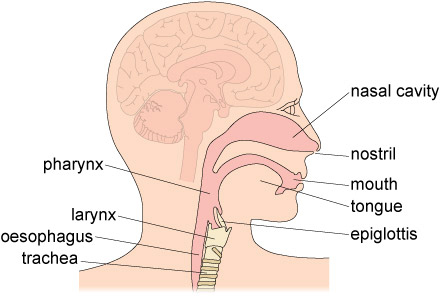
Upper Respiratory Tract
The upper respiratory tract (URT) comprises the nose, nasal cavity, sinuses, pharynx, and larynx, acting as the primary entry point for air, which it filters, warms, and humidifies.
The Nose
The nose is the most prominent anterior structure of the face, serving multiple vital roles.
Functions of the Nose:
- Olfaction (Smell): Houses olfactory receptors.
- Respiration: Provides the primary entry point for air into the respiratory system.
- Air Conditioning: Cleans, warms, and humidifies inspired air.
- Voice Resonance: Contributes to the timbre of the voice.
- Aesthetics: A key determinant of facial appearance.
Divisions: The nose is divided into the external nose and the nasal cavity.
External Nose
This is the visible part of the nose, projecting from the face. Its shape and size vary significantly among individuals due to genetics, sex, and ethnicity.
Key Features:
- Root: The superior attachment of the nose to the forehead.
- Bridge: The superior, bony part of the nose.
- Dorsum Nasi: The anterior border from the root to the apex.
- Apex (Tip): The free, rounded end of the nose.
- Nares (Nostrils): The two external openings of the nasal cavity, separated by the nasal septum.
- Alae Nasi: The flared, cartilaginous expansions that form the lateral boundaries of the nares.
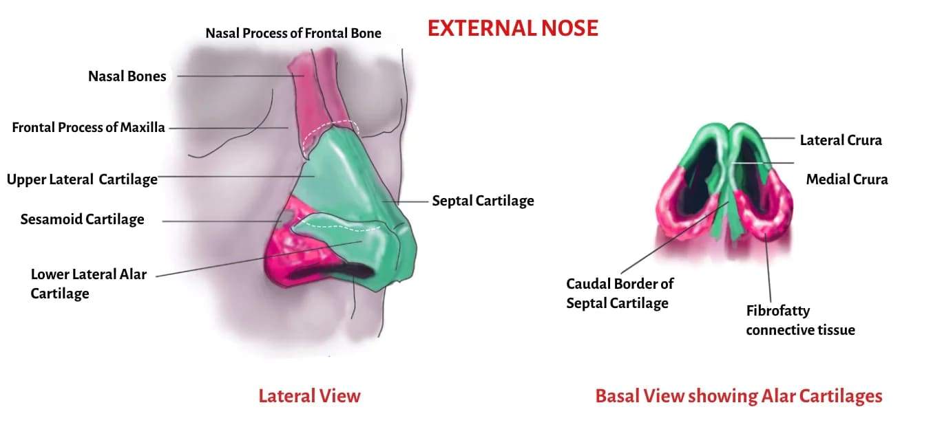
Framework of the External Nose
The external nose is supported by a combination of bone and hyaline cartilage.
Bony Framework (Superior Part - "Bridge"):
- Nasal Bones (paired): Form the superior part of the bridge.
- Frontal Processes of Maxillae (paired): Extend upwards along the sides of the nasal bones.
- Nasal Part of Frontal Bone: Forms the root of the nose.
Cartilaginous Framework (Inferior Part - "Apex and Alae"):
These are plates of hyaline cartilage that provide flexibility and shape.
- Septal Nasal Cartilage: Forms the anterior part of the nasal septum, extending from the perpendicular plate of the ethmoid bone and vomer, maintaining the midline structure.
- Lateral Nasal Cartilages (paired): Located superior to the major alar cartilages, contributing to the side walls of the nose.
- Major Alar Cartilages (paired): Form the apex and alae of the nose. Each has:
- Medial Crus: Forms part of the mobile nasal septum.
- Lateral Crus: Forms the ala of the nose.
- Minor Alar Cartilages (variable): Small, accessory cartilages within the alae.
- Alar Fibrofatty Tissue: Connective tissue and fat that contribute to the shape and flexibility of the alae, especially in the most inferior part.
Summary of Support:
- Bones: Support the upper one-third (bridge).
- Upper Cartilages (Lateral Nasal): Support the sides of the mid-nose.
- Lower Cartilages (Major Alar): Primarily support the tip and help define the shape and patency of the nostrils.
- Skin and connective tissue: Also contribute to the overall shape and covering.
Nasal Cavity
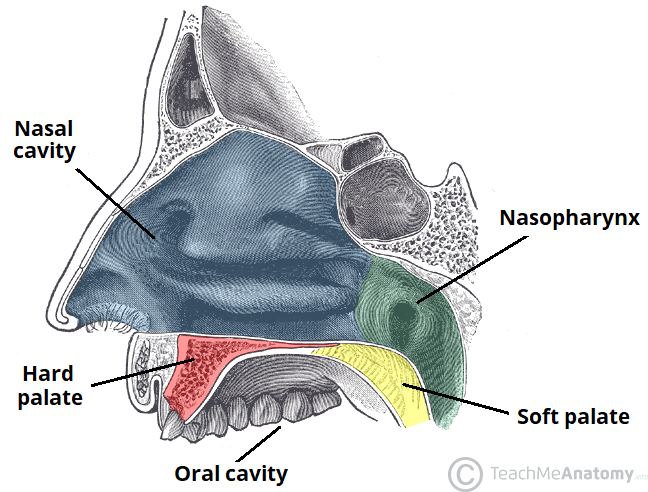
The nasal cavity is the internal space within the external nose, extending posterior to the pharynx.
Boundaries:
- Anteriorly: Communicates with the exterior via the nares (nostrils).
- Posteriorly: Opens into the nasopharynx via the choanae (posterior nasal apertures).
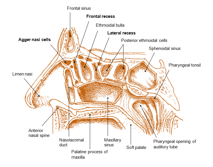
Walls of the Nasal Cavity:
Floor: Formed primarily by the hard palate (maxilla and palatine bones) and, to a lesser extent, the anterior part of the soft palate. This separates the nasal cavity from the oral cavity.
Roof: Narrow and arched, composed of several bones:
- Nasal Bone: Anteriorly.
- Frontal Bone: Anteriorly, between the nasal bones and ethmoid.
- Cribriform Plate of Ethmoid Bone: Mid-portion, perforated by olfactory nerve filaments. This is a critical anatomical landmark as it is thin and can be damaged.
- Body of Sphenoid Bone: Posteriorly.
Medial Wall (Nasal Septum): Divides the nasal cavity into right and left halves. It has both bony and cartilaginous components:
- Bony Parts:
- Perpendicular Plate of Ethmoid Bone: Forms the superior posterior part.
- Vomer: Forms the inferior posterior part.
- Cartilaginous Part:
- Septal Nasal Cartilage: Forms the anterior superior part, making up a significant portion of the septum.
- (Add: "Vomeronasal cartilage" is often a historical or minor finding; focus on the main three components for clarity.)
Lateral Wall: Complex and irregular, characterized by three shelf-like bony projections:
- Superior Nasal Concha (Turbinate): Part of the ethmoid bone.
- Middle Nasal Concha (Turbinate): Part of the ethmoid bone.
- Inferior Nasal Concha (Turbinate): A separate bone, not part of the ethmoid. These conchae increase the surface area of the nasal cavity and create turbulent airflow, facilitating air conditioning.
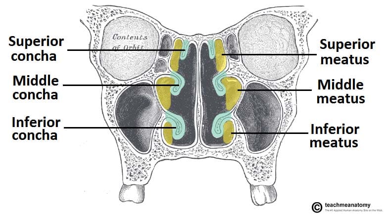
Nasal Meatus (Air Passages):
These are the spaces inferior to each concha.
- Spheno-ethmoidal Recess:
- Location: A small area located postero-superior to the superior nasal concha.
- Opening: Receives the opening of the sphenoidal air sinus.
- Superior Meatus:
- Location: Lies inferior to the superior nasal concha.
- Opening: Receives the opening of the posterior ethmoidal air cells (part of the ethmoid sinus).
- Middle Meatus: This is the most complex and clinically important meatus.
- Location: Lies inferior to the middle nasal concha.
- Key Structures:
- Bulla Ethmoidalis: A prominent bulge on the lateral wall, formed by the expansion of the middle ethmoidal air cells, which open onto or near it.
- Hiatus Semilunaris: A curved, crescent-shaped groove located inferior to the bulla ethmoidalis.
- Ethmoidal Infundibulum: A funnel-shaped channel that extends anteriorly and superiorly from the hiatus semilunaris.
- Openings: The hiatus semilunaris (or directly into the infundibulum) receives the drainage from: Maxillary Sinus, Frontal Sinus, & Anterior Ethmoidal Air Cells.
- Inferior Meatus:
- Location: Lies inferior to the inferior nasal concha.
- Opening: Receives the opening of the nasolacrimal duct, which drains tears from the eye into the nasal cavity.
Clinical Significance: The complex arrangement of conchae and meatuses, particularly the middle meatus, is crucial for understanding sinus drainage and the pathology of sinusitis. Blockage of these openings can lead to infection.
The Palate
The palate forms the roof of the oral cavity and the floor of the nasal cavity, acting as a crucial separator between these two spaces.
Divisions:
Hard Palate (Anterior 2/3):
Bony and immovable.
- Composition: Formed by the horizontal plates of the palatine bones posteriorly and the palatine processes of the maxillae anteriorly. (The term "premaxilla" is often used in developmental contexts for the anterior part of the maxilla forming the primary palate).
- Boundaries: Bounded anteriorly and laterally by the alveolar processes containing the teeth and their associated gingivae (gums).
- Covering: Covered by a thick, keratinized mucous membrane that is firmly attached to the underlying periosteum, making it resilient.
- Continuity: Continuous posteriorly with the soft palate.
Soft Palate (Posterior 1/3):
Fibromuscular and highly mobile. Lacks any bony support.
- Attachments: Attached to the posterior edge of the hard palate.
- Muscles: Contains several muscles that allow for its movement during swallowing, gag reflex, and speech.
- Uvula: A conical, fleshy projection hanging from the free posterior border of the soft palate.
- Functions:
- Swallowing (Deglutition): During swallowing, the soft palate and uvula elevate to close off the nasopharynx, preventing food and liquids from entering the nasal cavity.
- Speech: Modifies the resonance of speech sounds.
A congenital malformation where there is an incomplete fusion of the palatine processes during embryonic development, resulting in an abnormal opening (communication) between the oral and nasal cavities.
Types: Can affect the hard palate, soft palate, or both. Often co-occurs with cleft lip.
Etiology (Causes): Multifactorial, involving both genetic predisposition and environmental factors. Risk factors include:
- Maternal Factors: Advanced maternal age, certain anticonvulsant medications during pregnancy, smoking, alcohol consumption, folate deficiency.
- Genetic Factors: Family history of clefting.
- Sex: Slightly more common in females (though cleft lip +/- palate is more common in males).
- Feeding Difficulties: Infants struggle with creating suction for feeding, leading to poor nutrition and potential aspiration.
- Speech Impairment: Difficulty forming certain sounds due to air escaping through the nose.
- Ear Infections: Increased risk of otitis media due to dysfunction of the Eustachian tube.
- Dental Problems: Misalignment of teeth.
- Psychological/Social: Affects aesthetics and can lead to self-consciousness.
- Respiratory Tract Infections (RTIs): Due to chronic oral breathing and potential for aspiration.
The nasal cavity is lined by two distinct types of mucous membrane, each with specialized functions:
Olfactory Mucous Membrane (Olfactory Epithelium):
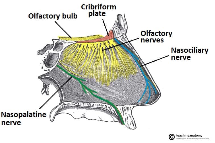
The olfactory mucous membrane (olfactory mucosa) is a specialized, yellowish-brown tissue lining the roof of the nasal cavity, superior conchae, and upper nasal septum. It contains bipolar receptor neurons that detect odors, along with supporting cells and glands, facilitating the sense of smell and serving as a barrier against pathogens.
- Location: Limited to the superior part of the nasal cavity, specifically covering:
- The superior nasal concha.
- The superior part of the nasal septum.
- The roof of the nasal cavity (cribriform plate area) and the spheno-ethmoidal recess itself
- Composition: Contains specialized olfactory receptor neurons (bipolar neurons).
- Function: Responsible for the sense of smell (olfaction).
- Pathway of Olfaction: Olfactory receptor neurons detect chemical odors. Their axons (fila olfactoria) pass superiorly through the small perforations (foramina) in the cribriform plate of the ethmoid bone to synapse with neurons in the olfactory bulb, which is part of the central nervous system located in the anterior cranial fossa.
Respiratory Mucous Membrane (Respiratory Epithelium):
Respiratory epithelium is a specialized ciliated pseudostratified columnar epithelium lining most of the conducting airways (nasal cavity, trachea, bronchi). It acts as a protective barrier, using mucus from goblet cells and mucociliary clearance to trap and expel pathogens/debris. It warms, humidifies, and filters inhaled air.
- Location: Lines the vast majority of the nasal cavity, covering all areas not occupied by the olfactory epithelium (i.e., inferior to the superior concha and olfactory region).
- Composition: Characterized by pseudostratified ciliated columnar epithelium with goblet cells (PCC with GC).
- Functions: Critically important for air conditioning:
- Warming Air: A rich vascular plexus (especially a dense network of veins known as the venous cavernous plexus or Kiesselbach's plexus in the submucosa) warms incoming air.
- Moistening Air: Seromucous glands and abundant goblet cells produce mucus, which adds moisture to the inhaled air.
- Cleaning Air: The sticky mucus traps airborne particles, dust, and pathogens. The cilia on the epithelial cells rhythmically beat, sweeping the contaminated mucus posteriorly towards the nasopharynx, where it is typically swallowed and destroyed by stomach acid. This is known as the mucociliary escalator.
Nerve Supply of the Nasal Cavity
The nasal cavity receives two main types of innervation: The nasal cavity receives sensory input from the olfactory nerve (CN I) for smell, and general sensation (pain, temperature, touch) from the trigeminal nerve (V1 and V2). Autonomic innervation controls mucosa blood flow and secretion via sympathetic (vasoconstriction) and parasympathetic (secretion) fibers.
Special Sensory (Olfaction): Olfactory Nerves (Cranial Nerve I): Responsible for the sense of smell. These fine nerve filaments arise from the olfactory epithelium, pass through the cribriform plate, and synapse in the olfactory bulb.
General Sensation:
- Provides touch, pain, and temperature sensation. Derived from branches of the Trigeminal Nerve (Cranial Nerve V).
- Ophthalmic Division (CN V1) through the Nasociliary Nerve:
- Anterior Ethmoidal Nerve: Supplies the anterior-superior part of the nasal septum and lateral wall, as well as the external nose.
- Posterior Ethmoidal Nerve: (Often overlooked but important) Supplies a small superior posterior part.
- Maxillary Division (CN V2) through the Pterygopalatine Ganglion: This ganglion receives fibers from CN V2 and provides a complex distribution of nerves to the posterior nasal cavity.
- Nasopalatine Nerve: Descends along the nasal septum, supplying the posterior-inferior part of the septum and eventually entering the incisive canal to supply the anterior hard palate.
- Posterior Superior Lateral Nasal Branches: Supply the posterior superior part of the lateral nasal wall and superior and middle conchae.
- Posterior Inferior Lateral Nasal Branches: Supply the posterior inferior part of the lateral nasal wall and inferior concha.
- (Note: The term "nasal" and "palatine" branches from the pterygopalatine ganglion are correct but more precisely broken down as above.)
Autonomic Innervation:
- Parasympathetic Fibers: Originate from the facial nerve (CN VII), travel via the greater petrosal nerve to the pterygopalatine ganglion. Postganglionic fibers then distribute with CN V2 branches to the nasal glands, causing vasodilation and increased mucous secretion (e.g., rhinorrhea).
- Sympathetic Fibers: Originate from the superior cervical ganglion. Postganglionic fibers also reach the pterygopalatine ganglion but pass through it without synapsing. They then distribute to nasal blood vessels, causing vasoconstriction (e.g., in response to cold or decongestants).
Blood Supply of the Nasal Cavity
The nasal cavity has a very rich and anastomosing blood supply, which is essential for warming air but also makes it prone to bleeding (epistaxis).
Arterial Supply: Primarily from branches of the Internal Carotid Artery and the External Carotid Artery.
From the Ophthalmic Artery (a branch of the Internal Carotid Artery):
- Anterior Ethmoidal Artery: Supplies the anterior-superior part of the lateral wall and septum.
- Posterior Ethmoidal Artery: Supplies the posterior-superior part of the lateral wall and septum.
From the Maxillary Artery (a terminal branch of the External Carotid Artery):
- Sphenopalatine Artery: This is the major arterial supply to the nasal cavity. It enters through the sphenopalatine foramen and branches extensively to supply the posterior-inferior part of the lateral wall and septum. It is often called the "artery of epistaxis."
- Greater Palatine Artery: Contributes some supply to the posterior inferior septum.
From the Facial Artery (a branch of the External Carotid Artery):
- Superior Labial Artery: Gives off a septal branch that contributes to the supply of the anterior part of the septum.
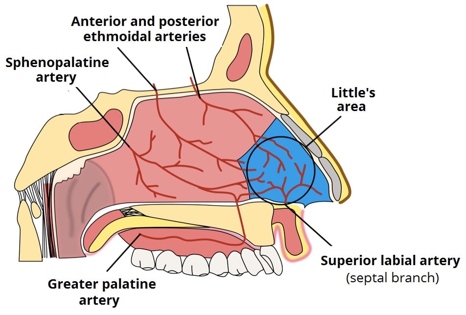
Location: A highly vascularized area on the anterior-inferior part of the nasal septum.
Composition: It's an anastomotic plexus formed by the convergence of branches from all five major arteries supplying the nasal septum:
- Anterior Ethmoidal Artery
- Posterior Ethmoidal Artery
- Sphenopalatine Artery
- Greater Palatine Artery
- Superior Labial Artery
Venous Drainage:
- Veins generally accompany the arteries and form a rich submucosal venous plexus.
- Blood drains posteriorly into the sphenopalatine vein (which leads to the pterygoid venous plexus) and anteriorly into the facial vein.
- Ethmoidal veins drain into the ophthalmic veins.
- Clinical note: Connections to the cavernous sinus via ophthalmic veins are important, as infections in the nasal cavity can potentially spread intracranially.

Lymphatic Drainage of the Nasal Cavity
The lymphatic drainage of the nasal cavity follows a pattern that reflects its anterior and posterior regions.
Anterior Nasal Cavity (including the Nasal Vestibule): Lymphatics drain into the submandibular lymph nodes.
Posterior Nasal Cavity (the larger part of the nasal cavity): Lymphatics drain primarily into the retropharyngeal lymph nodes (often then to deep cervical nodes) and the deep cervical lymph nodes (particularly the upper group).
- Clinical Significance: Understanding lymphatic drainage is for tracking the spread of infections or malignancies originating in the nasal cavity.
Paranasal Sinuses
The paranasal sinuses are air-filled, mucosa-lined extensions of the respiratory part of the nasal cavity that invaginate into four surrounding cranial bones: the frontal, ethmoid, sphenoid, and maxillary bones.
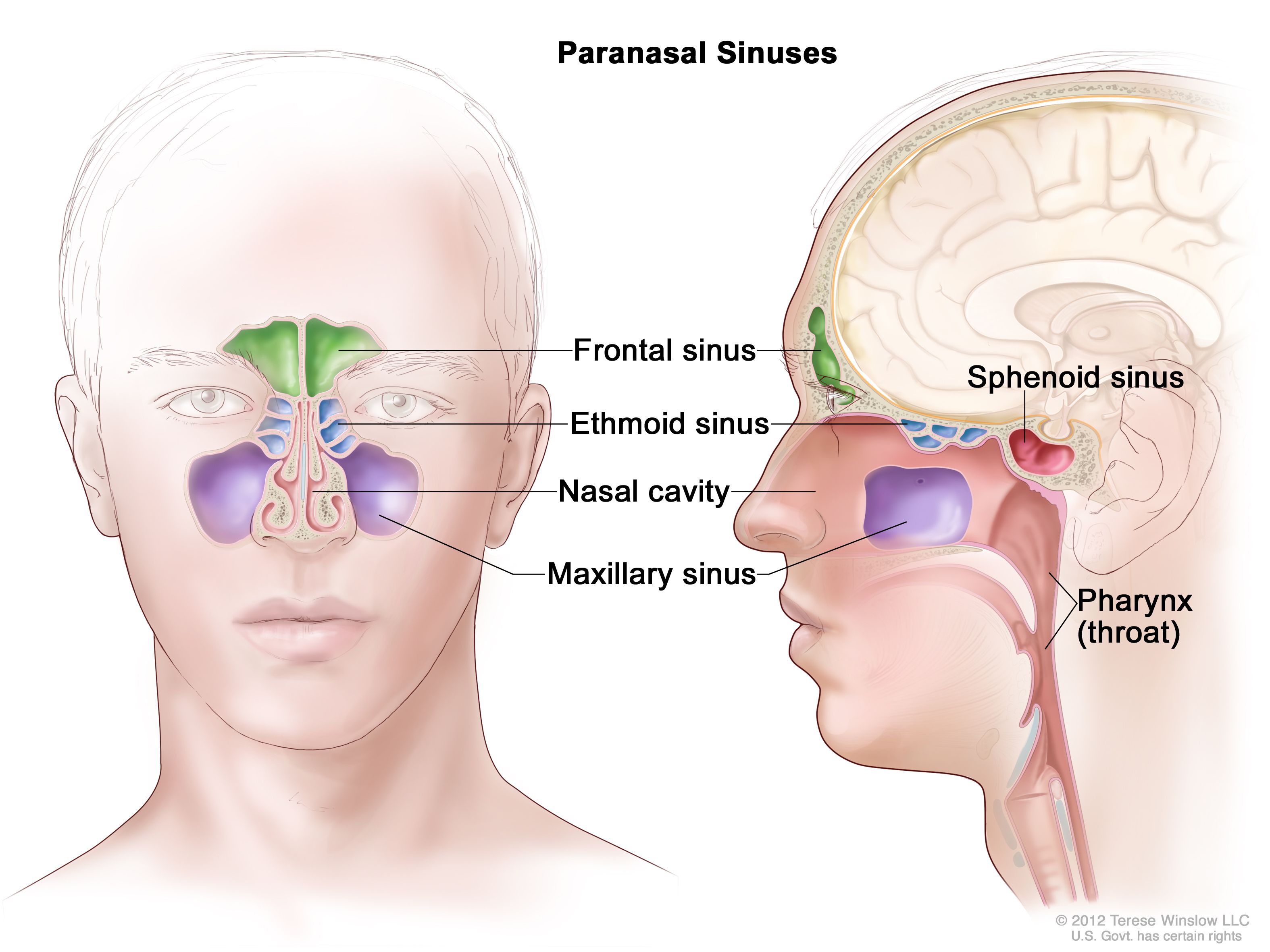
- Development: These sinuses begin to develop in fetal life but continue to expand (pneumatize) into the surrounding bones throughout childhood and even into adulthood. This expansion is more pronounced in older individuals.
- Lining: They are lined with mucoperiosteum, which is a specialized mucous membrane (respiratory epithelium: pseudostratified ciliated columnar epithelium with goblet cells) that is intimately adhered to the underlying periosteum of the bone. This continuity of the mucosal lining means that infections from the nasal cavity can easily spread to the sinuses.
Functions of Paranasal Sinuses
- Air Conditioning: The extensive mucosal lining contributes to the warming and humidifying of inspired air.
- Mucus Drainage (Mucociliary Clearance): The cilia on the columnar cells of the mucoperiosteum continuously beat, moving mucus (which traps particulate matter and pathogens) towards the openings (ostia) of the sinuses, from where it drains into the nasal cavity.
- Voice Resonance: They act as resonating chambers, influencing the timbre and quality of the voice. Blockage or fluid accumulation within the sinuses can significantly alter voice quality (e.g., a "nasal" or "muffled" sound).
- Lightening the Skull: Being air-filled spaces, they reduce the overall weight of the skull, which makes it easier for neck muscles to support the head.
- Protection: They may play a role in cranial protection by absorbing forces during facial trauma, acting as "crumple zones."
- Immune Defense: The mucus and immune cells within the mucosa contribute to local defense against pathogens.
Frontal Sinuses
Location: Situated within the frontal bone, between its outer and inner cortical tables. They are typically found posterior to the superciliary arches (brow ridges) and the root of the nose.
Structure:
- Two sinuses, right and left, separated by a bony septum (which is rarely in the median plane).
- They are often asymmetrical and vary significantly in size and shape among individuals.
- Each sinus is roughly triangular.
Development: Begin to pneumatize during childhood and are radiographically detectable around 6-7 years of age, reaching full size in late adolescence.
Drainage: Each frontal sinus drains inferiorly through the frontonasal duct (or nasofrontal duct). This duct empties into the ethmoidal infundibulum, which in turn opens into the semilunar hiatus of the middle nasal meatus.
Innervation: Sensory innervation is primarily provided by branches of the supraorbital nerves, which are derived from the ophthalmic division of the trigeminal nerve (CN V1). This explains referred pain from frontal sinusitis to the forehead.
Variations of the Frontal Sinuses
- Asymmetry: The right and left frontal sinuses are rarely equal in size, and their separating septum is often deviated from the midline.
- Size: They can range from very small to extensively large, sometimes extending laterally into the greater wings of the sphenoid bone or superiorly towards the parietal bone.
- Compartmentalization: A frontal sinus may have two parts: a vertical component in the squamous part of the frontal bone and a horizontal component in the orbital part.
- Clinical Significance of Large Sinuses: When the orbital part is large, its roof forms part of the floor of the anterior cranial fossa, and its floor forms the roof of the orbit. This proximity is clinically important during surgery or in cases of severe infection.
Ethmoidal Sinuses (Ethmoidal Air Cells)
- Location: These are not single large sinuses but a collection of multiple, small, interconnected air cells located within the ethmoid bone. They lie between the nasal cavity medially and the orbit laterally.
- Development: Present at birth but continue to grow. They are generally not well visualized on plain radiographs before 2 years of age but are readily identifiable on CT scans.
- Grouping and Drainage: They are typically divided into three main groups based on their drainage patterns:
- Anterior Ethmoidal Cells:
- Drainage: Drain directly or indirectly into the ethmoidal infundibulum, which then opens into the middle nasal meatus (via the semilunar hiatus).
- Middle Ethmoidal Cells:
- Drainage: Typically open directly onto the middle nasal meatus, often forming the prominent bulge known as the bulla ethmoidalis on the superior border of the semilunar hiatus.
- Posterior Ethmoidal Cells:
- Drainage: Open directly into the superior nasal meatus.
- Anterior Ethmoidal Cells:
- Innervation: Sensory innervation is provided by the anterior and posterior ethmoidal branches of the nasociliary nerve (a branch of the ophthalmic division of the trigeminal nerve, CN V1).
Vulnerability: The ethmoidal cells have thin bony walls, especially medially (lamina papyracea of the orbit) and superiorly (cribriform plate).
Spread of Infection:
- Orbital Complications: Infections can easily erode through the fragile medial wall of the orbit, leading to serious complications such as orbital cellulitis or even orbital abscess.
- Visual Impairment: Some posterior ethmoidal cells are in close proximity to the optic canal, which transmits the optic nerve (CN II) and ophthalmic artery. Spread of infection or inflammation here can compress the optic nerve, potentially causing optic neuritis or even blindness.
- Intracranial Spread: Infection can also spread superiorly through the cribriform plate to the anterior cranial fossa, leading to meningitis or brain abscess, though this is less common than orbital spread.
Sphenoid Sinuses
- Location: Situated within the body of the sphenoid bone, often extending into its greater wings and pterygoid processes.
- Development: They begin to pneumatize around 2-3 years of age, often originating from a posterior ethmoidal cell that invades the sphenoid bone. They are fully developed by late adolescence.
- Structure:
- Usually two sinuses, separated by a bony septum, which is frequently asymmetrical.
- The extensive pneumatization makes the body of the sphenoid bone relatively thin and fragile.
- Drainage: Each sphenoid sinus drains into the spheno-ethmoidal recess, which is located superior and posterior to the superior nasal concha.
- Innervation: Sensory innervation is primarily from the posterior ethmoidal nerve (a branch of the ophthalmic division of CN V1) and branches from the maxillary nerve (CN V2) (specifically, the pharyngeal branch).
- Arterial Supply: Primarily by the posterior ethmoidal arteries and branches from the maxillary artery.
The thin walls of the sphenoid sinuses place them in close proximity to numerous critical neurovascular structures, making sphenoid sinusitis or trauma particularly dangerous:
- Superiorly:
- Optic Nerves (CN II) and Optic Chiasm: Inflammation or infection can affect vision.
- Pituitary Gland: Located in the sella turcica, directly superior to the sinus. This proximity allows for a transsphenoidal surgical approach to the pituitary gland, minimizing external incisions.
- Laterally:
- Cavernous Sinuses: Containing important cranial nerves (III, IV, V1, V2, VI) and the inter[Truncated]
Lesson map
Use this map to move through the lesson in order:
Further reading
For deeper study, compare this lesson with current guidance and standard references from recognised medical and public-health organisations.