Neoplasia
Learning Objectives
- Epidemiology
- Definition of Neoplasia
- Benign and Malignant Neoplasms
- Nomenclature
- Characteristics of Benign and Malignant Tumors
Cancer Statistics and Trends, Uganda 2013 – 2017
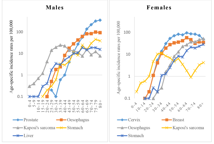
Cancer statistics and trends, Uganda 2013 - 2017
Definition of Neoplasia
- Etymology: Neoplasia means “new growth”.
- It is a clonal proliferation of cells that have escaped from normal growth control mechanisms as a result of the acquisition of genetic aberrations.
- It is the uncontrolled disorderly proliferation of cells, resulting in a benign or malignant tumor or neoplasm.
- Every cell within a given neoplasm shares a set of pathogenetic mutations, hence neoplasms are composed of genetically related “daughter” cells derived from a single aberrant founding cell.
- Because of their origin from a single progenitor cell, neoplastic cells are said to be clonal in origin.
Mutations
- The mutations that cause neoplasia alter gene function that regulates cell growth & survival, therefore conferring the most striking property of neoplasms.
- Tumor formation: Their ability to produce a localized mass referred to as a tumor (from the Latin word tumere, to swell).
- Oncology: The study of tumors is called oncology (from the Greek word oncos, “tumor” and logos, “study of”).
- The mutations that cause neoplasms are usually acquired but also may be inherited.
Hyperplasia
- In contrast to neoplasia, hyperplasia occurs when many cells within affected tissues begin to proliferate simultaneously in response to physiologic or pathophysiologic growth signals; as a result, hyperplasias are polyclonal.
- Examples of hyperplasias:
- Enlargements of lymphoid tissues caused by inflammatory mediators.
- Enlargement of the uterus in response to gestational hormones during pregnancy.
- Unlike neoplasias, hyperplasias are reversible. The lymph node returns to its normal size when the infection is cleared and the uterus returns to its prior state after delivery of the fetus.
| Feature | Hyperplasia | Neoplasia |
|---|---|---|
| Clonality | Polyclonal (simultaneous response of diverse cell lineages) | Monoclonal (descends from one mutated founder cell) |
| Trigger Dependence | Dependent on sustained physiologic or pathologic stimulus | Autonomous; continues despite stimulus removal |
| Reversibility | Reversible upon resolution of inciting signal | Irreversible progressive cellular growth |
Dysplasia
- Is disordered growth of epithelial cells that are abnormal but not malignant.
- It is important to recognize because it is a well-documented precursor of malignancy in many tissues such as:
- Cervix
- Endometrium
- Gastrointestinal tract
Features Used to Assess Dysplasia
- Cellular & Nuclear Pleomorphism: Typically abnormally large, hyperchromatic nuclei.
- Abnormal Mitotic Activity: Including more numerous mitotic figures and mitoses in superficial layers of stratified epithelium where they are not normally seen.
- Architectural Disarray:
- Partial or complete loss of the usual progressive maturation of cells in stratified squamous epithelium or transitional epithelium in bladder, resulting in a disorganized arrangement with dark-staining basal-appearing cells.
- When dysplastic changes are severe & involve the entire thickness of the epithelium, the lesion is referred to as carcinoma in situ, a preinvasive stage of cancer.
- Shown histopathologically is the disorderly maturation and spatial arrangement of cells, marked variability in nuclear size and shape (pleomorphism), and increased, often abnormal mitosis.
- Examples: Dysplasia occurring in the squamous epithelium of the uterine cervix.
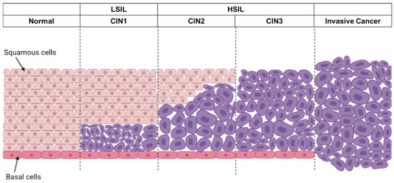
Squamous Intraepithelial Lesion (SIL)
Carcinoma in Situ
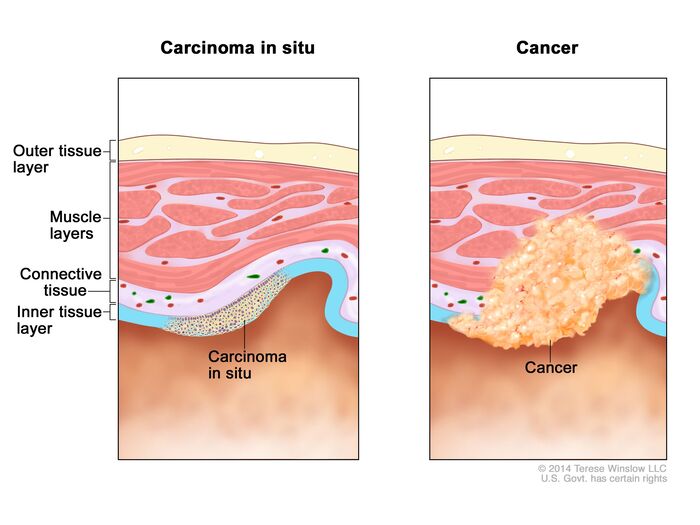
Carcinoma in situ
Carcinoma in Situ vs. Microinvasive Carcinoma
Carcinoma in situ represents severe dysplasia involving 100% of the epithelial thickness with zero breach of the basement membrane. Because lymphatics and capillaries reside in the subepithelial stroma, carcinoma in situ possesses zero metastatic potential until the basement membrane is physically penetrated.
Differentiation of Neoplasm
- Definition: Refers to the extent to which tumor cells resemble their parenchymal cells of origin both morphologically and functionally.
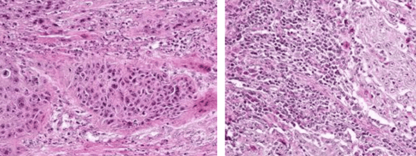
- Well-differentiated: If the resemblance to the tissue of origin is close.
- Poorly differentiated: If little resemblance to the tissue of origin is seen.
- Behavioral consequence: Neoplasms grow at the expense of function and vitality of normal tissue without benefit to the host and are largely independent of host control mechanisms.
- Benign neoplasms: Composed of well-differentiated cells that closely resemble their normal counterparts.
- Malignant neoplasms: Most exhibit morphologic alterations and many are undifferentiated.
- Anaplasia: Tumors composed of undifferentiated cells are said to be anaplastic, a feature which is a reliable indicator of malignancy.
- Anaplasia literally means “backward formation”, implying dedifferentiation or loss of structural & functional characteristics of differentiated cells.
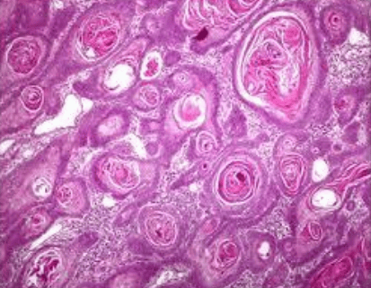
Neoplasia: Well differentiated squamous cell carcinoma
Moderately & Poorly differentiated squamous cell carcinoma
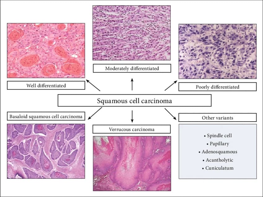
Squamous cell carcinoma morphological variants
Anaplastic Cells
Anaplastic cells display one or more of the following classical cytological hallmarks:
- Pleomorphism (variation in cell size & shape): Examples include tumor giant cells that are considerably larger than their neighbors, with each possessing either one enormous nucleus or several nuclei.
- Nuclear Abnormalities:
- Hyperchromatism (dark staining of chromatin due to abundant DNA).
- Variation in nuclear size & shape.
- Unusually prominent single or multiple nucleoli.
- Enlargement of nuclei resulting in an increased nuclear-to-cytoplasmic ratio (approaching 1:1, compared to the normal 1:4 or 1:6).
- Nucleoli may attain astounding sizes that approach the diameter of normal lymphocytes.
- Atypical Mitoses: Which may be numerous. Abnormal separation of chromatids during cell division may produce tripolar or quadripolar mitotic figures.
- Loss of Polarity: Groups of neoplastic cells lack normal patterns of orientation to one another. In most anaplastic tumors, cells grow in disorganized sheets, with total loss of organized structures such as glands or stratified squamous architecture.
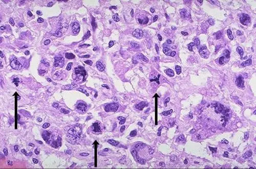
High power detail view of anaplastic tumor cells shows cellular & nuclear variation in size & shape. The prominent cell in the center field has an abnormal tripolar spindle
Functional Differentiation and Paraneoplastic Syndromes
- Functional retention in well-differentiated tumors: Most retain the function of their normal counterparts, whereas anaplastic tumors are much less likely to do so.
- Benign neoplasms & well-differentiated cancers of endocrine glands frequently produce the hormones of the cell of origin.
- Well-differentiated squamous cell carcinomas produce keratin.
- Well-differentiated hepatocellular carcinomas produce bile.
- Emergence of unanticipated functions: Cancers of nonendocrine origin may cause signs & symptoms by secreting ectopic hormones.
- Adrenocorticotrophic hormone (ACTH) (e.g., small cell carcinoma of the lung causing Cushing syndrome)
- Parathyroid hormone-related protein (PTHrP) (e.g., squamous cell lung carcinoma causing hypercalcemia)
- Insulin
- Glucagon, and others.
- These clinical manifestations constitute the paraneoplastic syndromes.
Classification & Nomenclature of Tumors
Neoplasms are classified as either benign or malignant, based on their behavior. They are also described by terms derived from the appearance of the neoplasm, tissue of origin, or degree of differentiation.
A. Malignant Tumors (Cancer)
- General Considerations:
- Invasion (spread of neoplasm into adjacent tissues) and metastasis (implantation of the neoplasm into non-contiguous sites) are characteristic.
- Metastasis is the most defining characteristic of malignancy, although there are some malignant tumors such as basal cell carcinoma of the skin that rarely metastasize.
- Malignant tumors are usually less differentiated than benign tumors.
- In anaplasia, tumors are poorly differentiated; there is pleomorphism (different cellular shapes & sizes), hyperchromatism (dark staining nuclei), increased nuclear-cytoplasmic ratio, abnormal mitoses, cellular dyspolarity, and prominent nucleoli.
- Highly anaplastic tumors are very aggressive, and well-differentiated tumors are less aggressive.
- Therapeutic Paradox: Paradoxically, the most aggressive tumors often respond well to chemotherapy & radiotherapy because these treatment modalities are most effective against rapidly dividing cells.
- Carcinoma: Malignant tumor of epithelial origin.
- Squamous cell carcinoma: Originates from stratified squamous epithelium (e.g., skin, mouth, esophagus, vagina) as well as areas of squamous metaplasia (e.g., bronchi or squamocolumnar junction of the uterine cervix). It is marked by the production of keratin.
- Transitional cell carcinoma: Arises from the transitional cell epithelium (urothelium) of the urinary tract.
- Adenocarcinoma: Carcinoma of glandular epithelium; includes malignant tumors of gastrointestinal mucosa, endometrium, and pancreas. It is frequently associated with desmoplasia (tumor-induced proliferation of non-neoplastic connective tissue/stroma), especially in adenocarcinoma of the breast, pancreas, and prostate.
- Sarcoma: Malignant tumor of mesenchymal origin. Named using a tissue-specific prefix:
- Osteosarcoma → Bone
- Rhabdomyosarcoma → Skeletal muscle
- Leiomyosarcoma → Smooth muscle
- Liposarcoma → Fatty tissue
- Eponymic Tumors: Named after the physicians who originally characterized them:
- Burkitt lymphoma
- Hodgkin disease
- Wilms tumor (nephroblastoma)
- Teratoma: A neoplasm derived from all three germ cell layers (ectoderm, mesoderm, endoderm). May contain structures such as skin, bone, cartilage, teeth, and intestinal epithelium. It may be benign (mature) or malignant (immature), and usually arises in the ovaries or testes.
B. Benign Tumors
- General Considerations:
- Benign tumors are usually well-differentiated and closely resemble the tissue of origin.
- They grow slowly and do not metastasize.
- If their growth compresses adjacent critical tissues, they can be harmful; for example, benign intracranial tumors (e.g., meningioma compressing the brainstem) can be more lethal than some malignant skin tumors.
- They tend to be encapsulated by a rim of compressed fibrous connective tissue.
- Nomenclature Suffix “–oma”: Typically denoted by the suffix –oma attached to the cell of origin (e.g., lipoma, fibroma).
Malignant Neoplasms Masquerading with the “-oma” Suffix
Although the suffix –oma typically indicates a benign tumor, several deadly malignancies and non-neoplastic conditions carry this suffix:
- Malignant tumors ending in –oma: Melanoma, Lymphoma, Seminoma, Hepatoma (Hepatocellular carcinoma), and Mesothelioma.
- Non-neoplastic swellings ending in –oma: Granuloma (focal aggregate of chronic inflammatory macrophages) and Hematoma (extravasated mass of blood).
- Papilloma: Arises from surface epithelium such as squamous epithelium of the skin, larynx, or tongue; may also develop from transitional epithelium of the urinary bladder, ureter, and renal pelvis. It consists of delicate finger-like epithelial processes overlying an axis of vascularized connective tissue stroma.
- Adenoma: Benign epithelial neoplasm of glandular origin. Occurs in several variants:
- Papillary cystadenoma: Characterized by adenomatous papillary processes projecting into cystic spaces, as observed in cystadenoma of the ovary.
- Fibroadenoma: Marked by proliferation of connective tissue surrounding glandular epithelium (e.g., fibroadenoma of the breast). In this instance, either the epithelial or the stromal elements may represent the primary proliferating component.
- Benign Tumors of Mesenchymal Origin: Most often designated by attaching “–oma” to the tissue of origin:
- Leiomyoma (smooth muscle) — includes the most common neoplasm in women: uterine leiomyoma (fibroid tumor)
- Rhabdomyoma (skeletal muscle)
- Lipoma (adipose tissue)
- Fibroma (fibrous tissue)
- Chondroma (cartilaginous tissue)
- Choristoma: A small non-neoplastic area of normal tissue misplaced within another organ (heterotopic rest), such as pancreatic tissue within the wall of the stomach or duodenum.
- Hamartoma: A non-neoplastic, disorganized tumor-like overgrowth of cell types regularly found within that specific organ; for example, a hemangioma (an irregular accumulation of blood vessels) or pulmonary chondroid hamartoma.
Hamartoma vs. Choristoma
Hamartoma = Home Tissue: Disorganized overgrowth of mature native tissue elements indigenous to that exact site.
Choristoma = Cleaved / Chopped away: Normal, mature tissue residing in an abnormal, foreign anatomical site.
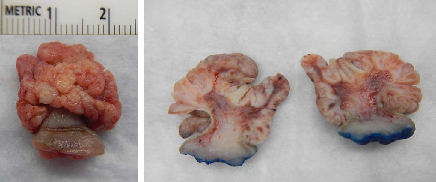
Squamous cell papilloma
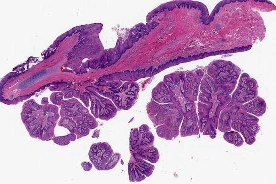
Microscopic image: squamous cell papilloma
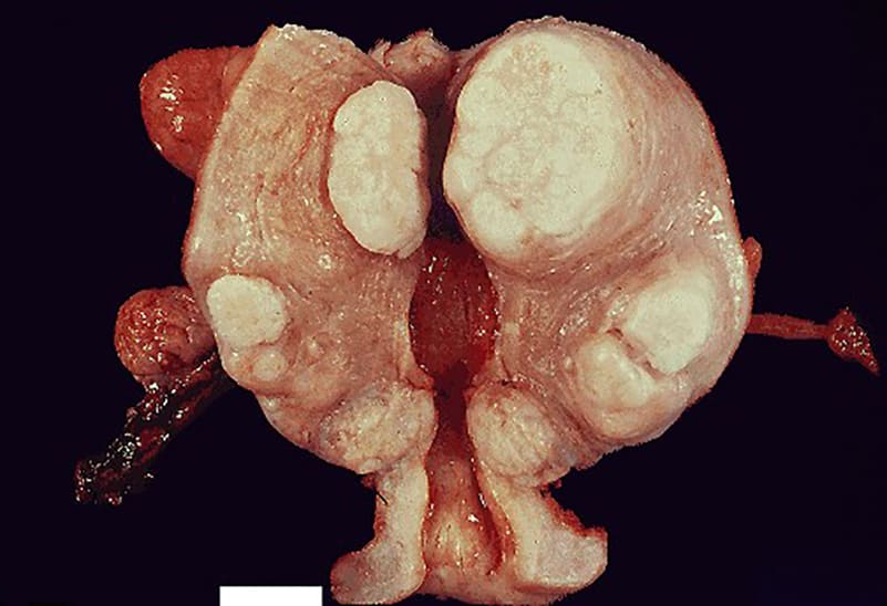
Uterine leiomyomas (fibroids)
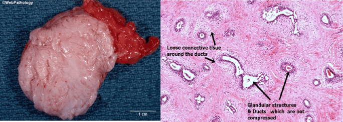
Fibroadenoma of the breast; gross & microscopic images
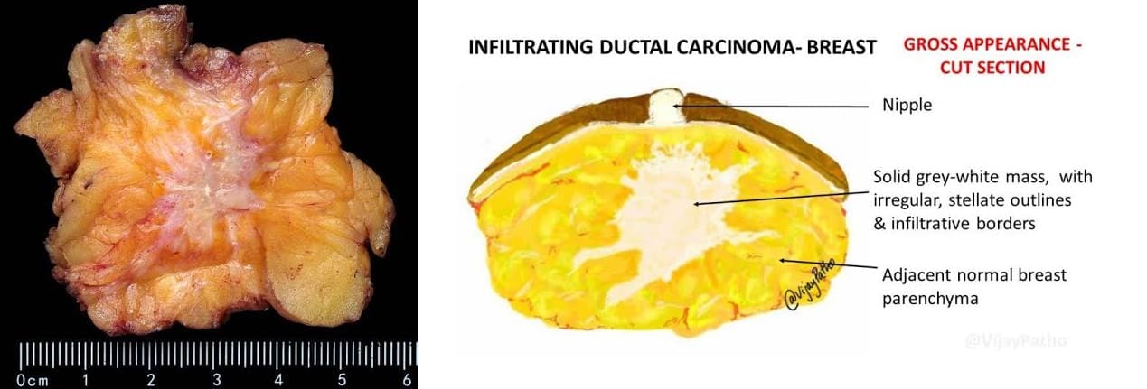
Invasive ductal carcinoma of the breast
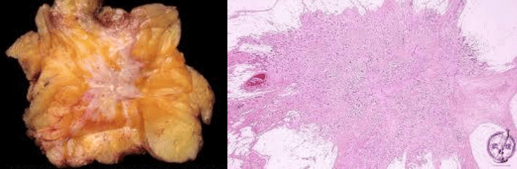
Invasive ductal carcinoma of the breast
Master Classification Table of Human Tumors
| Tissue of Origin | Benign Neoplasm | Malignant Neoplasm |
|---|---|---|
| I. Composed of One Parenchymal Cell Type | ||
| Connective tissue & derivatives: • Adipose tissue • Cartilage • Bone |
Lipoma Chondroma Osteoma |
Liposarcoma Chondrosarcoma Osteosarcoma |
| Blood vessels | Hemangioma | Angiosarcoma |
| Mesothelium | — | Mesothelioma |
| Brain coverings | Meningioma | Invasive meningioma |
| Epithelial lining of glands or ducts | Adenoma Papilloma Cystadenoma |
Adenocarcinoma |
| Liver cells | Hepatic adenoma | Hepatocellular carcinoma |
| Urinary tract epithelium (transitional) | Urothelial papilloma | Urothelial carcinoma |
| Placental epithelium | Hydatidiform mole | Choriocarcinoma |
| Testicular epithelium (germ cells) | — | Seminoma Embryonal carcinoma |
| Tumors of melanocytes | Nevus | Malignant melanoma |
| II. Composed of More Than One Neoplastic Cell Type: Usually Derived from One Germ Cell Layer (Mixed Tumors) | ||
| Salivary gland | Pleomorphic adenoma (mixed tumor of salivary gland) | Malignant mixed tumor of salivary gland |
| Renal anlage | — | Wilms tumor |
| III. More Than One Neoplastic Cell Type Derived from More Than One Germ Cell Layer (Teratogenesis) | ||
| Totipotential cells in gonads or embryonic rests | Mature teratoma, dermoid cyst | Immature teratoma, teratocarcinoma |
Properties of Neoplasms
- Monoclonality
- Invasion & Metastasis
- Other Clinical Manifestations of Malignancies
- Paraneoplastic Syndromes
A. Monoclonality
Definition: Monoclonality means that the origin of the neoplasm is from a single precursor cell (or a single clone).
- Most neoplasms are monoclonal. Polyclonal cellular proliferations are almost always non-neoplastic.
- Monoclonality is assessed by a variety of methods using isoenzyme patterns or other markers of clonality:
- Monoclonal tumors express only one of the isoenzymes or other markers of clonality.
- Polyclonal cellular proliferations exhibit both isoenzymes or markers.
- Glucose-6-Phosphate Dehydrogenase (G6PD) & Other X-Linked Markers:
- These were the first described & classic indicators of monoclonality in humans.
- Because of lyonization (random X-inactivation) in early embryonic life, tissues of females heterozygous for G6PD isoenzymes consist of a mosaic of all cell types, with random cells expressing one or the other of the two isoenzymes.
- Limitation: In the case of G6PD, one of the two polymorphic forms of these isoenzymes occurs with significant frequency only in women of African lineage, limiting the widespread clinical application of this technique.
- Other polymorphic X-linked markers such as iduronate-2-sulphate and phosphoglycerate kinase can be utilized across a broader spectrum of patients.
- Human Androgen Receptor Gene (HUMARA):
- This gene has now become the most common marker to determine monoclonality.
- This method evaluates methylation patterns adjacent to highly polymorphic short tandem repeats on the active vs. inactive X chromosome across diverse populations.
- Specific Translocations: If present, cytogenetic rearrangements such as the t(8;14) translocation in Burkitt lymphoma can be directly utilized to assess clonality.
- Monoclonality of Cells of Lymphoid Origin:
- Malignancies of B-cell Origin:
- Immunoglobulin Light Chain Restriction: Immunoglobulins are produced by B-cell malignant tumors and are demonstrable as surface or cytoplasmic immunoglobulins or, in the case of multiple myeloma, are secreted into the serum.
- If monoclonal: The resultant population of immunoglobulin molecules exhibits either kappa (κ) or lambda (λ) light chain specificity, but never both (normal κ:λ ratio of 3:1 is severely skewed).
- If polyclonal: Reactive B-cell or plasma cell proliferations yield heterogeneous immunoglobulins expressing a mixed distribution of both kappa and lambda light chains.
- Immunoglobulin Gene Rearrangement: Unique V(D)J somatic recombination is characteristic of normal B-cell maturation. While each normal B cell displays a unique rearrangement sequence, neoplastic proliferations produce an enormous population of cells all demonstrating the identical pattern of immunoglobulin gene rearrangement, proving clonal origin from a single progenitor cell.
- Immunoglobulin Light Chain Restriction: Immunoglobulins are produced by B-cell malignant tumors and are demonstrable as surface or cytoplasmic immunoglobulins or, in the case of multiple myeloma, are secreted into the serum.
- Malignancies of T-cell Origin:
- Surface Antigens (Markers): Expressed during T-cell maturation reflecting differentiation stages or functional subclasses. Clonal expansions share identical surface immunophenotypic profiles (e.g., predominant CD4 helper vs. CD8 cytotoxic marker expression).
- T-Cell Receptor (TCR) Gene Rearrangement: Analogous to immunoglobulin gene rearrangement; serves as a definitive molecular tool to demonstrate both the T-cell lineage of a tumor and its monoclonal origin.
- Malignancies of B-cell Origin:
B. Invasion & Metastasis
Local Invasion
- Definition: Invasion is the aggressive infiltration of adjacent tissues by a malignant tumor.
- It distinguishes benign from malignant tumors.
- Benign tumor growth: Benign tumors grow slowly and expand by pushing; most develop a rim of condensed fibrous tissue called a capsule.
- The capsule is formed by deposition of collagen by host stromal cells, such as fibroblasts.
- Benign encapsulated tumors are discrete, movable (non-fixed to underlying fascia), and usually easy to enucleate or excise surgically.
- Invasive cancer behavior:
- Invasive cancers can also induce an intense stromal response associated with fibrosis (desmoplasia); thus, slowly growing malignant tumors on naked-eye (gross) inspection may falsely appear to be encapsulated.
- However, cancers, even those that appear circumscribed, lack true histological capsules and progressively invade, infiltrate, and destroy surrounding tissues.
- This infiltrative growth pattern necessitates the surgical removal of a wide margin of surrounding normal tissue during radical excision.
- Exceptions to the capsule rule: These distinguishing features are not absolute; certain benign tumors such as hemangiomas completely lack fibrous capsules and interdigitate irregularly with adjacent normal tissues.
- Vascular Invasion vs. Metastasis:
- Local invasion often extends into lymphatics and blood vessels, resulting in the shedding of tumor emboli that can travel to distant anatomical sites.
- Critical Pathological Principle: Not all tumor emboli result in metastatic tumor implants; the presence of tumor cells within blood vessels or lymphatics indicates only the penetration of basement membranes and is not synonymous with metastasis.
- Metastasis strictly requires extravasation, colonization, and implantation at distant non-contiguous sites.
Metastasis
- Definition: The spread of a tumor to sites that are physically discontinuous with the primary tumor.
- The ability to metastasize unequivocally marks a tumor as malignant.
- The invasive phenotype of cancer cells permits penetration into blood vessels, lymphatic channels, and body cavities, facilitating systemic dissemination.
- Clinical Epidemiology at Diagnosis: Approximately 30% of patients with newly diagnosed non-skin solid malignancies (excluding non-melanoma skin cancer) present with clinically evident metastases, and an additional 20% harbor occult (microscopic/hidden) metastases at initial diagnosis.
- Tumor Size vs. Metastatic Risk: While large, poorly differentiated anaplastic tumors are statistically more prone to metastasize, extremely small occult primary tumors may present with widespread metastatic disease.
- Invasion Without Metastasis: Certain aggressive malignancies, such as basal cell carcinoma of the skin and primary gliomas of the central nervous system, are highly destructive and locally invasive but rarely metastasize. Thus, local invasiveness is not universally predictive of metastatic capability.
Multistep Process of Metastasis
Systemic metastatic dissemination proceeds through a defined sequence of biological cascades:
- Growth & vascularization of the primary tumor (sustained neoangiogenesis).
- Invasiveness & penetration of basement membranes into local lymphatics or blood vessels (intravasation).
- Transport & survival of tumor cells in the circulation (evasion of immune surveillance, mechanical shear stress, and aggregation with host platelets to form tumor emboli).
- Arrest of tumor emboli in the target tissue & passage again across basement membranes (adhesion to vascular endothelium, extravasation into parenchyma).
- Overcoming of target tissue local defense mechanisms.
- Development of successive metastatic implants (micrometastatic colony outgrowth supported by secondary angiogenesis).
Preferential Routes of Metastasis
- Routes of dissemination vary by cellular histogenesis:
- Carcinomas tend to metastasize predominantly via lymphatic spread to regional draining lymph node basins.
- Sarcomas tend to invade thin-walled venules early, resulting in widespread blood-borne (hematogenous) dissemination.
- Notable Carcinoma Exceptions: Certain epithelial carcinomas violate this rule and preferentially demonstrate early venous invasion with hematogenous spread:
- Renal cell carcinoma (propensity to invade the renal vein and inferior vena cava)
- Hepatocellular carcinoma (propensity to invade hepatic and portal venules)
- Target Organs of Hematogenous Metastasis: Most commonly involve organs receiving massive capillary blood flow:
- Liver (portal drainage filter)
- Lungs (systemic venous drainage filter)
- Brain
- Adrenal glands
- Lymph nodes
- Bone marrow
- Rare Sites: Metastatic colonization rarely occurs in skeletal muscle or the spleen despite their massive perfusion, owing to unfavorable microenvironmental factors (lactic acid turnover, mechanical contractions, and splenic macrophage surveillance).
Carcinomas Spreading Hematogenously ("Four Carcinomas Route Hematogenously")
While carcinomas classically favor lymphatics, remember the four notable exceptions that invade veins early:
- Follicular thyroid carcinoma
- Choriocarcinoma
- Renal cell carcinoma
- Hepatocellular carcinoma
Tumor Progression
- Definition: Characterized by the progressive accumulation of successive cytogenetic or molecular abnormalities over time, endowing subclones with increased growth rate, invasiveness, and resistance to therapy.
- Adenoma-to-Carcinoma Sequence: Exemplified by the stepwise progression of colorectal neoplasia:
Normal colonic epithelium → Early adenoma → Late adenoma → Invasive adenocarcinoma → Metastatic disease - Parallel Molecular Mutations: This morphologic cascade is driven by sequential genetic hits in:
- APC (loss of adenomatous polyposis coli gene on chromosome 5q → mucosal proliferation)
- K-ras (oncogene activation → adenoma formation)
- DCC (“deleted in colorectal cancer” on chromosome 18q → late adenoma progression)
- p53 (loss of tumor suppressor on chromosome 17p → frank carcinoma conversion)
- and possibly other collaborating genomic abnormalities.
- Clonal Heterogeneity: As a consequence of ongoing genomic instability during tumor progression, individual neoplastic sub-cells within a single primary tumor mass exhibit heterogeneous metastatic potential, antigenicity, growth rates, and therapeutic susceptibilities.
Colorectal Adenoma-to-Carcinoma Progression ("AK-53")
Follow the chronological order of chromosomal mutations:
APC (loss of 5q → epithelium at risk) → K-ras (proto-oncogene activation → adenoma outgrowth) → DCC (loss of 18q → late adenoma) → p53 (loss of 17p → frank malignant carcinoma).